Ebook Surgical review an integrated basic and clinical science study: Part 2
Bạn đang xem bản rút gọn của tài liệu. Xem và tải ngay bản đầy đủ của tài liệu tại đây (9.69 MB, 219 trang )
Porrett_ch19.qxd
6/2/09
6:38 PM
Page 263
SECTION
IV
Cardiovascular and
Respiratory Systems
Porrett_ch19.qxd
6/2/09
6:38 PM
Page 264
Porrett_ch19.qxd
6/2/09
6:38 PM
Page 265
CHAPTER
19
Cardiovascular Disease
and Cardiac Surgery
PAVAN ATLURI AND Y. JOSEPH WOO
KEY POINTS
• On taking initial breaths the neonatal pulmonary vascular
resistance drops, pressure in the left atria exceeds that in
the right atria, and spontaneous closure of the foramen
ovale occurs.
• The anterior leaflet of the mitral valve is in proximity to the
aortic valve.
• The coronary arteries are the first branches of the aorta.
• Cardiac cells can maintain prolonged action potentials, conduct from cell to cell via gap junctions, and self-generate.
roper function of the cardiovascular system is essential to
normal homeostasis. Alterations in the cardiovascular system’s ability to supply oxygen- and nutrient-rich blood result in multiple organ dysfunction. The heart is a complex pump
with many intricate components. A thorough understanding of normal cardiovascular physiology allows for an intricate understanding
of cardiovascular disease processes. Normal cardiovascular physiology as well as disease processes will be discussed in detail.
P
CARDIOVASCULAR PHYSIOLOGY
Fetal Circulation
Oxygenated blood from the placenta is brought to the fetus via the
umbilical vein. Roughly half of the blood from the placenta passes
through hepatic sinusoids, while the remainder bypasses hepatic
circulation flowing directly into the inferior vena cava (IVC) via the
ductus venosus. In the IVC, oxygenated placental blood mixes with
deoxygenated venous blood from the lower extremities before entering the right atrium. Once in the right atrium, the majority of
blood passes directly to the left atrium via the foramen ovale,
thereby bypassing the pulmonary circulation. Left atrial blood
mixes with the small amount of deoxygenated blood in the fetal
pulmonary circulation before entering the left ventricle and ultimately the ascending aorta.
A small portion of right atrial blood mixes with superior vena
caval (SVC) blood from the head and upper extremities as well as
coronary sinus blood and passes into the right ventricle (5% to
10% of total cardiac output). Since there is very high pulmonary
vascular resistance (PVR) in the fetus, the majority of right ventricular blood enters the pulmonary artery (PA) and is shunted to the
descending aorta via a patent ductus arteriosus (PDA). Roughly
• Coronary perfusion occurs during diastole.
• The major resistance to blood flow occurs at the level of
penetrating arteries.
• Myocardial oxygen demand is dependent on myocardial
oxygen tension.
• VSD is the most common congenital heart defect.
• New onset murmur following a myocardial infarction may
signify either a postinfarction VSD or papillary muscle
rupture.
• Type A dissections require emergent operation, while type
B dissections are managed conservatively.
half of the descending aortic blood passes into paired umbilical arteries and is returned to the placenta. These two fetal shunts, a
patent foramen ovale (PFO) and PDA, allow many neonates born
with cyanotic congenital heart disease to survive. Figure 19.1 illustrates the fetal circulation.
At birth, as the placental circulation is no longer present and
the neonatal lungs are expanded, the PVR is greatly reduced. This
allows increased pulmonary blood flow. With increased pulmonary
blood flow, left atrial pressure is greater than right atrial pressure.
This allows closure of the foramen ovale by the septum primum
pressed against the septum secundum. During the first days of life,
this closure is reversible. When an infant cries, an increase in pulmonary pressure with a right to left shunt through the foramen
ovale may be present. This is manifested as cyanosis in newborns.
Closure of the ductus arteriosus results from the release of
bradykinin, which mediates contraction of the muscular ductus
wall. Functional closure of the ductus typically occurs within the
first 15 hours after birth, and anatomic closure occurs by day 12 of
parturition. Prior to birth, locally produced prostaglandins maintain patency of the ductus. The fibrotic, atrophied remnant of the
ductus arteriosus is referred to as the ligamentum arteriosum.
Anatomy
The human cardiovascular system is composed of the systemic circulatory system, pulmonary circulation, and heart at the center of
the circulatory system. The heart is situated obliquely within the
pericardial sac, with one third situated to the right of the median
plane and two thirds to the left. The right ventricle abuts the sternocostal surface and forms the anterior surface of the heart. The
right side of the heart receives deoxygenated systemic blood via the
265
Porrett_ch19.qxd
266
6/2/09
6:38 PM
Page 266
Section IV • Cardiovascular and Respiratory Systems
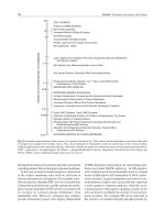
FIGURE 19.1. Diagram of the human circulation
before birth. Arrows indicate the direction of blood
flow. Note where oxygenated blood mixed with deoxygenated blood: in the liver (I), in the inferior vena
cava (II), in the right atrium (III), in the left atrium
(IV), and at the entrance of the ductus arteriosus into
the descending aorta (V). (From Sadler TW. Langman’s
Medical Embryology. 7th ed. Baltimore: Williams &
Wilkins; 1995:225, with permission.)
superior and IVC as well as deoxygenated blood from the coronary
circulation via the coronary sinus. The right heart then pumps this
blood through the low-pressure, high-flow pulmonary arteries.
Once the blood has circulated through the pulmonary circulation,
it is returned to the left atrium via four posteriorly situated pulmonary veins (two superior and two inferior pulmonary veins).
Blood from the left heart is ejected from the left ventricle into the
systemic circulation via the aorta.
Valvular Anatomy
The mammalian heart is composed of four one-way valves. Two
atrioventicular valves (mitral and tricuspid) provide unidirectional
diastolic flow from the atria to the ventricles and allow a systolic pressure gradient between the atria and ventricles. The semilunar valves
(aortic and pulmonary) allow systolic flow and maintain a diastolic
pressure gradient between the ventricles and outflow circulations.
The tricuspid and mitral valves are fibrous endocardium–lined
valves. The tricuspid valve separates the right atrium from the right
ventricle and consists of a large anterior leaflet attached to the anterior wall of the heart, a posterior leaflet at the right margin, and a
septal leaflet attached to the septum. Three chordae tendinae are
attached to the free surface of the leaflets and to the papillary muscles
at the right ventricular base. This apparatus prevents prolapse of the
tricuspid valve leaflets into the right atrium during systole. The mitral valve, located at the orifice of the left ventricle, consists of a large
anterior leaflet in continuity with the posterior wall of the aorta and
a smaller posterior leaflet. The anterior leaflet of the mitral valve is
anatomically in proximity to the aortic valve. Chordae tendineae
(Fig. 19.2) secure the leaflets to the anterior and posterior papillary
muscles and ensure coaptation of the valve leaflets during systole.
The aortic and pulmonic valves are situated at the outflow of the
left and right ventricles, respectively. The aortic valve is a trileaflet
valve. These leaflets are named according to the origin of the coronary arteries, namely the right coronary, left coronary, and noncoronary leaflets (Fig. 19.3). Similarly, the pulmonic valve is a trileaflet
valve with a right, left, and noncoronary leaflet.
Coronary Anatomy
The coronary circulation (Fig. 19.4) supplies oxygen-rich blood to
the myocardium and epicardium. The endocardium is in continuous contact with intracardiac blood and does not require additional blood flow. The right and left coronary arteries of the heart
arise just superior to the aortic valve in the coronary sinuses and are
the first branches of the aorta.
The right coronary artery arises from the anterior (right) sinus
of Valsalva in the aorta and runs along the atrioventricular (AV)
Porrett_ch19.qxd
6/2/09
6:38 PM
Page 267
Chapter 19 • Cardiovascular Disease and Cardiac Surgery
267
coronary arteries supply the posterior descending artery, branches
to the septum, and AV node (Fig. 19.5).
The left coronary artery arises from the left sinus of Valsalva and
passes between the left auricle (atrial appendage) and pulmonary
trunk toward the anterior AV groove. In 40% of the patients, the SA
branch arises from the left coronary artery. The left coronary artery
divides at the AV groove to give off the left anterior descending artery
(LAD) and circumflex coronary artery (Fig. 19.5). The LAD passes anteriorly along the interventricular groove to the apex and provides
septal branches that supply the anterior two thirds of the interventricular septum and diagonals that supply the anterior-lateral wall of
the left ventricle. The circumflex coronary artery follows the AV
groove around the left border of the heart to the posterior surface of
the heart and provides marginal branches (i.e., obtuse marginal) that
supply the posterior left ventricle. In 10% of the population, the circumflex coronary artery ends in the posterior descending artery, providing blood flow to the posterior one third of the interventricular
septum and AV node, defining a left-side dominant circulation.
The venous drainage of the heart is via veins that drain into the
coronary sinus as well as into smaller venae cordis minimae and anterior cardiac veins that drain into the right atrium. The coronary
sinus is a large vein that receives coronary venous blood from the
left (great cardiac, left marginal, and left posterior ventricular
veins) and right (middle and small cardiac veins) side veins. It runs
in the posterior AV groove.
FIGURE 19.2. Chordae tendineae tether the leaflets of the mitral and
tricuspid valves, allowing precise coaptation during systole. (From
Chitwood WR Jr. Mitral valve repair: ischemic. In: Kaiser LR, Kron
IL, Spray TL, eds. Mastery of Cardiothoracic Surgery. Philadelphia:
Lippincott–Raven Publishers; 1998:312, with permission.)
(coronary) groove. In about 60% of the population, the right coronary artery gives off a sinoatrial (SA) branch near its origin to supply the SA node. It traverses posteriorly toward the apex of the
heart and gives off a right marginal artery, which supplies the right
ventricle. After giving off this branch it continues in the posterior
interventricular groove. In roughly 85% of patients, the posterior
descending artery arises from the right coronary artery and defines a
right-side dominant circulation. In approximately 5% of patients, a
balanced pattern exists in which the right coronary and circumflex
Left coronary
cusp
Electrophysiology
As with any striated muscle, cardiac muscle contraction is initiated
by action potentials (rapid voltage changes of the cell membrane).
Certain cells within the cardiac muscle are capable of acting as the
pacemaker and spontaneously initiate action potentials. The action
potentials of cardiac muscle are special in that they can self-generate,
conduct from cell to cell via gap junctions, and are long in duration.
Action potentials of the myocardium can be classified as either
fast action potentials or slow action potentials. Fast action potentials
occur in normal myocardium of atria, ventricle, bundle of His, and
Purkinje fibers. Slow action potentials are seen in the pacemaker
cells of the SA and AV nodes. As seen in Figure 19.6 (solid line), fast
action potentials are characterized by a rapid depolarization (phase
Left coronary
artery
Anterior mitral
leaflet
Right coronary
cusp
Right coronary
artery
Noncoronary
cusp
Bundle
of His
FIGURE 19.3. Normal aortic valve from a
surgeon’s point of view. (From Damiano RJ.
Aortic valve replacement: prosthesis. In:
Kaiser LR, Kron IL, Spray TL, eds. Mastery
of Cardiothoracic Surgery. Philadelphia:
Lippincott–Raven Publishers; 1998:362, with
permission.)
Porrett_ch19.qxd
268
6/4/09
3:24 PM
Page 268
Section IV • Cardiovascular and Respiratory Systems
FIGURE 19.4. Anatomy of the coronary arteries and
cardiac veins. A. Anterior view. The origin of the left
main coronary artery is left lateral and somewhat posterior with respect to the aorta; it courses behind the
pulmonary artery and then divides into the left anterior
descending and circumflex coronary arteries. The origin of the right coronary artery is almost directly anterior, and it runs in the atrioventricular groove.
B. Posterior view. The great, middle, and small cardiac
veins come together at the level of the coronary sinus,
which lies in the left inferior atrioventricular groove
and empties into the right atrium. (From Greenfield LJ,
Mulholland MW, Oldham KT, et al. Surgery: Scientific
Principles and Practice. 3rd ed. Philadelphia: Lippincott
Williams & Wilkins; 2001:1487, with permission.)
A
B
Right
0—transient increase in Naϩ conductance), partial repolarization
(phase 1—outward movement of Kϩ), a plateau (phase 2—inward
Ca2ϩ), membrane repolarization (phase 3—decreased Ca2ϩ conductance and increased Kϩ conductance), and a resting membrane
potential (phase 4—equal inward and outward currents). In contrast, slow action potentials demonstrate a slower depolarization
phase (phase 0), and shorter plateau and repolarization (phase 3) to
an unstable slow depolarization resting phase (phase 4). The alterations in the membrane potential are a factor of a cell membrane’s
permeability to particular ions (Naϩ, Kϩ, Ca2ϩ) and the resulting
gradients that exists.
During an action potential, cardiac myocytes are in an effective
refractory period (ERP) and cannot be stimulated by another action potential. This occurs during phases 1 and 2, and at the beginning of phase 3. Shortly after this period is a relative refractory
period (RRP, late phase 3), during which a supranormal action
potential is needed for excitation. Immediately after the action potential, before return to a normal resting state (phase 4), is the
supranormal period during which the cells are hyperexcitable and
require a lower than normal action potential for stimulation.
Once an action potential arises, it is conducted across the cell
membrane to adjacent cells via gap junctions. The speed of transmission of the action potential is determined by a combination of
cell size and rate of depolarization. The smaller cells of the pacemaker cells demonstrate a slower conduction velocity than the
larger Purkinje cells. Similarly, the slow response of the pacemaker
cells mediates a slower conduction velocity when compared with
the fast response of ventricular myocardial cells.
SA nodal cells demonstrate the most rapid spontaneous depolarization and hence act as the pacemaker under routine conditions. This tissue lies within the wall of the right atrium at the
junction of the right atrium and SVC. Once the action potential is
initiated in the SA node, it is propagated via the atria to the AV
node. The AV node is located in the interatrial septum above the
tricuspid valve near the coronary sinus. In pathologic conditions
with SA nodal discontinuity, the AV node can act as a pacemaker.
The AV node protects the ventricle from excess stimulation in the
case of increased atrial rates, allowing the ventricle adequate
diastolic filling. From the AV node, the action potential is sent to
the ventricle via the bundle of His. The bundle of His splits into
Porrett_ch19.qxd
6/2/09
6:38 PM
Page 269
Chapter 19 • Cardiovascular Disease and Cardiac Surgery
269
FIGURE 19.5. Coronary anatomy: RCA, right coronary artery; PDA, posterior descending artery; LAD, left
anterior descending artery; OM, obtuse marginal artery. (From P Atluri, YJ Woo. The cardiovascular system. In: A.
Atluri, GC Karakousic, PM Porrett et al., eds. The Surgical Review. 2nd ed. Philadelphia: Lippincott Williams &
Wilkins; 2005, with permission.)
+25
1
2
Transmembrane potential (mV)
0
3
−25
1 gm
−50
ERP
RRP
SNP
−75
4
0
−100
Na+
influx
Ca2+
influx
K+
efflux
Na+
efflux
right and left bundle branches and ultimately into Purkinje fibers,
which conduct to the subendocardial surfaces (Fig. 19.7).
The autonomic nervous system (sympathetic and parasympathetic nervous systems) innervates the SA node and controls heart
rate by modifying SA nodal activity. The sympathetic nervous system
increases heart rate by increasing the rate of depolarization. In
contrast, the parasympathetic nervous system increases potassium
K+
influx
FIGURE 19.6. Schematic fast action potential
of human ventricular myocardium (solid) with
electrolyte movements, refractory periods (see
text) and force generated (dashed line). The five
phases of fast cardiac action potential are indicated as numbers. Phase 4: the resting membrane potential. Potassium conductance is high
and sodium conductance is low. Phase 0: Upstroke of the action potential due to membrane
depolarization. An increase in sodium conductance due to the opening of voltage dependent
fast sodium channels causes depolarization.
There is a simultaneous decrease in potassium
conductance. Phase 1: Period of partial repolarization due to a dramatic decrease in sodium
conductance and a brief increase in chloride
conductance. Phase 2: Plateau phase during
which changes in potassium efflux (conductance decrease and then plateaus) is matched
by calcium influx (conductance increases and
then plateaus). Phase 3: Membrane repolarization phase due to an increase in potassium
efflux (increase potassium conductance) and a
decrease in calcium influx (decreased calcium
conductance). (From P Atluri, YJ Woo. The
cardiovascular system. In: A. Atluri, GC
Karakousic, PM Porrett et al., eds. The Surgical
Review. 2nd ed. Philadelphia: Lippincott
Williams & Wilkins; 2005, with permission.)
conductance, increases the magnitude of hyperpolarization, slows
down the rate of spontaneous depolarization, decreases the rate of
closure of potassium channels, and slows down the heart rate. In addition to increasing heart rate (positive chronotropic effect), the
sympathetic nervous system increases the rate of conduction of action potentials through the conduction system. The parasympathetic
nervous system, in contrast, acts to slow down conduction.
Porrett_ch19.qxd
270
6/2/09
6:38 PM
Page 270
Section IV • Cardiovascular and Respiratory Systems
FIGURE 19.7. Structure of conduction system of
the heart. (From Johnson LR. Essential Medical
Physiology. 2nd ed. Philadelphia: Lippincott–Raven;
1998:166, with permission.)
Superior vena cava
SA node
Right
atrium
AV node
Left
atrium
Tricuspid valve
Bundle of His
Left bundle branch
Right ventricle
Septum
Right bundle branch
Left ventricle
Purkinje fibers
The electrical activity of the heart can be interpreted utilizing an electrocardiogram (ECG). The normal ECG demonstrates
P waves and QRS complexes, which represent atrial and ventricular
depolarization, respectively. Ventricular repolarization is demonstrated by the T wave.
Circulatory Physiology
As previously stated, the cardiovascular system is composed of
the pulmonary circulation to provide perfusion to the lung
parenchyma and the systemic circulation to provide systemic perfusion (and a very small degree of pulmonary circulation via the
bronchial vessels). The pulmonary circuit is a low-pressure (mean
PA pressure of 15 mm Hg), high-flow system. As compared to the
systemic circulation, the pulmonary vessels contain very little
smooth muscle and are much shorter. This results in highly compliant (compliance [mL/mm Hg] ϭ volume [mL]/pressure [mm Hg];
inversely proportional to elastance), low-resistance vessels. It
should be remembered that the pulmonary circulation must be
capable of handling the same volume as the systemic circulation, as
right heart output is equal to left heart output.
The pulmonary circulation is capable of handling increased
cardiac output as seen with exercise by both recruiting additional
pulmonary capillaries that are not normally utilized as well as distending the pulmonary vessels. PVR is able to decrease with increasing cardiac output because of these two mechanisms. This
drop in resistance maintains low PA pressures, thereby preventing
pulmonary edema and decreasing right heart cardiac work. Other
regulators of pulmonary blood flow are lung volume, hypoxia
(which causes pulmonary vasoconstriction), and hypercapnea
(which results in pulmonary vasodilation).
In contrast to the pulmonary circuit, the systemic circulation
operates at a high pressure, with high resistance to blood flow. The
flow of blood is from the left heart (left ventricle) to the aorta.
From the aorta, blood flows down a pressure gradient through various branches to arterioles and capillary beds. The large and small
arteries are thick-walled vessels with extensive elastic tissue and
smooth muscle. They are under high pressure but offer little resistance to blood flow. Resistance can be calculated using the following equation derived from the work of Jean Leonard Marie
Poiseuille on flow mechanics:
Resistance ϭ 8(viscosity of blood) (length of vessel)
⌸(radius of blood vessel)4
Aortic and arterial elasticity maintains perfusion during the
diastolic/filling phase of left ventricular cycling. Arterioles, the
short, terminal branches of the arteries, are the principal resistance
vessels of the systemic circulation. They comprise a large percentage of vascular smooth muscle innervated by the autonomic nervous system within the vessel wall that can constrict and impede
the flow of blood. Arterioles provide the largest pressure drop in the
circulation. Arteriolar resistance is regulated by the autonomic
nervous system.
As arterial structures progressively branch from the aorta ultimately to the capillary bed, the cross-sectional area of the vascular
bed continues to increase. On the outflow side of the capillary bed,
the cross-sectional area decreases as capillaries drain into venules
that merge into small veins, large veins, and ultimately the vena
cava. The velocity of blood flow is directly proportional to volume
of blood flow and inversely proportional to cross-sectional area.
Velocity of blood flow (cm/sec) ϭ Flow (cm3/sec)/cross-sectional
area (cm2)
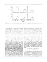
As illustrated in Figure 19.8, there is a decrease in the velocity
of blood flow as the cross-sectional area of the vascular bed increases. This is ideal at the capillary level (high surface area, low
velocity), where a high contact surface area and low velocity provide for optimal exchange of metabolic products at a cellular level.
Cardiac Mechanics
The heart is a biomechanical pump. The mechanical force generated by the heart is utilized to eject blood from the heart to either
the pulmonary or systemic circulations providing perfusion to end
organs. There must be synchrony of the cardiac myocytes, valves,
Porrett_ch19.qxd
6/2/09
6:38 PM
Page 271
Chapter 19 • Cardiovascular Disease and Cardiac Surgery
125
Area
100
y
cit
55
5,000
45
4,000
FIGURE 19.8. Pressure, area, and velocity relationship across the systemic circulation. (From
Kreisel D, Krupnick AS, Kaiser LR, eds. The Surgical Review. 1st ed. Philadelphia: Lippincott
Williams & Wilkins; 2001:308, with permission.)
25
3,000
2,000
Area (cm2)
50
re
ssu
Velocity (cm/s)
35
P re
Pressure (mm Hg)
lo
Ve
75
271
25
15
0
0
Vena cava
Large veins
Small veins
Venules
Capillaries
Aorta,
large arteries,
small arteries
Arterioles
0
1,000
and four chambers of the heart for maximum efficiency. The heart
is in a constant state of flux to ensure that adequate end organ perfusion is achieved. The primary variables that alter cardiac function
are preload, afterload, and autonomic nervous system stimulation.
A proper understanding of these forces is a prerequisite to an adequate understanding of cardiac mechanics.
The left and right ventricles function in a cyclical manner.
Contraction and ejection of blood occurs during systole. Myocardial
perfusion as well as filling of the ventricles occurs during the relaxation phase known as diastole. To simplify the discussion all references to ventricular function will focus on left ventricular mechanics.
The left ventricular intracavitary volume and pressure at end diastole (immediately prior to contraction) determine the preload of
the heart. There are several factors that affect preload. Increasing venous return increases preload, while fibrotic, hypertrophied, and
aging hearts become increasingly stiff and limit left ventricular filling
and preload. As described earlier, relaxation is an energy-dependent
process (calcium-ATPase), which is augmented by adrenergic stimulation, but is impaired in ischemia, hypothyroidism, and congestive
heart failure—all conditions that limit preload.
The afterload of a muscle is the pressure against which it must
contract. For the left ventricle, this is equivalent to the aortic pressure against which it must eject blood during systole. Afterload for
the right ventricle is equal to the PA pressure. The greater the afterload, the greater the potential energy the heart must generate to
provide adequate ejection into the aorta, and subsequently the
greater the cardiac work (described in the following text). Maximal
velocity of contraction is achieved when afterload is minimal.
Within normal physiologic ranges, the heart is able to accommodate a broad range of end-diastolic volume by altering contractility. This dynamic activity is described by the Frank–Starling
relationship, which describes the interplay between ventricular
filling and contractility. With increased ventricular filling the sarcomeres are stretched to an optimal length, thereby facilitating
increased contractility. Adrenergic stimulation can further increase
contractility (inotropy) of the heart, thereby increasing the stroke
volume (volume of blood ejected from the heart with each beat).
Parasympathetic innervation decreases inotropy. Additionally,
right atrial stretch leads to an increase in heart rate with subsequent
increase in cardiac output.
Cardiac output (l/min) ϭ stroke volume (l/beat)
ϫ heart rate (beats/min)
The cardiac cycle, as well as the interplay between preload and
afterload on stroke volume, can best be described using
pressure–volume loops (Fig. 19.9). These pressure–volume loops are
constructed by combining systolic and diastolic pressure curves. The
diastolic component (dotted line) is determined by diastolic filling
(preload). The shape of the loop is determined by both contractility
and the afterload against which the ventricle must contract. The cardiac cycle begins at end diastole when the left ventricle is filled with
left atrial blood and the cardiac muscle is relaxed. On excitation the
muscle begins to contract and generate force against closed valves
(isovolumetric contraction). Once the pressure in the left ventricle
exceeds aortic pressure, the blood is ejected into circulation during
systole. This volume ejected is the stroke volume (depicted by the
width of the pressure–volume loop). The remaining volume at the
end of contraction is the end-systolic volume. At the end of contraction the ventricle begins to relax (isovolumetric relaxation) and the
aortic valve closes as the pressure in the aorta exceeds that of the left
ventricle. With a drop in left ventricular pressure the mitral valve
opens and left atrial blood begins to fill the left ventricle during diastole. It should be noted that in the ideal system following passive
flow of atrial blood, atrial contraction near the end of diastole optimizes filling of the left ventricle (atrial kick), thereby optimizing the
Frank–Starling relationship. Loss of this end-diastolic atrial contraction as in atrial fibrillation in a heart with ventricular hypertrophy
can have adverse systemic hemodynamic consequences.
There are several factors that affect the pressure–volume loops.
Increased preload increases end-diastolic volume and stroke volume.
Increased afterload increases pressure that is required to be generated
during isovolumetric contraction to eject blood and decreases the
stroke volume. Increased contractility, as with adrenergic stimulation, increases stroke volume and decreases end-systolic volume. The
ability of a hypertrophic heart to increase stroke volume is severely
limited by its decreased diastolic compliance, limiting preload.
272
6/2/09
6:38 PM
Page 272
Section IV • Cardiovascular and Respiratory Systems
FIGURE 19.9. Pressure–volume loop
of one cardiac cycle. (From Mohrman
DE, Heller LJ. Cardiovascular Physiology. 3rd ed. New York: McGraw-Hill;
1991:54, with permission.)
120
Intraventricular pressure (mm Hg)
Porrett_ch19.qxd
Ejection
Reaches endsystolic volume
Aortic valve opens
80
Systole
Isovolumetric relaxation
Isovolumetric contraction
Diastolic filling
Mitral valve opens
60
Reaches end-diastolic volume
130
Stroke volume
Intraventricular volume (mL)
Oxygen utilization by the heart is twofold. A small amount of
oxygen is utilized for cellular homeostasis and a large amount is utilized during contraction. Changes in myocardial oxygen consumption are directly related to the work of the heart and changes in
contractility. Cardiac work can be quantified as stroke work, or work
that the heart performs with each beat (stroke work ϭ aortic pressure ϫ stroke volume). The minute work of the heart is equal to the
product of heart rate times the stroke volume multiplied by the aortic pressure (or cardiac output ϫ aortic pressure), so an increase in
any of these three variables will increase cardiac work and ultimately
increase myocardial oxygen consumption and demand.
The major determinant of oxygen demand is myocardial wall
tension. Tension in the wall of the ventricle is determined by both
the pressure in the ventricle and the geometry of the ventricle. The
normal left ventricle is a pressurized irregularly shaped chamber. If
we were to consider the ventricle as a cylinder then the law of
Laplace states that wall tension is proportional to internal pressure
times the radius. Increasing the wall thickness decreases the wall
tension by distributing the internal pressure over a greater number
of muscle fibers. In other words, wall tension equals pressure times
radius divided by wall thickness. Altering the geometrical configuration of the ventricle (as with cardiomyopathy), increasing the radius, decreasing the wall thickness, and increasing ventricular
pressure all increase wall tension and myocardial oxygen demand.
Changing the geometry of the ventricle requires extra energy consumption to realign the myocytes prior to each systolic contraction.
As stated previously, cardiac output is equal to the product of
stroke volume multiplied by the heart rate. A clinically feasible
means of calculating cardiac output is to utilize the Fick equation:
Cardiac output =
Total body oxygen consumption
3O2] arterial blood - [O2] venous blood
Dye dilution and thermal dilution of heat are other clinically
utilized methods of calculating cardiac output. Given the varying
sizes of patients (varying body surface area), simply calculating a
cardiac output may not provide enough information regarding
cardiac function and adequate systemic perfusion. The calculated
parameter of cardiac index factors in patient size and expresses
cardiac output per square meter of surface area, thereby eliminating the variable of patient size (cardiac index ϭ cardiac output/
body surface area). A cardiac index greater than 2 L/min/m2 is
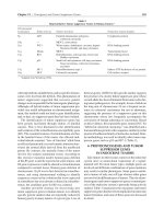
accepted as adequate. Figure 19.10 demonstrates the mechanical
and electrical events during the various phases of the cardiac cycle
(Table 19.1).
Coronary Physiology
Coronary blood flow follows the major vessels into smaller penetrating arteries, which provide the majority of the resistance to
blood flow. There is a dense capillary network by which the extensive metabolic demands of the heart can be provided. At rest coronary blood flow is approximately 1 mL/g of myocardium, but with
demand this flow is capable of increasing nearly fourfold. The increase in blood flow is accomplished with a combination of local
vasodilatation of the penetrating arteries as well as recruitment of
vessels that are collapsed at rest. Since nearly 70% of the oxygen is
derived from delivered coronary blood, there exists a very tight regulatory system to ensure adequate perfusion of the myocardium.
The myocardial tissue functions most optimally under aerobic conditions and is capable of sustaining only a few minutes of anaerobic
activity.
Coronary perfusion is accomplished during the relaxing diastolic phase. During systole the compressive forces within the myocardial wall are powerful enough to collapse the penetrating
vessels and prevent myocardial perfusion. Therefore, increasing
heart rate will not only increase myocardial oxygen demand but
also decrease myocardial perfusion. Regulation of coronary blood
flow is accomplished by a combination of the autonomic nervous
system, metabolic vascular mediators, and vascular endothelium–
mediated vasodilatation. There are a combination of ␣- and
-receptors on the conductance vessels, which regulate nervous
system–mediated vasoconstriction and vasodilatation, respectively.
Adenosine is produced by cardiac myocytes in response to ischemia
and is the primary metabolic vascular mediator. It acts locally on
vascular smooth muscle to cause vasodilatation. The vascular endothelium is capable of releasing both vasodilatory and vasoconstricting mediators.
Porrett_ch19.qxd
6/2/09
6:38 PM
Page 273
Chapter 19 • Cardiovascular Disease and Cardiac Surgery
A
B
C
D
E
F
G
A
mm Hg
Aortic
pressure
120
100
Ventricular
pressure
80
273
FIGURE 19.10. Mechanical and electrical events during a single cardiac cycle. The seven phases are denoted
by letters as follows: (A) atrial systole, (B) isovolumetric ventricular contraction, (C) rapid ventricular
ejection, (D) reduced ventricular ejection, (E) isovolumetric ventricular relaxation, (F) rapid ventricular filling, and (G) reduced ventricular filling. (From Johnson
LR. Essential Medical Physiology. 2nd ed. Philadelphia:
Lippincott–Raven; 1998:190, with permission.)
60
40
Atrial
pressure
20
0
Heart sounds
IV
I
II
III
IV
ml
60
Ventricular
volume
40
20
0
R
P
T
P
Q
TA B L E 19 . 1
S
Normal hemodynamic parameters.
Cardiac output
4.0–8.0 L/min
Cardiac index
2.5–4.5 L/min/m2
Stroke volume
60–130 mL
Systemic blood pressure
100–130/60–90 mm Hg
Mean arterial pressure (MAP)
70–105 mm Hg
Right atria/central venous pressure
2–10 mm Hg
Right ventricular pressure
15–30/0–18 mm Hg
Pulmonary artery pressure
15–30/6–12 mm Hg
Pulmonary capillary wedge pressure
5–12 mm Hg
Systemic vascular resistance (SVR)
700–1,600 dynes/sec/cm2
Pulmonary vascular resistance
20–130 dynes/sec/cm2
mm Hg, millimeters of mercury; L/min, liters/minute; m, meter, cm,
centimeter; sec, second.
CARDIOVASCULAR PATHOPHYSIOLOGY
Congenital Heart Disease
Atrial Septal Defect
Atrial septal defects (ASDs) account for 10% to 15% of cardiac
anomalies. Additionally, these are the most common congenital
conditions encountered in adults. ASDs may occur in association
with other complex congenital heart and genetic defects including
Down, Turner, Marfan, and Ehlers–Danlos syndromes.
A defect in formation of the septum primum results in ostium secundum–type ASD (Fig. 19.11). Ostium primum–type
ASDs are a result of malformation of the AV canal. Other less
common types of ASDs include sinus venosus–type ASD (defect
at the level of the SVC and IVC) and coronary sinus ASD (defect
in the wall between coronary sinus and left atrium). Roughly 20%
of adults have a PFO, which is clinically inconsequential as it
remains closed due to higher left atrial pressure compared with
right atrial pressures. But, with high right-sided pressures the
foramen ovale may become patent causing right to left shunting
of blood.
Porrett_ch19.qxd
274
6/2/09
6:38 PM
Page 274
Section IV • Cardiovascular and Respiratory Systems
FIGURE 19.11. Normal atrial septum formation (A) and ostium secundum–type atrial
septal defect caused by excessive resorption of
the septum primum (B, C). (From Sadler
TW Langman’s Medical Embryology. 5th ed.
Baltimore: Williams & Wilkins; 1985, with
permission.)
A
Often patients remain asymptomatic. Symptomatic patients
present with signs of heart failure, exercise intolerance, or
arrhythmias. Echocardiography is usually diagnostic. Many small
ASDs in children will close spontaneously and should be monitored. All symptomatic and large/significant ASDs should be closed
by either percutaneous or surgical means. Prior to closure it is
critical to measure PVR by cardiac catheterization. Elevated PVR
(Ͼ8 woods units) is a contraindication to closure.
Ventricular Septal Defect
Ventricular septal defects (VSDs) account for roughly 25% of congenital heart defects. VSDs can either occur singly or in combination. One half of patients with VSDs will also have another cardiac
anomaly, and should therefore undergo thorough evaluation. VSDs
are defined on the basis of their location in the ventricular septum,
i.e., outlet, septal, conoventricular, anterior muscular, midmuscular, apical muscular, and inlet septal (Fig. 19.12). Hemodynamically, VSDs result in left to right shunting of blood, thereby
resulting in elevated PVR and left atrial and ventricular overload.
With long-standing left to right shunting there is medial hypertrophy of the pulmonary vasculature and an increase in PA pressures.
B
C
Over time as the PVR increases, volume overload on the left heart
decreases and eventually there is a reversal of flow through the VSD.
With right to left shunting there is a worsening cyanosis that ensues, referred to as Eisenmenger syndrome. Once a diagnosis of
Eisenmenger syndrome has been made, operative repair is contraindicated, given the high risk of right heart failure. With an intracardiac defect, a functioning right ventricle, and failing lungs
secondary to elevated PVR, bilateral lung transplantion and intracardiac repair may be the only option. In the presence of a failing right
ventricle the only option would be a heart–lung transplant of which
only few are done currently because of the scarcity of donors.
VSDs can be diagnosed by echocardiography. Early surgical repair is indicated for children with large VSDs with a pulmonary
blood flow (Qp)–systemic blood flow (Qs) ratio greater than 2.
Moderate-sized defects are often monitored until childhood.
Patent Ductus Arteriosus
PDA results from failure of closure of the ductus arteriosus, which results in blood being shunted from the proximal descending thoracic
aorta to the PA bifurcation. PDAs take up to 3 months after birth to
close, and are therefore not considered pathologic until after this age.
The male–female ratio is 1:2. Failure of closure after 3 months is
thought to be secondary to immaturity of the medial muscular layer.
As with VSDs, left to right shunting of blood occurs, resulting
in increased pulmonary blood flow and left atrial and ventricular
overload. On auscultation a classic “machinelike” murmur is heard.
Closure of the PDA can be attempted by pharmacologic means in
newborns with inhibition of prostaglandins utilizing agents such as
indomethacin. PDAs that fail to close may be amenable to percutaneous embolization utilizing coils. In infants if operative closure is
required, these defects can often be approached thoracoscopically
or via a small left thoracotomy.
Tetralogy of Fallot
FIGURE 19.12. Major types of ventricular septal defects categorized
by anatomic location. (From Kaiser LR, Kron IL, Spray TL, eds. Mastery
of Cardiothoracic Surgery. Philadelphia: Lippincott–Raven Publishers;
1998, with permission.)
Tetralogy of Fallot (TOF) results from an anterior malalignment of
the infundibular septum. Classically this malformation results in a
VSD, overriding aortic valve, right ventricular outflow obstruction,
and resultant right ventricular hypertrophy (Fig. 19.13). With the addition of an ASD, the condition is referred to as pentalogy of Fallot.
Twenty-five percent of TOF patients have a right-sided aorta, and in
addition, anomalies in the coronary circulation may also exist.
The extent of the cyanosis depends on the severity of the tetralogy. In severe cases, increased cyanosis can occur with agitation or
crying. Older children often learn to squat to relieve cyanotic spells.
Physical examination reveals a systolic murmur over the left heart
Porrett_ch19.qxd
6/2/09
6:38 PM
Page 275
Chapter 19 • Cardiovascular Disease and Cardiac Surgery
Aorta
275
Patent ductus
arteriosus
Pulmonary
artery
FIGURE 19.13. Tetralogy of Fallot: schematic drawing. (From Sadler
TW. Langman’s Medical Embryology. 5th ed. Baltimore: Williams &
Wilkins; 1985, with permission.)
border and an accentuated second aortic heart sound. Chest
roentgenogram often demonstrates a boot-shaped heart. Conservative therapy includes a knee to chest position, supplemental oxygen, volume expansion, and sedation. Symptomatic infants should
undergo immediate repair with elective repair done at 1 year of age
for asymptomatic patients.
Tricuspid Atresia
Tricuspid atresia results from a lack of communication between the
right atrium and ventricle. Associated anomalies include an ASD,
enlarged mitral valve and left ventricle, and right ventricular hypoplasia. As with other congenital cardiac anomalies, echocardiography secures the diagnosis and nicely demonstrates the anatomic
abnormalites. These patients require surgical correction to increase
systemic oxygen saturation and avoid the development of heart
failure. Surgical repair requires a Fontan procedure, an operation
that results in the systemic venous return being connected directly
to the PA, resulting in increased pulmonary blood flow, a decreased
right to left shunt, and decreased volume overload of the left heart.
Transposition of the Great Vessels
Transposition of the great vessels (TGAs) occurs when the aorta
arises from the anatomic right ventricle and the PA arises from the
anatomic left ventricle (Fig. 19.14). TGA accounts for 8% to 10%
of all congenital heart defects. Associated cardiac anomalies can
include ASD, VSD, PDA, left ventricular outflow obstruction, abnormal coronary branching, or PFO. Normal physiologic closure
of the ductus arteriosus can result in profound cyanosis. In the
case of complete TGA, survival depends on early recognition and
the presence of a right to left shunt in the form of a PDA or ASD. A
closing ductus arteriosus can be maintained patent with infusion
of prostaglandin E1 to enhance the requisite left to right shunting,
thereby providing temporary palliation. Alternatively, an ASD can
be created percutaneously with a balloon septoplasty.
Diagnosis is confirmed by echocardiography. On physical examination a systolic murmur and loud single heart sound can be
appreciated. Chest roentgenogram reveals an oval or egg-shaped
heart, narrow superior mediastinum, and increased pulmonary
vascular markings. An arterial switch procedure in which the great
FIGURE 19.14. Transposition of the great vessels: schematic drawing.
(From Sadler TW. Langman’s Medical Embryology. 5th ed. Baltimore:
Williams & Wilkins; 1985, with permission.)
vessels are transposed to their appropriate anatomic positions is the
definitive operation for this anomaly.
Coarctation of the Aorta
Coarctation of the aorta is characterized by a focal narrowing of the
thoracic aorta, most frequently just distal to the origin of the left
subclavian artery usually near the ductus arteriosus. Coarctation
accounts for 5% to 8% of all congenital heart defects. Associated
anomalies include PDA, VSD, bicuspid aortic valve, subaortic obstruction, and mitral valve anomalies.
Coarctation has historically been categorized as either infantile
or adult. In infantile coarctation, the aortic obstruction is most
often preductal and leads to separation of the left ventricular flow
directed to the head and arms from the PA flow directed to the lower
body. This type of coarctation results in early left ventricular failure
and death if not surgically corrected. The more common adult type
of coarctation is postductal and leads to proximal hypertension and
eventual congestive heart failure over time, although patients may
remain asymptomatic and appear healthy well into adulthood.
Physical findings of absent femoral pulses with poor distal perfusion should warrant a workup for coarctation of the aorta. Findings on physical examination include upper extremity systolic
hypertension and a pressure differential between the left and right
upper extremity, absent or decreased lower extremity pulses, prominent pulsations at the sternal notch, and a systolic heart murmur
over the left sternal border that may be transmitted to the back. Chest
roentgenograms may reveal rib notching by the age of 10 years secondary to enlarged intercostal artery collateral circulation. Other radiologic findings include an indentation over the left border of the
heart at the site of coarctation, which results in the classic “3” sign.
In severe cases of aortic coarctation lower extremity blood flow
is entirely dependent on a PDA. With spontaneous ductal closure,
abdominal and lower extremity ischemia will ensue and prostaglandin E1 infusion to maintain patency of the ductus arteriosus
may be required. Severe cases require immediate operative repair,
whereas asymptomatic cases can be repaired on an elective basis.
Repair entails an end-to-end repair, bypass from the enlarged subclavian artery to the descending aorta, prosthetic flap with a synthetic graft, or subclavian flap aortoplasty.
Porrett_ch19.qxd
276
6/2/09
6:38 PM
Page 276
Section IV • Cardiovascular and Respiratory Systems
Total Anomolous Pulmonary Venous Connection
Total anomalous pulmonary venous connection (TAPVC) is a condition in which there is abnormal drainage of the pulmonary veins
into the right atrium. The presence of either a PFO or ASD is required to maintain blood flow to the left heart and thus the systemic circulation. Severity of symptoms depends on whether there
is obstructed or nonobstructed TAPVC. With obstructed TAPVC,
the obstruction causes pulmonary hypertension, decreased venous
return, low cardiac output, venous congestion, and acidosis. Obstructed TAPVC is a surgical emergency. Unobstructed cases present similar to that of an ASD. Operative repair is recommended for
unobstructed TAPVC once diagnosed to prevent pulmonary hypertension and minimize mortality. Up to 80% of infants with TAPVC
will die by 1 year of age if the condition is not surgically repaired.
Hypoplastic Left Heart Disease
Hypoplastic left heart syndrome (HLHS) accounts for 7% of congenital cardiac anomalies and 25% of deaths within the first week
of life. HLHS is a complex anomaly with aortic and aortic valve hypoplasia, mitral valve stenosis, and a hypoplastic left ventricle. A
PDA is essential for survival of the neonate and mandates infusion
of prostaglandin E1. Systemic blood flow is dependent on the parallel circulation that exists from the right ventricle to the systemic circulation via the PDA. Once the lungs expand and the PVR drops,
blood flow preferentially flows to the pulmonary circulation. To
balance pulmonary and systemic circulations, PVR should be controlled by adjusting ventilation, hematocrit should be increased,
and SVR should be altered pharmacologically.
Newborns typically present within the first 48 hours of life
with tachypnea and cyanosis. Echocardiography is almost universally diagnostic. Initial management includes prostaglandin E1 infusion and pharmacologic balancing of the systemic and
pulmonary circulations. Operative repair is carried out in three
stages. The first operation, the Norwood procedure, involves connection of the diminutive aorta to the proximal PA; at the same
time a graft is placed between the innominate artery and pulmonary trunk. The second stage, performed at 3 to 10 months of
age, comprises a hemi-Fontan procedure whereby SVC blood is
directed exclusively into the PA. The final stage, performed at 18 to
24 months, involves redirection of the IVC and SVC blood flow into
the pulmonary circulation, the Fontan procedure. Some centers
TA B L E 19 . 2
prefer to immediately list these neonates for heart transplantations
and reserve the three-stage procedure if a donor cannot be found.
Acquired Heart Disease
Cardiovascular disease is the number one killer, accounting for
37.3% of all deaths in the United States. The 2007 American Heart
Association Heart Disease and Stroke Update estimates that there
are 15.8 million Americans suffering from coronary heart disease
and 5.2 million suffering from heart failure. With an increasing
prevalence of diabetes, obesity, and a sedentary lifestyle the incidence of cardiovascular disease is expected to increase dramatically.
Coronary Artery Disease
Coronary artery disease (CAD) is the leading cause of mortality in
the United States. Similar to peripheral vascular disease, CAD is due
to luminal narrowing with a resultant decrease in blood flow secondary to progressive atherosclerotic disease. Risk factors for atherosclerotic disease include hypertension, diabetes, hypercholesterolemia,
smoking, sedentary lifestyle, and family history. Men are at higher
risk than women for developing premature coronary artery, but after
menopause, the risk is equivalent. Patients with CAD can present
with a spectrum of signs and symptoms ranging from asymptomatic
to chronic severe angina, depending on the extent of disease and degree of luminal narrowing. Diabetic patients often have no symptoms until a major cardiovascular event ensues. The Canadian
Cardiovascular Society Functional Classification has been developed
to classify anginal symptoms related to CAD (Table 19.2). A second
similar grading system for heart failure is the New York Heart Association Heart Failure Functional Classification, which is a subjective
classification system (Table 19.3). Asymptomatic patients may present with myocardial infarction (MI) or even sudden death related to
malignant arrhythmias. Roughly one half of all fatal heart attacks
occur in previously asymptomatic individuals.
Over 10% of patients undergoing noncardiac surgical procedures in the United States are estimated to be at risk for CAD. More
than 15% of these patients suffer from cardiovascular complications
in the postoperative period. This risk is even greater in patients with
peripheral vascular disease. It is critical to appropriately identify
patients at risk for CAD and evaluate them for critical disease.
Diagnostic studies traditionally utilized to identify patients at
risk for CAD have included stress echocardiography, stress
Canadian Cardiovascular Society functional classification.
Class I
Ordinary physical activity does not cause angina. Angina may occur with strenuous
or prolonged exertion.
Class II
Slight limitation of ordinary activity. Angina may occur with walking or climbing
stairs rapidly, walking uphill, walking or stair climbing after meals or in the cold, in
the wind, or under emotional stress, or walking more than two blocks on the level or
climbing more than one flight of stairs under normal conditions at a normal pace.
Class III
Marked limitation of ordinary physical activity. Angina may occur after walking one
or two blocks on level ground or climbing one flight of stairs under normal
conditions at a normal place.
Class IV
Inability to carry out any physical activity without anginal discomfort; angina may
be present at rest.
From Braunwald E. The history. In: Braunwald E, ed. Heart Disease: A Textbook of Cardiovascular Medicine.
5th ed. Philadelphia: WB Saunders; 1997:1–14, with permission.
Porrett_ch19.qxd
6/2/09
6:38 PM
Page 277
Chapter 19 • Cardiovascular Disease and Cardiac Surgery
TA B L E 19 . 3
277
New York Heart Association heart failure functional classification.
Class I
Patients with cardiac disease but without resulting limitation of physical activity.
Class II
Patients with cardiac disease resulting in slight limitation of physical activity. Ordinary
physical activity causes fatigue, palpitations, dyspnea, or angina. No symptoms at rest.
Class III
Patients with cardiac disease resulting in marked limitation of physical activity. Less
than ordinary physical activity results in fatigue, palpitations, dyspnea, or angina. No
symptoms at rest.
Class IV
Patients with cardiac disease who are unable to carry on any physical activity without
discomfort. Symptoms of cardiac insufficiency or angina may be present even at rest.
Any physical activity increases discomfort.
From Braunwald E. The history. In: Braunwald E, ed. Heart Disease: A Textbook of Cardiovascular Medicine.
5th ed. Philadelphia: WB Saunders; 1997:1–14, with permission.
electrocardiography and thallium tests, and intravenous dipyridamole thallium-201 or technetium-99m scintigraphy (DTS).
Newer modalities, including high-resolution cardiac computed tomography and magnetic resonance imaging, are gaining popularity
and may soon become accepted as routine screening studies. DTS is
the best initial preoperative noninvasive screening test. As compared
with exercise stress tests, DTS can be performed on patients who are
unable to perform the exercise portion of the study. In DTS, a finding of reversible defects following infusion of the radiotracer at
stress when compared with resting images reflects reversible defects
and viable myocardium, whereas a nonreversible defect signifies
that the defect is fixed likely resulting from scar and thus neither
amenable nor responsive to revascularization. Results obtained by
either thallium or technetium scintigraphy are 90% sensitive and
75% specific. The findings of CAD on DTS warrants a coronary
angiogram to define any coronary lesions and appropriate therapy
either in the form of coronary artery bypass grafting (CABG) or a
percutaneous coronary intervention (PCI) to enhance myocardial
perfusion. Coronary revascularization is generally performed to alleviate increasing anginal symptoms, preserve at-risk myocardium,
and prevent MI and damage. Noncritical coronary lesions can often
be managed medically until progression of disease ensues.
On the basis of the large body of literature comparing medical
therapy, PCI, and CABG, the American College of Cardiology and
the American Heart Association have established guidelines for surgical revascularization. These in-depth criteria are beyond the scope
of this chapter. The general guidelines include left main stenosis, disease in three or more vessels, proximal LAD stenosis, and failure of
PCI. Diabetic patients have been shown to fare better with CABG as
compared to PCI. In general, CABG is not performed on vessels with
lesions less than 70% because of decreased patency rates related to
outflow obstruction from competitive flow in the native circulation.
The long-term benefits of CABG are primarily related to patency
of the conduit. Vein grafts develop intimal hyperplasia that limits
long-term patency to 50% to 60% at 10 years. In contrast, the internal mammary artery (IMA) has been reported to have patency rates
upwards of 95% as far out as 20 years following operation. Statistically significant improvements in patient survival have been demonstrated in patients receiving an IMA to LAD bypass (Fig. 19.15).
allows for a still operative field and optimal circulatory management.
The CPB circuit is utilized to isolate the cardiopulmonary system and
thereby provide optimal, blood-free operative exposure for cardiovascular surgery. The CPB circuit must perform the functions of the
cardiovascular system. It must oxygenate blood, remove carbon dioxide, and provide adequate perfusion to end organs. The cardiovascular surgeon can utilize either total or partial CPB. During total CPB,
the venous return of the heart is circulated through the CPB circuit
in its entirety, whereas during partial CPB, a fraction of the blood is
allowed to circulate to the right ventricle and pulmonary circulation.
The basic components of the CPB circuit include the venous
reservoir, oxygenator, heat exchanger, and pump. The venous reservoir stores systemic venous return. The oxygenator both adds oxygen and removes carbon dioxide from the blood. Thermoregulation
of blood is controlled by the heat exchanger. Blood is returned to the
systemic circulation via the ascending aorta or femoral artery by
the pump. The pump can either be a kinetic centrifugal pump or the
more common electric motor driven, load-independent roller
pump. It is important to note, however, that CPB activates the complement cascade, triggers release of pro-inflammatory cytokines,
Alternatives to Traditional Coronary Artery Bypass Grafting
CABG accounted for 427,000 operative procedures in 2004. CABG
has traditionally been performed with the assistance of the
cardiopulmonary bypass (CPB) circuit with an arrested heart. This
FIGURE 19.15. Coronary artery bypass grafting performed for atherosclerotic coronary artery disease.
Porrett_ch19.qxd
278
6/2/09
6:38 PM
Page 278
Section IV • Cardiovascular and Respiratory Systems
upregulates inflammatory mediators (IL-1, TNF-␣, IL-6, IL-8, IL-10),
initiates the systemic inflammatory response syndrome (SIRS),
stimulates oxygen-free radical generation, and increases oxidative
stress. To minimize these systemic manifestations a renewed interest
in beating heart surgery has arisen.
Off-pump coronary artery bypass grafting (OPCAB) is performed on a beating heart with the use of stabilization devices to
minimize motion at the site of anastomoses. Blood flow to the affected myocardium can be sustained with the use of an intraluminal
shunt, which additionally minimizes blood in the operative field. Alternatively, silastic tapes can be utilized as a tourniquet for proximal
and/or distal control. The most critical vessels that supply the greatest amount of myocardium at risk are traditionally grafted first, i.e.,
LAD, to maximize perfusion to the heart throughout the case. Randomized, controlled trials have demonstrated significantly lower
transfusion requirements, decreased systemic inflammation, shorter
hospital stay, and decreased cost. Trends toward lower renal complications have been observed. Circulatory management is much more
difficult in OPCAB and requires very close communication between
the surgeon and the anesthesiologist. OPCAB is associated with a significant learning curve and is therefore performed at the discretion
of the surgeon based on experience. Heart failure, hemodynamic instability, severe left ventricular dysfunction, cardiomyopathy, frequent arrhythmias, and emergent operations were once absolute
contraindications for OPCAB, but are now relative contraindications
based on surgeon experience and preference.
The rapid development of minimally invasive techniques in gynecologic, urologic, and general surgery has stimulated an interest in
revascularizing myocardium utilizing smaller incisions. Initially this
consisted of performing beating heart revascularization through
small partial sternotomies or anterolateral thoracotomies, depending
on the target vessels of interest. This approach was originally termed
minimally invasive direct coronary artery bypass (MIDCAB). Often
these incisions can be limited to between 8 and 10 cm and yield excellent cosmetic results. MIDCAB is amenable to single- as well as
multivessel coronary disease. However, it is most ideally suited for an
isolated left internal mammary artery (LIMA) to LAD anastomosis.
Clinical trials have demonstrated excellent patency and rapid recovery following MIDCAB.
The development of robotic technology has further advanced
minimally invasive techniques in cardiac surgery. Robotic platforms provide 3-D vision, magnification, miniature instruments,
elimination of tremor, and mobility through multiple degrees of
movement, thereby allowing very precise and controlled motion
(Fig. 19.16). Several investigators have demonstrated feasibility of
performing precise coronary anastomoses with the robotic platform. Randomized clinical trials of robotically assisted totally endoscopic coronary artery bypass grafting (TECAB) performed
using CPB with peripheral cannulation have demonstrated TECAB
to be a safe procedure with angiographic patency, mortality, and
morbidity equivalent to standard CABG procedures. Further advances in technology, surgical expertise, and reduced cost will be
required before TECAB can become widespread.
Myocardial Infarction
The American Heart Association estimates that 700,000 Americans
will have a heart attack, 500,000 will have a recurrent attack, and an
additional 175,000 will have a silent heart attack. About 38% of
patients suffering an MI will die the ensuing year. Roughly every
FIGURE 19.16. The da Vinci robotic telemanipulation system. A. The
operative console at which the surgeon is seated. B. The instrument
cart with two instrument arms and a camera arm that stands next to
the operating room table. (From From Kaiser LR, Kron IL, Spray TL,
eds. Mastery of Cardiothoracic Surgery. 2nd ed. Philadelphia:
Lippincott–Raven Publishers; 2007, with permission.)
60 seconds an American will die from a heart attack. Advances in
medical management and interventions for MI have reduced the
mortality from acute MI by 24% since 1989. The goal of therapy is
to rapidly salvage as much myocardium as is feasible. Loss of more
than 40% of functional left ventricular mass often results in cardiogenic shock. Reperfusion of myocardium 40 minutes after onset of
acute ischemia results in salvage of 60% to 70% of affected myocardium, while as little as 3 hours following ischemia only 10% of
myocardium can be salvaged.
Medical management of MI necessitates rapid intervention.
Treatment should include decreasing myocardial oxygen demand,
increasing arterial oxygen delivery, maintaining perfusion, and protecting the threatened myocardium. Early reperfusion should be
the goal. Depending on the expertise of a given medical facility
thrombolytics or angioplasty can be utilized. Thrombolytic therapy
is easy to perform in most community settings by trained health
care professionals. Since time to reperfusion is essential, this is
often the strategy utilized in facilities lacking cardiac catheterization laboratories. If feasible, the preferred approach to myocardial
salvage is rapid evaluation and transfer to a cardiac catheterization
lab for PCI with the potential for emergent CABG in the event of
left main CAD or if the lesions are not revascularizable by PCI.
Vasopressors and inotropic agents are the first-line therapy for
cardiogenic shock. Maintenance of optimal filling pressures is essential and may require insertion of a PA catheter to optimize management. Ventilatory support and/or diuresis may be necessary to
maintain proper oxygenation in the setting of acute cardiogenic
pulmonary edema. While medical management is essential, early
revascularization with either PCI or CABG is critical and has been
shown to significantly improve long-term survival.
Complications of Myocardial Infarction
A number of structural sequelae may ensue in the early- or latepostinfarction period, which require prompt surgical intervention.
These complications include VSD, ventricular free wall rupture, left
ventricular aneurysm, and ischemic mitral regurgitation (MR).
Early recognition and treatment of these complications is critical to
maximizing survival. Overall these postinfarction complications
are responsible for 20% of deaths following MI.
Porrett_ch19.qxd
6/2/09
6:38 PM
Page 279
Chapter 19 • Cardiovascular Disease and Cardiac Surgery
279
Ventricular Septal Defect
Ventricular Free Wall Rupture
Postinfarction VSDs complicate 1% to 2% of MIs, accounting for
5% of deaths following an MI. Roughly 60% of postinfarction VSDs
occur in the anteroapical septum as a result of a full-thickness anterior wall MI secondary to an LAD occlusion with limited collateral
vessel formation. The remainder of patients have posterior septal
VSDs resulting from occlusion of either a dominant right or circumflex coronary artery. Postinfarction VSDs occur most frequently
2 to 4 days following an acute MI, but can occur between a few
hours and a few weeks following infarction. The VSD may be a simple rupture or may develop a serpigenous dissection tract.
Typically, patients present with a new-onset harsh holosystolic
murmur that radiates to the axilla and is often associated with chest
pain and a thrill. The gold standard for diagnosis of a postinfarction
VSD is a right heart catheterization with a greater than 9% “step-up”
in oxygen saturation between the right atrium and PA. Color flow
Doppler echocardiography is also a good diagnostic modality for
VSD. Once diagnosed, immediate placement of an intra-aortic balloon pump (IABP) and early surgical intervention are necessary. Preoperative management centers on reducing systemic vascular
resistance while maintaining cardiac output and systemic perfusion.
Without an operation this condition is almost universally fatal, with
7% survival at 1 year if left untreated. Patients in cardiogenic shock
should be immediately taken to the operating room. Operative repair
depends on the location of the defect, but in general involves endocardial patch repair with possible exclusion of the infracted myocardium.
Postinfarction ventricular free wall rupture is more frequent than
VSDs, occurring in 11% of patients following an acute MI. Ventricular rupture and cardiogenic shock are the leading causes of mortality following an MI. Postinfarction ventricular free wall rupture
is more common in elderly women suffering their first MI. In the
present era, ruptures occur most frequently in hypertensive patients within 5 days of infarction. Rupture can affect the anterior,
lateral, and posterior walls. LV ruptures are divided into three categories: acute, subacute, and chronic. Acute ruptures result in sudden chest pain, profound shock, electromechanical dissociation,
and rapid death. Subacute rupture is characterized by a smaller defect that may be sealed by clot or fibrinous pericardial adhesions.
They usually present with signs of tamponade and cardiogenic
shock and may remain stable for several hours or days prior to intervention. A chronic rupture presents as a false aneurysm of the
left ventricle with adhesions containing the aneurysm. Diagnosis of
rupture is best made with echocardiography. Operative repair involves mattress closure of the defect buttressed with Teflon felt or a
Dacron patch.
Left Ventricular Aneurysm
Left ventricular aneurysms affect between 10% and 35% of patients
following an MI (Fig. 19.17). Aneurysm formation occurs in 50% of
patients by 48 hours following an MI. Aneurysm formation is
FIGURE 19.17. The pathophysiology of LV aneurysm formation. A. Area of infarction. B. True aneurysm.
C. False aneurysm. (From Kaiser LR, Kron IL, Spray TL, eds. Mastery of Cardiothoracic Surgery. Philadelphia:
Lippincott–Raven Publishers; 1998, with permission.)
Porrett_ch19.qxd
280
6/2/09
6:38 PM
Page 280
Section IV • Cardiovascular and Respiratory Systems
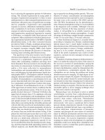
FIGURE 19.18. Chest radiograph demonstrating large ventricular
aneurysm. (From Kaiser LR, Kron IL, Spray TL, eds. Mastery of Cardiothoracic Surgery. Philadelphia: Lippincott–Raven Publishers; 1998, with
permission.)
thought to occur as a result of early infarct expansion and late
remodeling of the aneurysmal wall with scar. Asymptomatic patients have an excellent prognosis following aneurysm formation
with a 90% 10-year survival. Symptomatic patients have a much
poorer prognosis. Angina related to underlying CAD and dyspnea
are the most common presenting symptoms. Diagnosis can be made
using multiple diagnostic modalities. ECGs frequently demonstrate
Q waves with persistent ST elevation. Chest radiographs may
demonstrate LV enlargement (Fig. 19.18). Echocardiography can
often detect a paradoxical bulge during systole of the aneurysm. The
gold standard for diagnosis remains left ventriculography. There are
no absolute indications for operative repair. Symptomatic patients
with angina, congestive heart failure (CHF), or arrhythmias appear
to do better following repair. Surgical intervention involves either
simple plication of the aneurysm, linear closure, or closure with a
Dacron patch. In the absence of thrombus there is a low thromboembolic risk, 0.35%/patient-year, and therefore anticoagulation is
not required. In the setting of large LV thrombus or diminished left
ventricular function long-term anticoagulation is recommended.
Ischemic Mitral Regurgitation
MI or papillary muscle ischemia results in ischemic MR. By definition the leaflets and subvalvular apparatus are normal in ischemic
MR. The disease is a manifestation of postischemic myocardial remodeling. The presentation may be acute and immediately lifethreatening or may present in a chronic fashion with an insidious
onset of heart failure. The incidence of ischemic MR is between
17% and 55% following an MI, with up to 18% of patients having
evidence of MR within 6 hours of the onset of ischemia. In many
patients, however, the MR is mild and may be transient and improve over time. The development of ischemic MR is dependent on
transmural involvement, location, and extent of infarction or resultant papillary muscle ischemia, with posteroinferior MIs having
the highest likelihood of MR secondary to papillary muscle dysfunction. Ruptured papillary muscles can lead to life-threatening
acute MR, with the posterior papillary muscle involved three to six
times more commonly than the anterior. Complete rupture usually
occurs within the first 7 days after MI but may be delayed by up to
3 months. The presentation of acute MR represents only 1% to 2%
of all cases of ischemic MR. A murmur may be absent following
papillary muscle rupture given the rapid equalization of pressure
between the left atrium and ventricle. Rapid diagnosis is essential to
survival. Patients usually present with acute chest pain and shortness of breath and typically have a loud apical holosystolic murmur
that radiates to the left axilla. Transesophageal echocardiography is
the diagnostic tool of choice and can document the degree of MR,
wall motion abnormalities, and papillary muscle function. Medical
therapy includes afterload reduction with vasodilators and/or insertion of an IABP, although these patients often suffer from severe
cardiogenic shock that is unresponsive to either inotropic support
or therapy with an IABP. Mitral valve replacement is associated
with 10% to 40% mortality depending on comorbidities. The natural history of untreated papillary muscle rupture is death within 3
to 4 days, although patients with partial rupture may survive for
weeks. For patients with chronic ischemic MR, indications for operation include symptomatic coronary disease, severe MR (3ϩ or
4ϩ), or significant LV dysfunction secondary to MR. Surgical intervention consists of either valve replacement or repair with potential
CABG for severe CAD.
Valvular Heart Disease
Aortic Stenosis
The majority of aortic stenosis (AS) within the United States is a
result of either degenerative or congenital AS, with rheumatic AS
representing a small subset. Age-related degenerative AS is secondary to protein and lipid infiltration of the aortic valve leaflet with
subsequent cellular infiltration and ultimately calcification. This
results in increased valve stiffness and increasing valvular obstruction. Risk factors for calcific AS are the traditional risk factors for
atherosclerosis, including hypertension, hypercholesterolemia, diabetes, and smoking. Calcified bicuspid AS is the most common
form of congenital aortic valve disease, with an incidence of 0.9%
to 2.0% of the general population. The bicuspid structure of the
valve results in turbulent flow, which disrupts the valve resulting in
fibrosis and calcification. Clinically evident stenosis is present by
the age of 50 to 60. Calcifications of bicuspid aortic valves occur
more commonly at the commisures and often extend to the valve
annulus. Rheumatic AS results in fusion of the aortic valve leaflets
and subsequent narrowing of the outflow tract.
Physiologically, as the valve area narrows, the left ventricle hypertrophies (with resultant decreasing diastolic compliance) to
generate increased pressures for ventricular ejection, thereby maintaining cardiac output. Over time, the left ventricle is no longer able
to compensate for progressively decreasing valve area and eventually begins to dilate, resulting in decreased cardiac output, increased pulmonary pressures, and heart failure. Patients often
remain asymptomatic until they develop one or more classic symptoms associated with AS: syncope, angina, dyspnea/congestive heart
failure. Most patients will become symptomatic at aortic valve areas
of 1.0 cm2 (normal, 2.5 to 3.5 cm2; mild AS, Ͼ1.5 cm2; moderate
AS, 1.0 to 1.5 cm2; severe AS, Ͻ1.0 cm2; and critical AS, Ͻ0.7 cm2).
Porrett_ch19.qxd
6/2/09
6:38 PM
Page 281
Chapter 19 • Cardiovascular Disease and Cardiac Surgery
281
Idiopathic Hypertrophic Subaortic Stenosis
FIGURE 19.19. The natural history of medically treated aortic stenosis. (From Kaiser LR, Kron IL, Spray TL, eds. Mastery of Cardiothoracic
Surgery. 2nd ed. Philadelphia: Lippincott–Raven Publishers; 2007, with
permission.)
A weak arterial pulse that rises slowly (“parvus and tardus”) is
indicative of AS. On auscultation a harsh systolic aortic murmur
and loud S4 signifying the vigorous atrial contraction against the
noncompliant left ventricle are audible. ECG is consistent with left
ventricular and atrial hypertrophy. Echocardiography is helpful in
visualizing the aortic valve and measuring aortic valve area. Cardiac
catheterization is required to assess pressure gradients and flow
across the aortic valve. Indications for valve replacement are symptomatic patients or asymptomatic patients with a valve area less
than 1.0 to 0.7 cm2 depending on the clinical scenario. AS patients
have increased risk of myocardial ischemia related to LV hypertrophy and are therefore at increased risk of sudden death (Fig. 19.19).
Aortic Regurgitation
Aortic regurgitation/aortic insufficiency (AI) results from inadequate coaptation of the valve leaflets. Inadequate coaptation allows
ejected blood to return to the LV during diastole, thereby increasing
diastolic stress and resulting in concentric LV hypertrophy. Etiology
of AI includes rheumatic heart disease, dilatation of the aortic root,
aortic dissection, infective endocarditis, myxomatous degeneration, bicuspid aortic valve, rheumatoid arthritis, and systemic lupus
erythematosus. With acute AI the LV is unable to dilate to accommodate the large regurgitant flows, leading to a low cardiac output
state with elevated heart rate and diastolic ventricular pressures.
Physical findings with AI vary depending on the duration of
symptoms. For example, in acute AI the pulse pressure is not
widened, resulting in a lack of clinical symptoms. A classic “waterhammer pulse” is present with chronic AI. Auscultation reveals a
prominent S3 and other symptoms of heart failure, including rales.
Most patients with chronic AI remain asymptomatic for years
until there is an increase in the size of the regurgitant orifice leading to the onset of symptoms and heart failure. The onset of symptoms is associated with nearly 60% to 70% of regurgitant stroke
volume to the left ventricle. Echocardiography is the most helpful
diagnostic modality, since it allows for measurement of the regurgitant jet and left ventricular geometry and function. Asymptomatic patients may be followed with close observation and serial
echocardiograms until they experience symptoms or non-invasive
modalities demonstrate LV dilatation. Patients with acute AI and
those with chronic disease and symptoms should undergo valve
replacement.
Idiopathic hypertrophic subaortic stenosis (IHSS) is an asymmetrical, obstructive hypertrophic cardiomyopathy in which there is
anatomic and physiologic obstruction of the left ventricular outflow tract. Pathologically, IHSS results in marked thickening of the
middle and upper ventricular septum. Histologic examination reveals an atypical whorled configuration of myocytes and connective tissue elements described as myocardial disarray.
Left ventricular outflow obstruction is dynamic, increasing
with decreased ventricular volume and the use of inotropes. Patients with IHSS can be asymptomatic or present with symptoms of
left ventricular outflow tract obstruction (dyspnea, angina) or even
sudden death. On physical examination, a systolic murmur can be
heard over the left sternal border. ECG and chest radiographs
demonstrate LV hypertrophy. Echocardiograms display various
patterns of hypertrophy and mitral valve function. Cardiac
catheterization provide pullback gradient measurements across the
outflow tract as well as coronary arteriograms.
Usually, symptomatic patients can be treated nonsurgically
with -blockers and calcium channel blockers. Operation is reserved for patients with severe symptoms and resting or provocative gradients despite maximal medical therapy. Operative
intervention is also indicated in patients who have survived sudden
death episodes and have significant resting or provocative gradients. Surgical treatment of IHSS may involve left ventricular myotomy and myomectomy or, in certain cases, elimination of systolic
anterior motion of the mitral valve by mitral valve replacement or
Alfieri repair of the mitral valve. Modern innovations also include
alcohol ablation of the hypertrophic septum by injection into the
first septal perforator using percutaneous techniques and synchronized AV pacing to reduce dynamic outflow obstruction.
Mitral Stenosis
Rheumatic heart disease is the most common cause of mitral stenosis (MS). In the United States and other developed countries, the incidence of MS has decreased dramatically. Pathologic changes
include commissural fusion, leaflet fibrosis, and chordal fusion and
shortening. With progression of disease and narrowing of the valve
area chronic pulmonary venous obstruction ensues with elevations
of left atrial pressures, pulmonary hypertension, right ventricular
enlargement, and congestive heart failure. Classically, patients present with dypsnea (initially on exertion but eventually even at rest),
orthopnea, paroxysmal nocturnal dyspnea, and fatigue. Systemic
thromboembolism may be the presenting symptom and occurs in
up to 20% of patients. Auscultory findings include a presystolic
murmur, opening snap, and diastolic rumble. Chest radiography
often reveals left atrial enlargement and pulmonary congestion.
Echocardiography is the primary means for assessing mitral valve
anatomy and flow dynamics. Symptomatic patients often have mitral valve areas of 1.5 to 2.0 cm2 (normal, 4.0 to 6.0 cm2), whereas
valve areas of 1.0 cm2 or less are associated with severe symptoms.
Surgery is indicated for patients with hemodynamically significant
valve obstruction and New York Heart Association (NYHA) class III
to IV symptoms, onset of atrial fibrillation regardless of symptoms,
increasing pulmonary hypertension, episodes of systemic embolization, or infective endocarditis. Operative intervention consists of
mitral valve replacement (Fig. 19.20). The choice of a mechanical or
tissue valve depends on the age of the patient, the probability of
Porrett_ch19.qxd
282
6/2/09
6:38 PM
Page 282
Section IV • Cardiovascular and Respiratory Systems
valve repair, replacement, and, in rare cases, commissurotomy
(Figs. 19.21 and 19.22).
Tricuspid and Pulmonic Valves
Tricuspid regurgitation commonly occurs in the setting of heart
failure with annular dilatation. Rheumatic heart disease can affect
the tricuspid valve, thereby leading to tricuspid stenosis and/or regurgitation. Acquired pulmonary valve disease is rare, although
rheumatic involvement can occur. Valve fibrosis secondary to carcinoid syndrome most commonly affects right-sided valves.
Infective Endocarditis
FIGURE 19.20. Conventional mitral valve replacement with complete
excision of the leaftlets and the entire subvalvular apparatus. The mitral
prosthesis is implanted using a series of horizontal mattress sutures.
(From Kaiser LR, Kron IL, Spray TL, eds. Mastery of Cardiothoracic
Surgery. 2nd ed. Philadelphia: Lippincott–Raven Publishers; 2007, with
permission.)
long-term survival, and risks/desire of anticoagulation. Mechanical
valves have good long-term durability but require life-long anticoagulation. Tissue valves often last between 10 and 14 years and require redo cardiac surgery and valve replacement at that point.
Infection of the heart most commonly affects the valves. Predisposing factors for development of infective endocarditis include previous congenital or acquired cardiac lesions, immunocompromised
status, IV catheters, and IV drug abuse. Gram-positive organisms
are the most common cause of bacterial endocarditis (i.e., Streptococcus viridans, Staphylococcus aureus, and Staphylococcus epidermis), although gram-negative bacteria, fungi, and viruses can also
result in endocarditis.
Classic presenting symptoms of endocarditis include fever,
weakness, night sweats, and anorexia. Physical examination commonly reveals a cardiac murmur, splinter hemorrhages, Osler nodes,
Janeway lesions, and Roth spots. Persistent bacteremia results in positive blood cultures in 85% to 95%. Echocardiography, either
transthoracic or transesophageal, can provide visualization of resultant valvular vegetations. Medical management with appropriate IV
antibiotics is the treatment of choice and is often successful in clearing the bacteremia and vegetation. Valve replacement is reserved for
prosthetic valve endocarditis, failure of medical management, lifethreatening emboli, severe valvular insufficiency or obstruction, and
congestive heart failure. Localized mitral valve endocarditis can occasionally be treated with partial leaflet resection and valve repair.
Mitral Regurgitation
Mitral valve competence depends on the coordinated function of
the annulus, leaflets, chordae tendineae, papillary muscles, left
atrium, and left ventricle. Dysfunction of any of these components
can result in MR. The most common etiology of MR is myxomatous degeneration. Other causes include ischemic heart disease, dilated cardiomyopathy, rheumatic heart disease, mitral annular
calcification (MAC), endocarditis, chordal rupture, and collagen
vascular disorders.
Clinically, left atrial pressures are elevated secondary to regurgitant blood flow. Progressive disease results in pulmonary venous
obstruction, pulmonary hypertension, and right heart failure. Additionally, the left ventricle is chronically subjected to volume overload, resulting in left ventricular dilatation and left heart failure.
Patients may remain asymptomatic for years until the heart is no
longer able to compensate. Symptoms occur when the regurgitant
volume approaches 50% and can include dyspnea, weakness, fatigue, and palpitations. Physical examination reveals an apical pansystolic murmur and S3 gallop. Patients often will have atrial
fibrillation secondary to atrial dilatation. Operative intervention is
recommended for symptomatic patients with compromise of their
lifestyle and for asymptomatic patients with progression of pulmonary hypertension, atrial fibrillation, or LV dilatation. Any patient with acute MR should undergo an urgent operation. The type
of operation performed is dependent on surgical expertise and
patient comorbidities. Operative strategies include complex mitral
Heart Failure
Heart failure is a major global health concern. It is estimated that
there are 5 million cases of congestive heart failure in the United
States alone, with 550,000 new cases diagnosed each year. The major
cause of heart failure is ischemia, with idiopathic dilated cardiomyopathy being the second leading cause. The majority of these patients are medically managed with angiotensin-converting enzyme
(ACE) inhibitors, -blockers, and diuresis to optimize preload, afterload, and contractility. Medical management has been shown to
have beneficial effects on ventricular remodeling. Percutaneous and
surgical interventions allow optimization of myocardial function by
revascularization, mitral valve repair, resynchronization therapy
with biventricular pacemakers, myocardial reconstruction, and passive ventricular restraint devices. Mechanical ventricular restraints
and surgical resections have attempted to restore myocardial efficiency and function by restoring ventricular geometry and preventing the progression of adverse ventricular remodeling. One such
mechanical ventricular restraint is the Acorn Cardiac Support Device. This polyester mesh fabric cradle maintains ventricular conformation, reduces dilatation, and diminishes wall stress. Surgical
resections, such as the Dor procedure, have attempted to restore
ventricular geometry and resect nonviable myocardium that hinders normal, efficient myocardial contractility. Multiple studies have
reported variable success from improvements in left ventricular
function and geometry following these reconstructive procedures.
Porrett_ch19.qxd
6/2/09
6:38 PM
Page 283
Chapter 19 • Cardiovascular Disease and Cardiac Surgery
A
C
283
B
FIGURE 19.21. Technique of mitral ring annuloplasty. A. Placement of
annular sutures. B. Placement of sutures on the annular ring prosthesis.
C. Completed ring annuloplasty. (From Kaiser LR, Kron IL, Spray TL, eds.
Mastery of Cardiothoracic Surgery. Philadelphia: Lippincott–Raven Publishers; 1998, with permission.)
Orthotopic heart transplant remains the gold standard and definitive therapy for end-stage heart failure. Unfortunately, access to
heart transplantation is limited by the shortage of available donor
hearts, with only about 2,400 heart donors annually. Therefore,
careful selection criteria and rational allocation of the organs have
been developed. Traditionally heart transplants were performed in
a biatrial fashion with anastomoses performed between left and
right atria, aorta, and PA (Fig. 19.23). Newer techniques utilize bicaval anastomosis (SVC and IVC, left atrium, aortic, and PA anastomosis) in an attempt to diminish tricuspid regurgitation and the
need for pacemakers (Fig. 19.24). Long-term survival is institution
specific, but can approach 90% at 1 year and 85% at 5 years. Longterm graft failure is most often secondary to accelerated coronary
artery atherosclerotic disease.
FIGURE 19.22. Quadrangular resection of the
posterior mitral valve leaflet and mitral valve
annuloplasty for mitral valve prolapse. The free
edges of the resected margin are reapproximated in the midline and the posterior valve is
sutured to the annulus. (From Kaiser LR, Kron
IL, Spray TL, eds. Mastery of Cardiothoracic
Surgery. Philadelphia: Lippincott–Raven Publishers; 1998, with permission.)
A
B
Porrett_ch19.qxd
284
6/2/09
6:38 PM
Page 284
Section IV • Cardiovascular and Respiratory Systems
FIGURE 19.24. Bicaval orthotopic heart transplant starts with the left
atrial anastomosis. (From Kaiser LR, Kron IL, Spray TL, eds. Mastery of
Cardiothoracic Surgery. 2nd ed. Philadelphia: Lippincott–Raven Publishers; 2007, with permission.)
FIGURE 19.23. Orthotopic implantation of a cardiac allograft. The
aortic anastamosis is being completed. (From Kaiser LR, Kron IL,
Spray TL, eds. Mastery of Cardiothoracic Surgery. Philadelphia:
Lippincott–Raven Publishers; 1998, with permission.)
Given the limited number of donor hearts available for transplantation, a great deal of interest has developed in mechanical assist devices and totally artificial hearts. At present, mechanical
cardiac assistance can be utilized either as a bridge to transplantation or as destination therapy. The longevity of mechanical devices
is currently limited, thereby necessitating the need for either device
replacement, transplantation, or end-of-life decisions.
Myxomas should be resected once discovered. Newer minimally invasive approaches have allowed for resection with small incisions
and rapid recovery (Fig. 19.25).
Primary malignant tumors of the heart include angiosarcoma,
malignant fibrous histiocytoma, and rhabdomyosarcoma. These
aggressive tumors grow rapidly and invade surrounding structures.
Metastatic lesions are present in 80% of all cases. The long-term
prognosis is poor with median survival less than 1 year following
resection. Primary malignancies that can metastasize to the heart
include bronchogenic carcinoma, melanoma, leukemia, lymphoma, and carcinoma of the breast.
Cardiac Neoplasms
Neoplasms of the heart are either primary cardiac tumors or
metastatic tumors. Seventy-five percent of primary cardiac tumors
are benign with the half of these being myxomas, while 75% of malignant primary cardiac tumors are sarcomas. Cardiac myxomas
occur roughly twice as frequently in women than in men, with a
peak incidence between the third and sixth decades of life. Seventyfive percent of myxomas occur in the left atrium, with 5% demonstrating an autosomal dominant pattern of inheritance.
Atrial myxomas generally arise from the interatrial septum, but
have been demonstrated to arise from heart valves and vasculature.
They appear as round, smooth tumors with a lobulated surface.
Most of these lesions are asymptomatic and are incidentally discovered by echocardiography or computed tomography. Symptoms
can include malaise, valve orifice obstruction, or embolism.
FIGURE 19.25. Gross pathologic appearance of this myxoma, which
consists of large, mottled-tan hemorrhagic tissue, somewhat gelatinous
and myxoid, measuring 6 cm in maximal dimension. (From Kaiser LR,
Kron IL, Spray TL, eds. Mastery of Cardiothoracic Surgery. 2nd ed.
Philadelphia: Lippincott–Raven Publishers; 2007, with permission.)
Porrett_ch19.qxd
6/2/09
6:38 PM
Page 285
Chapter 19 • Cardiovascular Disease and Cardiac Surgery
Thoracic Aortic Disease
Aortic Dissection
Aortic dissection occurs three times as frequently as rupture of the
abdominal aorta. Up to 40% of patients suffering from an acute aortic dissection die immediately. Fifty percent of patients with an acute
type A dissection die within 48 hours. There are roughly 2,000 new
cases of aortic dissection diagnosed in the United States annually.
Aortic dissections arise from an intimal disruption that permits
blood to form a plane of separation within the media of the aortic
wall, creating a false lumen. Patients with connective tissue disorders,
i.e., Marfan disease, undergo cystic medial necrosis related to a tissue
factor defect as the inciting event for dissection. Iatrogenic causes,
i.e., catheterization and cannulation, are also potential causes of aortic dissection. Dissection of the aorta has been linked to bicuspid
aortic valve, hypertension, and AS. Dissections are classified as either
acute (Ͻ2 weeks) or chronic (Ͼ2 weeks). By the Stanford classification system type A dissections involve the ascending aorta and classically propagate to the arch and descending aorta, whereas type B
dissections involve only the descending aorta (Fig. 19.26). Type A
Intimal
tear
Intimal tear
285
dissections are usually located in the right anterior aspect of the aorta
from which they extend to involve the ascending aorta, arch, and descending aorta. Retrograde propagation can also occur whereby the
coronary ostia are involved, resulting in myocardial ischemia and infarction. Type B dissections begin distal to the left subclavian artery
origin and involve the descending thoracic and abdominal aorta.
Aortic dissection should always be considered in patients with
severe, unrelenting chest or back pain that is described as ripping or
tearing. Pain is usually mid-sternal for ascending dissections and
mid-back for descending dissections. Findings may also include
signs of malperfusion to the brain, viscera, limbs, or heart. Perfusion
to end organs will be maintained as long as flow to the major vasculature remains patent through a true (native) or false (new, artificial)
lumen. Malperfusion will occur with occlusion of aortic branches
secondary to dissection. Hypotension and tachycardia can be signs
of free rupture, pericardial tamponade, acute aortic insufficiency, or
myocardial ischemia and should be immediately investigated.
ECG findings consistent with acute ischemia will be present if
the dissection involves the coronary ostia with resultant limitation
of coronary perfusion. The classic finding of a widened medistinum by chest x-ray should prompt further investigation, but this
finding is not necessarily always present. Diagnosis is established by
either high-resolution computed tomography or transesophageal
echocardiography. Magnetic resonance imaging, intravascular ultrasound, and aortography are second-line modalities for diagnosis. Initial management involves tight blood pressure control with
the goal of minimizing the change in pressure over the change in
time (⌬P/⌬t) thereby reducing aortic wall stress. Acute type A dissections mandate immediate operative treatment given the high
rate of mortality. Acute type B dissections are initially treated medically with control of hypertension unless there is evidence of aortic
rupture into the left chest or severe major organ or limb ischemia
from aortic branch obstruction.
The indications for surgical repair of chronic dissections differ.
Type A dissections that are not recognized acutely are repaired to
prevent late development of aortic insufficiency and congestive heart
failure or aneurysmal dilation of the ascending aorta exceeding 5 cm.
Chronic type B dissections are repaired for aneurysmal dilation of
the descending aorta greater than 6 cm or end-organ malperfusion.
The goal of surgical repair is to replace the segment of aorta
containing the intimal tear with a prosthetic graft while maintaining or restoring perfusion of the heart, carotid and subclavian arteries, spine, and lower body. In acute type A dissections, aortic
replacement is limited to the ascending aorta and proximal aortic
arch, even when the dissection extends distally. This procedure effectively eliminates the causes of death related to type A dissection
without exposing the patient to the morbidity of replacement of
the entire aorta. Lifetime follow-up with serial cross-sectional imaging is necessary to identify and follow the development of
aneurysmal dilation of the remaining dissected aorta.
Thoracic Aortic Aneurysm
FIGURE 19.26. The Stanford classification of aortic dissections. (From
Kaiser LR, Kron IL, Spray TL, eds. Mastery of Cardiothoracic Surgery.
Philadelphia: Lippincott–Raven Publishers; 1998, with permission.)
Aneurysm is defined as dilatation of a vessel by 50% or more of the
normal diameter. Patients with connective tissue disorders and inherent vascular wall abnormalities have a greatly increased predisposition to aneurysm formation. The incidence of thoracic aortic
aneurysms are estimated to be roughly 5.9/100,000 person-years.
Risk of rupture is directly proportional to the size of the aneurysm.
For the scope of this discussion, thoracic aneurysms will be
Porrett_ch19.qxd
6/2/09
286
6:38 PM
Page 286
Section IV • Cardiovascular and Respiratory Systems
Crawford I
Crawford II
Left
subclavian
artery
Left
subclavian
artery
Aorta
Aorta
Celiac artery
Celiac artery
Diaphragm
Diaphragm
Renal artery
Renal artery
Superior
mesenteric
artery
Superior
mesenteric
artery
A
B
Crawford III
Left
subclavian
artery
Left
subclavian
artery
Crawford IV
Aorta
Aorta
Celiac artery
Diaphragm
Renal artery
Superior
mesenteric
artery
C
FIGURE 19.27. Crawford classification of thoracoabdominal aneurysms. (From Kaiser LR, Kron IL, Spray TL,
eds. Mastery of Cardiothoracic Surgery. Philadelphia: Lippincott–Raven Publishers; 1998, with permission.)
Celiac artery
Diaphragm
Renal artery
Superior
mesenteric
artery
D
Porrett_ch19.qxd
6/2/09
6:38 PM
Page 287
Chapter 19 • Cardiovascular Disease and Cardiac Surgery
classified as ascending aortic aneurysms, aortic arch aneurysms, or
descending thoracic and thoracoabdominal aneurysms.
Ascending aortic aneurysms are most often asymptomatic but
occasionally can present with chest pain. These aneurysms are often
found incidentally. On physical examination a clinician may detect a
diastolic murmur or widened pulse pressure that indicates aortic insufficiency secondary to dilatation of the aortic root. A chest x-ray
may reveal a widened mediastinum, thereby raising concern for an
ascending aortic aneurysm or dissection. Concern of an aneurysm
warrants further diagnostic workup. Classically, this consisted of an
aortogram to define the aneurysm and relation of the great vessels.
This has now been replaced by high-resolution helical CT with contrast, MRI, or transesophageal echocardiography. CT is the imaging
study of choice given its ability to evaluate the entire aorta, dissections, and mural thrombus. Unfortunately, CT is contraindicated in
patients with renal insufficiency. Emergent repair is indicated with
rupture, impending rupture, or concomitant dissection. Symptomatic aneurysms, valvular dysfunction, an aortic diameter greater
than 5 cm, or growth greater than 1 cm/y warrant elective repair.
There are many choices for repair including simple tube-graft replacement, composite valve-graft conduit, or valve-sparing operations with or without aortic root replacement. Smaller aneurysms
should be closely monitored for progression of disease.
Aortic arch aneurysms have a pathophysiology similar to that of
isolated ascending aortic aneurysms. However, involvement of the
aortic arch branch vessels carries an increasingly greater risk with
operative repair. Therefore, elective repair for arch aneurysms is reserved for patients with aortic diameters greater than 6 cm, saccular
aneurysms, or asymmetric aneurysms. Repair of arch aneurysms requires a period of deep hypothermic circulatory arrest, in which the
core body temperature is cooled to 10oC and circulatory flow is temporarily arrested to allow repair. The procedure involves resection of
the aneurismal aorta, leaving behind a patch(es) containing the arch
vessels, and replacement of the diseased vessel with a synthetic graft
to which the arch vessels are incorporated. Cerebral protection is
achieved either by isolated hypothermia alone or by antegrade
and/or retrograde perfusion of cold blood.
Thoracoabdominal aneurysms are traditionally classified according to the Crawford Classification System. By this system type I
aneurysms are isolated to the thoracic aorta, and type II–IV involve
varying portions of the thoracic and abdominal aorta (Fig. 19.27).
As compared to ascending aortic aneurysms, roughly 50% of patients are symptomatic at the time of diagnosis of thoracoabdominal aneurysms. Diagnosis can be made with CT, MRI, or rarely
aortogram. Repair of thoracoabdominal aneurysms carries a high
morbidity, and for this reason surgical intervention is reserved for
aneurysms greater than 6 cm. All symptomatic aneurysms should be
repaired, regardless of size. Thoracoabdominal aneurysms are traditionally repaired utilizing a prosthetic tube graft. Circulatory management consists of partial CPB (left atrium to femoral artery) or a
shunt to provide blood flow to the viscera and lower extremities
during cross-clamping and repair. The major morbidity associated
with this procedure is spinal cord ischemia resulting in paralysis. In
addition, pulmonary insufficiency is also commonly seen following
this procedure. The clamp and sew technique, the original repair
strategy, was associated with a high rate of spinal cord ischemia. To
minimize spinal cord ischemia all large intercostal arteries should
be preserved to maximize perfusion to the spinal cord. Intraoperative management includes utilization of a lumbar drain to maximize
perfusion pressure to the spinal cord. Postoperatively, perfusion
287
FIGURE 19.28. A. CT angiogram demonstrating a thoracoabdominal
aortic aneurysm, preoperative. B. CT angiogram following placement
of a thoracic endovascular stent graft, demonstrating successful exclusion of the aneurysm.
pressure should be maintained by increasing mean arterial pressure
and decreasing spinal cord pressure.
Endovascular Repair of Aortic Aneurysms
Endovascular repair of thoracic aortic aneurysms is a direct development of endovascular technology for repairing infrarenal AAA. Thus
far, endovascular stent grafts are utilized for repair of thoracic aortic
aneurysms that are distal to the left subclavian artery and proximal to
the visceral segment (Fig. 19.28). As with open repair, there is a high
incidence of spinal cord ischemia and paralysis, which mandates
close postoperative monitoring, with or without a spinal drain. In the
high-risk patients, morbidity and mortality are markedly improved
following endovascular thoracic aortic repair as compared with traditional open repair. Additionally, endovascular stent grafts have
been used in the setting of isolated type B dissections and in combination with type A dissections to stent the descending thoracic aorta.
Thus far, this modality is limited to segments of aorta without critical
branches. Strategies to overcome this shortcoming have involved
performing bypass procedures to allow for coverage of the orifice of a
desired vessel (i.e., left carotid to subclavian artery bypass; bypass
grafts to visceral vessels, also known as debranching). Newer experimental technologies are attempting to utilize fenestrated endografts
to incorporate segments of aorta involving either the arch vessels or
visceral and renal arteries.
CARDIOVASCULAR DEVICES
Intra-Aortic Balloon Pumps
A failing heart can benefit from both decreased myocardial work as
well as increased perfusion. An IABP can help satisfy these needs
(Fig. 19.29). The IABP is positioned in the descending thoracic
aorta just below the left subclavian artery takeoff. The IABP is
timed to inflate during diastole. This allows for increased retrograde flow of aortic blood through the coronary ostia, enhancing