Ebook Principles of ambulatory medicine (7th edition): Part 2
Bạn đang xem bản rút gọn của tài liệu. Xem và tải ngay bản đầy đủ của tài liệu tại đây (21.2 MB, 1,016 trang )
P1: PNW
GRBT129-62
GRBT129/Barker- 2568G GRBT129-Barker-v14.cls
•
S E C T I O N
May 6, 2006
11:34
Char Count= 0
•
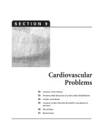
9
Cardiovascular
Problems
62: Coronary Artery Disease
63: Postmyocardial Infarction Care and Cardiac Rehabilitation
64: Cardiac Arrhythmias
65: Common Cardiac Disorders Revealed by Auscultation of
the Heart
66: Heart Failure
67: Hypertension
947
P1: PNW
GRBT129-62
GRBT129/Barker- 2568G GRBT129-Barker-v14.cls
May 6, 2006
948
11:34
Char Count= 0
P1: PNW
GRBT129-62
GRBT129/Barker- 2568G GRBT129-Barker-v14.cls
•◗
C h a p t e r
May 6, 2006
•
6 2
Coronary Artery Disease
Nisha Chandra-Strobos and
Glenn A. Hirsch
Pathogenesis
949
Risk Factors
950
Diagnosis
951
History
951
Physical Examination
952
Electrocardiography
952
Cardiac Stress Testing
953
Ambulatory Electrocardiography
955
Electron-Beam Computed Tomography
955
Cardiac Catheterization and Coronary Angiography
956
Computed Tomography Coronary Angiography
Treatment of Angina Pectoris
957
958
General Therapeutic Considerations
958
Lipids and Diet
958
Alcohol
959
Antioxidants
959
Fish Oil and ω-3 Fatty Acids
959
Postmenopausal Hormone Replacement Therapy
959
Physical Conditioning
960
Medical Treatment
960
Percutaneous Coronary Intervention
964
Surgical Management
965
Other Therapies
966
Unstable Angina
966
Variant Angina
967
Angina with Normal Coronary Arteries
967
Silent Ischemia
967
Coronary Artery Disease in Women
967
Summary
968
11:34
Char Count= 0
Chest pain is one of the most common complaints of patients in an ambulatory practice. The major early objective
in the diagnosis of patients with chest pain is separating
noncardiac from cardiac etiologies. Chapters 42 and 59
describe the various causes of noncardiac chest pain. This
chapter describes the pathogenesis of coronary artery disease (CAD) and its most common clinical symptom, angina
pectoris. Chapter 63 describes the posthospital medical
care and rehabilitation of patients who had a myocardial
infarction (MI).
CAD caused by atherosclerosis is one of the most common ailments in the Western world, and it remains the
leading nontraumatic cause of disability and death in the
United States. Increased public awareness and health education have reduced CAD mortality by >20% in the last
25 years. However, CAD still affects approximately
13,000,000 Americans. Cardiovascular disease accounts
for 38% of the total mortality in the United States or approximately the same number of deaths as the next five
leading causes combined (cancer, chronic lower respiratory diseases, accidents, diabetes mellitus, and influenza
and pneumonia). Of these cardiovascular deaths, coronary
heart disease accounts for 53% (1). Chest pain is one of the
most common presenting symptoms of patients with CAD
who seek medical attention. Health care providers must
understand the appropriate diagnostic evaluation and subsequent therapeutic options for patients with chest pain.
A detailed history and physical examination are essential
when evaluating patients with chest pain. They cannot be
replaced by sophisticated procedures; rather, they guide
the clinician in selecting the most appropriate diagnostic
evaluation.
PATHOGENESIS
CAD presents in a variety of ways, largely related to
the underlying pathophysiology of plaque formation and
atherosclerosis. The endothelium plays an integral role
in defending against atherosclerosis, modulating vascular
tone, and preventing intravascular thrombosis. These endothelial functions are adversely affected by CAD risk factors, even before the development of overt atherosclerosis.
In the earliest stages of disease, circulating monocytes adhere to vascular endothelial cells (via adhesion molecules)
and migrate into the intima of the blood vessel, where they
949
P1: PNW
GRBT129-62
GRBT129/Barker- 2568G GRBT129-Barker-v14.cls
950
May 6, 2006
11:34
Section 9 / Cardiovascular Problems
ingest oxidatively modified low-density lipoprotein (LDL)
and become trapped as foam cells. Collections of foam
cells, known as fatty streaks, may be present even in early
childhood. Foam cells die, leading to the development of
a lipid core. Smooth muscle cells are signaled to migrate
from the media, destroying the internal elastic lamina of
the vessel in the process. Calcification of the plaque occurs early and can be visualized noninvasively by electronbeam computed tomography (EBCT; see later discussion).
The arterial wall progressively thickens and remodels. Encroachment of plaque into the lumen of a coronary artery
occurs late in the atherosclerotic process, reflecting advanced disease. Arterial cross-sectional area is reduced by
approximately 40% before a lesion is visible as “significant”
CAD on catheterization, a finding demonstrated by use of
in vivo intravascular ultrasound (2).
Atherosclerotic progression is accelerated by three processes: endothelial dysfunction, inflammation, and thrombosis. Advanced lesions may be calcified and fibrotic, but
more concerning are plaques that have a core of lipid and
necrotic tissue surrounded by a thin fibrous cap. This cap
contains collagen, and its characteristics are closely related to the risk of plaque rupture, the major cause of
acute coronary syndromes. Specifically, a thinner fibrous
cap is more likely to rupture. A ruptured plaque exposes
the highly thrombogenic underlying collagen matrix and
leads to rapid thrombus formation. Complete occlusion
of a coronary vessel by thrombus on a ruptured plaque
typically causes an acute transmural MI characterized by
ST-segment elevation on the electrocardiogram (ECG).
Nonocclusive thrombus can cause unstable angina or an
MI without ST-segment elevation. Nonocclusive thrombus
may not cause symptoms but instead may change plaque
geometry and lead to rapid plaque growth.
MIs are classified by their appearance on 12-lead ECG
during the acute phase as either ST-segment elevation
or non–ST-segment elevation and are treated differently
(3–6). It is important to recognize that an acute MI often arises from rupture of an atherosclerotic plaque that
caused <50% luminal reduction by angiography prior to
plaque rupture (7,8). On the other hand, a coronary artery
that is narrowed by ≥70% is more likely than is a less
severe narrowing to cause exertional angina. The discordance between plaque severity and the development of an
acute MI indicates that coronary disease is not simply a
mechanical problem but instead occurs as the end result
of the interplay between mechanical stresses, inflammation, cholesterol deposition, and thrombosis.
Most patients with classic exertional angina by history
have fixed atherosclerotic lesions of ≥70% in at least one
major coronary artery. Fundamentally, angina is caused
by a mismatch between myocardial oxygen supply and demand. Supply is affected by coronary perfusion pressure,
coronary vascular resistance, and the oxygen-carrying capacity of blood. Flow is autoregulated over a wide vari-
ety of perfusion pressures; therefore, most of the changes
in flow result from changes in resistance (i.e., vasodilation). However, the coronary bed beyond a significant flowlimiting stenosis already is maximally vasodilated such
that small increases in demand (e.g., increased heart rate
and blood pressure during exercise) may result in myocardial ischemia. Oxygen demand is related to heart rate,
systolic blood pressure, and wall tension. Wall tension is
determined by ventricular pressure, cavity size, and wall
thickness. Physical exertion and emotional stress have potent effects on these variables and, not coincidentally, are
the common triggers for ischemic chest pain.
RISK FACTORS
Both genetic and environmental risk factors influence the
development of atherosclerotic heart disease. The recognition of risk factors is especially important because many
of these conditions can be modified to prevent disease.
Landmark epidemiologic surveys, such as the Framingham Heart Study, have helped to define levels of risk for
individual risk factors. Treatment guidelines have been
revised to include the important interactions between
individual risk factors and age. Risk calculators (CAD
event risk over 10 years) are available on the Internet at
The
27th Bethesda Conference was designed to bring attention to specific patients at high risk for development of
CAD events (9). This work has been incorporated into the
National Cholesterol Education Program (NCEP) Expert
Panel on Detection, Evaluation and Treatment of High
Blood Cholesterol in Adults (Adult Treatment Panel III
[ATP-III]) (see Chapter 82) (10). The concepts of “risk”
and “risk factor” are important in understanding and using the guidelines. The Bethesda Conference outlined four
categories of risk based on observational studies and efficacy studies (clinical trials). Table 62.1 summarizes these
risk factors.
Category I risk factors are those for which interventions
have been proven to reduce the risk of CAD events. They
include smoking, elevated LDL cholesterol, diet high in
saturated fat, hypertension, left ventricular hypertrophy,
and “thrombogenic factors,” which are unnamed but have
the potential of being reduced by aspirin.
Category II risk factors are those for which interventions
are likely to lower CAD risk. They include diabetes mellitus,
physical inactivity, low levels of high-density lipoprotein
(HDL) cholesterol, increased levels of triglycerides, obesity, and postmenopausal estrogen deficiency. Since the
publication of these findings, diabetes has been reclassified as a CAD “risk equivalent” based on data suggesting
that diabetic patients without known CAD have survival
rates similar to those of nondiabetic patients who have experienced an MI. The ATP-III guidelines focus attention
Char Count= 0
P1: PNW
GRBT129-62
GRBT129/Barker- 2568G GRBT129-Barker-v14.cls
May 6, 2006
11:34
Chapter 62 / Coronary Artery Disease
◗ TABLE 62.1
Risk Factors for Cardiovascular
Disease
Category I (Factors for which Interventions Have Been Proved to
Lower CVD Risk)
Cigarette smoking
Elevated LDL cholesterol
High-fat/high-cholesterol diet
Hypertension
Left ventricular hypertrophy
Thrombogenic factors (as affected by aspirin)
Category II (Factors for which Interventions Are Likely to Lower
CVD Risk)
Diabetes mellitus
Physical inactivity
Low levels of HDL cholesterola
Elevated triglycerides
Small, dense LDL particle size
Obesity
Postmenopausal status (women)
Category III (Factors Associated with Increased CVD Risk That,
if Modified, Might Lower Risk)
Psychosocial factors
Elevated lipoprotein (a)
Elevated homocysteine
Oxidative stress
No alcohol consumption
Category IV (Factors Associated with Increased Risk That
Cannot Be Modified)
Age
Male gender
Low socioeconomic status
Family history of early-onset coronary artery disease
a May now be considered a category I risk factor; see text.
CVD, cardiovascular disease; HDL, high-density lipoprotein LDL;
low-density lipoprotein.
Adapted from Pasternak RC, Grundy SM, Levy D, et al. 27th Bethesda
Conference: matching the intensity of risk factor management with the
hazard for coronary disease events. Task Force 3. Spectrum of risk
factors for coronary heart disease. J Am Coll Cardiol 1996;27:978.
on the “metabolic syndrome,” which incorporates abdominal obesity, atherogenic dyslipidemia (elevated triglycerides, small LDL particles, low HDL cholesterol), elevated
blood pressure, insulin resistance (with or without glucose intolerance), and prothrombotic and proinflammatory states. Patients with this syndrome now are appropriately targeted for intensive risk factor modification. Low
HDL cholesterol, with the publication of the Veterans Affairs High-Density Lipoprotein Intervention Trial (VA-HIT)
(11), now may be considered a category I risk factor, because an intervention to raise HDL cholesterol (i.e., with
gemfibrozil) in this trial reduced the incidence of cardiovascular events (12). Although postmenopausal status correctly identifies a cardiac risk factor, evidence from randomized trials demonstrates that hormone replacement
therapy may actually increase the risk of cardiovascular
Char Count= 0
951
events and therefore is not recommended for treatment or
prevention of CAD (13,14).
Category III risk factors are those associated with increased CAD risk that may, if modified, lower risk. These
include the “emerging” risk factors such as depression,
elevated lipoprotein (a) levels, and hyperhomocysteinemia. This list probably should be expanded to include
inflammatory markers (elevated white blood cell count,
high-sensitivity C-reactive protein, serum fibrinogen, soluble adhesion molecules), thrombotic risk factors (plasminogen activator inhibitor-1), and sleep apnea. Coronary
calcification as measured by EBCT (15) can correctly be
considered a category III risk factor for now, but it may
need to be reclassified (like diabetes mellitus) as a CAD
risk equivalent because it is a measure of the subclinical
coronary artery plaque burden.
Category IV risk factors are those that are associated with
increased risk but cannot be modified. They include age,
male gender, low socioeconomic status, and family history
of early-onset CAD. Positive family history has been defined as CAD in a male first-degree relative younger than
55 years or in a female first-degree relative younger than
65 years. These factors usually are taken into consideration
with the available risk scoring systems.
DIAGNOSIS
History
Character and Location of Ischemic Pain
The discomfort of myocardial ischemia can be described
in a variety of ways. Classically, the term angina pectoris
describes a “strangulation of the chest,” a helpful point to
remember because many individuals describe something
other than “pain” and instead mention chest tightness or
heaviness. Often it is more effective to ask the patient to
describe the discomfort. Some patients may simply hold
their clenched fist in the middle of their chest (Levine sign).
Angina typically begins and ends gradually over 2 to
5 minutes and usually is steady in character, although occasionally it waxes and wanes. If ischemic pain continues
for >20 minutes, myocardial necrosis (i.e., an MI) is more
likely to have occurred. The discomfort of angina pectoris
usually is midline and substernal, sometimes with radiation to the shoulder, arm, hand, or fingers, usually to the
left. Radiation down the inside of the arm into the fingers supplied by the ulnar nerve is classic. Pain also may
radiate into the neck, lower jaw, or interscapular region.
Occasionally, a patient has pain only in a referred location and experiences no chest discomfort at all. The pain
of myocardial ischemia is diffuse and cannot easily be localized. Rarely is the patient able to point with one finger
to the location. When pain can be localized in this way,
it likely is noncardiac in origin. The elderly, especially the
P1: PNW
GRBT129-62
GRBT129/Barker- 2568G GRBT129-Barker-v14.cls
952
May 6, 2006
11:34
Section 9 / Cardiovascular Problems
frail elderly, are more likely than are younger patients to
experience atypical symptoms such as dyspnea, confusion,
or dyspepsia rather than pain.
The Canadian Cardiovascular Society (CCS) Classification System was designed to provide a simple way of
grading anginal symptoms (16). Class I angina occurs with
strenuous, rapid, or prolonged exertion but not with ordinary physical activity. Patients with class II angina experience slight limitation of ordinary activity. Class II angina
occurs on walking or climbing stairs rapidly; walking uphill; walking or climbing stairs after a meal, in cold, or in
wind; or under emotional stress. Class III angina produces
marked limitations of ordinary physical activity. Angina
occurs on walking one or two blocks on level terrain or
climbing one flight of stairs under normal conditions and
at a normal pace. With class IV angina, the most severe
type, the patient is unable to carry on any physical activity without discomfort, and anginal symptoms may be
present at rest. A higher CCS class is associated with more
extensive CAD and a higher risk of CAD events.
Precipitating Factors
The single most important diagnostic feature of the discomfort of myocardial ischemia is its predictable relationship to exertion, emotional stress, or other situations that
may either increase myocardial oxygen demand or reduce
supply. The cause of atypical pain, pain in an unusual location or of an unusual character, may be clarified by this
relationship. Pain that is experienced at rest, if it is caused
by ischemia, suggests unstable angina or MI.
Anxiety and mental stress are important and often overlooked provoking factors in many patients. Angina is more
likely to occur during cold or windy weather because of increased peripheral vascular resistance and, consequently,
increased myocardial work. Other triggers include sexual
intercourse or a heavy meal.
Relief of Ischemic Pain
Because angina is fundamentally caused by a discrepancy between oxygen supply and demand, relief of pain
is achieved by increasing coronary blood flow or decreasing oxygen demand. Most people must stop or at least slow
the activity responsible for precipitating the pain before it
is relieved. Angina often is relieved by sublingual nitroglycerin, but the practitioner and the patient both need to
realize that relief of chest pain by nitroglycerin is not specific for myocardial ischemia (17). For example, the pain of
esophageal spasm can also be relieved by nitroglycerin.
Physical Examination
The physical findings in patients with CAD are nonspecific. A complete cardiovascular examination should focus
on identifying markers of hypertension and dyslipidemia,
peripheral vascular disease, or diabetes mellitus. Severe
aortic valve disease (stenosis or regurgitation) or pulmonary hypertension without CAD can cause angina pectoris either from left or right ventricular wall strain,
respectively, leading to myocardial ischemia.
Electrocardiography
A 12-lead ECG should be obtained as soon as possible in
a patient with suspected CAD, although in many cases the
ECG is completely normal. The most reliable ECG sign of
chronic ischemic heart disease is the presence of a prior MI
as manifested by two or more pathologic Q waves in a particular myocardial territory (e.g., anterior, lateral, inferior,
etc.) (Fig. 62.1A). The differential diagnosis of Q waves on
ECG includes prior MI, healed myocarditis, hypertrophic
cardiomyopathy, an infiltrative myocardial disorder such
as amyloidosis or sarcoidosis, and Wolff-Parkinson-White
syndrome (usually with characteristic findings of preexcitation; see Chapter 64). Nonspecific ST-T wave changes,
conduction abnormalities (except for left bundle-branch
block [LBBB], discussed later), and arrhythmias do not
help establish the diagnosis of myocardial ischemia. However, ST-segment depression with a flat or downsloping ST segment is suggestive of subendocardial ischemia
(Fig. 62.1B). It is seldom present on the resting ECG of
patients with ischemic heart disease unless they are experiencing angina at the time the tracing is recorded. On
the other hand, transient ischemic changes are seen commonly when a patient with CAD is exercised to a point
at which chest pain develops. Such ECG changes, appearing with exercise or pain and resolving with rest or with
the resolution of pain, usually are an indication of myocardial ischemia. Therefore, the necessity of repeating the
ECG at rest or after the chest pain has resolved cannot be
overemphasized. ST-segment elevation during chest pain
(Fig. 62.1C) suggests acute myocardial injury (e.g., MI) or
variant angina (discussed later). T-wave inversion on an
ECG taken at rest is a nonspecific finding but can occur
after infarction or as a specific transient finding in a patient experiencing angina. Therefore, ECG changes noted
during episodes of chest pain not only can confirm the
diagnosis of myocardial ischemia but also may indicate
the extent and location of the ischemic myocardium. As
a general rule, the more widespread the changes on ECG,
the greater the extent of myocardium that is involved. STsegment elevation in the absence of chest pain is common
on the resting ECG of healthy young adults and is caused
by rapid or “early” repolarization of the ventricle. This pattern (Fig. 62.1D) usually is noted in the mid–left chest
leads (V2 –V4 ) but may be more widespread. ST-segment
elevation from pericarditis is diffuse and can be associated with PR-segment depression in the limb leads (except
aVR, which may show PR-segment elevation).
Char Count= 0
P1: PNW
GRBT129-62
GRBT129/Barker- 2568G GRBT129-Barker-v14.cls
May 6, 2006
11:34
Chapter 62 / Coronary Artery Disease
Char Count= 0
953
A
B
C
D
FIGURE 62.1. Electrocardiographic strips from patients with suspected ischemic heart disease. A: Q
waves suggestive of prior myocardial infarction. B: ST-segment depression developing after exertion.
C: ST-segment elevation during coronary artery spasm (variant angina). D: Early repolarization
(a normal variant).
The presence of ST-T abnormalities in an otherwise
healthy person is a nonspecific finding and should not be
considered confirmation of CAD. There is a high association of LBBB with organic heart disease (see Chapter 64),
especially CAD. Right bundle-branch block (RBBB), on the
other hand, is seen commonly in the absence of other cardiac abnormalities.
Cardiac Stress Testing
Exercise Electrocardiography
The exercise stress test is a means of establishing the
diagnosis of myocardial ischemia. It also can be used
to assess the efficacy of antianginal therapy, to identify
patients who are likely to have more severe CAD and a
large area of myocardium at risk, and to assess serially
the degree of conditioning or exercise capacity in patients
of all age groups. The American College of Cardiology
(ACC)/American Heart Association (AHA) exercise testing
guidelines outline the recommendations for the use of
exercise testing in establishing the diagnosis of CAD, in
assessing risk and prognosis in patients with symptoms
or a prior history of CAD, and the use of exercise testing
after MI (18,19). The usefulness of exercise testing in establishing the diagnosis of CAD is based in part on the likelihood that the patient has this condition (i.e., the “pretest
probability” of CAD). This can be determined by the patient’s age, gender, and symptoms. For example, exercise
testing would not be expected to greatly improve the ac-
curacy of diagnosing CAD in an older patient with typical angina (who has a high pretest probability of CAD)
nor in a young, asymptomatic individual (who has a low
pretest probability of CAD). The usefulness of stress testing
in these situations would be limited by false-negative and
false-positive findings, respectively. The ACC/AHA guidelines recommend exercise testing to diagnose CAD in adult
patients with an intermediate pretest probability of CAD
based on gender, age, and symptoms (18,19). For patients
with known CAD, the guidelines recommend stress testing for those with a significant change in clinical status.
Patients with unstable angina, decompensated heart failure, severe aortic stenosis, or uncontrolled hypertension
should not be referred for stress testing because of an unacceptably high risk for provoking a cardiac event during
exercise.
Exercise stress testing is based on the rationale that,
as the work performed by the patient increases, cardiac
work is increased. The increased cardiac work results in increased myocardial oxygen utilization, with a subsequent
increased demand in coronary blood flow. If narrowed or
obstructed coronary arteries prevent the required increase
in coronary blood flow, myocardial ischemia may occur
and be manifested as chest pain and/or ECG changes (20).
The simplest and least expensive exercise stress test is
the graded, symptom-limited exercise treadmill test. The
test requires 12-lead ECG monitoring of the patient while
walking on a treadmill at workloads that can be progressively increased by increasing the speed and inclination of
the treadmill. A stationary bicycle ergometer (with hand
P1: PNW
GRBT129-62
GRBT129/Barker- 2568G GRBT129-Barker-v14.cls
954
May 6, 2006
11:34
Section 9 / Cardiovascular Problems
FIGURE 62.2. Algorithm for determining the appropriate stress
test. See text for a description of the procedures.
pedals) can be substituted for a treadmill, permitting the
patient to exercise with his or her arms instead of legs.
Although it is not commonly used, this method of stress
testing permits exercise by a patient who may otherwise
be unable to do so because of lower-extremity claudication, arthritis, or amputation. It also may be useful in
the evaluation of patients who have chest pain predominantly or exclusively with work that involves the arms and
shoulders.
A simple algorithm can be used to decide the type of
stress test to recommend (Fig. 62.2). First, the patient’s
ability to exercise should be assessed. If the patient can
walk up a flight of stairs carrying laundry or groceries,
for example, a treadmill exercise protocol can generally be
chosen to allow the patient to achieve a level of cardiac
work that permits meaningful information to be obtained
from the test. If the patient cannot perform this task, or
one that is comparable, a pharmacologic stress test with
cardiac imaging (discussed later) should generally be recommended. The patient’s baseline ECG should be reviewed
to determine the presence of baseline ST-segment abnormalities that might lower the predictive value of exerciseinduced changes. False-positive stress tests are often encountered in women, in patients taking medications such
as digoxin or amiodarone, and in patients with left ventricular hypertrophy or mitral valve prolapse (21). For these
patients and in those with baseline ST-segment abnormalities, intraventricular conduction defects (i.e., LBBB or
RBBB), or other conduction system disorders (e.g., WolffParkinson-White syndrome), the diagnostic accuracy of
the exercise stress test can be enhanced by concurrent radioisotopic or echocardiographic imaging (see later discussion). The choice between radioisotopic or echocardiographic imaging depends largely on the expertise of local
laboratories.
Radioisotope Imaging
Radioisotope imaging can enhance the specificity of stress
testing by evaluating myocardial function or flow (22). Radioisotope imaging can be used in conjunction with either treadmill exercise testing or pharmacologic stress testing, using either dobutamine to increase cardiac work or
adenosine or dipyridamole to alter coronary blood flow
(see later discussion). Commonly used imaging modalities
include radioisotope imaging with thallium 201 (201 Tl)–
and/or technetium 99 (99 Tc)–based agents (e.g., 99m Tcsestamibi). The usefulness of 201 Tl as a perfusion tracer
is based on its ability to function as an analogue of ionic
potassium. It is very efficiently extracted by healthy myocardial cells, and uptake is proportional to regional perfusion and myocardial viability. 99m Tc-sestamibi has a
shorter half-life (6 hours) than does 201 Tl (73 hours), allowing administration of a larger tracer dose. This and
its higher emission energy make it an excellent agent for
cardiac imaging. 99m Tc-sestamibi is particularly useful in
obese patients and in patients with large breasts (because
of possible attenuation of the radioisotopic images in the
area of the anterior myocardium).
Both 201 Tl and 99m Tc-sestamibi can be used to assess
regional myocardial blood flow, either by planar imaging or by single-photon emission computed tomography
(SPECT). Imaging usually occurs at two separate times:
the stress scan, obtained very shortly after the patient has
exercised or received a pharmacologic agent, and the rest
scan, obtained either before or several hours after stress.
The radioisotope is injected intravenously at the time of
peak exercise (or at the time of peak infusion during a
pharmacologic stress test), and scintigraphic images are
obtained shortly thereafter, depicting regional myocardial
perfusion at the time of peak stress. The rest scan typically
is obtained several hours later and shows redistribution
of the isotope. Ischemia is indicated by the filling in of a
cold spot defined on the stress images (i.e., normalization
or “redistribution” of a radioisotopic defect), and infarction is indicated by a persisting cold spot or one with only
partial redistribution.
Radioisotope imaging with stress gated blood pool
scans (multiple-gated acquisition [MUGA]) also can be
used to assess myocardial ischemia. To allow for continuous imaging during exercise, stress MUGA is performed
with the patient exercising on a semirecumbent bicycle. The rationale for this test is based on the fact that
myocardium that becomes ischemic during graded exercise develops regional wall-motion abnormalities that can
be detected by sequential image analyses. This type of
imaging labels the blood pool with a radioisotope and gates
image acquisition to the ECG. Right and left ventricular
volumes, regional left ventricular wall motion, and global
and regional ejection fractions can be measured, both at
rest and with stress.
The cost of stress testing with radioisotope scanning
usually is several times that of a standard exercise test.
Stress Echocardiography
Two-dimensional echocardiography can be used instead
of radioisotope scanning to detect areas of regional myocardial dysfunction (as evidenced by a wall-motion
Char Count= 0
P1: PNW
GRBT129-62
GRBT129/Barker- 2568G GRBT129-Barker-v14.cls
May 6, 2006
11:34
Chapter 62 / Coronary Artery Disease
abnormality) with exercise or pharmacologic stress
(23,24). Typically, baseline images are first obtained at
rest to determine the adequacy of the echocardiographic
images. If these images are technically inadequate (e.g.,
because of obesity or severe obstructive lung disease),
an intravenous ultrasound contrast agent can be used if
available; if not, radioisotope images are preferable. If the
rest images are technically adequate, the patient undergoes treadmill exercise stress and then images are reacquired immediately, using special software to allow for
direct comparison of pre-exercise and postexercise images.
If pharmacologic stress testing with dobutamine (see Pharmacologic Stress Testing) is used, the dose of dobutamine
is increased in stepwise fashion, and echocardiographic
images typically are obtained each time the dose is increased. The safety of dobutamine stress echocardiography is comparable to that of a routine exercise stress test
(23,25,26). The sensitivity, specificity, and cost of the test
are similar to those of radioisotopic stress testing. Stress
echocardiography may be preferred in some cases because
additional information is provided that is not obtained
with radioisotopic scanning (e.g., presence of pericardial
effusion, ventricular hypertrophy, or valvular abnormality). It also avoids exposure to radioactivity.
Pharmacologic Stress Testing
Patients who are unable to exercise because of physical limitations can be evaluated after intravenous administration of dipyridamole, adenosine, or dobutamine in
conjunction with an imaging modality. Dipyridamole and
adenosine dilate all coronary vessels and generally increase
flow to all areas of the heart. Enhanced dilation of normal coronary arteries, compared to that of significantly
narrowed vessels, augments differences in flow that usually are not apparent at rest. These agents are suitable for
use with radioisotopic imaging modalities that may readily demonstrate this flow heterogeneity. After administration of dipyridamole or adenosine followed by either 201 Tl
or 99m Tc-sestamibi (i.e., the stress image), myocardium
supplied by a narrowed coronary artery typically demonstrates a perfusion defect that “fills in” during the rest image. Because of its ultrashort duration of action, adenosine
is preferable to dipyridamole for this test.
Dobutamine is a β 1 -receptor agonist that at high
dosages (20–40 μg/kg/min intravenously) increases
myocardial contractility and heart rate in a similar manner and extent to exercise. Heart rate may not be affected
to the same extent as contractility, and atropine often is
administered intravenously to increase the heart rate to
the maximal predicted heart rate for age. Dobutamine
can be used in conjunction with either echocardiography
or radioisotopic imaging for diagnosis of CAD.
Mild side effects (e.g., nausea, flushing, and headache)
are common with dipyridamole, adenosine, and dobutamine. Dipyridamole and adenosine (but not dobutamine)
Char Count= 0
955
can produce severe bronchospasm and therefore must be
used with caution or not at all in patients with asthma
or chronic obstructive pulmonary disease. Adenosine can
cause transient heart block, typically lasting several seconds. Because dobutamine increases atrioventricular conduction, it should not be used in patients with atrial flutter
and should be used carefully in patients with atrial fibrillation.
Implications of an Abnormal Stress Test
If treadmill exercise stress testing is performed, factors
affecting prognosis include the degree of ST-segment depression, time to development of ST-segment depression
during exercise, duration of the ST-segment depression in
recovery, and speed of heart rate decline during recovery.
In addition, an ischemic ECG response that is accompanied by hypotension generally implies a large amount of
myocardium at risk. Prognostic information from pharmacologic stress testing-induced ECG abnormalities is less
reliable. The number, size, and location of abnormalities
evident on stress imaging studies reflect the location and
extent of functionally significant coronary stenoses (27).
Both radioisotopic and echocardiographic imaging can
detect left ventricular dilation with stress, a finding that
suggests global, severe ischemia. Lung uptake of a radioisotopic tracer indicates stress-induced left ventricular
dysfunction and suggests multivessel CAD. Many studies
have shown that high-risk abnormal stress tests are associated with an increased risk for cardiac events. On the
other hand, normal radioisotopic or echocardiographic
stress tests are associated with a favorable prognosis. In
a review of 16 studies involving almost 4,000 patients over
2 years, a negative perfusion scan was associated with a
0.9% rate of cardiac death per year, similar to that of the
general population (28).
Ambulatory Electrocardiography
The ambulatory ECG (Holter monitor) may be useful
for detecting myocardial ischemia. However, it is not a
good tool for screening patients to make the diagnosis
of CAD. In patients with CAD who are symptomatic during ambulatory ECG monitoring, ST-segment elevation
or depression can be observed during episodes of pain
and at other times as well (silent ischemia; see later discussion). In patients with silent ischemia, the ambulatory ECG is particularly useful for quantifying the degree
and frequency of ischemia and assessing the efficacy of
therapy.
Electron-Beam Computed Tomography
Studies in the 1970s demonstrated that coronary calcification (detected by cardiac fluoroscopy) was useful in identifying patients with angiographically significant CAD (29).
P1: PNW
GRBT129-62
GRBT129/Barker- 2568G GRBT129-Barker-v14.cls
956
May 6, 2006
11:34
Char Count= 0
Section 9 / Cardiovascular Problems
A
B
FIGURE 62.3. Anatomic representation of the coronary arteries. These vessels are represented as
they would be seen on the angiogram. No attempt has been made to convey the third dimension.
Careful study of the changes in position of the various branches with rotation of the heart is essential
to intelligent interpretation of arteriograms. A: Anteroposterior. B: Lateral. (Modified from Abrams HL,
Adams DF. The coronary arteriogram: structural and functional aspects [First of two parts]. N Engl J
Med 1969;281:1276, with permission.)
EBCT is a highly sensitive technique for detecting coronary
artery calcium and may be useful for diagnosing CAD noninvasively (11). ECG gating allows data acquisition within
one or two breath-holds, making it a rapid test with limited
radiation exposure. The images obtained by this technique
allow the determination of a calcium score, which is an
index of calcium deposition in multiple arterial segments
and is a good approximation for overall plaque burden
in the coronary tree. High calcium scores are associated
with increased risk for MI (30). The test offers improved
discrimination over conventional risk factors in the identification of people with CAD (31). The negative predictive
value of EBCT is high. The test is particularly useful for
screening asymptomatic individuals with multiple risk factors, in whom an abnormal EBCT should prompt further
testing and/or treatment. A very low EBCT score would be
reassuring (32).
Cardiac Catheterization and
Coronary Angiography
Coronary angiography is defined as the radiographic visualization of the coronary vessels after injection of radiopaque contrast medium (33). This technique provides
direct information about the presence of CAD and defines
the distribution and severity of obstructive coronary lesions. It is considered the “gold standard” to confirm the
diagnosis of CAD. The images obtained are stored as either 35-mm cine film or, more commonly, a digital recording. Percutaneous or cutdown techniques of the femoral or
brachial arteries allow insertion of sheaths for the intro-
duction of selective catheters for the right and left coronary
ostia, saphenous bypass grafts, or internal mammary arteries. Arteriography is performed as part of cardiac catheterization, which may include left ventriculography and
hemodynamic assessment. Figure 62.3 shows diagrammatically the coronary arteries and their branches as they
appear on coronary arteriography. The three major coronary arteries are the left anterior descending, left circumflex, and right coronary artery. The coronary tree can be
divided into 29 segments, but the extent of disease usually is defined as one-vessel, two-vessel, three-vessel, or left
main disease, with significant disease taken to mean the
presence of ≥50% reduction in diameter (some operators
and texts use ≥70% reduction in diameter).
The 1999 ACC/AHA Guidelines for Coronary Angiography outline the indications and contraindications for
the procedure (33). The guidelines recommend arteriography for patients with CCS class III or IV angina while
receiving medical treatment (marked limitations of ordinary physical activity because of angina or angina at rest,
discussed earlier) and those with high-risk criteria on noninvasive testing regardless of angina severity. It may be reasonable to consider coronary arteriography for patients
whose angina has improved with medical treatment but
remains present, those in whom noninvasive testing has
shown evidence of worsening disease, those who cannot
tolerate medical therapy, those with angina who cannot be
adequately risk stratified because of disability or illness,
and those whose occupation involves the safety of others
(e.g., pilots, bus drivers) and who have abnormal, but not
high-risk, stress test results.
P1: PNW
GRBT129-62
GRBT129/Barker- 2568G GRBT129-Barker-v14.cls
May 6, 2006
11:34
Chapter 62 / Coronary Artery Disease
Inherent in the recommendation for coronary arteriography is the assumption that the patient is a potential
candidate for coronary revascularization. If the patient’s
general medical condition or other medical problems preclude revascularization, or if the patient refuses to consider
revascularization regardless of catheterization results, arteriography is ill advised.
Indications for percutaneous coronary intervention
([PCI] including angioplasty and stenting) (34) and coronary artery bypass surgery (35) are reviewed in separate ACC/AHA guidelines and are discussed later in this
chapter.
Patient Experience. The patient may undergo cardiac
catheterization as part of an evaluation during a hospitalization, but the test itself does not require that the
patient be admitted to the hospital. The procedure is
not painful, and the patient remains awake throughout the study. Approximately 1 hour before the procedure, the patient is given a sedative, often diazepam
(Valium), 5 to 10 mg orally. After the patient is brought
to the catheterization laboratory, either the area of the
brachial artery or the femoral artery is prepared for sterile procedure. The site of introduction of the catheter
usually is selected based on the preference of the operator but also is guided by the presence and extent of
peripheral vascular disease. Typically, a catheter is
introduced percutaneously through a wire that is
threaded through an introducer needle. Under fluoroscopic guidance, the catheter is threaded to the coronary sinuses, and the orifices of the right and left coronary arteries are injected sequentially with contrast
medium. The patient is asked to hold his or her breath
during the few seconds of the injection. In addition to
this part of the test, which visualizes the coronary arteries, studies are typically performed to measure ventricular pressures and to assess left ventricular contraction
during injection of dye directly into the left ventricular cavity. During ventriculography, focal wall-motion
abnormalities, ventricular aneurysms, and valvular lesions such as mitral regurgitation can be assessed in
addition to the measurement of overall left ventricular
function and ejection fraction. At the end of the procedure, the catheter is withdrawn, and pressure is applied
to the arteriotomy site to achieve hemostasis.
During the procedure, the patient should feel relaxed or
even slightly drowsy from the sedation. The patient usually
does not feel pain except for the moment when the needle is
initially introduced. There is some pressure as the catheter
is held in place. The patient may experience a sensation of
hot flushing when the dye is injected, particularly when the
larger bolus of dye is injected into the left ventricle during
ventriculography.
Risks and Relative Contraindications
The major complications of coronary arteriography are
MI, stroke, and death. These risks are related to the ex-
Char Count= 0
957
perience of the laboratory performing the study and to the
risk profile of the patient undergoing the test. Risks tend
to be lower in young, otherwise healthy patients. Risks
tend to be higher in older patients with poor left ventricular function, diabetes mellitus, or peripheral vascular disease, and those who are clinically unstable (e.g., patients
with cardiogenic shock, recent acute MI, or decompensated heart failure) at the time of the procedure. In a survey of almost 60,000 patients, mortality from angiography
was 0.11%, MI occurred in 0.05%, and stroke occurred in
0.07%. The most common complication was a problem
with vascular access, which occurred in 0.43% of patients
(36).
There are no absolute contraindications to coronary arteriography. Relative contraindications include renal failure, active gastrointestinal bleeding, acute stroke, severe
anemia, coagulopathy, unexplained fever or active untreated infection, severe uncontrolled hypertension, allergic reaction to angiographic contrast agents, and
decompensated congestive heart failure (CHF). Renal insufficiency has been the most well-studied complication.
It occurs in up to 5% of patients without preexisting renal dysfunction and in 10% to 40% of patients with baseline renal insufficiency. More than 75% of patients who
develop renal insufficiency recover normal renal function,
although 10% of these patients may require dialysis temporarily. Pretreatment with intravenous hydration (0.9%
saline) (37) and limiting the amount of intravenous contrast material used are effective means to avoid contrastinduced renal dysfunction.
For patients with underlying renal dysfunction, pretreatment with N-acetylcysteine (38) or intravenous
sodium bicarbonate (39) has been shown to reduce
contrast-induced acute renal failure following cardiac
catheterization. No direct comparison of these prophylactic measures has been performed to date. Patients taking
the oral hypoglycemic metformin should be asked to withhold it for 48 hours prior to the procedure, because the use
of iodinated contrast dye in patients taking metformin has
been associated with development of lactic acidosis (40).
The major predictors of contrast allergy are prior contrast
allergy (50% risk of subsequent reaction), iodine allergy,
and shellfish allergy. These conditions should be discussed
with the patient before referral for angiography. The use
of nonionic contrast medium along with pretreatment using corticosteroids and antihistamines may reduce allergic
complications.
Computed Tomography
Coronary Angiography
High-definition rapid CT scanning has evolved as a potent
diagnostic tool for identifying CAD noninvasively. Newer
CT devices are able to rapidly scan through a patient’s
chest using many slices for image acquisition (the current
P1: PNW
GRBT129-62
GRBT129/Barker- 2568G GRBT129-Barker-v14.cls
958
May 6, 2006
11:34
Section 9 / Cardiovascular Problems
state of the art is to use a 64-slice scanner), quickly and
accurately identifying unique features of coronary and cardiac anatomy. Multislice cardiac CT scanning is extremely
accurate in detecting coronary narrowings in the proximal
two thirds of the coronary tree that are demonstrated by
conventional coronary angiography, but its resolution of
the distal third is less accurate. However, it is superior to
conventional coronary angiography in identifying extraluminal vascular abnormalities that result in coronary narrowings but cannot be seen by conventional techniques.
Additionally, other noncardiac causes of chest pain, such
as aortic dissection, pneumonia, or pulmonary embolus,
may be diagnosed by this imaging technique. CT coronary angiography is particularly useful in patients with
peripheral vascular disease because it can minimize or
avoid catheter-related complications. During CT angiography, the patient receives intravenous radiographic contrast, and the total scanning time usually is ≤15 minutes.
Image quality is improved at slower heart rates and patients may receive low doses of β-blockers to facilitate
this. Because iodinated contrast is used for this procedure, the risks and precautionary treatment associated
with such therapy is the same as for cardiac catheterization.
TREATMENT OF ANGINA PECTORIS
General Therapeutic Considerations
In evaluating and treating patients with angina, it is of
paramount importance to identify and treat underlying
contributing factors and to modify cardiac risk factors that
promote CAD progression if possible.
Hypertension often is present in patients with angina.
There is a linear relationship between left ventricular work
and myocardial oxygen demand. Left ventricular systolic
pressure increases in response to an increase in peripheral
vascular resistance. Both systolic and diastolic hypertension can increase myocardial oxygen demand. An attempt
should always be made to reduce resting blood pressure to
normal in patients with chronic hypertension, including
those with isolated systolic hypertension. This can be of
crucial importance in reducing the frequency and severity
of angina pectoris in the hypertensive patient. β-Blockers
and calcium channel blockers (see Chapter 67) are excellent choices in such patients because these agents have
other antianginal properties as well. Agents such as hydralazine and minoxidil, which cause a reflex tachycardia,
are less desirable.
It is important to achieve a maximal level of pulmonary
compensation in patients with angina and coexisting lung
disease (see Chapter 60). Chronic hypoxemia, acidosis,
and the increased work of breathing in patients with pulmonary disease increase myocardial oxygen demand, de-
crease myocardial oxygen delivery, or both. Unfortunately,
the treatment of angina in patients with severe lung disease often is limited by a real, or perceived, need to avoid
the use of β-blockers (see later discussion).
Abstinence from tobacco products is essential because
nicotine in tobacco can cause coronary vasoconstriction. Chapter 27 describes techniques used to achieve
this goal. Similarly, passive tobacco smoke should be
avoided.
The possibility of hyperthyroidism (see Chapter 80) in
patients with angina should never be overlooked, particularly in older patients or in those with increasing angina.
Often, particularly in the older patient, other obvious
signs of hyperthyroidism are not present. For example,
hyperthyroidism may be manifested only by an increased
frequency or severity of angina, an increase in heart
rate in people with atrial fibrillation, or increasing heart
failure.
Anemia is important to consider in patients with angina,
particularly if the hemoglobin concentration falls to
<7 g/dL, when cardiac output must increase to maintain adequate peripheral oxygen delivery at rest. Obviously,
this problem is exacerbated in patients with concomitant
chronic lung disease and hypoxemia.
Heart failure (see Chapter 66) in patients with angina
should always be optimally treated. The real possibility
that heart failure is producing angina at rest (see later
discussion) or nocturnal angina should be considered. Diuretics, vasodilators, and β-blockers may be useful in patients with rest or nocturnal angina and may reduce the
frequency and severity of angina. The calcium channel
blocker amlodipine has been shown to be safe in patients
with left ventricular dysfunction and may be useful for patients with angina in this setting because it has little negative inotropic effect, reduces preload and afterload, helps
decrease left ventricular end-diastolic pressure, and lowers
peripheral vascular resistance.
Lipids and Diet
Most of the recent decline in mortality from heart disease is
believed to be related to primary and secondary risk factor
reductions (41,42). Numerous randomized controlled trials involving cholesterol reduction have been performed
and have supported the ability to reduce CAD morbidity
and mortality with both primary and secondary prevention strategies. The West of Scotland Coronary Prevention Study demonstrated significant mortality reduction
with treatment of hyperlipidemia with pravastatin in
asymptomatic people; the greatest benefit occurred in patients with other risk factors for CAD (43). The landmark
Heart Protection Study in the United Kingdom randomized subjects with CAD, peripheral vascular disease, or
diabetes to 40 mg of simvastatin or placebo and demonstrated reductions in mortality in simvastatin-treated
Char Count= 0
P1: PNW
GRBT129-62
GRBT129/Barker- 2568G GRBT129-Barker-v14.cls
May 6, 2006
11:34
Chapter 62 / Coronary Artery Disease
patients regardless of baseline LDL cholesterol levels (44).
The value of secondary prevention was established by
the Scandinavian Simvastatin Survival Study (45) and
the Cholesterol and Recurrent Events (CARE) trial (46).
Both trials demonstrated a significant reduction in mortality when LDL cholesterol levels were lowered to approximately 100 to 120 mg/dL. Other trials also have
clearly demonstrated that coronary artery lesions did not
progress when elevated LDL cholesterol levels were reduced to <100 mg/dL (47,48). More recent trials have
compared the effects of more aggressive to less aggressive lipid-lowering strategies, usually by examining
the effects of high-dose and lower-dose therapy with a
β-hydroxy-β-methylglutaryl-coenzyme A (HMG-CoA) reductase inhibitor or “statin.” One of these trials demonstrated that 80 mg of atorvastatin reduced the frequency of
cardiovascular events to a greater degree than did 40 mg of
pravastatin by more intensive lowering of LDL cholesterol
(mean LDL cholesterol lowered to 62 mg/dL) (49). Another
study compared the effects of 80 mg and 10 mg of atorvastatin in patients with stable CAD and demonstrated
clinical benefit with the more aggressive lipid-lowering approach, achieving mean LDL cholesterol levels of 77 mg/dL
and 101 mg/dL in the 80-mg and 10-mg groups, respectively (50).
The NCEP guidelines indicate that the desirable LDL
cholesterol level is <100 mg/dL in patients with established CAD or with coronary heart disease risk equivalents
including diabetes mellitus, multiple risk factors that confer a 10-year CAD risk >20%, or other clinical forms of
atherosclerotic disease (i.e., peripheral arterial disease, abdominal aortic aneurysm, or symptomatic carotid artery
disease) (10). Updates recommend considering an LDL
cholesterol target <70 mg/dL in very-high-risk patients,
defined as those with an acute coronary syndrome or with
established CAD and multiple major CAD risk factors (especially diabetes mellitus), severe and poorly controlled
risk factors (especially cigarette smoking), or the metabolic
syndrome (51). Treatment of patients having low HDL
cholesterol levels with the fibrate gemfibrozil was shown to
reduce the risk of major cardiovascular events in patients
with CAD (12). In addition to pharmacologic options for
lipid-lowering drug therapy, the guidelines recommend a
multifaceted lifestyle approach to reduce CAD risk. This
approach calls for reducing the intake of saturated fats
to <7% of total calories and reducing dietary cholesterol
to <200 mg/day. Achieving an ideal body weight and increasing physical activity also are advised. These lifestyle
recommendations are an essential part of treatment for all
patients with coronary disease. Chapter 82 discusses these
changes in more detail. Obesity has emerged as a national
epidemic, with several studies confirming the increased
mortality and morbidity from this condition (52). Chapter 83 discusses in detail the various treatment options
for this condition.
Char Count= 0
959
Alcohol
Alcohol is an acute pressor agent and may be responsible for as many as 10% of all cases of hypertension (53).
However, moderate drinking (1–3 drinks per day) is accompanied by an increase in HDL cholesterol level (54).
The extent to which the increase in blood pressure associated with heavy drinking mitigates the beneficial effect on
HDL remains to be determined (55). A review of lifestyle
recommendations for patients with CAD estimated a 20%
reduction in mortality with moderate alcohol use (compared with 24% reduction from physical activity and 36%
reduction with smoking cessation) (56).
Antioxidants
Although antioxidants may be important in inhibiting
atherosclerosis, clinical trials of antioxidant therapy have
not demonstrated conclusive long-term benefit. In the
Heart Outcomes Prevention Evaluation (HOPE) study, for
example, approximately 9,500 patients at high risk for cardiovascular events were randomly assigned to therapy with
either 400 IU of vitamin E or placebo for an average of
4.5 years. There was no apparent effect of treatment with
vitamin E on cardiovascular outcomes in this study (57).
More recently, a meta-analysis of 19 trials suggested the
possibility of increasing mortality with high-dosage vitamin E supplementation for CAD prevention, with risk increasing as the dosage of vitamin E exceeded 150 IU/day
(58).
Fish Oil and ω-3 Fatty Acids
Fish oils (ω-3 fatty acids) have demonstrated cardiovascular benefit in people who have taken them by decreasing
the risk of potentially fatal arrhythmias, slowing plaque
progression, decreasing levels of triglycerides, and mildly
decreasing blood pressure. Currently, the AHA recommends two servings of fish per week. Similarly, other foods
that contain α-linolenic acid, which can be metabolized
into ω-3 fatty acids by the body, such as flaxseed, walnuts,
soy products, and tofu, are recommended, but the benefit
of ω-3 fatty acid production via α-linolenic acid intake is
not well delineated (59).
Postmenopausal Hormone
Replacement Therapy
Earlier studies demonstrated improvements in surrogate
measures such as endothelial function from hormone replacement therapy (HRT). Observational studies suggested
a decreased risk for cardiovascular events in women taking HRT compared to women who did not (60,61). This
finding led to two randomized, placebo-controlled studies
to definitively evaluate the role of HRT in postmenopausal
women with chronic stable CAD. The Heart and Estrogen/
P1: PNW
GRBT129-62
GRBT129/Barker- 2568G GRBT129-Barker-v14.cls
960
May 6, 2006
11:34
Section 9 / Cardiovascular Problems
Progestin Replacement Study (HERS) showed that HRT
did not result in a reduced risk for cardiovascular death
or nonfatal MI (13). The Estrogen Replacement and
Atherosclerosis Study (ERAS) failed to show an effect
of HRT on the angiographic progression of atherosclerotic heart disease (62). There also is evidence that postmenopausal HRT increases the risk of venous thromboembolic disease (13,63) and gallbladder disease (13) in
women with CAD. Therefore, HRT is not recommended
for reducing cardiovascular morbidity or mortality in postmenopausal women.
Physical Conditioning
Physical conditioning can improve the exercise tolerance and psychological well-being of patients with stable angina. Additionally, improvements in atherosclerotic
risk factors, such as hypertension, glucose intolerance, low
HDL cholesterol concentrations, elevated triglyceride levels, and obesity, reduce CAD risk from the perspective of
both primary and secondary prevention. The combination
of weight reduction and exercise lowers LDL cholesterol
concentrations (64). Studies confirm that moderate exercise (20 minutes three times per week) is as effective for
weight loss as more vigorous exercise (65). Most large communities have developed supervised exercise programs
for patients with CAD. Chapter 63 details the benefits of
physical conditioning and exercise programs for patients
with heart disease. The AHA-published guidelines for exercise in various patient groups are available on their
website (www.americanheart.org). Patients with angina
should be counseled to avoid physical activities that are
known to provoke their symptoms. Health care providers
should specifically discuss the safety of sexual intercourse,
a subject that people often are reluctant to broach (see
Chapter 63). The appropriate level of sexual activity or participation in any stressful physical activity ideally should
be based on the results of an exercise stress test. The energy requirements for a broad range of activities are summarized in Table 63.5.
Medical Treatment
The basic objective in treating patients with angina pectoris is not only to relieve or prevent symptoms but also
to prevent disease progression. The former goal may be
achieved by medical therapy that improves the relationship between myocardial oxygen demand and supply. The
latter goal may be accomplished by preventing platelet aggregation and by decreasing the growth of atherosclerotic
plaque and the risk of plaque rupture. The major advance
in the medical management of angina has been the demonstration that long-acting antiplatelet and antithrombotic
agents and vigorous lipid-lowering therapy can improve
outcomes in selected patients with CAD. Table 62.2 lists
practical information about the drugs used most often for
treatment of angina.
Nitrates
Traditionally, nitroglycerin and related compounds have
been an inexpensive mainstay of treatment of patients
with angina pectoris. Nitrates increase coronary blood
flow in patients with spasm, but the predominant mechanism of action in most patients is not an increase in blood
flow but rather a decrease in myocardial oxygen demand
and peripheral vascular resistance. These compounds produce dilation of the venous circulation, reduced venous
return, decreased ventricular volume, and decreased wall
tension. These effects ultimately reduce myocardial oxygen demand. Nitrates also produce arterial dilation to a
lesser degree and thereby reduce the resistance to ventricular ejection. Therefore, the beneficial antianginal effect of
nitrates is caused primarily by peripheral vasodilation.
Sublingual nitroglycerin is still the drug of choice in
most patients for the relief and prevention of discrete
episodes of angina pectoris. The initial dose should be
small (0.4 mg) to minimize unpleasant side effects
(flushing, headache, light-headedness). Patients should be
taught the importance of relieving their pain as soon as
possible, and they should be instructed to take nitroglycerin whenever such symptoms appear. If pain is not relieved by two to three tablets of nitroglycerin (the patient
should wait at least 5 minutes between doses) or if the
need for nitroglycerin increases suddenly and dramatically, the patient should be instructed to call his or her
health care provider or go to an emergency facility immediately because of the danger of impending MI. Because
nitroglycerin may lose potency on storage, patients should
be advised not to keep tablets longer than 3 to 4 months
after opening the bottle. If the use of nitroglycerin does
not result relieve the angina and the usual side effects are
not experienced, the problem may be caused by outdated
medicine that has lost its potency rather than by a change
in cardiac status. Prophylactic use of nitroglycerin is of
particular value in patients who have angina in response
to specific and reproducible stress despite other therapies.
For example, the patient who develops angina after walking from a car to a place of work can be instructed to take
nitroglycerin after the car is parked, wait a few minutes,
and then walk to work, thereby preventing pain altogether.
The most common side effects of nitroglycerin therapy are
flushing and headache. A nitroglycerin sublingual spray
has been developed that is designed to deliver 0.4 mg of nitroglycerin with each compression of the nebulizer. Some
patients find this preparation more acceptable and more
reliable than the tablet.
Long-acting nitrates are available in a variety of preparations (Table 62.2). Careful studies confirm the clinical efficacy of both nitroglycerin ointment and isosorbide tablets
Char Count= 0
Nitro-Bid, Nitrol
Topical
Ointment
Calan, Isoptin
Isoptin SR, Calan SR
Cardizem
Cardizem CD
Cardene
Norvasc
Verapamil
Verapamil SR
Diltiazem
Diltiazem CD
Nicardipine
Amlodipine
80-, 120-mg tablets
120, 180, 240 mg
30-, 60-, 90-, 120-mg tablets
120, 180, 240, 300, 360 mg
20-, 30-mg capsules
2.5, 5, 10 mg
10-mg capsule
30, 60, 90 mg
10-, 20-, 40-, 80-mg tablets, oral
40-, 80-, 120-mg tablets, oral
50-, 100-mg tablets, oral
50, 100 mg, or 100 mg XL
5-, 10-, 20-mg tablets, oral;
40-mg tablets or capsules,
oral
20 mg
2.5-, 5-, 10-, 15-mg/24 h rated
release (0.1, 0.2, etc., mg/h)
2% ointment
0.15-, 0.3-, 0.4-, 0.6-mg
tablets
Available Strengths
10 mg t.i.d. or q.i.d.
30 mg q.d. (for converting pts
from t.i.d. to XL, add up mg
dose, e.g., 30 mg t.i.d.
= 90 mg XL)
80 mg t.i.d. or q.i.d.
120–180 mg q.d.
30 mg q.i.d.
180–240 mg q.d.
20 mg t.i.d.
5 mg q.d., increase dosage after
3–5 days, small or elderly pts
start 2.5 mg q.d.
10–20 mg t.i.d. or q.i.d.
40 mg q.d.
50 mg q.d.
100 mg in two divided doses; in
older persons, 25 mg b.i.d.
20 mg b.i.d. given 7 h apart
10 mg b.i.d. or t.i.d.
5 mg
/2 inch every 4–6 h as needed
1
1 tablet (0.4 mg) at time of, or in
anticipation of, pain
Usual Starting Dosage
shown are those most often used.
30–45 min
1–2 h
30–45 min
1–2 h
30–120 min
Several hours
20–30 min
1–2 h
1–1.5 h
1–2 h
1–2 h
1–2 h
60 min
15–30 min
30 min
30–60 min
30 s
Onset
6–8 h
24 h
6–8 h
24 h
8h
24 h
8h
>24 h
4–6 h
24 h
24 h
24 h
5 h after
second dose
4–6 h
3–6 h as
needed
24 h
3–5 min
Duration
May 6, 2006
number refers to milligrams per 24 hours (Transderm-Nitro or Nitrodisc) or to square centimeters of the patch
(Nitro-Dur). Nitro-Dur contains 4 mg/cm of patch, reported now as release per hour.
d Other brands may be available.
b Generic available.
c The brand name of these preparations is followed by a number (5, 10, 15, 20). It is important to know whether that
120 mg q.i.d.
240 mg b.i.d.
60 mg q6h
360 mg q.d.
40 mg t.i.d.
10 mg q.d.
40 mg q6h
90 mg q.d.
320 mg/day in divided doses
240 mg
100–150 mg
200 mg b.i.d.
40 mg b.i.d. given 7 h apart
60–80 mg b.i.d. or t.i.d.
2–3 patches that deliver
15 mg/24 h
4–5 inches q3-4h
2–3 tablets at time of pain
over 15 min
Usual Maximum
Dosage
GRBT129/Barker- 2568G GRBT129-Barker-v14.cls
a Other drugs, other dosages of drugs listed, and combinations of different drugs are marketed. Drugs and dosages
Procardia
Procardia XL, Adalat CC
Inderal
Corgard
Tenormin
Lopressor
β -Adrenergic Blockersb
Propranololb
Nadolol
Atenolol
Metoprolol
Calcium Channel Blockers
Nifedipine
Nifedipine,
extended-release
Ismo
Isordil, Sorbitrate,
others
Isosorbide mononitrate
Long-Acting
Isosorbideb dinitrate
Transderm Nitro,
Nitro-Dur, Nitrodisc
Nitrostat and others
Nitrates
Nitroglycerin (sublingual)b
Patchc
Brand Name
Selected Drugs Used in the Treatment of Anginaa
Class
◗ TABLE 62.2
P1: PNW
GRBT129-62
11:34
Char Count= 0
961
P1: PNW
GRBT129-62
GRBT129/Barker- 2568G GRBT129-Barker-v14.cls
962
May 6, 2006
11:34
Section 9 / Cardiovascular Problems
(66,67). When selecting from among available oral preparations, the major considerations should be efficacy, convenience, and cost. Using these criteria, long-acting isosorbide probably is the best choice for ambulatory patients.
A nitrate patch for once-daily use is available. It provides
controlled release of 0.2, 0.4, or 0.6 mg of nitroglycerin
per hour through a semipermeable membrane applied to
the skin by means of an adhesive tape. The patch delivers
a standardized dose, but constant serum levels of nitrate
predispose to the development of tolerance; therefore, the
patch should be removed for a period of the day (e.g., at
night) (68).
The side effects of all long-acting nitrates are similar
to those produced by sublingual nitrates. Some patients
are unable to take long-acting nitrates because of persistent headache, but for most patients this is not a problem.
Nitrates can produce orthostatic hypotension and occasionally syncope.
Sildenafil (Viagra) is a drug used for treatment of
erectile dysfunction. The drug inhibits cyclic guanosine monophosphate (cGMP)-specific phosphodiesterase
type 5, allowing accumulation of cGMP in the corpus cavernosum of the penis. Because nitrates increase cGMP
levels but sildenafil inhibits cGMP breakdown, the combination of sildenafil and nitrates may result in severe hypotension. Therefore, sildenafil should not be used by men
who are taking nitrates of any kind (see Chapter 63).
β-Blocking Agents
A number of β-blockers are currently available in the
United States. These agents vary in their cardioselectivity,
their metabolism, and, to some degree, their side effects
(see later discussion and Chapters 64 and 67).
In many respects, β-blockade is an ideal approach to the
treatment of angina. It decreases heart rate, myocardial
contractility, and systemic blood pressure. These effects,
alone or in combination, significantly reduce myocardial
oxygen consumption and thereby attenuate the frequency
or severity of angina in most patients.
An added benefit for patients with ischemic heart
disease is that β-blockade often effectively prevents arrhythmias (see Chapter 64). It may decrease or eliminate
premature ventricular contractions (PVCs), and the ventricular rate in patients with atrial fibrillation also may
be decreased. Furthermore, when PVCs are frequent, the
number of hemodynamically effective ventricular contractions is diminished, which decreases coronary as well as
peripheral perfusion. In patients who are in atrial fibrillation, decreasing the ventricular response improves left
ventricular dynamics by decreasing heart rate, increasing
diastolic filling period, and decreasing myocardial oxygen
consumption.
The dosage of a β-blocker can be rapidly increased over
hours or days until the desired effect is obtained. The heart
rate is a useful guide to treatment, with sinus rhythm at a
resting rate of 50 to 60 bpm a reasonable goal. However,
the ideal dosage is one that not only results in mild sinus
bradycardia at rest but also blocks an increase in heart
rate with exercise. The dosage necessary to produce this
effect and that necessary to relieve angina pectoris may
vary considerably.
Although β-blockers are an important part of the management of CHF (see Chapter 66), the acute effect of
these drugs is to decrease myocardial contractility, and
they should not be used in patients with decompensated
CHF.
β-Blockers are contraindicated in a patient with
second- or third-degree block (see Chapter 64) because
life-threatening bradycardia can be precipitated in such
patients.
The nonselective β-blockers (propranolol, nadolol, pindolol, timolol, carvedilol) are relatively contraindicated
in patients with intrinsic asthma. A history of allergic
asthma or bronchospasm during pulmonary infections
should be sought in all patients for whom β-blockers
are being considered. Patients with chronic obstructive
lung disease may develop increased bronchospasm from
β-blockers even if they have no history of allergic or intrinsic asthma. In such patients, a selective β-blocker with
minimal β 2 -blocking effects should be used. Metoprolol
and atenolol both are cardioselective and often can be used
safely in such patients and in patients with peripheral arterial disease, particularly Raynaud disease, in whom nonselective β-blockers may exacerbate symptoms. However,
even these agents have β 2 -blocking effects at moderate
and high dosages and should be used cautiously in these
situations.
Although impotence occurs in ≤1% of the susceptible population, it is a major reason for discontinuing
the drug in young and middle-age men. This side effect
can sometimes be overcome by prescribing a β-blocker
with poor lipid solubility and therefore less penetration of
the nervous system (e.g., atenolol instead of propranolol).
Atenolol may be less likely to cause depression and confusion or to alter sleep patterns, occasional reported side
effects of other β-blockers.
Calcium Channel Blockers
Calcium channel blockers reduce the influx of calcium into
the slow channels of the myocardium and smooth muscle (see Chapter 64) and thereby cause several important
hemodynamic effects, including dilation of coronary arteries, prevention of coronary vasospasm, and production
of systemic vasodilation, thus effectively reducing preload
and afterload. They have been shown to be effective in the
treatment of both stable and unstable angina, and they
are effective antihypertensive agents. A number of calcium channel blockers are currently available (Table 62.2).
Char Count= 0
P1: PNW
GRBT129-62
GRBT129/Barker- 2568G GRBT129-Barker-v14.cls
May 6, 2006
11:34
Chapter 62 / Coronary Artery Disease
Although many are effective in the treatment of hypertension (see Chapter 67), only a few are currently approved
for use in patients with angina: nifedipine, nicardipine,
amlodipine, verapamil, and diltiazem. A meta-analysis
suggested that use of short-acting calcium blockers,
when used to treat hypertension, is associated with adverse outcomes (69). More recently, another meta-analysis
compared patients treated with diuretics, β-blockers,
angiotensin-converting enzyme (ACE) inhibitors, or clonidine to those treated with intermediate-acting or longacting calcium channel blockers and showed that those
treated with calcium antagonists had a higher risk for MI,
CHF, and major cardiovascular events (70). Although calcium channel blockers are effective in treating patients
with angina, it seems reasonable to consider other therapies first and to use calcium antagonists only if other antianginal medications do not relieve symptoms.
Nifedipine is a potent coronary and systemic vasodilator and may be used for angina and for treatment of hypertension. The common side effects of nifedipine are dizziness, flushing, headache, nausea, diarrhea, and peripheral
edema. The major adverse effect is significant hypotension, which, in association with a reflex tachycardia, can
actually intensify myocardial ischemia in a few patients.
All side effects usually can be controlled by reducing the
dosage of the drug. Nifedipine and other calcium channel blockers should be used cautiously in patients taking
digoxin because digoxin excretion may be inhibited and
digitalis toxicity may be induced. At higher dosages in patients with reduced left ventricular function, negative inotropy may be observed with nifedipine.
Nicardipine is structurally similar to nifedipine but is
less likely to cause hypotension or left ventricular dysfunction. It may be useful in patients with angina and borderline blood pressure.
Amlodipine has been shown to be an effective antianginal and antihypertensive agent. Its safety in patients with
significant left ventricular dysfunction makes it a particularly attractive anti-ischemic agent in patients with angina
and reduced left ventricular ejection fraction (71). Added
advantages are its once-daily dosing and the infrequent
incidence of side effects. It has few, if any effects, on the
atrioventricular (AV) node. Reflex tachycardia after administration is unusual.
Verapamil is often prescribed for the treatment of hypertension or arrhythmia but is also an effective antianginal
agent. However, it has a more potent negative inotropic effect than other calcium channel blockers and significantly
retards AV conduction. Therefore, it should not be used
in patients with compromised left ventricular function or
in those with sinus bradycardia, sick sinus syndrome, or
AV block (see Chapter 64). In these situations, amlodipine
or nicardipine are safer choices. Verapamil may be particularly beneficial in the patient with a supraventricular
arrhythmia who also has angina.
Char Count= 0
963
Diltiazem also significantly retards AV conduction, but it
has less of a negative inotropic effect than does verapamil
and, in contrast to nifedipine, is unlikely to cause hypotension or other side effects (e.g., flushing, headache, edema).
It is available as a twice-daily or once-daily preparation.
Caution must be exercised when treating older patients
with calcium blockers, especially if they are used in conjunction with a β-blocker or other agents that slow AV
conduction (e.g., digitalis) or if used in patients with preexisting conduction system disease. In such patients, significant heart block and bradycardia can be precipitated
but usually resolve after stopping administration of the calcium blocker or after the administration of calcium intravenously. This effect, most commonly seen with verapamil
and diltiazem, may occur with other calcium blockers but
not with amlodipine.
Anticoagulants and Antiplatelet Drugs
Aspirin (81–325 mg) remains the least expensive agent
for reducing platelet aggregation. The AHA/ACC guidelines on the management of chronic stable angina recommend daily aspirin, in the absence of contraindications,
for all patients with the condition (72). If aspirin is absolutely contraindicated, clopidogrel (75 mg), an inhibitor
of adenosine 5 -diphosphate (ADP)-induced platelet aggregation, may be used. It is generally preferable to ticlopidine, a drug with a similar mechanism of action, because
ticlopidine has a slower onset of action and is more often associated with the development of neutropenia and,
rarely, thrombotic thrombocytopenic purpura. Clopidogrel is commonly used for at least several months in patients following PCI and has been shown to decrease the
rate of restenosis (73). Studies in patients with acute coronary syndromes have demonstrated improved clinical outcomes at 1 year in those taking clopidogrel (74). As with
all antiplatelet drugs, clopidogrel is associated with an increased risk for bleeding. Other than the increased bleeding risk, the most common side effect is a skin rash.
Despite its significant cost compared to aspirin, several
studies have demonstrated the cost effectiveness of clopidogrel in acute coronary syndromes and for percutaneous
interventions (75).
Angiotensin-Converting Enzyme Inhibitors
ACE inhibitors reduce morbidity and mortality in patients
with CHF (see Chapter 66) and should be part of the treatment regimen of patients with CHF and angina. The HOPE
study demonstrated that the ACE inhibitor ramipril reduced mortality and the risk of MI and stroke in patients
with vascular disease or diabetes plus one other cardiovascular risk factor who were not known to have left ventricular dysfunction or CHF (57). However, a later study
examined the use of ACE inhibitors for patients having
P1: PNW
GRBT129-62
GRBT129/Barker- 2568G GRBT129-Barker-v14.cls
964
May 6, 2006
11:34
Section 9 / Cardiovascular Problems
stable CAD with preserved left ventricular function and observed no cardiovascular benefit during almost 5 years of
followup (76). Although the routine use of ACE inhibitors
cannot be recommended for all patients with CAD and preserved left ventricular function, it certainly should be considered, particularly in patients with coexistent hypertension or diabetes mellitus.
Initiating and Adjusting Long-Acting
Drugs for Angina
The choice of an antianginal regimen should be made after
evaluation of the patient’s age, angina frequency, lifestyle,
and possible mechanism of angina. In addition, the patient’s financial resources should be considered because
many drugs, although effective, are expensive and are not
available in generic form. In patients with stable angina,
a β-blocker is the best initial therapy. A nitrate preparation may be used if β-blockers are contraindicated or if
β-blockers alone fail to prevent angina (Table 62.2). A calcium channel blocker may be prescribed to patients who
have variable chest pain, in whom coronary artery spasm
(see Variant Angina) may be playing a role, and to those
intolerant of β-blockers. If the patient does not improve,
the dosage may be increased weekly until a response is
achieved. If the type of treatment selected initially fails to
help at a maximally tolerated dosage, another agent can
be added or substituted. Because nitrates and β-blockers
decrease myocardial oxygen demand by different mechanisms, concomitant use of the two types of therapy is
reasonable. If it is necessary to stop β-blocker therapy, it
should be tapered over several days to avoid the risk of
precipitating angina, which may occur when a β-blocker
is abruptly discontinued.
Patients who do not improve after maximal medical
management for angina pectoris often are considered
for coronary arteriography and possible revascularization.
Therefore, it is important to ensure that maximally tolerated doses of medications are used before medical therapy
is considered unsuccessful. In general, maximal medical
therapy for angina consists of a β-blocker, a long-acting
nitrate, and a calcium channel blocker in doses that either
achieve the desired effect or cannot be increased because
of the side effects. In addition, all patients with angina
should take daily aspirin (or clopidogrel if aspirin is absolutely contraindicated).
Percutaneous Coronary Intervention
PCI offers an important option for the treatment of CAD
that cannot be controlled by medical therapy. PCI has the
ability to restore nearly normal coronary flow in diseased
native coronary arteries, without the cost and morbidity
of bypass surgery. Various strategies are available, but all
generally involve mechanical treatment of the coronary le-
sion with balloon angioplasty alone, stenting with mechanical devices, varying combinations of atherectomy, plus
stenting or laser-guided vessel recanalization. Patients are
identified as candidates for PCI based on their coronary
anatomy determined by coronary angiography. PCI initially was used to treat patients with only single-vessel,
proximal, discrete, noncalcific coronary lesions. However,
in skilled hands and through improvements in the stents
themselves, PCI has evolved as an appropriate treatment
for multivessel CAD. Although there are no absolute contraindications to the procedure, patients with arterial dissection or eccentric calcific and long stenotic lesions are
poor candidates and have a higher risk for complications
and restenosis. These patients are best treated with surgery
if medical therapy alone is ineffective.
Patients who are appropriate candidates for PCI undergo the procedure either immediately after cardiac
catheterization or at a later time, depending on the clinical situation and the needs of the particular patient. The
patient’s experience with PCI is similar to that for cardiac
catheterization and coronary arteriography (described earlier in this chapter). The major complications of PCI are
abrupt vessel closure and restenosis. The usual restenosis
rate after conventional balloon angioplasty is 30%, but the
rate can be as high as 40% to 50%. Restenosis most commonly occurs within the first 6 months and can be successfully treated by a second angioplasty. A lack of symptoms
after 6 to 8 months usually indicates a favorable long-term
prognosis. The use of intracoronary stents has significantly
reduced the rates of both abrupt vessel closure and restenosis (77,78). Intracoronary stents typically are stainless steel
cylindrical structures and often are self-expanding. They
are available in a variety of lengths and sizes, and several
types allow treatment of the target lesion with pharmacologic agents to retard restenosis. Evidence demonstrates
that such drug-eluting stents have a lower rate of occlusion versus bare-metal stents (79). Stents are delivered and
deployed on balloon catheters using guiding catheters and
guidewires.
Barring complications, the patient’s experience during
PCI (with or without intracoronary stent placement) and
the time required for the procedure are the same as for
coronary angiography (see previous discussion). The only
exception is that many patients experience chest pain during inflation of the balloon in the coronary artery. The incidence of major side effects (including coronary artery
dissection, MI, and sudden death) is related to the skill
and experience of the operator and can be as low as 1%
to 4%. Overall, the procedure is successful 80% to 90%
of the time. Patients usually can be discharged the day
after the procedure and often can return to work 1 week
later. After successful angioplasty and stent placement, patients are prescribed aspirin indefinitely and clopidogrel
or ticlopidine for at least 4 to 6 weeks and often for at
least several months, depending on the type of procedure
Char Count= 0
P1: PNW
GRBT129-62
GRBT129/Barker- 2568G GRBT129-Barker-v14.cls
May 6, 2006
11:34
Chapter 62 / Coronary Artery Disease
and the patient’s clinical condition. Patients who cannot
take clopidogrel and who require ticlopidine can develop
leukopenia, so blood counts should be checked at 2-week
intervals.
Surgical Management
Coronary artery bypass graft (CABG) surgery is one of
the most common surgical procedures performed in the
United States country today. However, advances in PCI
technology and expertise have reduced the number of patients referred for CABG. It is generally accepted that patients with incapacitating angina pectoris who have good
left ventricular function and who have not responded to
maximal medical therapy should be considered candidates
for coronary arteriography and subsequent surgery. Early
studies demonstrated that CABG surgery prolongs survival
compared with medical therapy in patients with stable
angina who have ≥50% stenosis of the left main coronary artery (80) or who have triple-vessel CAD and a left
ventricular ejection fraction between 35% and 50% (81).
Long-term followup indicates that, in patients with normal left ventricular function, surgery does not result in a
survival benefit compared to medical therapy, even in patients with left main or triple-vessel CAD (82). In diabetics,
CABG yields better long-term outcomes compared to (usually multiple) PCIs (83).
CABG surgery usually involves the use of saphenous
vein bypass grafts and implantation of an internal mammary artery into the native coronary artery circulation or
the placement of radial artery conduits as bypass grafts.
Some surgeons also use radial or gastric arterial conduits
in patients undergoing repeat surgical procedures. The
technique used is often based on the surgeon’s preference
and experience.
CABG usually is performed through a median sternotomy using cardiopulmonary bypass and cardioplegic
arrest. Minimally invasive techniques use a limited thoracotomy incision and are associated with less postoperative pain, a shorter stay in the intensive care unit, earlier
discharge from the hospital, and a more rapid recuperation. Alternatives to standard bypass procedures can be
performed on the beating heart using “off pump bypass
surgery” in an effort to avoid some of the embolic complications of traditional cardiopulmonary bypass surgeries.
Some centers have little, if any, experience with these
techniques, and they are not available in every community. These operations may be technically more challenging and, understandably, there is a shortage of long-term
outcome data compared with conventional bypass techniques.
CABG surgery results in complete (or nearly complete)
relief of angina pectoris initially in approximately 60% of
properly selected patients, and another 20% have a significant decrease in their angina (84). There is a demonstra-
Char Count= 0
965
ble increase in exercise tolerance after surgery in approximately 60% to 80% of such patients.
Patients with good left ventricular function have a 1%
to 2% mortality rate from surgery, and <4% of patients
develop evidence of MI during the perioperative period.
Perioperative MI is more likely to occur in older patients
and in patients with severe disease distal to a proximal obstruction. Another major complication of bypass surgery
is stroke, which may be caused by cerebral hypoperfusion, arterial embolization, or both. The risk of perioperative stroke varies from <1% to approximately 6%, depending on the patient’s risk factors (85,86). The risk
is highest in patients older than 70 years and in those
with pre-existing cerebrovascular disease or a previous
stroke (87–89). Patients with significant atherosclerosis of
the proximal or ascending aorta or of the carotid or intracranial cerebral arteries are at increased risk. Neuropsychiatric complications of CABG include problems with
memory and other cognitive functions. The prevalence of
these disturbances varies from approximately 10% to 80%
soon after surgery, depending on the manner in which
neurocognitive function is assessed (90,91). In general,
neuropsychiatric disturbances resolve slowly over several
months.
Historically, up to 30% of patients developed recurrent
angina within 5 years after bypass surgery. The diagnosis
of recurrent angina should be confirmed by exercise stress
testing. The initial treatment is the same as it is for patients who have not undergone bypass surgery: β-blockers,
nitrates, or calcium channel blockers. Patients who prove
to be unresponsive to medical treatment should undergo
coronary arteriography in an effort to delineate new lesions that could be amenable to PCI or repeat CABG. In an
attempt to prevent formation of such lesions, it is critical
to administer aspirin to patients after bypass surgery (see
Chapter 57). In all patients undergoing CABG, it is imperative to implement strict risk factor modification, specifically smoking cessation and treatment of hyperlipidemia
to achieve an optimal LDL cholesterol level.
The postpericardiotomy syndrome may develop after bypass surgery, usually within 2 to 4 weeks (but sometimes
as early as a few days or as late as 6 months after the operation). The syndrome is characterized by fever, fatigue,
pleuritic chest pain, and often pleural and pericardial effusions. Laboratory examination shows leukocytosis and an
elevated erythrocyte sedimentation rate. Large effusions
may require drainage, but most patients respond to diuretics and a nonsteroidal anti-inflammatory drug (e.g.,
indomethacin 25–50 mg three times per day for 1–2 weeks).
Patients who are refractory to such treatment usually respond to prednisone, initially 60 mg/day for 2 to 3 days,
with tapering of the dosage over 7 to 10 days. Constrictive
pericarditis is a late rare complication of the postpericardiotomy syndrome; when it occurs, pericardial stripping
is often necessary.
P1: PNW
GRBT129-62
GRBT129/Barker- 2568G GRBT129-Barker-v14.cls
966
May 6, 2006
11:34
Section 9 / Cardiovascular Problems
Atrial fibrillation is common after CABG surgery, occurring in 30% to 40% of patients (and in >50% in
those older than 75 years). The arrhythmia often resolves
spontaneously without specific therapy, but it may persist
and require anticoagulation, cardioversion, or both. The
incidence of postoperative atrial fibrillation may be reduced by prophylactic use of β-blockers or amiodarone
(92,93).
Dysesthesia, swelling, and itching are common in the
leg from which the vein was harvested and can persist for
several months. The swelling usually responds to the use
of support hose or elevation of the legs periodically during
the day. If the itching is severe and there is no evidence of
local infection, topical corticosteroid ointments often are
effective.
CABG surgery has been shown to prolong life in several patient subsets (see earlier discussion). In addition,
60% of patients who either were working just before bypass surgery or had discontinued work because of cardiac
symptoms return to work after surgery. Early ambulation
is advisable, and the role of cardiac rehabilitation, as early
as 6 to 8 weeks postoperatively, cannot be overemphasized
(see detailed discussion in Chapter 63).
Patient Experience. Most patients are discharged within
5 to 7 days after CABG if the operation and postoperative recuperation were uncomplicated. Usually, the
patient is transferred from an intensive care unit to
an intermediate care unit within 24 hours; early and
aggressive mobilization is standard. After discharge, a
structured, self-directed exercise program is commonly
used. It is recommended that patients not operate a motor vehicle for 6 to 8 weeks after surgery.
Other Therapies
Conventional medical therapy and improved revascularization techniques result in improvement of angina in most
patients, but some patients do not improve despite these
therapies. Others cannot be treated with certain classes
of medications or cannot tolerate maximal doses because
of the side effects. In addition, some patients, particularly
those with diffuse and/or distal coronary disease, are not
appropriate candidates for either PCI or CABG. If conventional treatments cannot be used or fail to relieve symptoms, alternative therapies should be considered.
Enhanced external counterpulsation (EECP) may be
considered in patients with class III or IV angina who
remain symptomatic despite maximally tolerated medical
therapy and who are not believed to be candidates for either PTCA or CABG. EECP is available only in practices
with specialized equipment. It involves the use of inflatable pneumatic cuffs that are wrapped around the patient’s
lower legs and thighs and are sequentially inflated and deflated (using compressed air) in relation to the cardiac cycle. The use of high pressure (300 mm Hg) allows blood
to be pumped back to the heart during early diastole in
an attempt to increase coronary blood flow and possibly
to improve endothelial function and to promote the development of collateral coronary circulation. The patient
undergoes therapy for 1 hour per day, 5 days per week in
the office setting, usually for a total of 7 weeks. EECP appears to decrease the number of anginal episodes and to
improve exercise tolerance (94). EECP is reimbursed by
many insurance companies and by Medicare.
Chelation therapy is designed to “leach” calcium out
of atherosclerotic plaque by repeated intravenous administration of ethylenediamine tetraacetic acid (EDTA). Although many patients with refractory angina undergo, or
are interested in, this form of treatment, a review of published clinical studies of chelation therapy indicates that
it is of no clinical benefit (95). Because chelation therapy
may produce a number of serious adverse effects, it is not
recommended for treatment of patients with angina.
Many new techniques are being studied in an attempt
to improve coronary blood flow in patients with refractory angina who are not candidates for conventional revascularization procedures. These include therapeutic angiogenesis (gene therapy to stimulate blood vessel growth in
the heart), stem cell therapies, and percutaneous in situ
coronary venous arterialization (percutaneous catheterbased coronary bypass). The percutaneous coronary bypass procedure is a new, experimental approach that uses
a catheter and a self-expanding connector to create a fistula between a critically narrowed coronary artery and an
adjacent coronary vein (96). Whether these experimental
techniques become part of the treatment regimen for patients with angina will depend on the results of ongoing
trials.
Prompted by earlier animal and clinical studies that
implied a potentially infectious contribution to CAD formation and progression, various antibiotic therapies have
been tested in patients with CAD, but the results have
been disappointing. Therefore, use of antibiotic therapy
for treatment of CAD is not recommended.
UNSTABLE ANGINA
Unstable angina is a term used to describe pain caused by
cardiac ischemia that is becoming more intense, is occurring more frequently (often provoked by diminishing effort, perhaps even at rest), and is relieved less readily by nitroglycerin. The syndrome has also been called “crescendo
angina” and “preinfarction angina.” Most patients with unstable angina have atherosclerotic plaque rupture and consequent platelet aggregation, leading to coronary hypoperfusion. The term acute coronary syndrome (ACS) is now
used to encompass the spectrum of conditions ranging
from rest angina to non–ST-segment elevation MI. Patients
with ACS are at very high risk and should be hospitalized.
Char Count= 0
P1: PNW
GRBT129-62
GRBT129/Barker- 2568G GRBT129-Barker-v14.cls
May 6, 2006
11:34
Chapter 62 / Coronary Artery Disease
Although some cases of unstable angina can be managed successfully with aspirin, heparin, β-blockers, and
other antianginal therapy alone (which may include
calcium channel blockers or nitroglycerin, as discussed
previously), others may benefit from more aggressive antiplatelet therapy (e.g., an intravenous glycoprotein IIbIIIa inhibitor) and/or from early coronary arteriography
and revascularization. Consultation with a cardiologist is
advised when evaluating and treating a patient with ACS
to assess risk and to decide on the optimal therapeutic
strategy. Urgent PCI should be considered for patients
having rest angina on heparin, ST-segment changes on
β-blockers, those who do not respond to aspirin, and those
with extensive ECG changes, heart failure, or a high serum
troponin level.
VARIANT ANGINA
Coronary artery spasm often plays a major role in
the pathogenesis of variant angina (also referred to as
Prinzmetal angina). This type of angina typically occurs at
rest and is not precipitated by exertion or emotional stress.
Most patients with variant angina have a fixed, often proximal obstruction of a major coronary artery. Angina in this
group often is associated with spasm of the artery near the
site of obstruction and is marked by ST-segment elevation
at the time of the attack that resolves when the symptoms
abate. The variant syndrome in this group of patients commonly occurs after months or years of stable typical angina
pectoris or after an MI.
Some patients with variant angina (15%) have normal
coronary arteries but have spasm of one of the arteries,
which reduces blood supply to the myocardium, resulting
in ischemic pain. These patients usually are younger and
predominantly are women. These patients usually have no
history of typical angina or MI and rarely have a history of
a systemic arteritis syndrome. The ST-segment elevation
observed during the anginal attack is a manifestation of
coronary artery spasm. It occasionally can be confirmed
by arteriography. It is important to perform arteriography
in such patients because those with normal coronary arteries obviously are not candidates for CABG or PCI, and
most respond favorably to treatment with calcium channel
blockers, the drugs of choice in such patients. If used without calcium channel blockers, β-blockers may potentiate
coronary artery spasm because of unopposed α-adrenergic
vasoconstriction.
Angina and MI, probably caused by coronary vasospasm, have been reported in otherwise healthy patients who use cocaine. Coronary vasospasm usually occurs acutely after ingestion or inhalation of cocaine, but
it can be observed up to 1 hour after use as a result of
vasoactive byproducts of cocaine metabolism. Prolonged
episodes of angina in such patients usually respond well
Char Count= 0
967
to treatment with calcium channel blockers and nitrates
because concurrent hypertension is common.
ANGINA WITH NORMAL
CORONARY ARTERIES
Some individuals have chest pain that is characteristic
of angina but on arteriography are found to have normal coronary arteries. Many different possible causes have
been described, including coronary vasospasm, abnormal coronary vasodilator reserve (i.e., disease of small
coronary vessels), and noncardiac chest pain (especially
esophageal disease; see Chapter 42). The prognosis of these
patients is generally favorable, with survival comparable to
that of age- and sex-matched controls (97). However, chest
pain often is recurrent and may result in frequent visits to
the emergency department or practitioner’s office.
SILENT ISCHEMIA
Many episodes of myocardial ischemia are painless. Such
“silent ischemia” may be detected either during exercise treadmill testing or by continuous ECG monitoring. Asymptomatic ischemic ST-segment changes on ECG
monitoring are common in patients with CAD and have
been correlated with transient abnormalities in myocardial perfusion and function.
The presence of silent ischemia within the first 3 days
after an MI has been shown to be associated with a greater
frequency of recurrent ischemic events. Although several
studies have identified silent ischemia to be a poor prognostic factor for patients with CAD (98), whether treatment
of such patients in an attempt to eliminate these episodes
improves prognosis is unknown.
CORONARY ARTERY DISEASE
IN WOMEN
Women with CAD have worse outcomes than do men with
comparable disease (21,99). CAD is the leading cause of
death in women in the United States. The diagnosis of
CAD in women often is harder to establish because symptoms often are atypical (i.e., different from those in men),
and false-positive stress tests are more common. Clinicians must be alert to these potential differences in clinical presentations in women, and, if noninvasive testing
is warranted, stress testing with radioisotopic or echocardiographic imaging should be considered. The treatment
of angina in women is the same as that previously described. Many studies confirm that women who sustain an
MI are less likely than men to receive treatments known
to improve survival after MI (e.g., aspirin and β-blockers)
and are less likely to achieve optimal lipid control. These
P1: PNW
GRBT129-62
GRBT129/Barker- 2568G GRBT129-Barker-v14.cls
968
May 6, 2006
11:34
Section 9 / Cardiovascular Problems
discrepancies are more evident in minority populations.
Women are less likely to undergo PCI or CABG and, if referred, generally have more advanced coronary disease at
the time of referral than do men.
SUMMARY
Angina remains the cardinal manifestation of coronary
disease. The mortality rate of patients with angina depends
on a number of factors, including age, the extent and severity of CAD, left ventricular function, and medical comorbidity. The prognosis for older patients, those with diabetes mellitus, and women (see Coronary Artery Disease
In Women) is worse than for others with angina.
In general, a 2-year mortality rate of approximately
1.3% has been reported (100). In contrast, the crude first-
year mortality rate remains between 10% and 30% for patients with unstable angina (see previous discussion), 8%
to 10% for patients surviving 30 days after MI, and 5%
to 10% for those with stable angina of 2.5 years’ duration
(101–104).
Newer diagnostic strategies permit earlier diagnosis
of CAD. An improved understanding of the pathophysiology of CAD has led to a major emphasis on disease prevention and modification, with the cornerstones
of therapy being early identification of high-risk patients (particularly those with diabetes mellitus) and aggressive risk factor modification. Therapy must include
strategies to reduce platelet aggregation. Specific therapy for patients with angina includes β-blockers, nitrates,
and calcium blockers. This early and aggressive CAD
management strategy is rewarded by improved clinical
outcomes.
SPECIFIC REFERENCES
1. American Heart Association. Heart Disease and
Stroke Statistics—2005 Update. Dallas, TX:
American Heart Association, 2005.
2. Glagov S, Weisenberg E, Zarins CK, et al.
Compensatory enlargement of human
atherosclerotic coronary arteries. N Engl J Med
1987;316:1371.
3. Ryan TJ, Antman EM, Brooks NH, et al. 1999
Update: ACC/AHA guidelines for the
management of patients with acute myocardial
infarction. A report of the American College of
Cardiology/American Heart Association Task
Force on Practice Guidelines (Committee on
Management of Acute Myocardial Infarction).
J Am Coll Cardiol 1999;34:890.
4. Antman EM, Anbe DT, Armstrong PW, et al.
ACC/AHA guidelines for the management of
patients with ST-elevation myocardial
infarction: executive summary: a report of the
ACC/AHA Task Force on Practice Guidelines
(Committee to Revise the 1999 Guidelines on the
Management of Patients With Acute Myocardial
Infarction). J Am Coll Cardiol 2004;44:671.
5. Braunwald E, Antman EM, Beasley JW, et al.
ACC/AHA guidelines for the management of
patients with unstable angina and
non–ST-segment elevation myocardial
infarction: a report of the American College of
Cardiology/American Heart Association Task
Force on Practice Guidelines (Committee on the
Management of Patients With Unstable Angina).
J Am Coll Cardiol 2000;36:970.
6. Braunwald E, Antman EM, Beasley JW, et al.
ACC/AHA 2002 guideline update for the
management of patients with unstable angina
and non-ST-segment elevation myocardial
infarction: a report of the American College of
Cardiology/American Heart Association Task
Force on Practice Guidelines (Committee on the
Management of Patients With Unstable Angina).
2002. Available at: />guidelines/unstable/incorporated/index.htm.
7. Little WC, Constantinescu M, Applegate RJ,
et al. Can coronary angiography predict the site
of a subsequent myocardial infarction in
patients with mild-to-moderate coronary artery
disease? Circulation 1988;78:1157.
8. Ambrose JA, Tannenbaum MA, Alexopoulos D,
et al. Angiographic progression of coronary
artery disease and the development of
myocardial infarction. J Am Coll Cardiol
1988;12:56.
9. 27th Bethesda Conference: matching the
intensity of risk factor management with the
hazard for coronary disease events. J Am Coll
Cardiol 1996;27:957.
10. Expert Panel on Detection, Evaluation, and
Treatment of High Blood Cholesterol in Adults.
Executive summary of the Third Report of the
National Cholesterol Education Program
(NCEP) Expert Panel on Detection, Evaluation,
and Treatment of High Blood Cholesterol in
Adults (Adult Treatment Panel III). JAMA
2001;285:2486.
11. Boden WE. High-density lipoprotein cholesterol
as an independent risk factor in cardiovascular
disease: assessing the data from Framingham to
the Veterans Affairs High-Density Lipoprotein
Intervention Trial. Am J Cardiol 2000;86:19L.
12. Rubins HB, Robins SJ, Collins D, et al.
Gemfibrozil for the secondary prevention of
coronary heart disease in men with low levels of
high-density lipoprotein cholesterol. N Engl J
Med 1999;341:410.
13. Hulley S, Grady D, Bush T, et al. Randomized
trial of estrogen plus progestin for secondary
prevention of coronary heart disease in
postmenopausal women: Heart and
Estrogen/Progestin Replacement Study
(HERS) Research Group. JAMA 1998;280:
605.
14. Anderson GL, Limacher M, Assaf AR, et al.
Women’s Health Initiative Steering Committee.
Effects of conjugated equine estrogen in
postmenopausal women with hysterectomy: the
Women’s Health Initiative Randomized
Controlled Trial. JAMA 2004;291:1701.
15. O’Rourke RA, Brundage BH, Froelicher VF, et
al. American College of Cardiology/American
Heart Association Expert Consensus Document
on electron-beam computed tomography for the
diagnosis and prognosis of coronary artery
disease. J Am Coll Cardiol 2000;36:326.
16. Campeau L. Grading of angina pectoris [letter].
Circulation 1976;54:522.
17. Henrikson CA, Howell EE, Bush DE, et al. Chest
pain relief by nitroglycerin does not predict
active coronary artery disease. Ann Intern Med
2003;139:979.
18. Gibbons RJ, Balady GJ, Beasley JW, et al.
ACC/AHA guidelines for exercise testing:
executive summary. A report of the American
College of Cardiology/American Heart
Association Task Force on Practice Guidelines
19.
20.
21.
22.
23.
24.
25.
26.
27.
(Committee on Exercise Testing). Circulation
1997;96:345.
Gibbons RJ, Balady GJ, Bricker JT, et al.
ACC/AHA 2002 guideline update for exercise
testing: a report of the American College of
Cardiology/American Heart Association Task
Force on Practice Guidelines (Committee on
Exercise Testing). 2002. American College of
Cardiology website. Available at: www.acc.org/
clinical/guidelines/exercise/dirIndex.htm.
Detrano R, Gianrossi R, Mulvihill D, et al.
Exercise-induced ST segment depression in the
diagnosis of multivessel coronary disease: a
meta analysis. J Am Coll Cardiol 1989;14:
1501.
Wenger NK. Coronary heart disease in women:
clinical syndromes, prognosis, and diagnostic
testing. Cardiovasc Clin 1989;19:173.
Beller GA, Gibson RS. Sensitivity, specificity,
and prognostic significance of noninvasive
testing for occult or known coronary disease.
Prog Cardiovasc Dis 1987;29:241.
Dagianti A, Penco M, Agati L, et al. Stress
echocardiography: comparison of exercise,
dipyridamole and dobutamine in detecting and
predicting the extent of coronary artery disease.
J Am Coll Cardiol 1995;26:18.
Ryan T, Vasey CG, Presti CF, et al. Exercise
echocardiography: detection of coronary artery
disease in patients with normal left ventricular
wall motion at rest. J Am Coll Cardiol 1988;
11:993.
Mertes H, Saivada SG, Ryan T, et al. Symptoms,
effects, and complications associated with
dobutamine stress echocardiography:
experience in 1,118 patients. Circulation
1993;88:15.
Secknus MA, Marwick TH. Evolution of
dobutamine echocardiography protocol and
indications: safety and side effects in 3,011
studies over 5 years. J Am Coll Cardiol 1997;
29:1234.
Ritchie JL, Bateman TM, Bonow RO, et al.
Guidelines for clinical use of cardiac
radionuclide imaging. Report of the American
College of Cardiology/American Heart
Association Task Force on Assessment of
Diagnostic and Therapeutic Cardiovascular
Procedures (Committee on Radionuclide
Imaging), developed in collaboration with the
American Society of Nuclear Cardiology. J Am
Coll Cardiol 1995:25:521.
Char Count= 0
P1: PNW
GRBT129-62
GRBT129/Barker- 2568G GRBT129-Barker-v14.cls
May 6, 2006
11:34
Chapter 62 / Coronary Artery Disease
28. Brown KA. Prognostic value of thallium-201
myocardial perfusion imaging: a diagnostic tool
comes of age. Circulation 1991;83:363.
29. Bartel AG, Chen JTT, Peter RH, et al. The
significance of coronary calcification detected by
fluoroscopy: a report of 360 patients. Circulation
1974;49:1247.
30. Raggi P, Callister TQ, Cooil B, et al.
Identification of patients at increased risk of first
unheralded acute myocardial infarction by
electron-beam computed tomography.
Circulation 2000;101:850.
31. Guerci AD, Arad Y, Agatston A. Predictive value
of EBCT scanning. Circulation 1998;97:2583.
32. Budhoff MJ, Georgiou D, Brody A, et al.
Ultrafast computed tomography as a diagnostic
modality in the detection of coronary artery
disease: a multicenter study. Circulation
1996;93:898.
33. Scanlon PJ, Faxon DP, Audet AM, et al.
ACC/AHA guidelines for coronary angiography.
A report of the American College of
Cardiology/American Heart Association Task
Force on practice guidelines (Committee on
Coronary Angiography). Developed in
collaboration with the Society for Cardiac
Angiography and Interventions. J Am Coll
Cardiol 1999;33:1756.
34. Smith SC Jr, Dove JT, Jacobs AK, et al.
ACC/AHA guidelines of percutaneous coronary
interventions (revision of the 1993 PTCA
guidelines)—executive summary. A report of the
American College of Cardiology/American Heart
Association Task Force on Practice Guidelines
(Committee to Revise the 1993 Guidelines for
Percutaneous Transluminal Coronary
Angioplasty). J Am Coll Cardiol 2001;37:2215.
35. Eagle KA, Guyton RA, Davidoff R, et al.
ACC/AHA guidelines for coronary artery bypass
graft surgery: executive summary and
recommendations. A report of the American
College of Cardiology/American Heart
Association Task Force on Practice Guidelines
(Committee to Revise the 1991 Guidelines for
Coronary Artery Bypass Graft Surgery).
Circulation 1999;100:1464.
36. Noto TJ Jr, Johnson LW, Krone R, et al. Cardiac
catheterization 1990: a report of the Registry of
the Society for Cardiac Angiography and
Interventions (SCA&I). Cathet Cardiovasc Diagn
1991;24:75.
37. Trivedi HS, Moore H, Nasr S, et al. A
randomized prospective trial to assess the role
of saline hydration on the development of
contrast nephrotoxicity. Nephron 2003;93:C29.
38. Kay J, Chow WH, Chan TM, et al. Acetylcysteine
for prevention of acute deterioration of renal
function following elective coronary
angiography and intervention: a randomized
controlled trial. JAMA 2003;289:553.
39. Merten GJ, Burgess WP, Gray LV, et al.
Prevention of contrast induced nephropathy
with sodium bicarbonate. JAMA 2003;291:2328.
40. Misbin RI, Green L, Stadel BV, et al. Lactic
acidosis in patients with diabetes treated with
metformin. N Engl J Med 1998;338:265.
41. Hunink MGM, Goldman L, Tosteson ANA, et al.
The recent decline in mortality from coronary
heart disease, 1980–1990. The effect of secular
trends in risk factors and treatment. JAMA
1997;277:535.
42. Arciero TJ, Jacobsen SJ, Reeder GS, et al.
Temporal trends in the incidence of coronary
disease. Am J Med 2004;117:228.
43. Shepherd J, Cobbe SM, Ford I, et al. Prevention
of coronary heart disease with pravastatin in
men with hypercholesterolemia. West of
Scotland Coronary Prevention Study Group. N
Engl J Med 1995;333:1301.
44. Heart Protection Study Collaborative Group.
MRC/BHF Heart Protection Study of cholesterol
lowering with simvastatin in 20,536 high-risk
45.
46.
47.
48.
49.
50.
51.
52.
53.
54.
55.
56.
57.
58.
59.
60.
61.
62.
63.
individuals: a randomised placebo-controlled
trial. Lancet 2002;360:7.
Scandinavian Simvastatin Survival Study
Group. Baseline serum cholesterol and
treatment effect in the Scandinavian Simvastatin
Survival Study (4S). Lancet 1995;345:1274.
Sacks FM, Pfeffer MA, Moye LA, et al. The effect
of pravastatin on coronary events after
myocardial infarction in patients with average
cholesterol levels. Cholesterol and Recurrent
Events Trial Investigators. N Engl J Med
1996;335:1001.
Brown G, Albers JJ, Fisher LD, et al. Regression
of coronary artery disease as a result of intensive
lipid lowering therapy in men with high level of
apolipoprotein B. N Engl J Med 1990;323:1289.
Nissen SE, Tuzcu EM, Schoenhagen P, et al.;
REVERSAL Investigators. Effect of intensive
compared with moderate lipid-lowering therapy
on progression of coronary atherosclerosis: a
randomized controlled trial. JAMA 2004;291:
1071.
Cannon CP, Braunwald E, McCabe CH, et al.
Intensive versus moderate lipid lowering with
statins after acute coronary syndromes. N Engl J
Med 2004;350:1495.
LaRosa JC, Grundy SM, Waters DD, et al.
Treating to New Targets (TNT) Investigators.
Intensive lipid lowering with atorvastatin in
patients with stable coronary disease. N Engl J
Med 2005;352:1425.
Grundy SM, Cleeman JI, Merz CN, et al.
Implications of recent clinical trials for the
National Cholesterol Education Program Adult
Treatment Panel III guidelines. Circulation
2004;110:227.
Flegal KM, Graubard BI, Williamson DF, et al.
Excess deaths associated with underweight,
overweight, and obesity. JAMA 2005;293:1861.
Randin D, Vollenweider P, Tappy L, et al.
Suppression of alcohol-induced hypertension by
dexamethasone. N Engl J Med 1995;332:1733.
Langer RD, Criqui MH, Reed DM. Lipoproteins
and blood pressure as biological pathways for
effect of moderate alcohol consumption on
coronary heart disease. Circulation 1992;85:910.
Victor RG, Hansen J. Alcohol and blood
pressure: a drink a day. N Engl J Med 1995;
332:1782.
Iestra JA, Kromhout D, van der Schouw YT,
et al. Effect size estimates of lifestyle and dietary
changes on all-cause mortality in coronary
artery disease patients: a systematic review.
Circulation 2005;112:924.
Yusuf S, Dagenais G, Pogue J, et al. Vitamin E
supplementation and cardiovascular events in
high-risk patients. The Heart Outcomes
Prevention Evaluation Study Investigators. N
Engl J Med 2000;342:154.
Miller ER 3rd, Pastor-Barriuso R, Dalal D, et al.
Meta-analysis: high-dosage vitamin E
supplementation may increase all-cause
mortality. Ann Intern Med 2005;142:37.
AHA Scientific Statement: Fish consumption,
fish oil, omega-3 fatty acids and cardiovascular
disease, #71-0241 Circulation 2002;106:2747.
Grady D, Rubin SM, Petitti DB, et al. Hormone
therapy to prevent disease and prolong life in
postmenopausal women. Ann Intern Med
1992;117:1016.
Sullivan JM, Vander-Zwaag R, Hughes JP, et al.
Estrogen replacement and coronary artery
disease: effect on survival in postmenopausal
women. Arch Intern Med 1990;150:2557.
Herrington DM, Reboussin DM, Brosnihan KB,
et al. Effects of estrogen replacement on the
progression of coronary-artery atherosclerosis.
N Engl J Med 2000;343:522.
Grady D, Wenger NK, Herrington D, et al.
Postmenopausal hormone therapy increases risk
for venous thromboembolic disease. The Heart
and Estrogen/Progestin Replacement Study.
Char Count= 0
969
Ann Intern Med 2000;132:689.
64. Stefanick ML, Mackey S, Sheehan M, et al.
Effects of diet and exercise in men and
postmenopausal women with low levels of HDL
cholesterol and high levels of LDL cholesterol. N
Engl J Med 1998;339:12.
65. Jakicic JM, Marcus BH, Gallagher KI, et al.
Effect of exercise duration and intensity on
weight loss in overweight, sedentary women: a
randomized trial. JAMA 2003;290:1323.
66. Danahy DT, Aronow WS. Hemodynamics and
antianginal effects of high-dose oral isosorbide
dinitrate after chronic use. Circulation 1977;
56:205.
67. Thadani U, Fung HL, Darke AC, et al. Oral
isosorbide dinitrate in angina pectoris:
comparison of duration of action and
dose-response relation during acute and
sustained therapy. Am J Cardiol 1982;49:411.
68. Elkayam U. Tolerance to organic nitrates:
evidence, mechanisms, clinical relevance, and
strategies for prevention. Ann Intern Med
1991;114:667.
69. Psaty BM, Smith NL, Siscovick DS, et al. Health
outcomes associated with antihypertensive
therapies used as first-line agents: a systematic
review and meta-analysis. JAMA 1997;277:739.
70. Pahor M, Psaty BM, Alderman MH, et al. Health
outcomes associated with calcium antagonists
compared with other first-line antihypertensive
therapies: a meta-analysis of randomised
controlled trials. Lancet 2000;356:1949.
71. Packer M, O’Connor CM, Ghali JK, et al. Effect
of amlodipine on morbidity and mortality in
severe chronic heart failure. N Engl J Med
1996;335:1107.
72. Gibbons RJ, Chatterjee K, Daley J, et al.
ACC/AHA/ACP-ASIM guidelines for the
management of patients with chronic stable
angina: executive summary and
recommendations. A report of the American
College of Cardiology/American Heart
Association Task Force on Practice Guidelines
(Committee on Management of Patients with
Chronic Stable Angina). Circulation 1999;
99:2829.
73. Steinhubl SR.Berger PB, Mann JT, et al. Early
and sustained dual oral antiplatelet therapy
following percutaneous coronary intervention a
randomized controlled trial. JAMA 2002;
288:2411.
74. Yusuf S, Zhao F, Mehta SR, et al. Effects of
clopidogrel in addition to aspirin in patients
with acute coronary syndromes without
ST-segment elevation. N Engl J Med 2001;345:
494.
75. Beinart SC, Kolm P, Vedledar E, et al. Long-term
cost effectiveness of early and sustained dual
oral antiplatelet therapy with clopidogrel given
for up to one year after percutaneous coronary
intervention results from the Clopidogrel for the
Reduction of Events During Observation
(CREDO) Trial. J Am Coll Cardiol 2005;46:761.
76. Braunwald E, Domanski MJ, Fowler SE, et al.;
PEACE Trial Investigators.
Angiotensin-converting-enzyme inhibition in
stable coronary artery disease. N Engl J Med
2004;351:2058.
77. Fischman, DL, Leon, MB, Baim, DS, et al. A
randomized comparison of coronary stent
placement and balloon angioplasty in the
treatment of coronary artery disease. N Engl J
Med 1994;331:496.
78. Serruys PW, de Jaegere P, Kiemeneij F, et al. A
comparison of balloon expandable stent
implantation with balloon angioplasty in
patients with coronary artery disease. N Engl J
Med 1994;331:489.
79. Indolfi C, Pavia M, Angelillo IF. Drug-eluting
stents versus bare metal stents in percutaneous
coronary interventions (a meta-analysis). Am J
Cardiol 2005;95:1146.
P1: PNW
GRBT129-62
GRBT129/Barker- 2568G GRBT129-Barker-v14.cls
970
May 6, 2006
11:34
Section 9 / Cardiovascular Problems
80. Chaitman BR, Fisher LD, Bourassa MG, et al.
Effect of coronary bypass surgery on survival
patterns in subsets of patients with left main
coronary artery disease. Report of the
Collaborative Study in Coronary Artery Surgery
(CASS). Am J Cardiol 1981;48:765.
81. Passamani E, Davis KB, Gillespie MJ, et al. A
randomized trial of coronary artery bypass
surgery: survival of patients with a low ejection
fraction. N Engl J Med 1985;312:1665.
82. Caracciolo EA, Davis KB, Sopko G, et al.
Comparison of surgical and medical group
survival in patients with left main coronary
artery disease: long-term CASS experience.
Circulation 1995;91:2325.
83. The Bypass Angioplasty Revascularization
Investigation (BARI) Investigators. Comparison
of coronary bypass surgery with angioplasty in
patients with multivessel disease. The Bypass
Angioplasty Revascularization Investigation
(BARI) Investigators. N Engl J Med 1996;
335:217.
84. Hultgren HN, Peduzzi P, Detre K, et al. The 5
year effect of bypass surgery on relief of angina
and exercise performance. Circulation 1985;
72[Suppl 5]:79.
85. Puskas JD, Winston AD, Wright CE, et al. Stroke
after coronary artery operation: incidence,
correlates, outcome, and cost. Ann Thorac Surg
2000;69:1053.
86. Roach GW, Kanchuger M, Mangano CM, et al.
Adverse cerebral outcomes after coronary
bypass surgery. Multicenter Study of
Perioperative Ischemia Research Group and the
Ischemia Research and Education Foundation
Investigators. N Engl J Med 1996;335:1857.
87. McKhann, GM, Goldsborough, MA, Borowics,
LM, et al. Predictors of stroke risk in coronary
artery bypass patients. Ann Thorac Surg
1997;63:516.
88. Yoon BW, Bae HJ, Kang DW, et al. Intracranial
cerebral artery disease as a risk factor for central
nervous system complications of coronary
artery bypass graft surgery. Stroke 2001;32:94.
89. van der Linden J, Hadjinikolaou L, Bergman P,
et al. Postoperative stroke in cardiac surgery is
related to the location and extent of
atherosclerotic disease in the ascending aorta. J
Am Coll Cardiol 2001;38:131.
90. van Dijk D, Keizer AM, Diephuis JC, et al.
Neurocognitive dysfunction after coronary
artery bypass surgery: a systematic review. J
Thorac Cardiovasc Surg 2000;120:632.
91. Newman MF, Kirchner JL, Phillips-Bute B, et al.
Longitudinal assessment of neurocognitive
function after coronary-artery bypass surgery. N
Engl J Med 2001;344:395.
92. Daoud EG, Strickberger A, Man KC, et al.
Preoperative amiodarone as prophylaxis against
atrial fibrillation after heart surgery. N Engl J
Med 1997;337:1785.
93. Guarnieri T, Nolan S, Gottleib SO, et al.
Intravenous amiodarone for the prevention of
atrial fibrillation after open heart surgery. The
Amiodarone Reduction in Coronary Heart
(ARCH) trial. J Am Coll Cardiol 1999;34:343.
94. Arora RR, Chou TM, Jain D, et al. The
multicenter study of enhanced external
counterpulsation (MUSTEECP): effect of EECP
on exercise-induced myocardial ischemia and
anginal episodes. J Am Coll Cardiol
1999;33:1833.
95. Ernst E. Chelation therapy for coronary heart
disease: an overview of all clinical
investigations. Am Heart J 2000;140:139.
96. Oesterle SN, Reifart N, Hauptmann E, et al.
Percutaneous in situ coronary venous
arterialization: report of the first human
catheter-based coronary artery bypass.
Circulation 2001;103:2539.
97. Proudfit WL, Bruschke AVG, Sones FM. Clinical
course of patients with normal or slightly or
moderately abnormal coronary arteriograms:
10 year follow-up of 571 patients. Circulation
1980;62:712.
98. Gottlieb SO, Gottlieb SH, Achuff SC, et al. Silent
ischemia on Holter monitoring predicts
mortality in high-risk postinfarction patients.
JAMA 1988;259:1030.
99. Chandra NC, Ziegelstein RC, Rogers WJ, et al.
Observations of the treatment of women in the
United States with myocardial infarction: a
report from the National Registry of Myocardial
Infarction–I. Arch Intern Med 1998;158:981.
100. Thompson SG, Kienast J, Pyke SDM, et al.
Hemostatic factors and the risk of myocardial
infarction or sudden death in patients with
angina pectoris. N Engl J Med 1995;332:635.
101. Anderson HV, Cannon CP, Stone PH, et al.
One-year results of the thrombolysis in
myocardial infarction (TIMI) IIIB clinical trial: a
randomized comparison of tissue-type
plasminogen activator versus placebo and early
invasive versus early conservative strategies in
unstable angina and non-Q wave myocardial
infarction. J Am Coll Cardiol 1995;26:1643.
102. Califf RM, White HD, Van de Werf F, et al.
One-year results from the Global Utilization of
Streptokinase and TPA for Occluded Coronary
Arteries (GUSTO-I) Trial. Circulation 1996;
94:1233.
103. GUSTO Investigators. An international
randomized trial comparing four thrombolytic
strategies for acute myocardial infarction. N
Engl J Med 1993;329:673.
104. Hennekens CH, Buring JE, Sandercock P, et al.
Aspirin and other antiplatelet agents in the
secondary and primary prevention of
cardiovascular disease. Circulation 1989;80:749.
For annotated General References and resources related to this chapter, visit www.hopkinsbayview.org/PAMreferences.
Char Count= 0
P1: PNW
GRBT129-63
GRBT129/Barker- 2568G GRBT129-Barker-v14.cls
•◗
C h a p t e r
May 6, 2006
11:39
Chapter 63 / Postmyocardial Infarction Care and Cardiac Rehabilitation
•
6 3
Postmyocardial Infarction
Care and Cardiac
Rehabilitation
Kerry J. Stewart and
Roy C. Ziegelstein
Epidemiology of Myocardial Infarction
971
Overview
971
Demographic Subgroups
971
Prognosis of Patients Discharged from Coronary
Care Units
971
Survivors of Myocardial Infarction
971
Patients with Unstable Angina
973
Survivors of Cardiac Arrest Who Have Not Had a
Myocardial Infarction
973
Risk Stratification before Hospital Discharge
974
Rehabilitation and Management after Myocardial
Infarction
974
975
Postdischarge Appointments
976
Risk Stratification at 3 to 6 Weeks
976
Activity Schedule
976
Return to Work
978
Sexual Activity
979
Psychological Problems
979
Medical Therapy
981
Smoking, Hyperlipidemia, Hypertension, and
Exercise
982
Medical Complications
982
Referral for Cardiology Consultation
984
Physical Conditioning after Myocardial Infarction
971
lion deaths annually. These conditions are responsible for
approximately 40% of all deaths in this country (1). Approximately 35% of these deaths are caused by myocardial infarction (MI). Approximately 25% of men and 38%
of women will die within 1 year after having an MI (1). Although the overall death rate after MI has decreased, the
rate of hospitalizations for MI has been relatively stable.
The greater number of patients surviving an MI has increased the number of individuals with chronic heart failure (see Chapter 66) and the number of individuals who
should be considered for cardiac rehabilitation and secondary prevention (2). In both older and younger patients,
mortality after MI could be reduced still further with more
consistent use of interventions known to benefit patients
after MI, which is the focus of this chapter (3).
Patients who survive an acute MI are far more likely to
suffer recurring illness or death from coronary artery disease (CAD). Approximately 7.1 million people alive today
in the United States have a history of heart attack (1). Two
of every three survivors of MIs do not make a complete
recovery but still have a good long-term prognosis. The
longitudinal care of the patient who has survived an MI
usually is the responsibility of the patient’s primary care
provider.
Demographic Subgroups
Patient Education
Management of Unstable Angina after Discharge
from Hospital
Char Count= 0
The average age of a person having a first heart attack is
65.8 years for men and 70.4 years for women (1). Aging
of the population undoubtedly will result in greater numbers of individuals with chronic diseases, including CAD,
congestive heart failure (CHF), and stroke (4).
Death rates from CAD are highest among African
American men and women. Whereas the mortality from
cardiovascular disease has declined for men over the last
two decades, it has increased for women during this period (1). For patients hospitalized with an acute MI, women,
particularly African-American women, have higher case
mortality than men both during hospitalization and in the
48 months after discharge (5). The higher mortality in
women admitted for acute MI has been found in all age
groups irrespective of type of treatment (6).
984
984
Cardiac Rehabilitation
985
Long-Term Maintenance of Physical Conditioning
989
EPIDEMIOLOGY OF MYOCARDIAL
INFARCTION
Overview
Cardiovascular diseases are the major cause of mortality
in the United States, accounting for more than one mil-
PROGNOSIS OF PATIENTS
DISCHARGED FROM CORONARY
CARE UNITS
Survivors of Myocardial Infarction
Mortality
Over the last several decades, the in-hospital mortality
rate for patients with acute MI has decreased to approximately 10% (1,7). Although the in-hospital mortality rate
is higher for patients with Q-wave MI, there is a higher