Ebook ABC of COPD (2/E): Part 1
Bạn đang xem bản rút gọn của tài liệu. Xem và tải ngay bản đầy đủ của tài liệu tại đây (1.07 MB, 41 trang )
COPD
Second Edition
COPD
Second Edition
EDITED BY
Graeme P. Currie
Consultant in Respiratory and General Medicine
Aberdeen Royal Infirmary
Aberdeen, UK
A John Wiley & Sons, Ltd., Publication
This edition first published 2011, 2011 by Blackwell Publishing Ltd.
Blackwell Publishing was acquired by John Wiley & Sons in February 2007. Blackwell’s publishing program has been merged with Wiley’s
global Scientific, Technical and Medical business to form Wiley-Blackwell.
Registered office: John Wiley & Sons Ltd, The Atrium, Southern Gate, Chichester, West Sussex, PO19 8SQ, UK
Editorial offices: 9600 Garsington Road, Oxford, OX4 2DQ, UK
The Atrium, Southern Gate, Chichester, West Sussex, PO19 8SQ, UK
111 River Street, Hoboken, NJ 07030-5774, USA
For details of our global editorial offices, for customer services and for information about how to apply for permission to reuse the
copyright material in this book please see our website at www.wiley.com/wiley-blackwell
The right of the author to be identified as the author of this work has been asserted in accordance with the UK Copyright, Designs and
Patents Act 1988.
All rights reserved. No part of this publication may be reproduced, stored in a retrieval system, or transmitted, in any form or by any
means, electronic, mechanical, photocopying, recording or otherwise, except as permitted by the UK Copyright, Designs and Patents Act
1988, without the prior permission of the publisher.
Designations used by companies to distinguish their products are often claimed as trademarks. All brand names and product names used
in this book are trade names, service marks, trademarks or registered trademarks of their respective owners. The publisher is not associated
with any product or vendor mentioned in this book. This publication is designed to provide accurate and authoritative information in
regard to the subject matter covered. It is sold on the understanding that the publisher is not engaged in rendering professional services. If
professional advice or other expert assistance is required, the services of a competent professional should be sought.
The contents of this work are intended to further general scientific research, understanding, and discussion only and are not intended and
should not be relied upon as recommending or promoting a specific method, diagnosis, or treatment by physicians for any particular
patient. The publisher and the author make no representations or warranties with respect to the accuracy or completeness of the contents
of this work and specifically disclaim all warranties, including without limitation any implied warranties of fitness for a particular purpose.
In view of ongoing research, equipment modifications, changes in governmental regulations, and the constant flow of information relating
to the use of medicines, equipment, and devices, the reader is urged to review and evaluate the information provided in the package insert
or instructions for each medicine, equipment, or device for, among other things, any changes in the instructions or indication of usage and
for added warnings and precautions. Readers should consult with a specialist where appropriate. The fact that an organization or Website
is referred to in this work as a citation and/or a potential source of further information does not mean that the author or the publisher
endorses the information the organization or Website may provide or recommendations it may make. Further, readers should be aware
that Internet Websites listed in this work may have changed or disappeared between when this work was written and when it is read. No
warranty may be created or extended by any promotional statements for this work. Neither the publisher nor the author shall be liable for
any damages arising herefrom.
Library of Congress Cataloging-in-Publication Data
ABC of COPD / edited by Graeme P. Currie. – 2nd ed.
p. ; cm. – (ABC series)
Includes bibliographical references and index.
ISBN 978-1-4443-3388-6
1. Lungs – Diseases, Obstructive. I. Currie, Graeme P. II. Series: ABC series (Malden, Mass.)
[DNLM: 1. Pulmonary Disease, Chronic Obstructive. WF 600]
RC776.O3A23 2011
616.2 4 – dc22
2010029198
A catalogue record for this book is available from the British Library.
This book is published in the following electronic formats: ePDF 9781444329476; ePub 9781444329483
Set in 9.25/12 Minion by Laserwords Private Limited, Chennai, India
1
2011
Contents
Contributors, vii
Foreword, viii
Peter J. Barnes
1 Definition, Epidemiology and Risk Factors, 1
Graham S. Devereux
2 Pathology and Pathogenesis, 6
William MacNee
3 Diagnosis, 12
Graeme P. Currie and Mahendran Chetty
4 Spirometry, 17
David Bellamy
5 Smoking Cessation, 22
John Britton
6 Non-pharmacological Management, 26
Graeme P. Currie and Graham Douglas
7 Pharmacological Management (I) – Inhaled Treatment, 32
Graeme P. Currie and Brian J. Lipworth
8 Pharmacological Management (II) – Oral Treatment, 38
Graeme P. Currie and Brian J. Lipworth
9 Inhalers, 43
Graeme P. Currie and Graham Douglas
10 Oxygen, 49
Graham Douglas and Graeme P. Currie
11 Exacerbations, 53
Graeme P. Currie and Jadwiga A. Wedzicha
12 Non-invasive Ventilation, 59
Paul K. Plant and Graeme P. Currie
13 Primary Care, 64
Cathy Jackson
14 Death, Dying and End-of-Life Issues, 68
Gordon Linklater
15 Future Treatments, 72
Peter J. Barnes
Index, 77
v
Contributors
Peter J. Barnes
Cathy Jackson
Professor of Respiratory Medicine
Airway Disease Section
National Heart and Lung Institute
Imperial College London
London, UK
Professor of Primary Care Medicine;
Director of Clinical Studies
Bute Medical School
University of St Andrews
St Andrews, UK
David Bellamy
Gordon Linklater
Bournemouth General Practitioner (retired)
Bournemouth, UK
Consultant in Palliative Care Medicine
Roxburghe House
Aberdeen, UK
John Britton
Professor of Epidemiology
UK Centre for Tobacco Control Studies
University of Nottingham;
Consultant in Respiratory Medicine
City Hospital
Nottingham, UK
Mahendran Chetty
Consultant in Respiratory Medicine
Aberdeen Royal Infirmary
Aberdeen, UK
Brian J. Lipworth
Professor of Allergy and Respiratory Medicine
Asthma and Allergy Research Group
Ninewells Hospital and Medical School
Dundee, UK
William MacNee
Professor of Respiratory and Environmental Medicine
MRC Centre for Inflammation Research
Queen’s Medical Research Institute
University of Edinburgh
Edinburgh, UK
Graeme P. Currie
Consultant in Respiratory and General Medicine
Aberdeen Royal Infirmary
Aberdeen, UK
Paul K. Plant
Consultant in Respiratory Medicine
St James’s University Hospital
Leeds, UK
Graham S. Devereux
Professor of Respiratory Medicine
Division of Applied Health Sciences
University of Aberdeen;
Consultant in Respiratory Medicine
Aberdeen Royal Infirmary
Aberdeen, UK
Jadwiga A. Wedzicha
Professor of Respiratory Medicine
Royal Free and University College Medical
School
University College
London, UK
Graham Douglas
Consultant in Respiratory Medicine
Aberdeen Royal Infirmary
Aberdeen, UK
vii
Foreword
Chronic obstructive pulmonary disease (COPD) is a major global
epidemic. It already is the fourth commonest cause of death in
high income countries and is predicted to soon become the third
commonest cause of death worldwide. In the United Kingdom,
the mortality from COPD in women now exceeds that from breast
cancer. COPD is also predicted to become the fifth commonest
cause of chronic disability, largely because of the increasing levels
of cigarette smoking in developing countries in conjunction with
an ageing population. It now affects approximately 10% of men
and women over 40 years in the United Kingdom and is one
of the commonest causes of hospital admission. Because of this,
COPD has an increasing economic impact, and direct healthcare
costs now exceed those of asthma by more than threefold. Despite
these startling statistics, COPD has been relatively neglected and
is still underdiagnosed in primary care settings. This is in marked
contrast to asthma, which is now recognised and well managed in
the community. The new NHS National Strategy seeks to improve
diagnosis and management of COPD in the community and reduce
hospital admissions.
Highly effective treatment is now available for asthma, which
has in turn transformed patients’ lives. Sadly, this is not the case
viii
with COPD, where management is less effective and no drug
has so far been shown to convincingly slow progression of the
disease. However, we do now have effective bronchodilators and
non-pharmacological treatments, which can improve the quality
of life of patients. Many patients, however, are not diagnosed or
undertreated, so increased awareness of COPD is needed. There are
advances in understanding the underlying inflammatory disease,
so this may lead to more effective use of existing treatment and
the development of new drugs in the future. In this second edition
of the ABC COPD monograph, Graeme Currie and colleagues
provide a timely update on the pathophysiology, diagnosis, and
modern management of COPD. It is vital that COPD is recognised
and treated appropriately in general practice where the majority
of patients are managed, and this book provides a straightforward
overview of the key issues relating to this important condition.
Peter J. Barnes FRS, FMedSci
Head of Respiratory Medicine
National Heart & Lung Institute
Imperial College London
London, UK
CHAPTER 1
Definition, Epidemiology and Risk Factors
Graham S. Devereux
Division of Applied Health Sciences, University of Aberdeen, Aberdeen, UK and
Aberdeen Royal Infirmary, Aberdeen, UK
OVERVIEW
•
Chronic obstructive pulmonary disease (COPD) is characterised
by largely irreversible airflow obstruction and an abnormal
inflammatory response within the lungs
•
It is the fourth leading cause of death in the United States and
Europe
•
Cases of known COPD are likely to only represent the ‘tip of the
iceberg’ with as many individuals undiagnosed
•
Other conditions also cause progressive airflow obstruction and
these need to be differentiated from COPD
•
COPD is usually caused by cigarette smoking, but pipe, cigar and
passive smoking, indoor and outdoor air pollution, occupational
exposures, previous tuberculosis and repeated early life
respiratory tract infections have all been implicated in its
development
•
The prevalence of COPD in never smokers (estimated to be
between 25 and 45% worldwide) is higher than previously
thought; the use of biomass fuel (mainly in developing
countries) is one of the main risk factors
Definition
Chronic obstructive pulmonary disease (COPD) is a progressive
disease characterised by airflow obstruction and destruction of lung
parenchyma. The current definition as suggested by the American
Thoracic and European Respiratory Societies is as follows:
COPD is a preventable and treatable disease state characterised by
airflow limitation that is not fully reversible. The airflow limitation is
usually progressive and associated with an abnormal inflammatory
response of the lungs to noxious particles or gases, primarily caused
by cigarette smoking. Although COPD affects the lungs, it also
produces significant systemic consequences.
COPD is the preferred term for the airflow obstruction associated
with the diseases of chronic bronchitis and emphysema (Box 1.1).
A number of other conditions are associated with poorly reversible
airflow obstruction – for example, cystic fibrosis, bronchiectasis
ABC of COPD, 2nd edition.
Edited by Graeme P. Currie. 2011 Blackwell Publishing Ltd.
and obliterative bronchiolitis. These conditions need to be
considered in the differential diagnosis of obstructive airway
disease, but are not conventionally covered by the definition
of COPD. Although asthma is defined by variable airflow
obstruction, there is evidence that the airway remodelling processes
associated with asthma can result in irreversible progressive
airflow obstruction that fulfils the definition for COPD. Because
of the high prevalence of asthma and COPD, these conditions
co-exist in a sizeable proportion of individuals resulting in
diagnostic uncertainty.
Box 1.1 Definitions of conditions associated with airflow
obstruction
•
•
•
•
COPD is characterised by airflow obstruction. The airflow
obstruction is usually progressive, not reversible and does not
change markedly over several months. The disease is
predominantly caused by smoking.
Chronic bronchitis is defined as the presence of chronic productive
cough on most days for 3 months, in each of 2 consecutive years,
in a patient in whom other causes of productive cough have been
excluded.
Emphysema is defined as abnormal, permanent enlargement of
the distal airspaces, distal to the terminal bronchioles,
accompanied by destruction of their walls and without obvious
fibrosis.
Asthma is characterised by reversible, widespread and intermittent
narrowing of the airways.
Epidemiology
Prevalence
The prevalence of COPD varies considerably between epidemiological surveys. While this reflects the variation in the prevalence
of COPD between and within different countries, differences
in methodology, diagnostic criteria and analytical techniques
undoubtedly contribute to disparities between studies.
The lowest estimates of prevalence are usually based on
self-reported or doctor-confirmed COPD. These estimates are
usually 40–50% of the prevalence rates derived from spirometric
1
ABC of COPD
Prevalence rate (%)
2
Tip of the iceberg
(diagnosed COPD)
Sea level
Women
Men
1.8
1.5
1.2
0.9
0.6
0.3
Figure 1.1 Known cases of COPD may represent only the ‘tip of the iceberg’
with many cases currently undiagnosed.
Prevalence per 1000
(log scale)
indices. This is because COPD is underdiagnosed due to failure
to recognise the significance of symptoms and relatively late
presentation of disease (Figure 1.1). Estimates of the prevalence
of spirometric-defined COPD using UK criteria are less than the
estimates based on European and US criteria (Chapter 4).
In the United Kingdom, a national study reported that 10% of
males and 11% of females aged 16–65 years had an abnormally low
forced expiratory volume in 1 second (FEV1 ). Similarly, in Manchester, non-reversible airflow obstruction was present in 11% of
subjects aged >45 years, of whom 65% had not been diagnosed with
COPD. In Salzburg, Austria, doctor-confirmed COPD was reported
by 5.6% of adults aged ≥40 years in a population survey; however,
on evaluation using spirometric indices, 10.7% fulfilled UK criteria
and 26.1% fulfilled European/US criteria. In the United States, the
reported prevalence of airflow obstruction with an FEV1 < 80%
predicted was 6.8%, with 1.5% of the population having an FEV1 <
50% and 0.5% of the population having more severe airflow
obstruction (FEV1 < 35% predicted). As in the United Kingdom,
around 60% of subjects with airflow obstruction had not been
formally diagnosed with COPD.
In England and Wales, it has been estimated that there are about
900,000 patients with diagnosed COPD. However, after allowing
for underdiagnosis, the true number of individuals is likely to be
about 1.5 million, although a figure as high as 3.7 million has been
suggested. The mean age of diagnosis in the United Kingdom is
around 67 years, and the prevalence of COPD increases with age
(Figure 1.2). It is also more common in males and is associated
with socio-economic deprivation. In the United Kingdom, the
100
>65
45–65
10
20–44
1
1990
1991
1992
1993
1994
1995
1996
1997
Calendar year
Figure 1.2 Prevalence (per 1000) of diagnosed COPD in UK men ( ) and
women ( ) grouped by age, between 1990 and 1997. Reproduced with
permission from Soriano JB, Maier WC, Egger P, et al. Thorax 2000; 55:
789–794.
0.0
1990
1991
1992
1993
1994
1995
1996
1997
Calendar year
Figure 1.3 Prevalence of diagnosed COPD in UK men and women (per
1000) between 1990 and 1997. Reproduced with permission from Soriano
JB, Maier WC, Egger P, et al. Thorax 2000; 55: 789–794.
Standardised mortality/million
Main bulk of the iceberg
(undiagnosed COPD)
1000
Men
Women
800
600
400
200
0
1970
1980
1990
2000
2010
Year
Figure 1.4 UK death rates from COPD since 1971. Age-standardised
mortality rates per million: based on the European Standard Population.
Figure derived with data from Death registrations, selected data tables,
England and Wales 2008. Office for National Statistics, London.
health/DR2008/DR 08.pdf.
(Accessed 12/09).
prevalence of COPD in females is increasing (Figures 1.3 and 1.4).
For example, it was considered to be 0.8% in 1990 and had risen to
1.4% in 1997. In males, the prevalence appears to have plateaued
since the mid-1990s. Similar trends have been reported in the
United States. These time trends in prevalence probably reflect the
gender differences in cigarette smoking since the 1970s.
Mortality
COPD is the fourth leading cause of death in the United States
and Europe. Globally, COPD was ranked the sixth most common
cause of death in 1990; however, with increases in life expectancy
and cigarette smoking, particularly in developing countries, it is
expected that COPD will be the third leading cause of death
worldwide by 2020. In the United Kingdom in 2008, there were
approximately 25,000 deaths due to COPD; 13,000 of these deaths
were in males and 12,000 in females. These figures suggest that
COPD underlies 4.9% of all deaths (5.4% of male deaths and 4.4%
of female deaths) in the United Kingdom.
In the United Kingdom, over the last 30 years, mortality rates
due to COPD have fallen in males and risen in females. However,
it seems likely that in the near future, there will be no gender
Mortality rate (per 1000
person years)
Definition, Epidemiology and Risk Factors
400
3
Laboratory tests
Unscheduled GP,
A&E care
350
300
250
No COPD
200
Mild COPD
150
Scheduled GP and
specialist care
Inpatient
hospitalisation
Moderate COPD
100
Severe COPD
50
0
<45
45–65
>65
Age
Figure 1.5 UK deaths from COPD (per 1000 person years) by age and
severity of COPD. Figure derived with data from Soriano JB, Maier WC,
Egger P, et al. Recent trends in physician diagnosed COPD in women and
men in the UK. Thorax 2000; 55: 789–794.
difference. In the United States, the most recent data between
2000 and 2005 suggest that 5% of deaths are a consequence of
COPD. Although overall, the age-standardised mortality rate was
stable at about 64 deaths per 100,000, the death rate in males fell
from 83.8/100,000 in 2000 to 77.3/100,000 in 2005 and increased in
females from 54.4/100,000 to 56.0/100,000.
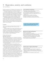
Mortality rates increase with age, disease severity and socioeconomic disadvantage (Figure 1.5). On average, in the United
Kingdom, COPD reduces life expectancy by 1.8 years (76.5 vs 78.3
years for controls); mild disease reduces life expectancy by 1.1
years, moderate disease by 1.7 years and severe disease by 4.1 years.
Morbidity and economic impact
The morbidity and economic costs associated with COPD are very
high, generally unrecognised and more than twice that associated
with asthma. The impact on quality of life is particularly high in
patients with frequent exacerbations, although even those with mild
disease have an impaired quality of life.
In the United Kingdom, emergency hospital admissions for
COPD have steadily increased as a percentage of all admissions
from 0.5% in 1991 to 1% in 2000. In 2002/2003, there were 110,000
hospital admissions for an exacerbation of COPD in England with
an average duration of stay of 11 days, accounting for 1.1 million
bed days. At least 10% of emergency admissions to hospital are as
a consequence of COPD and this proportion is even greater during
the winter. Most admissions are in individuals over 65 years of
age with advanced disease who are often admitted repeatedly and
use a disproportionate amount of resource. Approximately 25% of
patients diagnosed with COPD are admitted to hospital and 15%
of all patients are admitted each year.
The impact in primary care is even greater, with 86% of care
being provided exclusively in that setting. It has been estimated
that a typical general practitioner’s list will include 200 patients
with COPD (even more in areas of social deprivation), although
not all will be diagnosed. On average, patients with COPD make
six to seven visits annually to their general practitioner. It has been
estimated that each diagnosed patient costs the UK economy £1639
annually, equating to a national burden of £982 million. For each
patient, annual direct costs to the National Health Service (NHS)
Treatment
Figure 1.6 An analysis of the direct costs of COPD to the National Health
Service. A&E, accident and emergency; GP, general practitioner. Figure
derived with data from Britton M. The burden of COPD in the UK: results
from the Confronting COPD survey. Respiratory Medicine 2003; 97(suppl C):
S71–S79.
are £819, with 54% of this being due to hospital admissions and
19% due to drug treatment (Figure 1.6). COPD has further societal
costs; about 40% of UK patients are below retirement age and the
disease prevents about 25% from working and reduces the capacity
to work in a further 10%. Annual indirect costs have been estimated
at £820 per patient and encompass the costs of disability, absence
from work, premature mortality and the time caregivers miss
work. Within Europe, it has been estimated that in 2001 the overall
cost of COPD to the economy was ¤38.7 billion; this comprised
of ¤4.7 billion for ambulatory care, ¤2.7 billion for drugs, ¤2.9
billion for inpatient care and ¤28.4 billion for lost working days.
Risk factors
Smoking
In developed countries, cigarette smoking is clearly the single most
important risk factor in the development of COPD, with studies
consistently reporting dose–response associations. Cigarette
smoking is also associated with increased probability of COPD
diagnosis and death. Pipe and cigar smokers have significantly
greater morbidity and mortality from COPD than non-smokers,
although the risk is less than that with cigarettes. Approximately
50% of cigarette smokers develop airflow obstruction and 10–20%
develop clinically significant COPD. Maternal smoking during and
after pregnancy is associated with reduced infant, childhood and
adult ventilatory function, days, weeks and years after birth, respectively. Most studies have demonstrated that the effects of antenatal
environmental tobacco smoking exposure are greater in magnitude
and independent of associations with post-natal exposure.
Other factors
In the last 5 years, an increasing number of risk factors other than
smoking have been linked to the development of COPD, particularly
in developing countries. These include indoor (biomass) and outdoor air pollution, occupational exposures and early life factors such
as intra-uterine growth retardation, poor nutrition, repeated lower
respiratory tract infections and a history of pulmonary tuberculosis.
Many of these risk factors are inter-related. For example, biomass
ABC of COPD
Proportion of patients with COPD
who are non-smokers (%)
4
50
45
40
35
30
25
20
15
10
5
0
l
)
S) ey ica ina
ay den den tria taly
ay ela nd
ile ico
III) ia zi
an and HS
na ea
nd nd
I
S mb Bra
s
l
hi Kor Jap
R
RH urk Afr Ch
e
e
Ch ex ugu ezu gla inla inla orw
E
,
a
u
C
C
C
o
w
w
N ol
A
,
F
F
M Ur
ta
N
n
Ze (E
(E a, T th
En
el
, S , S rg,
ce
n,
d,
u
HA C
*
y
Ve
ew nal*
ge tten tten bu O D nal ata
(N
vin
an
So
r
N
o
p
l
P
z
e
,
A
o al
o
io
io a
B
pr
La
at M
on nat
US
rrb rrb S
g
i
gt
tin
on
No No
il n ult
ul
l
d
e
M
M
ng
W
ua
G
Figure 1.7 Proportion of patients with COPD who are non-smokers worldwide. ECRHS, European Community Respiratory Health Survey. Figure reproduced
with permission from Salvi SS, Barnes PJ. Chronic obstructive pulmonary disease in non-smokers. Lancet 2009; 374: 733–743. *Australia, Belgium, Denmark,
France, Germany, Iceland, Ireland, Italy, Netherlands, New Zealand, Norway, Spain, Sweden, Switzerland, United Kingdom and United States.
smoke exposure is associated with intrauterine growth retardation
and repeated early life lower respiratory tract infections. Accumulating evidence suggests that the prevalence of COPD worldwide in
never smokers may be as high as 25–45% worldwide (Figure 1.7)
with many risk factors and associations identified (Table 1.1).
Table 1.1 Non-smoking risk factors associated with the development
of COPD.
Indoor air pollution
• Smoke from biomass fuel: plant residues (wood, charcoal, crops, twigs,
dried grass) animal residues (dung)
• Smoke from coal
Occupational exposures
• Crop farming: grain dust, organic dust, inorganic dust
• Animal farming: organic dust, ammonia, hydrogen sulphide
• Dust exposures: coal mining, hard-rock mining, tunnelling, concrete
manufacturing, construction, brick manufacturing, gold mining, iron and
steel founding
• Chemical exposures: plastic, textile, rubber industries, leather
manufacturing, manufacturing of food products
• Pollutant exposure: transportation and trucking, automotive repair
Treated pulmonary tuberculosis
Repeated childhood lower respiratory tract infections
Chronic asthma
Outdoor air pollution
• Particulate matter (<10 µm or <2.5 µm diameter)
• Nitrogen dioxide
• Carbon monoxide
Poor socio-economic status
Low educational attainment
Poor nutrition
Table reproduced with permission from Salvi SS, Barnes PJ. Chronic
obstructive pulmonary disease in non-smokers. Lancet 2009; 374: 733–743.
Air pollution
It has been demonstrated that urban air pollution may affect lung
function development and consequently be a risk factor for COPD.
Cross-sectional studies have demonstrated that higher levels of
atmospheric air pollution are associated with cough, sputum production, breathlessness and reduced ventilatory function. Exposure
to particulate and nitrogen dioxide air pollution has been associated with impaired ventilatory function in adults and reduced lung
growth in children.
Worldwide, around 3 billion individuals are exposed to indoor
air pollution from the use of biomass fuel (wood, charcoal, vegetable
matter, animal dung) for cooking and heating; the smoke emitted
contains pollutants such as carbon monoxide, nitrogen dioxide,
sulphur dioxide, formaldehyde and particulate matter (Figure 1.8).
It has been estimated that biomass smoke exposure underlies about
50% of diagnosed COPD in developing countries, with it being a
particular problem in females and young children who are heavily
exposed during cooking in poorly ventilated areas. Exposure to
biomass smoke has been reported to increase the risk of COPD by
two to threefold.
Occupation
Some occupational environments with intense prolonged exposure
to irritating dusts, gases and fumes can cause COPD independently of cigarette smoking. However, smoking appears to enhance
the effects of these occupational exposures. It has been estimated
that about 15–20% of diagnosed cases are attributable to occupational hazards; in never smokers, this proportion increases to
about 30%. Occupations that have been associated with a higher
prevalence of COPD include coal mining, hard rock mining, tunnel
working, concrete manufacturing, construction, farming, foundry
Definition, Epidemiology and Risk Factors
5
glycoprotein responsible for the majority of anti-protease activity
in serum. The α1-antitrypsin gene is highly polymorphic, although
some genotypes (usually ZZ) are associated with low serum levels
(typically 10–20% of normal). Severe deficiency of α1-antitrypsin
is associated with premature and accelerated development of
COPD in smokers and non-smokers, although the rate of
decline is greatly accelerated only in smokers. The α1-antitrypsin
status of patients with severe COPD who are less than 40 years
old should be determined since over 50% have α1-antitrypsin
deficiency. The detection of affected individuals identifies family
members who in turn require genetic counselling and patients
who might be suitable for future potential treatment with
α1-antitrypsin replacement.
Further reading
Figure 1.8 Over 2 billion people rely on biomass fuel as their main source of
domestic energy; indoor air pollution associated with this, is an increasingly
important cause of COPD in developing countries. Figure reproduced with
permission from Dr Duncan Fullarton, Respiratory Infection Group, Liverpool
School of Tropical Medicine, Liverpool, UK.
working, the manufacture of plastics, textiles, rubber, leather
and food products, transportation and trucking. The increasing
recognition that occupation can contribute to the development of
COPD emphasises the importance of taking a full chronological
occupational history.
Alpha-1-antitrypsin deficiency
The best documented genetic risk factor for airflow obstruction is
α1-antitrypsin deficiency. However, this is a rare condition and is
present in only 1–2% of patients with COPD. α1-Antitrypsin is a
Britton M. The burden of COPD in the UK: results from the Confronting
COPD survey. Respiratory Medicine 2003; 97(suppl C): S71–S79.
Gibson PG, Simpson JL. The overlap syndrome of asthma and COPD: what
are its features and how important is it? Thorax 2009; 64: 728–735.
/>Halbert RJ, Natoli JL, Gano A, Badamgarav E, Buist AS, Mannino DM. Global
burden of COPD: systematic review and meta-analysis. The European
Respiratory Journal 2006; 28: 523–532.
Hu G, Zhou Y, Tian J et al. Risk of COPD from exposure to biomass smoke: a
metaanalysis. Chest 2010; 138: 20–31.
Lopez AD, Shibuya K, Rao C et al. Chronic obstructive pulmonary disease:
current burden and future projections. The European Respiratory Journal
2006; 27: 397–412.
Prescott E, Vestbo J. Socioeconomic status and chronic obstructive pulmonary
disease. Thorax 1999; 54: 737–741.
Pride NB, Soriano JB. Chronic obstructive pulmonary disease in the United
Kingdom: trends in mortality, morbidity and smoking. Current Opinion in
Pulmonary Medicine 2002; 8: 95–101.
Salvi SS, Barnes PJ. Chronic obstructive pulmonary disease in non-smokers.
Lancet 2009; 374: 733–743.
Viegi G, Pistelli F, Sherrill DL, Maio S, Baldacci S, Carrozzi L. Definition,
epidemiology and natural history of COPD. The European Respiratory
Journal 2007; 30: 993–1013.
CHAPTER 2
Pathology and Pathogenesis
William MacNee
MRC Centre for Inflammation Research, Queen’s Medical Research Institute, University of Edinburgh, Edinburgh, UK
OVERVIEW
•
The clinical sequelae of chronic obstructive pulmonary disease
(COPD) results from pathological changes in the large airways
(bronchitis), small airways (bronchiolitis) and alveolar space
(emphysema)
•
Combinations of pathological changes occur to varying degrees
in different individuals
•
Chronic inflammation – involving neutrophils, macrophages and
T-lymphocytes – is found in the airways and alveolar space
•
Small airways inflammation (bronchiolitis) can lead eventually to
scarring; this important pathological change is difficult to assess
by conventional lung function tests, but is a major source of
airway obstruction
•
In COPD, lungs show an amplified and persistent inflammatory
response following exposure to particles and gases, particularly
those found in cigarette smoke
molecular mechanisms that result in the pathological changes found
and how these lead to physiological abnormalities and subsequent
development of symptoms.
Pathology
The pathological changes in the lungs of patients with COPD are
found in the proximal and peripheral airways, lung parenchyma
and pulmonary vasculature. These changes are present to different
extents in affected individuals (Box 2.1, Figures 2.1–2.3).
Box 2.1 Pathological changes found in COPD
Proximal airways (cartilaginous airways >2 mm in
diameter)
•
•
Introduction
Chronic obstructive pulmonary disease (COPD) is characterised by
chronic airflow limitation that is not fully reversible and an abnormal inflammatory response in the lungs. The latter represents the
innate and adaptive immune responses to a lifetime of exposure to
noxious particles, fumes and gases, particularly cigarette smoke. All
cigarette smokers have inflammatory changes within their lungs,
but those who develop COPD exhibit an enhanced or abnormal
inflammatory response to inhaled toxic agents. This amplified or
abnormal inflammatory response may result in mucous hypersecretion (chronic bronchitis), tissue destruction (emphysema),
disruption of normal repair and defence mechanisms causing small
airway inflammation (bronchiolitis) and fibrosis.
These pathological changes result in increased resistance to
airflow in the small conducting airways and increased compliance
and reduced elastic recoil of the lungs. This causes progressive
airflow limitation and air trapping, which are the hallmark features
of COPD. There is increasing understanding of the cell and the
•
•
•
↑ Macrophages and CD8 T-lymphocytes
Few neutrophils and eosinophils (neutrophils increase with
progressive disease)
Submucosal bronchial gland enlargement and goblet cell
metaplasia (results in excessive mucous production or chronic
bronchitis)
Cellular infiltrates (neutrophils and lymphocytes) of bronchial
glands
Airway epithelial squamous metaplasia, ciliary dysfunction, ↑
smooth muscle and connective tissue
Peripheral airways (non-cartilaginous airways <2 mm
diameter)
•
•
•
•
•
•
•
Bronchiolitis at an early stage
↑ Macrophages and T-lymphocytes (CD8 > CD4)
Few neutrophils or eosinophils
Pathological extension of goblet cells and squamous metaplasia
into peripheral airways
Luminal and inflammatory exudates
↑ B-lymphocytes, lymphoid follicles and fibroblasts
Peribronchial fibrosis and airway narrowing with progressive
disease
Lung parenchyma (respiratory bronchioles and alveoli)
•
•
ABC of COPD, 2nd edition.
Edited by Graeme P. Currie. 2011 Blackwell Publishing Ltd.
6
↑ Macrophages and CD8 T-lymphocytes
Alveolar wall destruction due to loss of epithelial and endothelial
cells
Pathology and Pathogenesis
(a)
•
•
•
(b)
Development of emphysema (abnormal enlargement of airspaces
distal to terminal bronchioles)
Microscopic emphysematous changes:
◦ centrilobular (dilatation and destruction of respiratory
bronchioles – commonly found in smokers and predominantly in
upper zones)
◦ panacinar (destruction of the whole acinus – commonly found in
α-1-antitrypsin deficiency and more common in lower zones)
Macroscopic emphysematous changes:
◦ microscopic changes progress to bullae formation (defined as an
emphysematous airspace >1 cm in diameter)
(c)
(d)
Pulmonary vasculature
•
•
•
↑ Macrophages and T-lymphocytes
Early changes:
◦ intimal thickening
◦ endothelial dysfunction
Late changes:
◦ ↑ vascular smooth muscle
◦ collagen deposition
◦ destruction of capillary bed
◦ development of pulmonary hypertension and cor pulmonale
Figure 2.2 (a) Paper-mounted whole lung section of a normal lung;
(b) paper-mounted whole lung section from a lung with severe central
lobular emphysema. Note that the central lobular form is more extensive in
the upper regions of the lung; (c) histological section of a normal small
airway and surrounding alveoli connecting with attached alveolar walls;
(d) histological section showing emphysema with enlarged alveolar spaces,
loss of alveolar walls and alveolar attachments and collapsed airway.
Muscle
Glands
Gland
Duct
Muscle
(b)
Gland
Figure 2.1 (a) A central bronchus from a
cigarette smoker with normal lung function. Very
small amounts of muscle and small epithelial
glands are shown. (b) Bronchial wall from a
patient with chronic bronchitis showing a thick
bundle of muscle and enlarged glands. (c) A
higher magnification of the enlarged glands from
(b) showing chronic inflammation involving
polymorphonuclear (arrow head) and
mononuclear cells, including plasma cells (arrow).
Printed with kind permission from JC Hogg and S
Green. (d) Scanning electron micrograph of
airway from a normal individual showing flakes of
mucus overlying the cilia. (e) Scanning electron
micrograph of a bronchial wall in a patient with
chronic bronchitis. Cilia are covered with a
blanket of mucus.
Cartilage
(a)
(d)
(c)
(e)
Cartilage
7
8
ABC of COPD
(a)
(b)
(c)
Figure 2.3 Histological sections of peripheral airways. (a) Section from a cigarette smoker with normal lung function showing a nearly normal airway with
small numbers of inflammatory cells. (b) Section from a patient with small airway disease showing inflammatory exudate in the wall and lumen of the airway.
(c) Section showing more advanced small airway disease, with reduced lumen causing structural reorganisation of the airway wall, increased smooth muscle and
deposition of peribronchial connective tissue. Images produced with kind permission of Professor James C Hogg, University of British Columbia, Canada.
Pathogenesis
Inflammation is present in the lungs – particularly in the small
airways – of all smokers. This normal protective response to inhaled
toxins is amplified in COPD and leads to tissue destruction,
impairment of defence mechanisms that limit such destruction
and disruption of repair mechanisms. In general, the inflammatory and structural changes in the airways increase with disease
severity and persist even after smoking cessation. A number of
mechanisms are involved in intensifying lung inflammation, which
results in the pathological changes in COPD (Figure 2.4).
Innate and adaptive immune inflammatory
responses
The innate inflammatory immune system provides primary protection against the continuing insult of inhalation of toxic gases and
particles. The first line of defence consists of the mucociliary clearance apparatus and macrophages that clear foreign material from
the lower respiratory tract; both of these are impaired in COPD.
The second line of defence of the innate immune system is
exudation of plasma and circulating cells into both large and small
conducting airways and alveoli. This process is controlled by an
array of proinflammatory chemokines and cytokines (Box 2.2).
Inflammatory cells
COPD is characterised by increased neutrophils, macrophages,
T-lymphocytes (CD8 > CD4) and dendritic cells in various parts
of the lungs (Box 2.2). In general, the extent of inflammation is
related to the degree of airflow obstruction. These inflammatory
cells are capable of releasing a variety of cytokines and mediators
which participate in the disease process. This inflammatory cell
pattern is markedly different from that found in asthma.
Amplifying processes
Innate immunity
Acquired immunity
Oxidative stress
Genetics
Epigenetics
Cigarette smoke
(and other irritants)
Epithelial
cells
Alveolar macrophage
TGF-β
CTG
Fibroblast
Chemotactic factors
CD8
lymphocyte
Monocyte
Neutrophil
Oxidants
Fibrosis
Proteases
Alveolar wall destruction
(Emphysema)
Cellular processes
Inflammatory cell
recruitment/
activation
Mediator release
Transcription factor
activation
Autoimmunity
Impaired tissue repair
Cell senescence
Apoptosis
Mucus hypersecretion
(Chronic bronchitis)
Figure 2.4 Overview of the pathogenesis of chronic obstructive pulmonary disease (COPD). Cigarette smoke activates macrophages in epithelial cells to produce
chemotactic factors that recruit neutrophils and CD8 cells from the circulation. These cells release factors which activate fibroblasts, resulting in abnormal repair
processes and bronchiolar fibrosis. Imbalance between proteases released from neutrophils and macrophages and antiproteases leads to alveolar wall destruction
(emphysema). Proteases also cause the release of mucus. An increased oxidant burden resulting from smoke inhalation or release of oxidants from inflammatory
leucocytes causes epithelial and other cells to release chemotactic factors, inactivates antiproteases and directly injures alveolar walls and causes mucus secretion.
Several processes are involved in amplifying the inflammatory responses in COPD. TGF-β, transforming growth factor-β; CTG, connective tissue growth factor.
Pathology and Pathogenesis
Box 2.2 Inflammatory cells and mediators in COPD
•
•
•
•
•
Neutrophils – release reactive oxygen species, elastase and
cytokines that are important in the pathogenesis of COPD, with
effects on goblet cells, submucosal glands, the induction of
emphysema and inflammation. They are increased in the sputum
and distal airspaces of smokers; a further increase occurs in COPD
and is related to disease severity.
Macrophages – produce reactive oxygen species, lipid mediators
such as leukotrienes and prostaglandins, cytokines, chemokines
and matrix metalloproteases. They are found particularly around
small airways and may be associated with both small airway
fibrosis and centrilobular emphysema in COPD. They are increased
in number in airways, lung parenchyma and in bronchoalveolar
lavage fluid and increase further depending on disease
severity.
Eosinophils – increased numbers of eosinophils have been
reported in sputum, bronchoalveolar lavage fluid and the airway
wall in some patients with COPD and may represent a distinct
subgroup of COPD patients with a good clinical response to
corticosteroids.
T-lymphocytes (CD4 and CD8 cells) – increased in the airways and
lung parenchyma with an increase in CD8:CD4 ratio. Numbers of
Th1 and Tc1 cells, which produce interferon-γ, also increase. CD8
cells may be cytotoxic from the release of lytic substances such as
perforin and granzyme, cause alveolar wall destruction and induce
epithelial and endothelial apoptosis.
B-lymphocytes – increased in the peripheral airways and within
lymphoid follicles, possibly as a response to chronic infection or an
autoimmune process.
Inflammatory mediators
Many inflammatory mediators are increased in patients with COPD.
These include
•
•
•
•
leukotriene B4 (LTB4 ), a neutrophil and T-cell chemoattractant,
which is produced by macrophages, neutrophils and epithelial
cells;
chemotactic factors such as CXC chemokines, interleukin-8
(IL-8) and growth-related oncogene-α produced by macrophages
and epithelial cells; these attract cells from the circulation and
amplify proinflammatory responses;
proinflammatory cytokines such as tumour necrosis factor-α,
IL-1β and IL-6;
growth factors such as transforming growth factor-β (TGF-β),
which may cause fibrosis in the airways either directly or
through the release of another cytokine (connective tissue growth
factor).
An adaptive immune response is also present in the lungs of
patients with COPD, as shown by the presence of mature lymphoid
follicles. These increase in number in the airways according to
disease severity. Their presence has been attributed to the large
antigen load associated with bacterial colonisation or frequent lower
respiratory tract infections or possibly an autoimmune response.
Dendritic cells are major antigen-presenting cells and are increased
in the small airways, and provide a link between innate and adaptive
immune responses.
9
Protease/antiprotease imbalance
Increased production (or activity) of proteases or inactivation
(or reduced production) of antiproteases results in imbalance.
Cigarette smoke and inflammation per se produce oxidative stress,
which primes several inflammatory cells to release a combination
of proteases and inactivate several antiproteases by oxidation. The
major proteases involved in the pathogenesis of COPD are the
serine proteases produced by neutrophils, cysteine proteases and
matrix metalloproteases (MMPs) produced by macrophages. The
major antiproteases involved in the pathogenesis of emphysema
include α-1-antitrypsin, secretory leukoproteinase inhibitor and
tissue inhibitors of MMP (Box 2.3).
Box 2.3 Proteinases and antiproteinases involved in COPD
Proteinases
Serine proteinases
Neutrophil elastase
Cathepsin G
Proteinase 3
Cysteine proteinases
Cathepsins B, K, L, S
Matrix metalloproteinases
(MMP-8, MMP-9, MMP-12)
Antiproteinases
α-1-antitrypsin
Secretory leukoprotease inhibitor
Elafin
Cystatins
Tissue inhibitors of MMP (TIMP1–4)
Oxidative stress
The oxidative burden is increased in COPD. Sources of increased
oxidants include cigarette smoke and reactive oxygen and nitrogen
species released from inflammatory cells. This creates an imbalance
in oxidants and antioxidants (oxidative stress). Many markers of
oxidative stress are increased in stable COPD and are increased
further during exacerbations. Oxidative stress can lead to inactivation of antiproteinases and stimulation of mucous production.
It can also amplify inflammation by activating many intercellular
pathways, including kinases (e.g. P38 mitogen-activated protein
(MAP) kinase) enhancing transcription factor activation (e.g.
nuclear factor-κB (NF-κB)) and epigenetic events (such as decreasing histone deacetylates) that lead to increased gene expression of
proinflammatory mediators.
Emphysema is characterised by enlargement of the airspaces distal to the terminal bronchioles and is associated with destruction
of alveolar walls but without fibrosis. Paradoxically, fibrosis may
occur in the small airways in COPD. A number of mechanisms
are involved in the pathogenesis of emphysema, including protease/antiprotease imbalance, oxidative stress, apoptosis and cell
senescence (Box 2.4).
Box 2.4 Mechanism of emphysema in COPD
•
•
•
Protease/antiprotease imbalance – activation of MMPs such as
MMP-9 and -12, serine proteases such as neutrophil elastase and
inactivation of antiproteases such as α-1-antitrypsin
Activation of CD8 T-cells, which release perforin and granzymes
Apoptosis of alveolar cells resulting from a decrease in VEGF
signalling
10
•
•
•
ABC of COPD
Accelerated lung aging and cell senescence leading to failure of
lung maintenance and repair
Ineffective clearance of apoptotic cells (efferocytosis) by
macrophages leading to decreased anti-inflammatory mechanisms
Mitochondrial dysfunction with increased oxidative stress leading
to increased cell apoptosis, for example through SIRT-1
MMP, Matrix metalloproteinase; VEGF, vascular endothelial growth
factor; SIRT, sirtuin.
Pathophysiology
The pathogenic mechanisms described earlier result in the pathological changes found in COPD. These in turn cause physiological
abnormalities such as mucous hypersecretion, ciliary dysfunction,
airflow limitation and hyperinflation, gas exchange abnormalities,
pulmonary hypertension and systemic effects.
Mucous hypersecretion and ciliary dysfunction
Mucous hypersecretion results in a chronic productive cough.
This is characteristic of chronic bronchitis, but not necessarily
associated with airflow limitation, while not all patients with COPD
have symptomatic mucous hypersecretion. Mucous hypersecretion
is due to an increased number of goblet cells and increased size
of bronchial submucosal glands in response to chronic irritation
caused by noxious particles and gases. Ciliary dysfunction is due to
squamous metaplasia of epithelial cells and results in dysfunction
of the mucociliary escalator and difficulty expectorating.
anatomic alterations described in COPD – is the main mechanism
accounting for abnormal gas exchange. The extent of impairment
of diffusing capacity for carbon monoxide is the best physiological
correlate to the severity of emphysema.
Pulmonary hypertension
Pulmonary hypertension develops late in the course of COPD at
the time of severe gas exchange abnormalities. Contributing factors include pulmonary arterial vasoconstriction (due to hypoxia),
endothelial dysfunction, remodelling of the pulmonary arteries
(smooth muscle hypertrophy and hyperplasia) and destruction of
the pulmonary capillary bed.
The development of structural changes in the pulmonary arterioles results in persistent pulmonary hypertension and right
ventricular hypertrophy/enlargement and dysfunction (Figure 2.5).
Systemic effects
COPD is associated with several extra-pulmonary effects (Box 2.5).
The systemic inflammation and skeletal muscle wasting contribute
to limiting the exercise capacity of patients and worsens prognosis, irrespective of the degree of airflow obstruction. There is
an increased risk of cardiovascular disease in individuals with
COPD and, if present, it is associated with a systemic inflammatory
response and vascular dysfunction.
Box 2.5 Systemic features of COPD
•
•
•
•
Airflow limitation and hyperinflation/
air trapping
Chronic airflow limitation is the physiological hallmark of COPD.
The main site of airflow limitation occurs in the small conducting
airways that are <2 mm in diameter. This is because of inflammation, narrowing (airway remodelling) and inflammatory exudates
in the small airways. Other factors contributing to airflow limitation include loss of lung elastic recoil (due to destruction of
alveolar walls) and destruction of alveolar support (from alveolar
attachments).
The airway obstruction progressively traps air during expiration,
resulting in hyperinflation of the lungs at rest and dynamic hyperinflation during exercise. Hyperinflation reduces the inspiratory
capacity and, therefore, the functional residual capacity during
exercise. These features result in the breathlessness and impaired
exercise capacity typical of COPD.
Gas exchange abnormalities
Gas exchange abnormalities occur in advanced disease and are characterised by arterial hypoxaemia with or without hypercapnia. An
abnormal distribution of ventilation/perfusion ratios – due to the
•
•
•
Cachexia
Skeletal muscle wasting
Increased risk of cardiovascular disease
Normochromic normocytic anaemia
Osteoporosis
Depression
Secondary polycythemia
Chronic hypoxia
Pulmonary vasoconstriction
Pulmonary arteriole
Muscularisation
Intimal hyperplasia
Fibrosis
Obliteration
Pulmonary hypertension
Cor pulmonale
Death
Oedema
Renal and hormonal changes
Figure 2.5 The development of pulmonary hypertension in chronic
obstructive pulmonary disease (COPD).
Pathology and Pathogenesis
Pathology, pathogenesis and
pathophysiology of exacerbations
Exacerbations are often associated with increased neutrophilic
inflammation in the airways, and in some mild exacerbations, the
presence of increased numbers of eosinophils. Some exacerbations
are infectious in origin (either bacterial or viral), while other
potential mechanisms include air pollution and changes in ambient
temperature. Viruses and bacteria may activate transcription factors
such as NF-κB and the MAP kinases, leading to the release of
inflammatory cytokines.
In mild exacerbations, the degree of airflow limitation is often
unchanged or only slightly increased. Severe exacerbations are associated with worsening of pulmonary gas exchange due to increased
ventilation/perfusion inequality and subsequent respiratory muscle
fatigue. The worsening ventilation/perfusion relationship results
from airway inflammation, oedema, mucous hypersecretion and
bronchoconstriction. These reduce ventilation and cause hypoxic
vasoconstriction of pulmonary arterioles, which in turn impairs
perfusion.
11
Respiratory muscle fatigue and alveolar hypoventilation can
contribute to hypoxaemia, hypercapnia, respiratory acidosis and
lead to severe respiratory failure and death. Hypoxia and respiratory
acidosis can induce pulmonary vasoconstriction, which increases
the load on the right ventricle, and together with renal hormonal
changes, can result in peripheral oedema.
Further reading
Chung KF, Adcock IM. Multifaceted mechanisms in COPD: inflammation,
immunity, and tissue repair and destruction. The European Respiratory
Journal 2008; 31: 1334–1356.
Hogg JC. Lung structure and function in COPD. The International Journal of
Tuberculosis and Lung Disease 2008; 12: 467–479.
Hogg JC, Timens W. The pathology of chronic obstructive pulmonary disease.
Annual Review of Pathology 2009; 4: 435–459.
MacNee W. Pathogenesis of chronic pulmonary disease. Clinics in Chest
Medicine 2007; 28: 479–513.
Rodriguez-Roisin R, MacNee W. Pathophysiology of chronic obstructive pulmonary disease. The European Respiratory Monograph 2006; 11: 177–200.
CHAPTER 3
Diagnosis
Graeme P. Currie and Mahendran Chetty
Aberdeen Royal Infirmary, Aberdeen, UK
OVERVIEW
•
Chronic obstructive pulmonary disease (COPD) may be
undiagnosed in its initial phase due to paucity of clinical features
•
The diagnosis should be considered in any individual >35 years
with breathlessness, chest tightness, wheeze, cough, sputum
production and reduced exercise tolerance and who has a
history of smoking (or other significant risk factor)
•
Clinical examination may be normal in early disease
•
All patients with suspected COPD need spirometry to confirm
the diagnosis and grade the severity of airflow obstruction
•
A chest X-ray and full blood count are mandatory at the time of
diagnosis
•
Other investigations such as detailed lung function tests,
electrocardiogram, echocardiogram, chest computed
tomography, pulse oximetry and arterial blood gases may be
required in selected cases
As with most medical conditions, a thorough history should be
taken and examination performed before embarking on investigations in a patient with possible or suspected chronic obstructive
pulmonary disease (COPD). The diagnosis should be considered
in individuals over 35 years of age with any relevant respiratory
symptom and history of smoking (or other significant risk factor,
e.g. exposure to indoor biomass fuels in developing countries)
(Figure 3.1). The presence of airflow obstruction and severity of
COPD are confirmed by spirometry; this remains the gold standard
diagnostic test (Chapter 4).
Clinical features
Typical presenting symptoms
COPD may occur in any current or former smoker over the age of
35 years who complains of breathlessness, chest tightness, wheeze,
chronic cough, sputum production, frequent winter chest infections
or impaired exercise tolerance. The condition may also be present
in the absence of troublesome respiratory symptoms, especially in
those with a sedentary lifestyle or limited mobility.
Figure 3.1 An example of an indoor fire used for cooking in a rural Indian
village. Image reproduced with permission from Dr David Bellamy, retired
Bournemouth General Practitioner.
Breathlessness may initially be noticed only during exertion,
which later becomes progressive and persistent. It is useful to
determine how breathlessness affects daily living activities such as
walking on the flat (and walking distance), walking up inclines,
climbing flights of stairs, carrying bags, walking to the shops, washing and dressing, doing light housework and hobbies. The impact of
breathlessness on an individual’s day-to-day life can be objectively
assessed by the Medical Research Council (MRC) dyspnoea scale
(Table 3.1). Chest tightness and wheeze (the high-pitched noise
Table 3.1 MRC breathlessness scale.
Grade
1
2
3
4
5
ABC of COPD, 2nd edition.
Edited by Graeme P. Currie. 2011 Blackwell Publishing Ltd.
12
Degree of breathlessness related to activities
Not troubled by breathlessness except on strenuous exercise
Short of breath when hurrying or walking up a slight hill
Walks slower than contemporaries on the level because of
breathlessness, or has to stop for breath when walking at
own pace
Stops for breath after about 100 m or after a few minutes on
the level
Too breathless to leave the house, or breathless when dressing
or undressing
MRC, Medical Research Council.
Diagnosis
Table 3.2 Causes of a chronic (lasting >8 weeks) cough.
•
Airway
disorders
Parenchymal
disease
•
COPD
Lung cancer
Pleural disease Others
Pleural effusion Gastro-oesophageal
reflux disease
Asthma
Interstitial lung Mesothelioma Upper airway cough
disease
syndrome
Bronchiectasis
Chronic lung
Angiotensin-converting
including
infections (TB
enzyme inhibitor use
cystic fibrosis
or fungal
infections)
Smokers cough/
Exposure to irritant
chronic
dusts/chemicals/
bronchitis
fumes/particulate
matter
Postviral
hyperreactivity
COPD, chronic obstructive pulmonary disease; TB, tuberculosis.
produced by air travelling through an abnormally narrowed smaller
airway) may only be experienced during exertion or an exacerbation. Breathlessness, chest tightness and wheeze overnight are more
suggestive of asthma. Chronic cough (defined as lasting >8 weeks)
may be the presenting symptom in many other respiratory disorders
(Table 3.2), and in COPD, is usually associated with sputum production. In healthy individuals, around 100 ml of sputum is produced
daily, which is transported up the airway and swallowed; expectoration of excessive amounts of sputum is usually abnormal. In COPD,
excessive amounts of sputum are often expectorated in the mornings
and it is usually clear (mucoid), although it may also be light green
because of nocturnal stagnation of neutrophils. During an exacerbation, sputum may be a darker green because of dead neutrophils.
The initial presenting feature of COPD may also be with repeated
lower respiratory tract infections, especially during winter months.
Other features in the history
As part of the overall assessment of COPD, it is essential to find
out about symptoms of anxiety and depression, other medical
conditions, current medication, frequency of exacerbations (and
number of courses of steroids and antibiotics in the preceding year),
previous hospital admissions, exercise limitation, occupational and
environmental exposure to dust, chemicals and fumes, exposure to
biomass fuel, previous chest problems (chronic asthma or tuberculosis), number of days missed from work and financial impact
and the extent of social and family support. There is increasing evidence that COPD causes systemic effects, with anorexia and weight
loss being relatively common and under-recognised problems in
advanced COPD.
It is also important to determine when a patient started smoking,
when he/she stopped smoking, the number of cigarettes smoked
each day and the current smoking status. Patients should be asked
about willingness to quit smoking and considered for referral to
smoking cessation services. The number of smoking pack years can
be calculated as follows:
•
A one pack year is defined as 20 cigarettes (one pack) smoked per
day for 1 year.
13
Number of pack years = (number of cigarettes smoked per day
× number of years smoked)/20.
For example, a patient who has smoked 15 cigarettes per day for
40 years has a (15 × 40)/20 = 30 pack year smoking history.
Signs
Signs of respiratory disease tend to appear as COPD progresses.
Physical examination may therefore be normal, or reveal only
prolongation of the expiratory phase of respiration or an elevated
respiratory rate at rest in mild disease. In COPD, wheeze is usually
heard during expiration (as airways normally dilate during inspiration and narrow in during expiration) and may occur only during
exercise, in the morning (reflecting pooling of secretions blocking
off smaller airways), or during an exacerbation. Late course inspiratory crackles are occasionally found in COPD, especially when
excessive lower airway secretions are present.
As the disease progresses, examination may reveal a hyperinflated chest, increased anteroposterior diameter of the chest wall,
intercostal indrawing, diminished breath sounds, wheezing at rest
and faint heart sounds (due to hyperinflated lungs). Cyanosis
(blue discolouration of the skin, lips and mucous membranes
(Figure 3.2)), pursed lip breathing and use of accessory muscles
(sternocleidomastoids, platysma and pectoral muscles) are features
of advanced disease.
Cor pulmonale is present when right ventricular hypertrophy
and pulmonary hypertension occurs as a consequence of any
chronic lung disorder. Some patients with severe COPD may
therefore demonstrate signs consistent with cor pulmonale (raised
jugular venous pressure, loud P2 due to pulmonary hypertension,
tricuspid regurgitation, pitting peripheral oedema (Figure 3.3) and
hepatomegaly) and its presence usually indicates a poor prognosis. It
may occur alongside features of CO2 retention (warm peripheries,
peripheral vasodilation and bounding pulse). Finger clubbing is
not found in COPD and its presence should prompt thorough
evaluation to exclude a cause such as lung cancer, bronchiectasis
or idiopathic pulmonary fibrosis (Figure 3.4). Tar staining of the
nails and fingers is commonly found in current or previous heavy
cigarette smokers with COPD (Figure 3.5).
Figure 3.2 A patient with advanced hypoxic chronic obstructive pulmonary
disease with central cyanosis.
14
ABC of COPD
Figure 3.3 Pitting ankle oedema is a feature of cor pulmonale; other causes
or contributory factors such as oral corticosteroids, calcium antagonists,
excessive intravenous fluid administration, hypoalbumenaemia or dependant
oedema should be considered.
Figure 3.5 A right-handed patient with gross tar staining of the fingers due
to chronic cigarette smoking.
Table 3.3 Calculation of the BODE index.
Parameter
Points
0
BMI
≤21
≥65
FEV1 % predicted
Modified MRC scale 0–1
6-minute walk
≥350
distance (metres)
1
2
3
>21
50–64
36–49
≤35
2
3
4
250–349 150–249 ≤149
Score
0 or 1
0, 1, 2 or 3
0, 1, 2 or 3
0, 1, 2 or 3
Total (out of 10)
The modified MRC scale uses the same clinical descriptors as the original
MRC scale (1–5), although values are denoted as 0–4 in the calculation of
the BODE index.
BODE, BMI, Obstruction, Dyspnoea and Exercise; MRC, Medical Research
Council.
disorder (Table 3.4). Since asthma tends to be the main differential
diagnosis of COPD, a careful history should be taken in order to
help distinguish between either disorder (Table 3.5). Symptoms
such as haemoptysis, chest pain and weight loss require urgent
referral to secondary care to rule out lung cancer or an alternative
cardiorespiratory disorder.
Figure 3.4 A patient with tar staining and finger clubbing; chronic
obstructive pulmonary disease and non-small cell lung cancer were both
diagnosed in this patient.
Skeletal muscle wasting and cachexia may occur in advanced
disease, while some patients may also be overweight. The body mass
index (BMI; weight/height2 ) should be calculated during the initial
examination. The BODE index (BMI, Obstruction, Dyspnoea and
Exercise) is a grading system which predicts the risk of death from
any cause and from respiratory causes among patients with COPD
(Table 3.3). A BODE index of 0–2, 3–4, 5–6 and 7–10 is thought
to be associated with a 52-month mortality rate of approximately
10, 30, 50 and 80% respectively.
Differential diagnosis
Particular attention should be made to other features in the history
and examination, which may suggest an alternative or concomitant
Investigations
Lung function testing
Solitary peak expiratory flow (PEF) readings can significantly and
seriously underestimate the extent of airflow obstruction, while
serial monitoring of PEF is not generally useful in the diagnosis
of COPD. Demonstration of airflow obstruction with spirometry
is required to confirm the diagnosis. Spirometry is also useful in
assessing severity of the disease and in following its progress plus
response to treatment. The normal age-related decline in forced
expiratory volume in 1 second (FEV1 ) is around 20–40 ml/year and
this increases to 40–80 ml/year in current smokers. More detailed
lung function measurements such as lung volumes (total lung
capacity and residual volume), gas transfer and 6-minute walk test
can be performed if doubt exists in diagnosis or more thorough
evaluation is required (such as during assessment for surgery or
lung transplantation). Lung function testing in COPD is discussed
in detail in Chapter 4.
Diagnosis
15
Table 3.4 Conditions in the differential diagnosis of COPD.
Condition
Suggestive feature
Investigation
Asthma
Family history, atopy, non-smoker, young age, nocturnal
symptoms
Orthopnoea, history of ischaemic heart disease, fine lung crackles
Haemoptysis, weight loss, hoarseness
Copious sputum production, frequent chest infections, childhood
pneumonia, coarse lung crackles
Dry cough, history of connective tissue disease, use of drugs such
as methotrexate, amiodarone, etc., fine lung crackles
Dry cough, risk factors for immunosuppression, fever
Trial of inhaled corticosteroids, reversibility testing if airflow
obstruction present
Chest X-ray, electrocardiogram, echocardiogram
Chest X-ray, bronchoscopy, CT
Sputum microscopy, culture and sensitivity, high-resolution CT
Congestive cardiac failure
Lung cancer
Bronchiectasis
Interstitial lung disease
Opportunistic infection
Tuberculosis
Weight loss, haemoptysis, night sweats, risk factors for
tuberculosis and immunosuppression
Pulmonary function testing, chest X-ray, high-resolution CT,
lung biopsy, autoantibodies
Chest X-ray, sputum microscopy, culture and sensitivity,
induced sputum, bronchoalveolar lavage
Chest X-ray, sputum microscopy, culture and sensitivity
COPD, chronic obstructive pulmonary disease, CT, computed tomography.
Table 3.5 Clinical differences between COPD and asthma.
COPD
Asthma
Age
Cough
>35 years
Persistent and productive
Smoking
Breathlessness
Almost invariable
Progressive and persistent
Nocturnal symptoms
Uncommon unless in
severe disease
Uncommon unless family
members also smoke
Possible
Any age
Intermittent and nonproductive
Possible
Intermittent and
variable
Common
symptoms and identify complications related to COPD such as
bullae formation and pulmonary arterial hypertension (enlarged
central pulmonary arteries and peripheral arterial pruning). There
is no direct link between extent of chest X-ray abnormality and
degree of airflow obstruction. When there is doubt in diagnosis or
a surgical procedure contemplated (such as lung volume reduction
surgery or bullectomy), high-resolution computed tomographic
(HRCT) imaging of the chest is required (Figure 3.7).
Imaging
All patients with suspected COPD should have a posteroanterior
chest X-ray performed at diagnosis. This may be normal, although
as the disease progresses, hyperinflation (flattened diaphragms,
a narrowed heart, >6 anterior rib ends visible and ‘squared off
apices’) and hyperlucency of lung fields may be evident (Figure 3.6).
A chest X-ray also helps discount other causes of respiratory
Other investigations
All patients should have a full blood count checked; this may
show secondary polycythaemia, and excludes anaemia as a cause
of chronic breathlessness. The discovery of a raised eosinophil
count should raise the possibility of an alternative diagnosis such
as asthma or eosinophilic pneumonia.
In patients with signs of cor pulmonale, an electrocardiogram
may show changes of chronic right-sided heart strain (Figure 3.8).
However, an echocardiogram is more sensitive in detecting tricuspid
valve incompetence along with right atrial and ventricular hypertrophy and may also indirectly assess pulmonary artery pressure.
Moreover, echocardiography is also a useful tool to determine
whether left ventricular dysfunction is present, especially when
Figure 3.6 Chest X-ray showing typical changes of chronic obstructive
pulmonary disease (>6 ends of anterior ribs visible, flat diaphragms and
increased translucency of lung fields).
Figure 3.7 High-resolution computed tomogram of the chest showing
widespread upper lobe emphysematous bullae in a patient with advanced
COPD.
Family history
Concomitant eczema
or allergic rhinitis
Common
Common
COPD, chronic obstructive pulmonary disease.