Ebook ABC of sexually transmitted infections (5/E): Part 2
Bạn đang xem bản rút gọn của tài liệu. Xem và tải ngay bản đầy đủ của tài liệu tại đây (2.85 MB, 61 trang )
8 Pelvic inflammatory disease and pelvic pain
Helen Mitchell
Acute pelvic inflammatory disease (PID) is most commonly
caused by infection ascending from the vagina or cervix, which
causes inflammation of the upper genital tract. This can result
in any combination of salpingitis, endometritis, oophoritis,
parametritis, pelvic peritonitis, and tubo-ovarian abscess
formation.
The organisms commonly responsible for acute PID depend
on the local prevalence of sexually transmitted infections (STIs).
Chlamydia trachomatis is the most common treatable bacterial STI
in the United Kingdom and is implicated in more than 50% of
cases of acute PID. Ten to 20% of cases are associated with
Neisseria gonorrhoeae, this rate will be higher in areas with higher
local prevalence. Studies have shown that 8-39% of women with
C trachomatis related genital infection will develop acute PID.
In addition, it is estimated that for every overt case of chlamydial
pelvic infection there are three covert (asymptomatic) cases.
The role of Mycoplasma genitalium and Ureaplasma
urealyticum in acute pelvic infection is still unclear, but they
have been implicated in the pathogenesis of acute endometritis
and chorioamnionitis associated with pre-term labour.
Other organisms connected with acute pelvic infection
include anaerobes, Bacteroides fragilis, peptostreptococci,
Escherichia coli, and Lancefield group B haemolytic streptococci.
Bacterial vaginosis is associated with ascending infection and
acute PID after induced abortion and post partum.
Clinical diagnosis of PID
The most common presenting symptoms are lower abdominal
pain and abnormal vaginal discharge. Other symptoms associated
with PID include intermenstrual and post-coital bleeding, dysuria,
deep dyspareunia, and fever. Low backache and rectal discomfort
may also be present. Right upper quadrant pain from
perihepatitis is a feature of the uncommon Fitz-Hugh-Curtis
syndrome in association with C trachomatis related PID.
The history for pain should include onset, site, and nature,
as well as aggravating and relieving factors. A full menstrual,
contraception, and gynaecological history should be taken to
make a risk assessment for unplanned pregnancy, including
ectopic pregnancy, and ovarian disease. The sexual history will
provide a risk assessment for the presence of an STI. It is also
important to ask about urinary or bowel symptoms.
Differential diagnosis of lower abdominal
pain
●
●
●
●
●
●
●
●
Ectopic pregnancy
Urinary tract infection
Ovarian cyst complications—torsion and
rupture
Endometriosis
Ovarian malignancy
Bowel disease
Irritable bowel syndrome
Appendicitis
Risk factors for PID in patient’s history
Presence of an STI
● New sexual partner in past month
● Frequent change of sexual partner
● No condom use
● Age under 25 years
● Partner with symptoms
● Previous medical history of an STI
● Involuntary infertility
Gynaecological interventions that can cause
ascending infection
● Intrauterine contraceptive device insertion or
change in 20 days
● Termination of pregnancy—induced abortion
● Hysterosalpingogram
● Endometrial sampling
● Hysteroscopy
● Dilatation and curettage
● Evacuation of retained products of conception
Clinical diagnosis of PID
Presenting symptoms
● Lower abdominal pain
● Abnormal vaginal discharge
● Intermenstrual or post-coital bleeding (or
both)
Dysuria
Backache
Fever
With additional clinical signs from list below
Adnexal tenderness
● Cervical excitation pain
● Mucopurulent cervical discharge
Pyrexia above 38ЊC
Rebound
Guarding
Adnexal mass
●
Mucopurulent cervical discharge with cervicitis
30
Pelvic inflammatory disease and pelvic pain
A history of abdominal surgery for infertility, ovarian
disease, appendicectomy, and bowel disease can provide useful
diagnostic pointers. If the onset of lower abdominal pain has
occurred after a recent gynaecological intervention, then the
intervention may have introduced an infection or transmitted
an infection from the cervix to the upper genital tract.
Patient
Patient complains
complains of
of lower
lower abdominal
abdominal pain
pain
Take
Take history
history (including
(including gynaecological
gynaecological
history)
history) and
and examine
examine (abdomen
(abdomen and
and vagina)
vagina)
Any
Any of
of the
the following
following present?
present?
•• Missed
Missed or
or overdue
overdue period
period
No
No
•• Recent
Recent delivery,
delivery, abortion,
abortion, or
or miscarriage
miscarriage
•• Abdominal
Abdominal guarding
guarding or
or rebound
rebound
tenderness,
tenderness, or
or both
both
•• Abnormal
Abnormal vaginal
vaginal bleeding
bleeding
•• Abdominal
Abdominal mass
mass
Investigations and clinical decisions
It is most important to exclude ectopic pregnancy by testing
urine for  human chorionic gonadotrophin with a sensitive
pregnancy testing kit (if available).
Other immediate investigations that should be carried out
include dipstick urinalysis to exclude urinary tract infection. If
this is positive, a midstream urine sample should be sent for
microscopy and culture. The appropriate specimens should be
collected for Chlamydia nucleic acid amplification testing and
gonorrhoea culture. These results will not be available
immediately, so treatment needs to be started if the healthcare
professional suspects acute PID.
If the woman is seen at a genitourinary medicine (GUM)
clinic, immediate microscopy can exclude bacterial vaginosis
and may show gonorrhoea infection, but, again, treatment is
started once the clinical diagnosis is made.
In a hospital setting, a full blood count, blood chemistry,
and blood cultures should be carried out in all patients
with high fever or acute abdominal pain with peritonitis.
Ultrasonography can identify adnexal disease and exclude
ectopic pregnancy in a woman with a positive pregnancy test.
Clinical symptoms and signs of PID only have a 65%
positive predictive value when compared with laparoscopy. The
routine use of diagnostic laparoscopy to diagnose acute PID,
however, is limited by the risks and cost of this procedure.
Laparoscopy usually is carried out only in patients in whom the
diagnosis remains uncertain.
Treatment of acute PID
Treatment should be started immediately to reduce the risk of
long term sequelae. In the United Kingdom, the incidence of
gonorrhoea and genital chlamydial coinfection has increased
over the past decade; therefore, the antibiotic regimen used to
treat PID should cover N gonorrhoea, C trachomatis, and
anaerobic infections. There may be local variations in N
gonorrhoea antibiotic sensitivities, and the local microbiology
laboratory should be able to advise on appropriate antibiotic
choices. When prescribing for women it is important to check
Yes
Yes
Refer
Refer patient
patient for
for surgical
surgical or
or
gynaecological
gynaecological opinion
opinion and
and assessment.
assessment.
Before
Before referral,
referral, set
set up
up an
an intravenous
intravenous
line
line and
and apply
apply resuscitatory
resuscitatory measures
measures
ifif necessary
necessary
No
No Any
Is
Is there
there cervical
cervical
Any other
other
excitation
excitation tenderness
tenderness
illness
illness
or
or lower
lower abdominal
abdominal
found?
found?
tenderness
tenderness and
and
Yes
Yes
vaginal
vaginal discharge?
discharge?
Yes
Yes
Manage
Manage
approappropriately
priately
Manage
Manage for
for pelvic
pelvic
inflammatory
inflammatory disease
disease
Review
Review in
in three
three days
days
Has
Has patient
patient
improved?
improved?
No
No
Refer
Refer
patient
patient
Yes
Yes
Continue
Continue treatment
treatment until
until completed
completed
•• Educate
Educate and
and counsel
counsel
•• Promote
Promote and
and provide
provide condoms
condoms
•• Offer
Offer HIV
HIV counselling
counselling and
and testing
testing ifif both
both facilities
facilities are
are available
available
Lower abdominal pain flow chart
Indications for hospital admission for
women with acute PID
●
●
●
●
●
●
●
Uncertain diagnosis
High fever and rigors with dehydration
Diffuse peritonism
Adnexal mass
HIV positive women with immunosuppression
if pelvic abscess suspected
Intravenous drug users if poor treatment
compliance and social circumstances
Intercurrent medical illness, for example
sickle cell disease, insulin dependent diabetes
mellitus
Oral antibiotic regimens
●
●
Ofloxacin 400 mg twice daily for 14 days (U and C)
Metronidazole 400 mg twice daily 14 days
or
●
●
●
Doxycycline 100 mg twice daily 14 days
Metronidazole 500 mg twice daily 14 days
Ceftriaxone 250 mg intramuscular stat
or
Amoxyl 3 g orally with 1 g probenicid CW ϩ E
Where these specified antibiotics are not available, the alternative
regimen is used. It should
● Cover N gonorrhoeae according to local known antibiotic sensitivities
● Include appropriate treatment for 14 days to cover C trachomatis
and anaerobic bacteria
●
In pregnancy, erythromycin 500 mg twice daily for 14 days should
be used as an alternative to doxycycline. If a long acting
preparation is not available four times daily dosing is required
Adhesions over liver capsule associated with perihepatitis in chlamydial
pelvic infection
31
ABC of Sexually Transmitted Infections
the risk of early pregnancy, current combined oral
contraception use, and any history of antibiotic allergies.
Further management
The woman should be advised to return for review two or three
days after taking oral treatment if her symptoms are no better.
If the symptoms have worsened during this time, she should be
advised to visit the emergency department.
No evidence supports the routine removal of the
intrauterine contraceptive device (IUCD) in acute PID;
however, removal should be considered if no clinical response
to treatment is seen. In such situations, oral emergency
contraception may be required.
The patient must be advised to complete the full course of
antibiotics, abstain from sexual intercourse, and attend the
GUM clinic for a follow up appointment.
Admission to hospital will allow intravenous antibiotic
therapy and fluid rehydration, provision of adequate analgesia,
and regular clinical review of symptoms and signs.
Indications for laparotomy in acute pelvic infection include
generalised peritonitis, bilateral or enlarging abscess and where
the clinical condition has not improved or has deteriorated
after 48 hours on intravenous antibiotics.
Recommended parenteral treatment regimens include
cefoxitin with doxycycline and a combination of clindamycin
with gentamicin when a tubo-ovarian abscess is present.
Adverse sequelae of PID
Chronic PID
The risk of developing chronic PID increases with each episode of
acute PID. Chronic pelvic infection is a debilitating condition,
with general malaise and fatigue, that results in frequent time off
work and incapacity. Symptoms include irregular menses with
congestive dysmenorrhoea, secondary deep dyspareunia, chronic
pelvic pain, and low backache. Women with chronic PID have
increased hysterectomy rates.
Tubal factor infertility (TFI)
The risk of TFI increases with each episode of acute infection
● one episode
12% risk of TFI
● two episodes
35% risk of TFI
● three episodes
70% risk of TFI
95% of infertile women with a history of PID will have TFI and
30% of women with no history of PID will also have TFI, probably
as a result of “silent” subclinical infection
Ectopic pregnancy
Ectopic pregnancy can be life threatening. The risk of ectopic
pregnancy is 1:100 of all pregnancies, which is increased
sevenfold after acute PID
Partner notification and aftercare
Partner notification and epidemiological treatment is essential
to prevent reinfection, with the consequent increase in long
term sequelae.
Women with negative STI test results should be advised that
their diagnosis is non-specific PID and that because of the risks
of sequelae, doctors have a low threshold for starting antibiotic
treatment in sexually active women. Partner notification and
epidemiological treatment is still necessary because the male
partner may have non-specific urethritis.
Many women will express anxieties over future fertility and
may even request tests for tubal patency; however, these tests
should only be done in the course of formal investigation after
a period of involuntary infertility. It is important to emphasise
the need for continued contraception to avoid unplanned
pregnancy.
Laparoscopic view of ectopic pregnancy
Prevention of pelvic infection
Management of the complications and reproductive sequelae of
Chlamydia infection in women costs national health
programmes millions each year.
The introduction of screening programmes for genital
C trachomatis infection has reduced substantially the incidence of
acute PID and ectopic pregnancy. Screening programmes are
cost effective when the local prevalence rate is 6% and a nucleic
acid amplification diagnostic test assay is used.
Studies have shown that bacterial vaginosis is common in
women attending for legal abortion and, if left untreated, it is
associated with an increased risk of post-abortal pelvic
infection. Prophylaxis and treatment for bacterial vaginosis is
metronidazole (1 g suppository given rectally at time of
operation).
32
Blocked tube at laparoscopy
Pelvic inflammatory disease and pelvic pain
Evidence also shows that antibiotic prophylaxis effective
against bacterial vaginosis given before total abdominal and
vaginal hysterectomy prevents post-operative vaginal vault
infection.
Although PID can occur after the insertion of an IUCD no
evidence at present recommends routine screening or
antibiotic prophylaxis for bacterial vaginosis before insertion of
the device. Bacterial vaginosis does not affect conception rates
during in vitro fertilisation procedures, but it is an independent
risk factor for subsequent miscarriage.
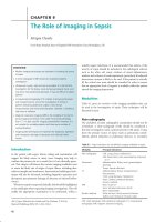
The photograph of mucopurulent cervical discharge with cervicitis is
the copyright of Dr Marc Steben, Clinique de l’Ouest, Montreal,
Canada. The photographs of adhesions over the liver capsule and the
ectopic pregnancy are courtesy of Mr Alfred Cutner
Opportunities for Chlamydia screening to prevent pelvic
infection*
All women and men
● Younger than 25 years
● Older than 25 years with a new sexual partner or two or more
partners in the previous year
● Of any age with symptoms
● Attending GUM clinics
All women
● Younger than 35 years before surgical uterine instrumentation—
for example, hysteroscopy
● Before IUCD insertion
● Before induced abortion (termination of pregnancy)
*British guidelines from Chief Medical Officer and Royal College of
Obstetricians and Gynaecologists
Further reading
Berger GS, Westrom LV, eds. Pelvic inflammatory disease. New York:
Raven Press, 1992
● Bevan CD, Johal BJ, Mumtaz G, Ridgway G, Siddle NC. Clinical,
laparoscopic and microbiological findings in acute salpingitis:
report on a United Kingdom cohort. Br J Obstet Gynaecol
1995;102:407-14
● Mann SN, Smith JR, Barton SE. Pelvic inflammatory disease.
Continuing medical education. Int J STD AIDS 1996;7:315-21
● Royal College of Obstetrics and Gynaecology’s website
www.RCOG.org.uk
●
Recommendations from the 31st RCOG study group. In:
Templeton A, ed. The prevention of pelvic infection. London: RCOG
Press, 1996:267-70
● Robinson AJ, Greenhouse P. Prevention of recurrent pelvic
infection by contact tracing: a common-sense approach. Br J
Obstet Gynaecol 1996;103:859-61
● Walker CK, Kahn JG, Peterson HB, Sweet RL. Pelvic
inflammatory disease: meta-analysis of antimicrobial regimen
efficacy. J Infect Dis 1993;168:969-78
●
33
9 Sexually transmitted infections in pregnancy
Helen Mitchell
Pregnant women may be unaware they have an existing
asymptomatic sexually transmitted infection (STI) or they may
be still at risk of acquiring an STI during pregnancy. Therefore,
it is necessary to overcome a natural hesitancy to discuss risk
factors for STIs. Infections at this time can affect the fetus and
neonate by vertical transmission, which may result in serious
and life threatening consequences. Screening for infections in
pregnancy and starting early treatment can prevent adverse
outcomes for the mother and neonate.
The management of STIs in pregnancy should be guided by
expert advice because certain treatments are contraindicated
during pregnancy. A test of cure should be carried out after
treatment and before delivery for women testing positive for
Chlamydia trachomatis, Trichomonas vaginalis, and Neisseria
gonorrhoeae.
Gonorrhoea
Mother
Uncomplicated gonorrhoea rates in young women have
increased dramatically over the past decade in the United
Kingdom. Worldwide gonorrhoea prevalence varies, with
particularly high rates reported in Africa.
Baby
Intrapartum infection occurs in about 30-50% of babies born
to untreated mothers and is associated with
●
●
●
●
Conjunctivitis (ophthalmia neonatorum) “sticky eye” with
onset of purulent conjunctival discharge between two and
five days after birth
Disseminated neonatal infection
Diagnosis is by Gram stained smear and culture of
conjunctival swab.
Treatment of established infection is with systemic antibiotics,
for example ceftriaxone.
C trachomatis
Mother
Genital chlamydial infection rates in young women have also
increased substantially in the United Kingdom. Non-invasive
testing for chlamydia using nucleic acid amplification tests, for
example polymerase chain reaction (PCR) on self taken
vulval-introital swabs, may be appropriate in late pregnancy and
in situations in which the woman declines a speculum
examination.
Baby
Intrapartum infection in babies born to untreated mothers is
associated with
●
●
●
Conjunctivitis (ophthalmia neonatorum) in 30-50% of babies
with onset occurring 3-14 days after birth
Otitis media
Nasopharyngitis
34
Screening in pregnancy guidelines
Routine antenatal screening
● In the United Kingdom the current programme includes
serology for syphilis, Hepatitis B, and HIV antibody testing with
a pre-test discussion
Hepatitis C
● Screening for anti-hepatitis C virus (anti-HCV) antibodies should
be done in high risk groups, such as intravenous drug users and
women that received organ transplant or blood transfusion
before HCV screening commenced
Other STIs
● Screening for gonorrhoea, chlamydia, and T vaginalis in
pregnancy should be considered in women with STI risk factors,
young women under 25 years and those with a history of STIs or
pelvic inflammatory disease, or both.
● Routine antenatal screening for gonorrhoea and chlamydia to
prevent complications of maternal infection in pregnancy and
neonatal infection is appropriate in high prevalence countries
● No evidence currently supports routine antenatal screening
using type specific antibody testing for herpes simplex virus
(HSV-1 and HSV-2)
Partner notification and epidemiological treatment is
essential to prevent reinfection during the antenatal period
and further risk of vertical transmission
Gonorrhoea
Gonorrhoea in pregnancy is associated with
● Low birth weight
● Premature delivery
● Pre-term rupture of membranes
● Chorioamnionitis
● Postpartum sepsis
● Secondary infertility
Appropriate treatment regimes include a single
intramuscular dose of ceftriaxone (250 mg),
cefotaxime (500 mg), and spectinomycin (2 g)
(see Chapter 5). Ciprofloxacin and tetracyclines
should be avoided in pregnancy.
C trachomatis
C trachomatis in pregnancy is associated with
● Low birth weight
● Premature delivery
● Pre-term rupture of membranes
● Chorioamnionitis
● Postpartum sepsis
Treatment in pregnancy is with erthromycin (500 mg twice daily)
for two weeks or amoxycillin (500 mg three times daily) for seven
days. Doxycyline and tetracycline are both contraindicated in
pregnancy
Sexually transmitted infections in pregnancy
●
Chlamydial pneumonitis, which presents with staccato cough,
tachypnoea, and failure to thrive, occurs after 4-12 weeks in
10-20% of exposed babies.
Diagnosis is by culture of C trachomatis or nucleic acid test
(NAAT) on conjunctival, nasopharyngeal, and rectal swabs.
Treatment of established infection is with systemic antibiotics,
for example erythromycin.
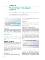
Ophthalmia neonatorum
●
●
●
●
●
●
●
Ophthalmia neonatorum is conjunctivitis that develops within
21 days of birth. In the United Kingdom it is a notifiable
condition
Chlamydial or gonococcal infection should always be excluded
Chlamydial ophthalmia is more common but it is not possible
to distinguish them clinically
Untreated gonococcal ophthalmia neonatorum can lead to
corneal ulceration and perforation with permanent loss of vision
Diagnosis is by Gram stained smear and culture of a swab from
the conjunctiva for N gonorrhoea, culture for C trachomatis, and
ligase chain reaction
Established infection is treated with systemic antibiotics
In areas of high STI prevalence without routine antenatal
screening ocular prophylaxis should be given routinely to all
newborn babies within one hour of birth, using a 1%
tetracycline or 0.5% erythromycin eye ointment
Prophylactic systemic ceftriaxone should be considered for
babies born vaginally to mothers with known untreated
gonorrhoea
Chlamydial pneumonitis
Genital herpes simplex infection
Mother
The diagnosis of genital herpes simplex infection (HSV-1 and
HSV-2) in women has seen a slow but steady increase and about
5% of antenatal attendees in the United Kingdom have a history
of symptomatic genital herpes. On serological testing, 25% of
genitourinary medicine clinic attendees and 20% of adult
Americans have type specific antibodies to HSV-2. However, only
35% of infected adults are aware that they have genital herpes.
Maternal primary HSV infection during pregnancy is
associated with
●
●
●
●
Spontaneous abortion
Low birth weight
Premature delivery
Stillbirth.
It is important to ascertain whether a pregnant women
presenting with genital ulceration has a recurrent infection or a
true primary HSV infection. In tropical countries it is important
to exclude other causes of genital ulceration (see Chapter 11).
Differentiation of primary from non-primary infection is by
serology because history is a poor indicator. Seroconversion in
primary infection takes between three and six weeks and can be
tracked using immunoglobulin G and immunoglobulin M type
specific antibody testing.
Ophthalmia neonatorum
Advice for pregnant women with known recurrent genital
herpes
●
●
●
●
●
Women with recurrent genital herpes can deliver vaginally if
they do not have overt genital ulcers at the time of delivery
Repeated viral cultures during pregnancy are of no clinical
value in predicting recurrences or viral shedding at the time of
delivery
Women with a recurrence at the time of delivery are currently
delivered by lower segment caesarean section to prevent
intrapartum viral transmission
If recurrent lesions are present at the time of delivery there is a
low risk of neonatal herpes even with vaginal delivery. This risk
must be offset against the maternal risks of surgical delivery and
some obstetricians may agree to vaginal delivery after discussion
with the pregnant woman to obtain her informed consent
Suppression therapy during the third trimester may reduce the
risk of recurrence at the time of delivery in women with
frequent recurrence but this does not reduce viral shedding so
the benefit is uncertain
35
ABC of Sexually Transmitted Infections
Women presenting with suspected primary genital herpes
acquired during the third trimester of pregnancy should be
offered aciclovir antiviral treatment and delivered by elective
lower segment caesarean section if labour commences within
a six week period after diagnosis.
The risks of primary HSV-2 are highest in the last trimester
and if, during this time, the male partner has an episode of
recurrent genital HSV-2 sexual intercourse should be avoided.
Pregnant women should be informed of the risks of
acquiring HSV infection during pregnancy. Receptive oral
sex with a partner with orolabial HSV-1 is a risk factor for
women with no personal history of orolabial or genital
herpes infection.
Baby
Antepartum HSV transmission is rare and may cause stillbirth.
Neonatal HSV infection is rare in the United Kingdom and the
United States (2 per 100 000 and 7 per 100 000 live births,
respectively). The highest risk of intrapartum transmission and
neonatal infection is 40% for babies born by vaginal delivery in
a woman with primary genital herpes infection at the time of
delivery. In women with recurrent herpes at vaginal delivery the
risk of neonatal herpes is less than 1%. Postnatal infection can
occur if a relative or caregiver with a herpetic whitlow or
orolabial HSV-1 handles or kisses the child.
Confirmation of diagnosis is essential and the method used
will depend on the laboratory services available from EM of
vesicle fluid to viral PCR testing.
Neonatal herpes simplex infection can be localised or
disseminated affecting multiple organs, including hepatitis
and encephalitis. If neonatal HSV is suspected immediate
intensive treatment with intravenous antiviral therapy
should be started. Disseminated infection has a high
mortality rate (70%) even with effective antiviral therapy.
Surviving neonates are at a high risk of neurological
sequelae.
HIV
Mother
By the end of 2002 an estimated 42 million adults and children
worldwide are living with HIV and 50% of infected adults are
women. In some of the countries in Sub-Saharan Africa one in
three women attending antenatal services will be HIV positive.
In the United Kingdom, data obtained by national
unlinked anonymous monitoring of HIV infection show that
one in 200 women attending antenatal clinics in Central
London are HIV positive, but in rural areas only one in 2500
women are HIV positive. During 2002, 720 births took place to
HIV positive women in the United Kingdom, of which 80%
were to previously diagnosed women.
Worldwide, HIV in pregnancy is associated with
●
●
●
HIV testing in pregnancy
●
●
●
●
●
●
Low birth weight
Premature delivery
Stillbirth.
Pregnancy does not seem to have an adverse effect on the
health of an HIV positive woman or her long term prognosis
unless she has AIDS or a concurrent infection, such as
tuberculosis.
Baby
Each day 2000 children in Africa are newly infected with HIV
and many millions of children have been orphaned by HIV.
The risk of mother-to-child transmission is related to the
maternal viral load, stage of HIV disease, duration of pregnancy
at the time of delivery and the risk is increased by vaginal
delivery.
The highest transmission rates occur in resource poor
countries with high HIV prevalence where interventions to
prevent transmission are not widely available. The additional
risks of transmission in resource poor countries include breast
feeding after delivery. In some societies, bottle-feeding is
associated with social stigma and a substantial risk of infant
death from acute gastroenteritis.
All babies born to infected mothers will exhibit maternal
HIV antibodies; in uninfected babies 50% will lose the
antibodies by 10 months. All uninfected babies should be
confirmed as HIV negative at six months using HIV PCR testing
and at 18 months by serial antibody titre.
36
●
All pregnant women should be offered HIV screening routinely
by HIV antibody testing with a pre-test discussion
Women may not be able to accurately assess their personal risk
of HIV infection
The universal offer of HIV testing in pregnancy that allows
women to opt out is more effective than selective offer or
allowing women to choose if they feel HIV testing is necessary
The medical benefit of knowing a women’s HIV status is that
women who test positive can be offered interventions that
effectively reduce the risks of vertical transmission of HIV
Mother-to-child transmission without interventions during
pregnancy is 15-30%, which is further increased by breast
feeding
The transmission risk can be effectively reduced to less than 1%
by the following interventions during pregnancy
Antiretroviral therapy for the mother which includes
zidovudine or nevirapine. Strong evidence shows that both
treatments effectively reduce the risk of vertical transmission
Elective caesarean section delivery
Avoiding breast feeding
Antiretroviral therapy for the neonate after delivery
In high prevalence countries women should be retested in the
third trimester
Total: 2.1 million–2.9 million
Eastern Europe and Central Asia
9000–15 000
Western Europe
5000–7000
East Asia and Pacific
6000–12 000
North America
8000–12 000
Caribbean
19 000–31 000
Latin America
37 000–50 000
North Africa and Middle East
31 000–49 000
Sub-Saharan Africa
2 million–2.2 million
South and South East Asia
110 000–190 000
Australasia
<200
Number of children (younger than 15 years) estimated to be living with HIV
and AIDS as of end 2003. Adapted from www.UNAIDS.org
Sexually transmitted infections in pregnancy
Syphilis
Mother
Worldwide, syphilis (Treponema pallidum) is still a common
infection in pregnancy. The rates are low in the United
Kingdom; nevertheless, routine antenatal screening is still
carried out. In high prevalence countries congenital syphilis can
occur as a result of acquisition in late pregnancy and infected
women not attending for antenatal care. The treatment regimen
used in pregnancy depends on the stage of maternal infection,
history of antibiotic allergy and is usually with intramuscular
benzathine penicillin injections. Effective maternal treatment
will prevent congenital syphilis in the unborn child except when
treatment has commenced late in the third trimester.
Baby
Syphilis is associated with 25% of stillbirths in rural SubSaharan Africa and congenital syphilis accounts for 30% of
perinatal deaths. The risk of congenital syphilis in untreated
cases is related to the stage of maternal syphilis with the risk
decreasing with advancing stage of maternal disease. Up to 50%
of babies born to mothers with untreated primary or secondary
infection will be infected compared with less than 5% of babies
born to mothers with late latent infection.
Transplacental transfer of maternal antibodies occurs but if
the baby is not infected the treponemal antibody will be lost by
six months. Diagnosis of congenital infection occurs by
demonstrating the presence of treponemes in lesions and by
serology using the fluorescent treponemal antibody absorption
test for immunoglobulin M. Treatment of an infected neonate
is with intravenous penicillin.
Clinical features of congenital syphilis
Early congenital syphilis is a multi-organ disease that can present
with hepatosplenomegaly
● Anaemia
● Petechiae
● Periostitis
Latent (early and late)
● No clinical signs of active infection
Late (more than two years is similar to adult late disease)
● Osteoperiostitis
● Joint effusions usually knees—Clutton’s joints
● Gummata
● Neurological and cardiovascular complications
The lesions of early and late syphilis may heal but result in classical
stigmata of congenital syphilis
● Sabre shaped tibial deformity
● Saddle nose deformity
● Frontal bossing of the skull
● Linear scars around the mouth
● Small notched incisors
● Corneal opacities
Hepatitis B
In the United Kingdom the prevalence of hepatitis B carriage
in the antenatal population is low. In women from endemic
areas carriage is higher and vertical transmission can occur,
especially when the mother is hepatitis Be antigen positive.
Parental consent for immunisation should be obtained
before birth so that babies born to high risk carriers can be
given hepatitis B virus immunoglobulin passive vaccination and
active immunisation shortly after birth to prevent both neonatal
infection and the risk of chronic carriage.
Congenital syphilis on teeth
Hepatitis C
The risk of vertical transmission with hepatitis C is estimated
to be 6%. Transmission may be increased in co-infection
with HIV. No specific intervention has been identified to
reduce the transmission rate.
Genital warts
Genital warts may appear for the first time or increase in size
and number during pregnancy as a result of changes in local
cellular immunity. There is a very small risk of vertical
transmission resulting in neonatal laryngeal, mucous
membrane, or genital human papillomavirus infection.
Imiquimod, podophyllin, and podophyllotoxin topical
treatments are all contraindicated in pregnancy.
T vaginalis
Trichomonae infection in pregnancy is associated with adverse
pregnancy outcomes, including pre-term delivery and low birth
Congenital syphilis on mouth
37
ABC of Sexually Transmitted Infections
weight. Pregnant women can be treated with oral
metronidazole treatment regimes but high dose metronidazole
treatment regimes should be avoided in the first trimester and
also during breast feeding because they may cause breast milk
to taste bitter to the infant.
Bacterial vaginosis
At present, no evidence supports routine antenatal screening
for bacterial vaginosis for all pregnant women or that treating
asymptomatic women with bacterial vaginosis in general
antenatal clinics reduces their risk of pre-term labour. However,
some evidence shows that treating bacterial vaginosis reduces
pre-term labour in women with a history of pre-term delivery
and it may be that this subgroup of women could benefit from
early screening and oral treatment. Further trials are needed to
show that such screening and antenatal treatment reduces
perinatal mortality and morbidity. Symptomatic pregnant
women should be treated with oral metronidazole (400 mg
twice daily) for between five and seven days.
38
Further reading
Genc M, Ledger, WJ. Syphilis in pregnancy. Sex Transm Inf
2000;76:73-9
● Guidelines on STIs in pregnancy. www.rcog.org.uk (accessed
26 Nov 2003) and www.bashh.org (accessed 26 Nov 2003)
● PHLS Communicable Disease Surveillance Centre and PHLS
Syphilis Working Group. Antenatal syphilis screening in the UK: a
systematic review and national options appraisal with recommendations.
London: Public Health Laboratory Service, 1998
www.hpa.org.uk (accessed 26 Nov 2003)
● Reducing mother to child transmission of HIV infection in the United
Kingdom. Recommendations of an intercollegiate working party for
enhancing voluntary confidential HIV testing in pregnancy. London:
Royal College of Paediatrics and Child Health, 1998
www.hpa.org.uk (accessed 26 Nov 2003)
●
The photograph of opthalmia neonatorium is reproduced from
King A, Nicol C. Venereal diseases. London: Baillière Tindall, 1969
10 Other conditions that affect the female
genital tract
Helen Mitchell
Bartholin’s gland conditions
A Bartholin’s cyst is a painless enlargement that may
increase or decrease in size over time, and the history is
often longer or intermittent
The Bartholin’s glands can become enlarged by abscess or cyst
formation. In abscess formation, common infecting pathogens
include Neisseria gonorrhoeae, Chlamydia trachomatis, Escherichia coli,
 haemolytic streptococci, Staphylococcus aureus, and anaerobes.
Investigations
All sexually active patients who present with a cyst or abscess
should be offered a full sexually transmitted infection (STI)
screen. However, if the client is too uncomfortable for an
examination, the screen can be deferred until follow up. If the
abscess is discharging pus, additional swabs of pus should be
taken for microscopy, culture, and sensitivity.
A Bartholin’s abscess is a painful genital swelling and on
examination the gland is tensely enlarged with pain, local
redness, and warmth. The swelling may become fluctuant
“pointing” and will eventually discharge pus, after which
the intense throbbing pain is relieved
Vulvar symptoms
Women may present with complaints of genital skin itching,
burning, soreness, and discomfort during sexual intercourse.
Some women experience longstanding symptoms and despite
frequent clinic attendances may fail to receive a diagnosis and
appropriate advice or treatment with consequent psychological
and psychosexual morbidity. In some women, relationship
difficulties, psychosexual problems, and depression can lead to
somatisation and genital symptoms with no clinically apparent
cause.
Discharging Bartholin’s abscess.
Reproduced from King A, Nicol C.
Venereal diseases. London: Baillière
Tindall, 1969
Clinical management
A detailed history is very important and should include onset
and duration of symptoms and whether any topical treatments
have been used and with what degree of success.
Details of personal habits and hygiene should be covered,
such as use of perfumed soaps, bath additives, douching,
depilatory preparations, alternative remedies, laundry
detergents, and fabric conditioners. If the patient admits to
scratching, ask whether this is worse at night and if they
regularly wear fingernail varnish, because this can contain
formaldehyde, which is a contact irritant. A personal and family
history of atopy, asthma, hayfever, eczema, other dermatological
conditions, and nickel and food allergies can be relevant.
General principles
●
●
●
●
●
●
The external genital area should be examined carefully. The skin
of the rest of the body, scalp, mouth, eyes, and fingernails may
need to be examined as appropriate. The inguinal lymph nodes
should be palpated and the vaginal mucosa inspected by
speculum examination
Vulvar symptoms often are caused by recurrent vulvovaginal
Candida infection
If symptoms persist and tests for STIs and other genital infections
are negative, it is important to consider whether there is an
underlying dermatological disorder
Scratching and rubbing to relieve symptoms can result in both
secondary skin changes and infection that can further alter the
clinical appearances
Vulvar skin biopsy may be required to make a definitive diagnosis
Referral to specialist services with the combined clinical
expertise of a dermatologist, gynaecologist, or genitourinary
medicine physician should be considered for all women with
persistent vulvar symptoms
Management of Bartholin’s gland conditions
Abscess
● Painful non-discharging abscess—refer urgently to on call
gynaecology for a marsupialisation or incision and drainage
procedure
● Abscess has spontaneously discharged and pus is weeping
freely—oral flucloxacillin (500 mg four times daily orally) should
be prescribed for five days. Refer the patient to routine
gynaecology outpatients because recurrence is common and may
require interval marsupialisation
● Advise rest, loose clothing, and analgesia as required, for
example, ibuprofen
● Use Sitz baths (one cup of salt in bowl of water) and cotton balls
to gently clear away pus
● Pat the area dry after washing or dry with a hairdryer on a low
heat setting
● Follow up appointment for results or to perform full STI screen
Cyst
● Offer full STI screen
● No antibiotics are required
● Referral to routine gynaecology outpatient appointment to
consider interval marsupialisation
Common causes of vulvar symptoms
●
●
●
●
●
Genital infections
STIs
Vulvodynia
Genital dermatoses
Psychosexual problems
39
ABC of Sexually Transmitted Infections
Vulvar and perianal itching
Threadworm infestation should be considered if the
itching is predominately perianal (pruritus ani) rather
than vulvar (pruritus vulvae). A “sticky tape” test should
carried out by applying a clear sticky tape strip to the
perianal skin in the morning before washing. The tape is
applied to a glass microscopy slide and examined for
threadworm ova.
General advice for patients with vulvar symptoms, including
genital itching
●
●
●
●
●
●
●
●
●
●
●
●
Aqueous cream can be used a soap substitute for washing
A bland emollient is useful as a skin moisturiser
Avoid perfumed products, bath additives, talcum powder,
vaginal deodorant sprays, and sanitary pads with perfume or
deodorisers
Change laundry detergent to a skin sensitive brand or a nonbiological brand
Do not use fabric conditioner for undergarments
Shaving or use of depilatory creams in the genital area may
exacerbate symptoms
Patients sensitive to spermicide or latex condoms can try using
washed latex condoms or those with only a lubricant
Perfumed oils and creams should not be used as lubricants
Avoid self treatment with over the counter or alternative
remedies
Try not to scratch because this can damage the skin and set up a
cycle of itch-scratch-itch, which then needs to be broken by using
a moderate potency topical steroid initially then reducing the
dose as symptoms resolve
A tepid bath, ice pack, or cold soaked cotton pad applied locally
may help reduce an intense need to scratch
Itching can often be worse at night. A mildly sedating
antihistamine, such as chlorpheniramine, at night may help
reduce nocturnal scratching
Genital dermatoses
Lichenification
This can occur in any itchy skin condition and describes
the appearance where the skin is thickened and pale with
accentuated skin line markings and folds. When scratching
is marked, evidence of excoriation with areas of broken skin
and traction hair loss will be seen. Post-inflammatory
hypopigmentation and hyperpigmentation can be
present.
Non-specific vulvar appearance with oedema, redness, and introital splitting
Causes of genital itching
Infection
● Candidiasis
● Trichomonas vaginalis
● Genital warts
● Genital herpes simplex
● Molluscum contagiosum
Infestation
● Pediculosis pubis (crab lice)
● Threadworms
Genital dermatoses
● Non-allergic contact or irritant dermatitis
● Eczema
● Psoriasis
● Lichen sclerosus
● Lichen planus
● Seborrhoeic eczema
Neoplastic conditions
● Pre-malignant intraepithelial neoplasia
● Invasive neoplasia
Systemic conditions
● Diabetes mellitus
● Renal or hepatic dysfunction
Irritant contact dermatitis
This is commonly caused by skin sensitisers present
in products used in general and genital hygiene. Avoidance
of some common contact irritants may relieve
symptoms.
Allergic contact dermatitis
This can occur with self treatment with essential oils, local
anaesthetic creams, and pile relieving ointments common in
patients with chronic symptoms. Contact dermatitis
medicamentosa is an allergic contact dermatitis usually caused
by excipients or additives in topical treatment.
Patch testing may be useful for identifying specific allergens
in atopic eczema and allergic contact dermatitis. Nickel allergy
is a form of allergic contact dermatitis and may be relevant in
women with poor quality genital piercings.
40
Hyperpigmentation secondary to contact dermatitis caused by the use of
depilatory creams in the genital area
Other conditions affecting the female genital tract
Psoriasis
The appearance of affected genital areas may be altered, with
red, glazed, well defined patches that are often not scaly. It is
important to examine the limb flexures for characteristic
“silvery” plaques and the nails for pitting.
Eczema
The characteristic appearance of eczema can be altered on the
vulva because it is a moist area prone to friction from clothing
and during sexual intercourse. Other skin sites may be affected
and there may be a personal or a family history of atopy, such
as hayfever and asthma.
Seborrhoeic eczema
This can affect the vulva and also may be evident on the face,
chest, scalp, and eyebrows. It is treated with a mild steroid
containing an antifungal component.
Early stages of lichen sclerosus in a young woman, affecting the labia minora
on left
Lichen simplex chronicus
Plaques of lichenification are seen in this condition, but it is
not a specific diagnosis. It is important to review the skin
appearance once symptoms are controlled by a topical steroid
to exclude an underlying dermatosis, particularly lichen
sclerosus.
Lichen sclerosus
Lichen sclerosus is an autoimmune condition linked with
alopecia areata and vitiligo. There may be predisposing genetic
factors and, in some cases, infective trigger agents. Lichen
sclerosus can occur at any age and affects both sexes, but is
most common in women over 50 years of age.
The anogenital area is commonly affected in a classic figure
of eight distribution around the vulva and anus. Common
presenting symptoms are itching, soreness, dyspareunia, and
painful fissures at the introitus. The affected skin is dull and
white, with horizontal skin wrinkling, telangiectasia, and small
ecchymoses. In chronic, untreated cases, loss of normal
anatomy may result with fusion of the clitoral hood, abnormal
clitoral sensation, resorption of the labia minora, and
narrowing of the introitus.
Diagnosis can be made clinically in overt cases or by skin
biopsy that shows characteristic histological appearances.
Treatment is with a potent topical steroid twice daily until
symptoms resolve and the condition is quiescent. Maintenance
treatment continues with weekly or fortnightly applications.
The lifetime risk of squamous cell carcinoma in lichen
sclerosus is 4-5% and women should be taught how to examine
themselves and when to seek medical attention, for example for
ulceration, raised lesions, and localised persistent symptoms.
Surgical treatment is indicated rarely but may be useful
when introital narrowing precludes satisfactory sexual
intercourse.
More advanced lichen sclerosus with loss of architecture and marked pallor
with telangiectasia
Lichen planus
Lichen planus is considered to be an autoimmune disorder
that affects skin or mucosal surfaces, or both. Women may
present with pruritus and dyspareunia with associated oral
symptoms. The classical appearances are itchy, purple papules
or plaques on the vulva, which can be white or have postinflammatory hyperpigmentation. These lesions may exhibit
Köebnerisation with local extension along trauma and scar
lines. Wickham’s striae is a lacy white appearance on the surface
of the affected genital mucosa and may also be identified on
the flexor aspects of the wrists, gingival margins, and oral
mucosa.
Suspicious lesion with pigmentation that should be referred for expert
opinion and histological diagnosis
Malignant melanoma is the second most frequent vulvar
malignancy, and it is important to refer any patient with a
suspicious pigmented genital lesion for an expert opinion to
exclude pre-malignant or malignant change
41
ABC of Sexually Transmitted Infections
Clinical findings are important to establish the diagnosis
because the histological appearances on skin biopsy often show
only non-specific inflammatory changes.
Treatment is with topical steroids. In vulvovaginal gingival
syndrome, the vagina is also affected with painful red erosions,
and consequent synechiae formation can distort the vaginal
anatomy, causing severe dyspareunia. In such cases, systemic
and topical intravaginal steroids are necessary.
Pigmentary changes
Areas of pigmentation change may be seen on examination,
and it is important to ascertain whether any localised symptoms
are present.
●
●
●
Lentigines are areas of darker pigmentation caused by a
localised increase in melanocytes
Post-inflammatory hypopigmentation and hyperpigmentation
can occur in women with chronic itching area with well
circumscribed areas of depigmentation and scratching
Vitiligo is an autoimmune skin condition with well
circumsribed areas of depigmentation that can involve the
genital area.
Suspicious lesion with variable pigmentation (VIN III) that should be
referred for expert opinion and histological diagnosis
Vulval intraepithelial neoplasia
and invasive vulval neoplasia
Vulval intraepithelial neoplasia (VIN) can be low grade (VIN I)
or high grade (VIN II/III). Pre-malignant lesions in the genital
area can be difficult to identify clinically because there are no
consistent diagnostic features, and VIN can be warty or flat,
single or multiple, asymptomatic or symptomatic, and varied in
coloration.
Squamous cell carcinoma is responsible for 90% of all vulvar
malignancies and is associated with the presence of a high risk
human papillomavirus (for example, types 16, 18, 33, and 35).
Specialist advice is recommended for persistent genital
skin lesions and genital warts that do not respond to
topical treatment. Urgent referral is required for suspicious
lesions with ulceration, bleeding, or dark or patchy
pigmentation.
Vulval pain syndromes
Vulval pain can be caused by local infection, trauma, topical
wart treatments, and pelvic floor disorders, and can occur in
association with systemic disease. Vulvodynia is defined by the
International Society for the Study of Vulvovaginal Disease
(ISSVD) as chronic burning, soreness, or rawness. Vulvodynia
has features in common with other pain syndromes and
psychological support and psychosexual counselling are
important in long term management.
Vulvar vestibulitis syndrome is a triad of symptoms and signs
with superficial dyspareunia on attempted penetration or
tampon insertion, erythema, and point tenderness localised in
the vestibule. The aetiology is uncertain, and it is thought to be
a self limiting condition. Approaches to treatment include
general vulvar symptoms advice, topical local anaesthetic, and
lubricants to facilitate sexual intercourse.
Cyclical vulvodynia occurs when recurrent vulval
symptoms happen in relation to menstruation and coitus. It
may be caused by changes in vaginal pH or associated
vulvovaginal candidiasis and bacterial vaginosis. Intravaginal
azole treatment at the cyclical trigger points may be
beneficial.
42
Red raised suspicious lesion (squamous cell carcinoma) that should be
referred urgently for expert opinion and histological diagnosis
ISSVD classification of vulval pain syndromes
●
●
●
●
●
Vulvar vestibulitis (provoked localised vulval dysaesthesia or
vestibulodynia)
Cyclical vulvodynia
Dysaesthetic vulvodynia (unprovoked generalised vulval
dysaesthesia)
Vulvar dermatoses
Vulvar papillomatosis
Vulvar papillomatosis with characteristic club shaped papillae
Other conditions affecting the female genital tract
Dysaesthetic vulvodynia is characterised by a history of
diffuse and constant burning pain and affects an older age
group of women. This condition has closer parallels with
glossodynia and is thought to be a disorder of cutaneous
sensory perception. Treatment is with tricyclic antidepressants
or the newer antiepileptic drugs, for example gabapentin.
Vulvar papillomatosis describes the appearance of small
lobular papillae on the inner surface of the labia minora and
around the vestibule. These papillae now are thought to be a
normal anatomical variant, and in most women are
asymptomatic and do not require treatment.
Causes of dyspareunia
Superficial
Infection
● Candidiasis, T vaginalis, and genital herpes simplex
Trauma
Episiotomy scars, introital fissures, or tears caused by sex toys
●
Vulval disorders
● Lichen sclerosus
● Lichen planus
● Vulval pain syndromes
● Post-menopausal vulvovaginal atrophy
● Iatrogenic self treatment, post-radiotherapy, and 5-fluorouracil
Psychosexual
● Vaginismus
Deep
● Ovarian disease
● Endometriosis
● Acute and chronic pelvic inflammatory disease
● Uterine fibroids
Further reading
Edwards A, Wojnarowska F. The vulval pain syndromes. Int J
STD AIDS 1998;9:74-9
● Institute of Psychosexual Medicine website www.ipm.org.uk
(accessed 26 Nov 2003)
● Nunns D. Vulval pain syndromes. Br J Obstet Gynecol
2000;107:1185-93
● Powell JJ, Wojnarowska F. Lichen sclerosus. Lancet
1999;353:1777-83
● Ridley CM, Robinson AJ, Oriel. Vulval disease: a practical guide
to diagnosis and management. London: Arnold Publishers, 2000
● Skrine R. Blocks and freedoms in sexual life. A handbook of
psychosexual medicine. Oxford: Radcliffe Medical Press, 1997
● Skrine R, Montford H, eds. Psychosexual medicine. An
introduction. London: Arnold, 2001
● Vulval pain patient information website
www.vul-pain.dircon.co.uk (accessed 7 Jan 2005)
●
Localised redness in the vestibule with associated point tenderness elicited
using a cotton tip swab
Psychosexual problems
Chronic vulvovaginal symptoms can interfere seriously with
sexual and emotional relationships, resulting in reduced libido
and avoidance of sexual intercourse if it exacerbates symptoms.
Psychosexual problems can occur after an acute STI diagnosis
or recurrent episodes of genital herpes or vaginal discharge.
Repeat clinic attendances by a woman complaining of
abnormal vaginal discharge or vulvar symptoms with no
apparent physical cause may be a covert way for the woman to
raise concerns or feelings about their genital area. Therefore, it
is important that all doctors are able to recognise psychosexual
problems and, where appropriate, offer referral for
psychosexual counselling.
43
11 Genital ulcer disease
Frances Cowan
Several sexually transmitted infections (STIs) can affect both
sexes and do not differ substantially in their presentation
between men and women. The next chapters deal with such
infections, namely genital ulceration, genital growths and
infestations, hepatitis, HIV, and AIDS.
Genital ulceration
Genital ulceration (or erosion) is a common symptom in both
sexes and may be caused by a sexually transmitted agent, other
infectious agents, a dermatological condition, or trauma.
Particular points that need to be elicited from the patient to
aid diagnosis are the number of ulcers, the time they have been
present, the degree of discomfort they cause, and when they
appeared in relation to sexual intercourse, trauma, or lesions
elsewhere on the body.
Most sexually transmitted causes of ulceration are said to be
either “multiple and painful” or “solitary and painless,”
although, of course, exceptions exist and it is unwise to
make a presumptive diagnosis on the basis of these signs
and symptoms alone
Multiple
Painful Herpes genitalis Balanitis or vulvitis
Herpes zoster (Candida, Trichomonas,
Vincent's organisms)
Behçet's
syndrome
Chancroid
Erythema multiforme
Stevens-Johnson
syndrome
Folliculitis
Furuncle
Scabies
Multiple painful ulcers
Multiple painful ulcers are most commonly caused by the
herpes simplex virus (discussed in detail below). The
first episode of genital herpes may occur within one to two
weeks of infection, but it also may occur some time later. It
may be associated with systemic symptoms in addition to
ulceration, including fever, headache, myalgia, and urinary
or faecal retention (or both). Some people get ulceration at
multiple sites (mouth, nipples, and fingers) during their
first episode. Occasionally, herpes zoster gives rise to genital
ulceration, but recurrent ulceration on the genitals,
buttocks, or thighs almost always is caused by herpes simplex
infection.
Other infections that can cause multiple painful ulcers or
erosions include balanititidis (due to Candida, Trichomonas, and
 haemolytic streptococci) and infestations with scabies or
pubic lice (in which the ulceration is secondary to scratching).
People (or sexual contacts) who have travelled or live in areas
in which chancroid occurs (parts of sub-Saharan Africa, the
Americas, and Asia) may have multiple painful ulcers. These
ulcers are caused by Haemophilus ducreyi, which has a short
incubation period of just two to five days.
A number of dermatological conditions can occur on the
genitalia (see Chapters 6 and 10) and many of these can cause
superficial ulceration or erosions (for example psoriasis,
Solitary
Tuberculosis
(recurrent herpes genitalis)
Painless Secondary syphilis
Crohn's
disease
Primary
Carcinoma
syphilis
Trauma
Gumma
Circinate balanitis
Lymphogranuloma
(Reiter's syndrome)
venereum
Granuloma inguinale
Leukoplakia
Lichen sclerosis et atrophicus
Balanitis xerotica obliterans
Carcinomas
Causes of genital ulceration and erosions
Chancroid (soft sore)
Cause
● Haemophilus ducreyi (Gram negative bacillus)
Distribution
● Widespread in tropical countries, occasional outbreaks in large
cities in wealthier countries. Large epidemic reported from
Greenland
Incubation period
Three to 10 days
●
Main symptoms
● Soft, painful, anogenital ulcers, painful inguinal adenopathy
(mostly unilateral). Ulcers single or multiple. Purulent base,
contact bleeding, and undermined edge characteristic
Complications
Destructive (phagaedenic) ulceration, inguinal abscess formation
●
Diagnosis
● Usually clinical in endemic areas. Can be confirmed by culture
on special media. Polymerase chain reaction tests have been
developed
Treatment
Ciprofloxacin: 500 mg orally twice daily for three days (C, E, U, W)
● Ceftriaxone 250 mg intramuscularly in a single dose (C, E, U, W)
● Azithromycin 1 g orally in a single dose (C, E, U, W)
● Erythromycin 500 mg orally four times daily for seven days
(E, U, W), three times daily for seven days (C)
● Abscesses—aspiration or incision and drainage indicated for
fluctuant lesions
● Resistance—commonly found to co-trimoxazole
● HIV co-infection—treatment failure possible and extended
therapy is sometimes required
●
Chancroid
44
C= Centers for Disease Control, USA; E=European STI guidelines;
U=UK National Guidelines; W= World Health Organization
Genital ulcer disease
dermatitis, lichen planus, and drug eruptions). These often but
not always are associated with dermatological problems
elsewhere. Behçets disease causes genital ulceration that is
usually associated with oral lesions.
Single painless ulcers
The most common cause of painless genital ulceration is
primary syphilis (see Chapter 12). The incubation period
is usually 21 days, but lesions may show from 9-90 days
after sexual intercourse with an infected partner. The
gumma that occur in tertiary syphilis are also solitary and
painless.
Other causes of solitary, painless ulcers are carcinoma,
circinate balanitis, or lichen sclerosis et atrophicus (previously
known as balanitis xerotica obliterans). Lymphogranuloma
venereum and donovanosis are two tropical STIs that should be
considered in people living in or travelling to endemic areas or
those who are in sexual contact with people from such areas.
Self inflicted trauma (dermatis artefacta) may result in large,
solitary, apparently painless, ulcers.
Behçets disease
Lymphogranuloma venereum (LGV)
Cause
● Chlamydia trachomatis, L1, L2, and L3 serotypes
Distribution
● Mainly tropical countries, rare compared with other STIs
Incubation period
● Three to 30 days
Main symptoms
● Characteristically a very small genital ulcer is the first sign. May
also start with urethritis or proctitis. Presentation is most
common at the next stage where painful, usually unilateral
inguinal adenopathy develops usually with fever and malaise.
Untreated patients may subsequently develop discharging
inguinal sinuses, genital lymphoedema, fistulas, and rectal
strictures
Diagnosis
● Usually clinical. The most specific confirmatory test is the
demonstration of high levels of antibody to L1-3 serotypes of C
trachomatis. The diagnosis may be supported by less specific forms
of chlamydia testing—for example, polymerase chain reaction
tests on material taken from ulcers or lymph nodes
Treatment
Doxycycline: 100 mg twice a day for 14 days (W), 21 days (E,
U, C)
● Azithromycin: 1 g weekly for three weeks
● Erythromycin: 500 mg four times daily for 14 days (W), 21 days
(E, U, C)
●
C= Centers for Disease Control, USA; E=European STI guidelines;
U=UK National Guidelines; W= World Health Organization
Multiple painless ulcers
Secondary syphilis can result in multiple eroded papules or
mucous patches.
Trauma as a result of sex or other causes can cause multiple
or solitary erosions or ulcers.
Primary syphilis
Donovanosis (Granuloma inguinale)
Cause
● Klebsiella (formerly Calymmatobacterium) granulomatis
Distribution
Localised areas of India, Brazil, South Africa, Papua New Guinea
●
Incubation period
● Two to 40 days
Main symptoms
● Slow-growing, painless, friable genital and inguinal lesions that
often stand out from the skin
Complication
● Genital lymphoedema, pelvic lesions in women, rarely
haematogenous dissemination to bone and other viscera
Diagnosis
Demonstration of intracellular bacteria (Donovan bodies) in
material taken from lesions
Treatment
All treatments given until cured or at least two weeks (E) or three
weeks(C):
● Azithromycin: 500 mg daily (C, E, U, W) or 1g weekly (C, E, U)
● Doxycyline: 100 mg twice daily (C, E, U, W)
● Erythromycin: 500 mg four times daily (C, E, U, W)
● Ceftriaxone: 1 g intramuscularly daily
● Ciprofloxacin: 750 mg daily (C)
●
C=Centers for Disease Control, USA; E=European STI guidelines;
U=UK National Guidelines; W= World Health Organization
45
ABC of Sexually Transmitted Infections
Diagnosis of genital ulcers
●
●
●
●
Patient complains of a
genital sore or ulcer
Although some people who present with genital ulceration have
the classic signs and symptoms described above, many individuals
present atypically
Basing the diagnosis on appearance alone has been shown to be
suboptimal
Where laboratory facilities exist, every attempt should be made
to confirm the diagnosis either microbiologically or
histologically, as appropriate
In the absence of laboratory facilities, syndromic management
should be used to cover treatment for the most probable
infectious causes, with onward referral if the ulceration fails to
respond to first and second line therapy
Genital herpes
Genital herpes is a common infection caused by the herpes
simplex virus (HSV). HSV has two viral subtypes: type 1
(HSV-1) and type 2 (HSV-2). Classically, genital herpes is caused
by infection with HSV-2. In recent years, however, childhood
infection with HSV-1, the cause of orolabial herpes (cold sores),
has become less common, at least in western countries. This
means that an increasing number of people are becoming
sexually active when they are uninfected with HSV-1 and hence
are susceptible to infection.
Genital HSV-1 acquired through orogenital contact is the
most common cause of first episode genital herpes in the
United Kingdom, particularly in young people. Genital
infection with HSV-1 is clinically indistinguishable from HSV-2.
Take history and examine
No
Yes
46
• Educate and counsel
• Promote and provide
condoms
• Offer HIV counselling
and testing if both
facilities are available
Yes
Treat for HSV-2. Treat for
syphilis if indicated*
Treat for syphilis
and chancroid
Treat for HSV-2†
• Educate and counsel on risk reduction
• Promote and provide condoms
• Offer HIV counselling and testing if both facilities are available
• Review in seven days
No
No
Ulcer(s) healed?
Ulcer(s) improving?
Refer
Yes
Yes
• Educate and counsel on risk reduction
• Promote and provide condoms
• Offer HIV counselling and testing if both facilities are available
• Partner management
Continue treatment
for a further seven
days
* Indications for syphilis treatment: RPR positive; no recent syphilis treatment
† Treat for HSV-2 where prevalence is 30% or higher, or adapt to local conditions
Genital ulcer disease flow chart
Duration of viral shedding
Natural course
Wet ulcer
Dry crusts
Symptoms
Vesticular
pustule
–4
–2
2
0
Sexual
contact
4
Lesions
noted
6
8
10
New
lesion
formation
common
12
14
16
Lesions Symptoms
start gone unless
to heal
lesions
irritated
18
20 Days
Lesions
healed
Course of first episode genital herpes
Vesticular
pustule
Duration of viral shedding
Wet ulcer
Dry crusts
Symptoms
The incubation period for HSV is one to two weeks; however,
only about half of the people that get infected have symptoms
of genital herpes at the time of their infection with either
HSV-1 or HSV-2. Some people will become symptomatic at
later date and others will remain asymptomatic. Therefore,
the reported cases of symptomatic disease greatly
underestimate the total burden of infection. Infected
individuals who are totally asymptomatic and unaware of their
infection can transmit the infection to their partners.
Seroepidemiological studies from the United States indicate
that 22% of the adult population are infected with HSV-2. The
rates in Europe are lower, with rates in the United Kingdom
around 7%. Studies from developing countries indicate very
high rates of infection, for example over 40% of Tanzanian
women have become infected by age 19 years.
Genital herpes is a lifelong chronic condition. After
infection, the virus becomes latent in the local sensory
ganglion, periodically reactivating to cause symptoms, such as
genital ulceration (a recurrence) or asymptomatic, but
nonetheless infectious, viral shedding.
Genital HSV-2 recurs and is shed more often than genital
HSV-1 (the converse is true for oral infection). On average,
people with symptomatic genital HSV-2 get a symptomatic
recurrence around four times per year (although the range is
wide—from none to more than twelve recurrences per year).
Asymptomatic shedding may be more frequent than this. As a
general rule, the frequency of recurrences and shedding
reduces over time.
People with symptomatic genital HSV-1 typically have
around one recurrence per year (again the range is wide).
Although symptoms usually occur at the site where HSV enters
the body, such as the genital area, recurrences may occur
anywhere in the distribution of that dermatome, typically on
the buttocks or thighs.
No
Sore or ulcer
present?
Only vesicles present?
–2
0
Prodromal Lesions
noted
signs
2
4
6
New
lesion
formation
common
Course of recurrent genital herpes
8
Symptoms
gone unless
lesions
irritated
10
12
Lesions
healed
Days
Genital ulcer disease
Clinical presentation: first episode infection
The first time a person has clinical symptoms of genital
herpes is called the “first episode.” It usually presents with
multiple painful genital ulcers. Typical lesions start as vesicles,
which then become superficial ulcers that crust and heal.
Separate lesions may coalesce to form substantial areas of
superficial ulceration. Viral shedding lasts until lesions have
crusted over.
More recently, it has been recognised that atypical
presentations are common. Small erosions or fissures may be
caused by HSV, as can dysuria in the absence of any obvious
lesions. One third of patients may have constitutional symptoms
including fever and malaise. About 10% of patients have
a headache and photophobia, and symptoms of viral
meningitis can occur. A few people complain of retention of
urine, either because it is too painful to pass urine over the
lesions (urinating in a bath of warm water, which dilutes the
urine as it passes over the ulcers, may help) or because of
temporary viral autonomic neuritis.
Clinical presentation: recurrent episodes
Recurrent episodes are generally less severe and are not
caused by reinfection. It is common not to have an identifiable
trigger, although trauma (for example due to sexual
intercourse) and ultraviolet light can both precipitate
infections. Recurrences generally occur more often in the first
year of infection, and genital HSV-2 infection is more likely to
become recurrent than genital HSV-1. Some people notice
prodromal symptoms before a recurrence, typically tingling in
the distribution of the sciatic nerve. However, prodromal
symptoms are not always followed by a clinical recurrence.
Infectious viral shedding can occur during prodromal
symptoms.
First episode
genital herpes
Clinical presentation of first episode genital herpes
Site
Symptoms
Pain Dysuria Retention Constipation Discharge None
Penis (glans,
coronal sulcus
and shaft)
Urethra (male)
Anus/rectum
Buttocks/thighs/
scrotum
Vulva/urethra
Vagina
Cervix
ϩ
ϩϩ
ϩ
ϩ
ϩϩ
ϩ
ϩ
ϩ
ϩ
ϩ
Ϯ
Ϯ
ϩ
ϩ
ϩ
Ϯ
ϩ
ϩ
Ϯ
ϩ
ϩϩ
Ϯ
Ϯ
ϩ
Diagnosis
Counselling
Genital herpes is diagnosed by isolating the virus directly
from genital lesions by culture, polymerase chain reaction, or
antigen detection. Other causes of genital ulceration may
need to be excluded. Infection can also be confirmed by
detecting antibodies to HSV-1 or HSV-2 in a blood sample
using type specific antibody tests. Although these antibody
tests can be used to confirm or refute infection, they do not
give information about the site of infection or whether
the individual is symptomatic. In people with their
first symptoms of genital herpes it can be determined
whether it was acquired recently by taking serial blood
samples. The first blood sample is taken at the time of
presentation (which will be negative if herpes is recently
acquired) and the second sample is taken three weeks later
(by which time it should be positive).
When counselling patients with first episode genital herpes, the
following issues should be discussed
● Possible source of infection
● Natural course, including risk of subclinical viral shedding
● Future treatment options
● Risk of transmission by sexual and other means
● Risks of transmission to the fetus during pregnancy and the
advisability of the obstetrician or midwife being informed
● Sequelae of infected men infecting their uninfected partners
during pregnancy
● The possibility of partner notification
Treatment: first episode
Patients who present within five days of the start of the episode or
while new lesions are still forming should be given oral antiviral
drugs, such as aciclovir, famciclovir, or valaciclovir, which are all
highly effective in reducing the severity and duration of the
episode. They should be started as soon as possible after the
start of symptoms. Even if the symptoms and signs of the first
episode seem to be minor, treatment should be started, as this
may prevent much more severe symptoms developing.
Supportive therapy, such as analgesics, should also be
considered.
Lay perceptions of herpes are that it is a severe and
stigmatising condition. Because infection can only be managed,
not eradicated, many people need time and support to come to
terms with the diagnosis.
A minority of people have persistent psychological distress
and need ongoing psychological support. Providing correct
information and support may prevent the development of
more severe psychological sequelae
Couples in which only one of the partnership is infected
with genital herpes need to decide how important it is to
prevent transmission and, therefore, to use condoms on a
long term basis. Some uninfected partners will prefer to
“risk” acquiring HSV rather than use condoms indefinitely,
whereas others will continue to use condoms for the
foreseeable future
47
ABC of Sexually Transmitted Infections
Treatment: recurrent infection
Genital herpes recurrences are self limiting and generally cause
minor symptoms. Decisions about how best to manage clinical
recurrences should be made with the patient. Treatment may
be supportive therapy only, episodic antiviral treatments, and
suppressive antiviral therapy. The most appropriate strategy
for managing an individual patient may vary over time,
according to recurrence frequency, symptom severity, and
relationship status.
Supportive treatment includes saline bathing and
application of petrolatum. Oral aciclovir, valaciclovir, and
famciclovir given at the time of the episode are effective at
reducing the duration and severity of a recurrence (the median
reduction in duration is one to two days for most patients). If
given early in the episode, treatment may abort the recurrence.
For patients with frequent recurrences, continuous daily
antiviral drugs greatly reduce the frequency of recurrences.
Transmission
Herpes simplex is transmitted when the infectious virus comes
in contact with mucous membranes or abraded skin. The
infectious virus can be shed during a period of clinical
symptoms, prodromal symptoms, or in the absence of
symptoms. Therefore, people infected with genital herpes
should be advised to abstain from sex during clinical
recurrences or when they have prodromal symptoms. However,
people should be aware that they may be infectious to their
sexual partners between recurrences. The frequency of
asymptomatic shedding, is linked closely to the frequency of
clinical shedding, so that people with frequently recurring
symptoms will probably shed virus often between clinical
recurrences.
Infection is much more easily transmitted from men to
women than from women to men. However, recent research
showed that male condom use can reduce the risk of male to
female transmission substantially. As female to male
transmission occurs much less often, it has been more difficult
to show whether condoms are effective in preventing
transmission.
Antiviral drugs such as aciclovir, valaciclovir, or famciclovir
dramatically reduce levels of asymptomatic genital shedding of
virus. Trials are underway to see if this results in a reduced
transmission risk. One large study of once daily valaciclovir has
confirmed that this reduction results in a reduced risk of
transmission between sexual partners.
Partner notification
Partners of people with first episode genital herpes may benefit
from partner notification because they may have unrecognised
genital herpes that can be appropriately diagnosed and
managed.
The line drawings showing the courses of first episode and recurrent
genital herpes are with permission of Dr L Corey
48
Overview of genital herpes
Cause
● HSV-1 and HSV-2
Site of infection
● Site of exposure
● HSV-1 acquired through orogenital contact
● HSV-2 through genital contact
Incubation period
● One to two weeks
● Asymptomatic infection can occur
● Genital herpes is a lifelong chronic condition
● The virus becomes latent in a local sensory ganglion
Main symptoms
First episode
● Multiple painful genital ulcers starting as vesicles
● Constitutional symptoms, for example fever, malaise, headache,
photophobia, and occasional retention of urine
Recurrent episodes
● Less severe ulceration, sometimes preceded by prodromal
symptoms, for example tingling
Diagnosis
● Isolate virus from genital lesions by culture, polymerase chain
reaction, or antigen detection
Treatment of first episode (all for five days)
● Aciclovir (200 mg five times daily)
● Famciclovir (250 mg three times daily)
● Valaciclovir (500 mg twice daily)
Episodic treatment (all for five days)
● Aciclovir (200 mg four times daily)
● Valaciclovir (500 mg twice daily)
● Famciclovir (125 mg twice daily)
Suppressive therapy
● Aciclovir (400 mg twice daily)
● Valaciclovir (250 mg twice daily or 500 mg once daily)
● Famciclovir (250 mg twice daily)
Further reading
American Social Health Association website
www.ashastd,org/hrc/educate/html (accessed 26 Nov 2003)
● Corey L, Wald A. Genital Herpes. In: Holmes KK, Mårdh PA,
Sparling PF, Lemon S, Stamm W, Piot P, et al. Sexually
Transmitted Diseases. 3rd ed. New York: McGraw Hill,
1999:285-315
● Corey L, Wald A, Patel R, Sacks S, Tyring S, Warren T, et al.
Once-daily valacyclovir to reduce the risk of transmission of
genital herpes. N Engl J Med 2004;350:11-20
● Herpes Virus Association (SPHERE), 41 North Road, London
N7 www.herpes.org.uk (accessed 26 Nov 2003)
● Wald A. Genital herpes. Clinical Evidence 2002;7:1416-25
● Wald A, Link K. Risk of human immunodeficiency virus
infection in herpes simplex virus type 2 seropositive persons:
a meta-analysis. J Infect Dis 2002;185:45-52
●
12 Syphilis—clinical features, diagnosis, and
management
The advent of penicillin had a dramatic and rapid impact on
the incidence of early infectious syphilis throughout the world
in the late 1940s. In England and Wales, the number of cases of
syphilis seen in the sexually transmitted infection (STI) clinics
has declined substantially since the peak after the second world
war. More recently, since 1998, the rate of infectious syphilis has
increased substantially. Outbreaks have occurred in Brighton,
Manchester, and London, mostly as a result of homosexual
transmission. Between 1996 and 2002, new diagnoses have
increased tenfold (122 to 1193 cases).
Elsewhere in the world, syphilis still presents a major
clinical problem and the World Health Organization
estimates that 12 million new cases of infectious syphilis are
diagnosed worldwide each year. Most of these cases occur in
South and South East Asia (4 million) and sub-Saharan Africa
(4 million). In other countries, such as the United States and
Russia, syphilis is still a major problem. In the United States,
infectious syphilis increased substantially during the 1990s,
particularly affecting the African-American community. The
numbers of cases are now declining but are still high.
Infectious syphilis has reached epidemic proportions in
Eastern Europe, particularly the newly independent states of
the former Soviet Union.
12 000
Men
Women
10 000
8000
6000
4000
2000
0
1931
1940
1950
1960
1970
1980
1990
2001
Year
New cases of infectious syphilis seen in genitourinary medicine clinics in
England and Wales, 1931-2001. Adapted from the PHLS Communicable
Diseases Surveillance Centre. Communicable Disease Report 1997;7:22
Acquired syphilis has been classified traditionally as either
early infectious or late non-infectious. The arbitrary cut off
point between these stages is usually two years
Time after exposure
Early infectious
Primary
Secondary
No of cases
Michael Adler, Patrick French
Latent (early)
9-90 days
Six weeks to six months
(Four to eight weeks after primary
lesion)
ϽTwo years
Late (non-infectious)
Latent (late)
Neurosyphilis
Cardiovascular syphilis
Gummatous syphilis
ϾTwo years
3-20 years
Ͼ10-40 years
3-12 years after primary infection
Primary syphilis
Sites of primary syphilis
Genital
● Shaft of penis
● Coronal sulcus
● Glans penis
● Prepuce
● Fraenum
● Urethral meatus
● Anal margin and canal
● Rectum
● Labia minora, labia majora
● Fourchette
● Clitoris
Vaginal wall
Cervix
Extragenital
● Lip
● Tongue
● Mouth, tonsil, pharynx
● Fingers
● Eyelid
● Nipple
● Any part of the skin or
mucous membranes
●
●
The incubation period for primary syphilis is 9-90 days (mean
21 days). Lesions are found at the site of inoculation, which
may sometimes be extragenital.
The lesion is normally solitary and painless. It first
develops as a red macule that progresses to a papule and
finally ulcerates. This ulcer is usually round and clean with
an indurated base and edges. Inguinal lymph nodes are
moderately enlarged, rubbery, painless, and discrete.
The primary lesions will heal within 3-10 weeks and may
go unnoticed by the patient. Lesions on the cervix, rectum,
and anal canal and margin may, in particular, be
asymptomatic.
Primary chancre of vulva
Primary chancre of penis
49
ABC of Sexually Transmitted Infections
Secondary syphilis
The lesions of secondary syphilis usually occur four to eight
weeks after appearance of the primary lesion. In about one
third of cases the primary lesion is still present. The lesions
are generalised, affecting both skin and mucous membranes.
The skin lesions are usually symmetrical and non-itchy.
They can be macular, papular, papulosquamous, and, very
rarely, pustular. The macular lesions (0.5-1 cm in diameter)
appear on the shoulders, chest, back, abdomen, and arms.
The papular lesions are coppery red and are the same size as
the macules. They may occur on the trunk, palms, arms, legs,
soles, face, and genitalia. Skin lesions are commonly a
mixture of macular and papular lesions (maculopapular).
In warm, opposed areas of the body, such as the anus and
labia, papular lesions can become large and coalesce to form
large, fleshy masses (condylomata lata). The papulosquamous
lesions are found when scaling of the papules occurs and can
be seen in association with straightforward papular lesions. If
papulosquamous lesions occur on the palms or soles they are
sometimes described as psoriasiform.
Pustular lesions are rare and occur when the papular
lesions undergo central necrosis. Mucous membrane lesions are
shallow, painless erosions that are usually found in association
with papular skin lesions and affect the mucous surface of the
lips, cheeks, tongue, face, pharynx, larynx, nose, vulva, vagina,
glans penis, prepuce, and cervix. They have a greyish
appearance and are sometimes described as “snail track” ulcers.
The lesions of the skin and mucous membrane may be
associated with non-specific constitutional symptoms of malaise,
fever, anorexia, and generalised lymphadenopathy. The
secondary stage is one of bacteraemia, and any organ may show
evidence of this, for example hepatitis, iritis, meningitis, and
optic neuritis with papilloedema.
Without treatment, the symptoms and signs of secondary
syphilis resolve. About one quarter of untreated patients have
recurrent episodes of secondary syphilis. Recurrent secondary
syphilis is rare after the first year of infection.
Clinical features of secondary syphilis
Skin lesions
Mucous membrane lesions
Generalised lymphadenopathy
Arthritis, arthralgia, and periostitis
Hepatitis
Glomerulonephritis and nephritic syndrome
Iridocyclitis and choroidoretinitis
Neurological disease (meningitis and
cranial nerve palsies)
Alopecia
75-80%
30%
50-60%
·
Rare
(Ͻ10%)
Lesions of secondary syphilis
Skin
Mucous membranes
Macular or papular
Condylomata lata
Papulosquamous
Pustular
Erosions
Maculopapular rash on hands
Syphilis in HIV positive patients
Syphilis enhances HIV acquisition and transmission. Although
most HIV positive patients with syphilis present with typical
features, the classical clinical features described previously can
be modified and altered. Features of syphilis can be mistaken
for clinical signs of HIV infection.
Clinical manifestations shared by syphilis and
HIV
●
●
●
●
●
●
●
●
Generalised lymphadenopathy
Skin rashes or alopecia or both
Oral manifestations (mouth ulcerations)
Cognitive impairment
Meningitis
Cranial nerve palsies
Myelopathies
Uveitis
Latent syphilis
People with untreated syphilis but no signs or symptoms of
infection have latent syphilis. This latent period is divided into
an early stage, in which the disease has been present for less
50
Maculopapular rash on chest (left) and condylomata lata (right)
Syphilis in HIV positive patients
●
●
●
Increased risk of multiple and larger ulcers in primary syphilis
Increased risk of genital ulceration in secondary syphilis
Possibly accelerated development of neurosyphilis, uveitis, and
gummata
Syphilis—clinical features, diagnosis, and management
than two years, and a late stage, in which the disease has been
present for more than two years. The condition is diagnosed by
●
●
●
●
Positive results from serological tests
No clinical evidence of early or late syphilis in any system
Normal results on chest radiography and screening
Examination of cerebrospinal fluid to exclude cardiovascular
syphilis or neurosyphilis.
About 65% of patients with untreated syphilis will not
develop late clinical sequelae of the disease. However, about
10% of patients will develop neurological lesions, 10% will
develop cardiovascular lesions, and 15% will develop
gummatous lesions. It is extremely rare to see late syphilis in
the developed world because of the decline in infectious
syphilis and improved clinics and treatment facilities.
Untreated primary or secondary syphilis
15% Gummatous syphilis
10% Neurosyphilis
10% Cardiovascular syphilis
65% No clinical sequelae
Course of untreated syphilis
Neurosyphilis
Neurosyphilis is classified as asymptomatic, meningovascular,
and parenchymatous (general paralysis of the insane and tabes
dorsalis). The widespread use of antibiotics for other unrelated
conditions has probably resulted in neurosyphilis that does not
always fit the older classical clinical forms and descriptions.
Epilepsy, confusion, aphasia, monoplegia,
hemiplegia, or paraplegia are just some of
the ways in which late meningovascular
syphilis can present
Meningovascular syphilis
This can be present in the early and late stages of syphilis.
Patients can present with acute meningeal involvement during
the secondary stages of the disease, which often coincides with
the development of skin lesions. Headache is the main symptom.
Signs of meningitis are found with third, sixth, and eighth
cranial nerve involvement, papilloedema, and, rarely,
homonymous hemianopia or hemiplegia. Late meningovascular
syphilis presents less acutely but headaches may still be a
presenting symptom. Cranial nerve palsies (third, sixth, seventh,
and eight) and pupillary abnormalities are seen. The pupils are
small and unequal in size and react to accommodation but not
light (Argyll Robertson pupils). Cerebral and spinal cord
(anterior spinal artery) vessels may be affected.
Parenchymatous neurosyphilis
This may present as general paralysis of the insane or tabes
dorsalis, or, rarely, as a combination of the two. General
paralysis with resulting cerebral atrophy occurs 10-20 years
after the original primary infection.
Tabes dorsalis is characterised by increasing ataxia, failing
vision, sphincter disturbances, and attacks of severe pain. These
pains are described as “lightning” because they occur as acute
stabbing pain mostly in the legs. The signs of tabes dorsalis are
largely caused by degeneration of the posterior columns: absent
ankle and knee reflexes (rarely biceps and triceps), impaired
vibration and position sense, and a positive Romberg’s sign.
Asymptomatic neurosyphilis
As the name implies, no neurological symptoms or signs are
detected in asymptomatic neurosyphilis and the diagnosis is
based entirely on changes in the cerebrospinal fluid and serum.
Cardiovascular syphilis
This most commonly occurs in large vessels, particularly the
aorta, but medium and small sized vessels may also be affected.
The aorta is affected by an aortitis (with or without coronary
ostial stenosis), aneurysm of the ascending part, and aortic
incompetence. The symptoms of an aneurysm affecting the
arch usually result from the pressure on structures within the
General paralysis of the insane
Early
● Irritability
● Fatigability
● Inefficiency
● Personality changes
● Headaches
● Impaired memory
● Tremors
Late
● Defective judgment
● Lack of insight
● Depression or euphoria
● Confusion and disorientation
● Delusions
● Seizures
● Transient paralysis and
aphasia
Signs
● Expressionless facies
● Tremor of lips, tongue, and
hands
● Dysarthria
● Impairment of handwriting
● Hyperactive tendon reflexes
● Pupillary abnormalities
● Optic atrophy
● Convulsions
● Extensor plantar responses
Tabes dorsalis
Symptoms
● Lightning pains
● Ataxia
● Bladder disturbance
● Paraesthesiae
● Tabetic crises
● Visual loss
● Rectal incontinence
● Deafness
● Impotence
Signs
Argyll Robertson pupils
● Absent ankle reflexes
● Absent knee reflexes
● Absent biceps and triceps
reflexes
● Romberg’s sign
● Impaired vibration sense
● Impaired position sense
● Impaired sense of touch and
pain
● Optic atrophy
● Ocular palsies
● Charcot’s joints
●
51
ABC of Sexually Transmitted Infections
superior mediastinum. Thus, stridor and cough (trachea),
dysphagia (oesophagus), breathlessness (left bronchus),
hoarseness (left recurrent laryngeal nerve), and Horner’s
syndrome (sympathetic chain) may occur. Finally, pressure on
the superior vena cava can result in congested veins in the head
and neck as well as cyanosis. The signs of cardiovascular disease
are no different from those of aortic incompetence and
aneurysms from other causes.
Gummas
These are granulamatous lesions that develop 3-12 years after
the primary infection. Gummas may occur on the skin or
mucous membranes and in bone or viscera. Skin lesions are
usually nodular. They can occur anywhere on the skin and are
found as small groups of painless lesions that are indolent,
firm, coppery red, and about 0.5-1 cm in diameter.
If subcutaneous tissue is affected, the lesions start as
smooth, hard swellings that eventually break down to well
circumscribed, punched out ulcers, which, when they heal,
leave typical tissue paper scarring. These often occur on the
leg, face, and scalp. Lesions in mucous membrane are punched
out ulcers on the hard and soft palate, uvula, tongue, larynx,
pharynx, and nasal septum. Bone and visceral gummas are
extremely rare, but affect the tibia, skull, clavicle, sternum,
femur, liver, brain, oesophagus, stomach, lung, and testes.
Cardiovascular
syphilis—
aneurysm of the
ascending aorta
and
cardiomegaly
Diagnosis and management
Establishing a diagnosis of syphilis can sometimes be difficult,
and it is reasonable for all suspected cases to be referred to or
discussed with an STI specialist. The diagnosis can be confirmed
by history, physical examination, and one or all of dark ground
microscopy, serology, examination of cerebrospinal fluid, and
radiology. The application and interpretation of these
investigations depend on the clinical stage of the syphilis.
Gummas on the
lower limb
History and examination
Assessment of an individual suspected to have syphilis should
(in addition to the assessment outlined in Chapters 3 and 4)
include a careful history of previous syphilis screening and
previous diagnosis of syphilis. If a diagnosis of syphilis has been
made in the past, then it is important to attempt to determine
the stage of disease, the treatment given, and the serological
response to treatment, particularly the venereal disease
research laboratory (VDRL) or the rapid plasmin reagin (RPR)
titre (see below). History taking should also inquire about
possible symptoms of early and late syphilis.
Dark ground microscopy
This test can be used to establish the diagnosis from the lesions
of primary and secondary syphilis or occasionally from material
obtained by puncture of the inguinal nodes (especially if a
topical antiseptic or antibiotic has been applied or if lesions are
healed or concealed). The presence of oral commensal
treponemes makes microscopy unreliable for mouth lesions.
Three separate specimens from the lesion(s) should be
examined by dark ground microscopy initially and, if necessary,
on three consecutive days. This is done by cleaning the lesion
with a gauze swab soaked in normal saline and squeezing it to
encourage a serum exudate. The serum is then scraped off the
lesion and placed on the three slides.
Dark ground microscopy is a vital test in primary syphilis
because it may be the only means of establishing a positive
diagnosis. Considerable experience is required to recognise
52
Diagnostic criteria for syphilis
●
●
●
●
●
●
History
Physical examination
Dark ground microscopy
Serology
Lumbar puncture
Chest radiography and screening
An examination should focus on determining whether the
patient has any signs of early syphilis or the manifestations
of late complications, particularly neurological and
cardiovascular disease
Dark ground microscopy of
Treponema pallidum
Syphilis—clinical features, diagnosis, and management
Treponema pallidum. It is bluish white, closely coiled (8-24 coils),
and 6-20 m long. The treponeme has three characteristic
movements: watch spring, corkscrew, and angular.
Serological tests
The serological tests used to diagnose syphilis are either nonspecific (non-trepomenal) or specific (trepomenal). Specific
tests for syphilis are useful for confirming the diagnosis
particularly at first presentation; however, these tests usually
remain positive throughout a patient’s life, even after successful
treatment. Non-specific tests are useful to monitor the response
to treatment and for diagnosing reinfection of syphilis. However,
they may also give false positive tests in a variety of conditions.
The most widely used non-specific tests are either the VDRL
test or the RPR test. These tests depend on the appearance of
antibody (reagin) in the serum, and this usually occurs between
three and five weeks after the patient has contracted the
infection. They are both quantitative tests and this can be useful
in assessing the stage and activity of the disease. Decreasing
titres are associated with treatment response and increasing
titres are associated with treatment failure and reinfection.
However, VDRL and RPR titres also decay naturally without
treatment, so untreated patients may have active disease despite
low titre or negative RPR and VDRL results.
Both tests may yield biological false positive reactions to
acute infections (such as herpes viruses, measles, and mumps)
or after immunisation against typhoid or yellow fever. Chronic
causes of biological false positive reactions include autoimmune
diseases and rheumatoid arthritis.
Serological tests
Non-specific
● Venereal Disease Reference Laboratory (VDRL)
● Rapid Plasmin Reagin (RPR)
Specific
● T pallidum EIA test
● Absorbed fluorescent treponemal antibody (FTA) test
● T pallidum haemagglutination (TPHA) test
Biological false positive reactions
Acute
Infections
After
immunisation
Autoimmune
disease
Leprosy
Biological false positive reactions to serological tests
It is possible that all serological tests may be negative in
early primary infection. The TPHA test is the last of the
commonly used tests to become positive (between four and
eight weeks after infection). The positive syphilis serology
can only be interpreted in the light of the history and
clinical findings so is important to use a systematic approach
to both the screening and subsequent confirmatory tests
before making a diagnosis
Specific tests
The specific tests include the more recently available T pallidum
enzyme immunoassay (EIA) tests that are beginning to replace
the fluorescent treponemal antibody test (FTA) and T pallidum
haemagglutination assay (TPHA) test as the specific tests of
syphilis screening. The EIA tests have the advantage of
becoming positive early on in the course of infection and are
easier to automate. The FTA and EIA tests are usually the first
to become positive—between three and four weeks after
infection. These tests are positive in 85-90% of cases of primary
syphilis. In early syphilis these may be the only positive
serological tests.
Specific and non-specific tests are also positive in other
trepomenal conditions that are similar to syphilis, such as yaws,
bejel, and pinta. Bejel and pinta are unusual conditions;
however, yaws remains endemic in a number of countries
around the world. Yaws is caused by the spirochaete T pertenue.
It is usually an infection acquired in childhood and is
characterised by skin ulceration, usually of the lower limbs.
Abnormalities of the cerebrospinal fluid may be found at
any stage of syphilis and are common in early syphilis
(particularly the secondary stage). Lumbar puncture is not
routinely required in early syphilis or in asymptomatic late
syphilis; however, it is important that all patients with suspected
neurosyphilis have a full neurological examination and
cerebrospinal fluid (CSF) assessment. Some specialists also
recommend that all patients with HIV infection and syphilis for
more than two years should have a lumbar puncture to assess
possible neurological involvement (see below).
Most patients with neurosyphilis will have a cell count above
5ϫ106 lymphocytes/l and a protein level above 40 g/l. Provided
that the CSF is not contaminated with macroscopic blood, the
trepomenal and non-trepomenal tests are useful to diagnose
neurosyphilis. Most patients with positive CSF RPR, or VDRL
tests will have neurosyphilis, although people with probable
neurosyphilis have negative non-specific tests. Although many
Chronic
Diagnosis and serological interpretation
Results positive
None
All
T pallidum EIA (or FTA) and
VDRL
T pallidum EIA (or FTA) and
TPHA
T pallidum EIA or FTA only
VDRL/RPR only
Diagnosis
Syphilis not present or very early
primary syphilis
Untreated, recently treated, or
latent syphilis
Primary syphilis
Treated syphilis or untreated late
latent or late syphilis
Early primary syphilis—untreated
or recently treated early syphilis
False positive reaction
Cerebrospinal fluid and radiology
●
●
●
●
CSF investigations
Cell count
●
●
Radiology
Chest x ray (posteroanterior
and lateral)
Total protein
VDRL or RPR, TPHA, and FTA
53
ABC of Sexually Transmitted Infections
Trepomenal antibody screening and confirmatory testing screening test—EIA (or TPHA and VDRL or RPR combination)
Reactive
Negative
Confirm with trepomenal test different from that used in screening (for example TPHA if
EIA screen). Perform quantitative non-trepomenal test (VDRL or RPR)*
Report: Trepomenal antibody NOT detected
but advise repeat if at risk of recent infection
Confirmatory test reactive
Non-trepomenal test reactive
Confirmatory test
reactive
Non-trepomenal
test negative
Confirmatory test negative
Non-trepomenal test negative or reactive
Consider Immunoglobulin M (IgM) EIA depending
on non-trepomenal test titre and clinical details
Perform additional confirmatory test(s) or refer
to reference laboratory for further testing
IgM EIA
Additional confirmatory
IgM reactive
IgM negative†
(Either) reactive
Both negative
EIA
Report: Consistent
with recent or active
trepomenal infection
Advise repeat to confirm
Report: Consistent
with trepomenal
infection**
Advise repeat to confirm
Report: Consistent with
trepomenal infection
at some time
Advise repeat to confirm
IgM reactive
IgM negative
Report: Consistent
with recent or active
trepomenal infection
Advise repeat to confirm
Report: Consistent with
trepomenal infection
at some time
Advise repeat to confirm
Non-trepomenal
test negative
Non-trepomenal
test reactive
Report: Trepomenal
antibody not detected
(false-positive
screening test)
Report: Trepomenal
antibody not detected
(biological false-positive
non-trepomenal test)
* Testing up to a dilution of 1 in 16 will detect a prozone; reactive sera should be titrated to the endpoint.
† In the absence of a history of adequate treatment, a negative result does not exclude the need for treatment.
** Add: “at some time” if VDRL titre less than one in 16.
Syphilis (treponemal) screening and interpretation algorithm, Public Health Laboratory Service, United Kingdom, 2000
individuals have positive FTA or TPHA in the CSF, negative
tests virtually rule out neurosyphilis.
The final diagnostic procedure in the assessment of a patient
with latent syphilis or suspected cardiovascular disease is chest
radiography (posterior and anterior and left lateral) to show the
arch of the aorta and to screen for aortic dilatation. If the
examination and investigations show aortic involvement then
more specialised tests and referral to cardiologists are usually
indicated.
Treatment and prognosis
Penicillin remains the cornerstone of treatment for all types of
syphilis. In primary and secondary syphilis, treatment can be
either given in a form of benzathine penicillin as a single
injection or 10 days of procaine penicillin. Patients with
penicillin allergy or patients who decline parenteral treatment
can be prescribed doxycycline therapy.
Some specialists recommend that steroids should be used at
the start of treatment for late syphilis because of a potential risk
that focal oedema and swelling may lead to cerebral or
coronary artery occlusion.
The prognosis of treated syphilis depends on the stage of the
disease and the degree of tissue damage in cardiovascular and
neurological syphilis. Adequate treatment of primary, secondary
and latent syphilis will always halt the progression of the disease.
The prognosis in symptomatic neurosyphilis is variable.
Although, in general, the inflammatory process is arrested by
adequate treatment, tissue damage may be too great to prevent
an improvement in symptoms. In cardiovascular disease, the
54
The Jarisch-Herxheimer reaction is common in primary
and secondary syphilis and patients must be warned that
fever and flu like symptoms may occur 3-12 h after the first
injection; occasionally the chancre or skin lesions enlarge
or become more widespread. Reassurance and antipyretics,
such as paracetamol and non-steroid anti-inflammatory
agents, are usually all that is required