Ebook Pediatric ultrasound: Part 2
Bạn đang xem bản rút gọn của tài liệu. Xem và tải ngay bản đầy đủ của tài liệu tại đây (20.09 MB, 247 trang )
6
Ultrasound of the Chest
Michael Riccabona
Contents
6.1 Requisites ........................................................................................................................
6.1.1 Transducers ..........................................................................................................
6.1.2 Positioning ...........................................................................................................
6.1.3 Indications ...........................................................................................................
6.1.4 How to Perform Chest US ...................................................................................
6.2 Normal Findings..............................................................................................................
6.2.1 Chest Wall............................................................................................................
6.2.2 Breast ...................................................................................................................
6.2.3 Pleural Space .......................................................................................................
6.2.4 Diaphragm ...........................................................................................................
6.2.5 Lung.....................................................................................................................
6.2.6 Mediastinum ........................................................................................................
6.2.7 CDS .....................................................................................................................
6.3 Pathology of Chest Wall ..................................................................................................
6.3.1 Aplasia, Variations of Ribs ..................................................................................
6.3.2 Congenital Malformations ...................................................................................
6.3.3 Traumatic Changes ..............................................................................................
6.3.4 Chest Wall Tumours ............................................................................................
6.3.5 Breast ...................................................................................................................
6.3.6 Role of US and Additional Imaging ....................................................................
6.4 Pathology of Pleural Space .............................................................................................
6.4.1 Pleural Effusion ...................................................................................................
6.4.2 Empyema .............................................................................................................
6.4.3 Other Pleural Pathology ......................................................................................
6.4.4 Role of Imaging ...................................................................................................
6.5 Pathology of Diaphragm .................................................................................................
6.5.1 Diaphragmatic Hernia .........................................................................................
6.5.2 Diaphragmatic Motion Disturbance ....................................................................
6.5.3 Role and Potential of Imaging .............................................................................
190
190
190
190
191
191
191
191
192
192
192
193
195
195
195
195
195
196
196
198
198
198
198
200
200
201
201
202
202
M. Riccabona
Division of Pediatric Radiology, Department of Radiology,
University Hospital Graz, Auenbruggerplatz 3, Graz 8036, Austria
e-mail:
M. Riccabona, Pediatric Ultrasound,
DOI 10.1007/978-3-642-39156-9_6, © Springer Berlin Heidelberg 2014
189
190
6
Ultrasound of the Chest
6.6 Lung Pathology ...............................................................................................................
6.6.1 Pneumonia ...........................................................................................................
6.6.2 Lung Abscess.......................................................................................................
6.6.3 Atelectasis............................................................................................................
6.6.4 Respiratory Distress Syndrome (RDS)/Hyaline Membrane Syndrome ..............
6.6.5 Sequestration .......................................................................................................
6.6.6 Congenital Cystic Adenomatoid Malformation (CCAM) ...................................
6.6.7 Cysts ....................................................................................................................
6.6.8 Infarction .............................................................................................................
6.6.9 Tumours and Space-Occupying Lesions .............................................................
6.7 Other Miscellaneous and Rare Applications ...................................................................
6.7.1 US for Interstitial Lung Disease ..........................................................................
6.7.2 US for Pneumothorax ........................................................................................
6.8 Additional Imaging .........................................................................................................
6.1
Requisites
6.1.1
Transducers
203
203
203
204
205
206
207
207
208
210
210
210
211
211
Chest Wall
High-resolution linear arrays, plenty of US gel (stand-off pad sometimes helpful)
Deeper Structures
Sector and curved linear arrays – small surface helpful to properly insonate through
intercostal space for sufficient penetration into deeper structures
Frequency depends on age and depth of targeted structure
6.1.2
Positioning
Depends on area of interest: prone, supine, decubitus
• For jugular access extend head and neck, potentially put pillow below
shoulders
NOTE: For standardised assessment and measurement of pleural effusions standardised upright positioning (sitting) helpful – also improves comparability with
chest radiographs.
6.1.3
Indications
• Pleural and pericardial effusions
• Equivocal opacities on plain film
– e.g. tumour, malformation, cyst, pneumonia and effusion
• Diaphragm and diaphragmatic motion
6.2
Normal Findings
191
• Pathology of chest wall (soft tissue, cartilage, breasts etc.)
• Assessment of mediastinal structures
– Particularly thymus, central vessels
• Echocardiography addressed separately (see Chap. 5)
6.1.4
How to Perform Chest US
For large vessels: typical cardiologic planes
• Jugular, parasternal and intercostal access
For other chest areas:
• Upper abdomen with transdiaphragmatic access through liver and spleen
• Subxiphoid access
• Jugular access
• Intercostal access
NOTE: In neonates and infants, ossification of chest wall is not completed – access
through cartilaginous parts of sternum and ribs.
Documentation: Basic minimum documentation of all scanned areas is advisable,
even if normal. If lesion, image in longitudinal and axial sections:
• Additional sections should be obtained if necessary and with pathology
• Try to document all relevant structures with some neighbouring reference
structure
• Proper labelling, potentially using pictograms, is extremely helpful
NOTE: Even if only chest US is requested, orienting overview of cardiac structures or potential effusion (and upper abdominal “sonoscope” – brief survey
of particularly upper abdomen) is helpful. Detailed course of investigation depends
on individual query.
6.2
Normal Findings
6.2.1
Chest Wall
Below typical multilayer structures of skin and subcutaneous tissue, large chest and
intercostal muscles seen
Ribs seen as echogenic surfaces with shadowing in ossified parts, hypoechoic in
cartilaginous aspects (see Fig. 1.8):
• Continuity of ribs/sternum easy to follow, subtle alterations depictable – helpful
for diagnosis of fractures/fissures – to be differentiated from physiologic gaps
such as additional ossification centres, syndesmoses or synchondroses
Below chest wall:
• Echogenic surface – reverberation echoes caused by air-filled lungs
• In more medial position – cardiac and mediastinal structures
192
6.2.2
6
Ultrasound of the Chest
Breast
Breast appearance varies with age, depending on hormonal status:
• Neonatally breast tissue seen, may appear large, eventually regresses
• In (pre)puberty breast tissue increases, typical change in echotexture
• Eventually typical adult breast US features
NOTE: Some minimal breast tissue even in male neonates physiologic; thereafter
no breast tissue should be seen at any stage of development in boys.
6.2.3
Pleural Space
Usually pleural space not accessible by US
Visualisation of both pleural sheets only achieved by high-resolution linear
arrays if some effusion present
NOTE: The two pleural surfaces move independently from each other.
6.2.4
Diaphragm
Seen as un-/hypoechoic muscular structure – particularly at origin and insertion
Majority of diaphragm usually only indirectly visible – by aerated lung surface:
• Movement/shape of diaphragm assessed using this pseudosurface
• With pleural effusion, even smaller parts of diaphragm visible
• Documentation of diaphragmatic motion: M-Mode, video clip (Fig. 6.1)
6.2.5
Lung
Normal lung is aerated and only seen indirectly by surface (echogenic structure with
reverberation echoes that change with respiration)
• Parts beyond aerated lung surface not visualised
NOTE: As soon as US can penetrate lung tissue, some pathology must be expected
(e.g. atelectasis, consolidation, effusion, other non-aerated space-occupying
process).
Respiratory motion of lung surface used to differentiate normal aerated lung from
pneumothorax, where no motion of reflecting surface/air space can be noted:
• Also seen in air-filled bronchogenic cysts and severe obstructive hyperinflation
• Documentation by video clip or M-Mode
Basal parts of lungs best seen by transabdominal access:
• Should be part of any standard abdominal US (as effusion, atelectasis and
pneumonia may cause abdominal complains, particularly in young children)
6.2
Normal Findings
a
193
b
c
Fig. 6.1 Diaphragm and diaphragmatic motion: (a) Normal diaphragmatic respiratory motion on
M-Mode – the echogenic border represents air-filled base of lung, not diaphragm itself, the inhomogeneous spots are minimal peripheral atelectatic areas. (b) No diaphragmatic motion, conspicuously documented by M-Mode, after surgery and postoperative pleural effusion in diaphragmatic
palsy. (c) M-Mode under respirator therapy: M-Mode trace reflects effect of mechanical ventilation and not patients’ own respiratory motion
6.2.6
Mediastinum
6.2.6.1 Anterior Mediastinum/Thymus
Mainly Thymus (Fig. 6.2):
• Physiologically large in neonates, then eventually regresses
• Shape and size variable
• Echogenicity: hypoechoic, mixed, with some septa (“dot-dash pattern”)
• Behaviour of soft tissue: not compressing or displacing other structures,
particularly vessels
• Size of thymus difficult to assess, reliable age-related normal values not
available
• CDS: some internal vascularity
Value of US:
• Differentiate from other mediastinal or chest masses (unclear opacification on
chest film)
• Demonstrate normal echogenicity and behaviour in relation to surrounding
structures of a large thymus
• Additionally: ideal acoustic window to deeper structures
NOTE: Large thymus at unusual age may point at diffuse infiltration or thymus hyperplasia; infiltration and tumours will cause increased stiffness and thus subsequent impression or displacement of surrounding structures or crossing vessels.
6.2.6.2 Middle Mediastinum
Contains – among others – large vessels, trachea, potential nodes may be visualised
by US (Fig. 6.3):
• Particularly feasible in neonates and infants
194
6
a
b
c
b
d
Ultrasound of the Chest
Fig. 6.2 Thymus: (a) Anterior mediastinum, axial section, linear transducer: Large neonatal
thymus, serving as window to deeper structures such as the great vessels. Note non-ossified
sternum with central ossification centre. (b) Sagittal section, anterior and middle mediastinum,
linear transducer in trapezoid format, paramedian view: Large neonatal thymus. Note anechoic
non-ossified parts of ribs and large, uncompressed vessels; behind one can see a feeding tube in the
oesophagus. (c) Left anterior mediastinum, axial section, sector transducer: Enlarged thymus with
inhomogeneous echogenicity in a child with Hodgkin lymphoma. (d) Right anterior mediastinum,
axial section, linear transducer in trapezoid format: US in mediastinitis, abscess-like pseudotumorous inflammatory lesions with nodular appearance in the mediastinum
Fig. 6.3 Middle mediastinum: vessels and lymph nodes. Parasternal (jugular) sagittal view, sector
transducer: thoracic aortic arch, supra-aortic vessels, two enlarged mediastinal lymph nodes (dotted circular lines)
6.3
Pathology of Chest Wall
195
• Large space-occupying lesions, tumours or lymph node enlargement visible
• Anatomy of large vessels addressed with echocardiography
6.2.6.3 Posterior Mediastinum
Difficult to visualise by US
Usually anterior access supplemented by posterior paravertebral access
Used for assessing tumours, particularly neuroblastoma
6.2.7
CDS
Except for assessment of vessels, CDS not very useful in normal situation
Indications for chest CDS for DDx in pathology mostly of the lung: e.g. abscess
or necrosis, tumour vascularisation, vascular malformations, suspected particularly
peripheral pulmonary artery embolism (PAE, resemble triangular subpleural pneumonic areas without depictable vascularistion), etc.
6.3
Pathology of Chest Wall
6.3.1
Aplasia, Variations of Ribs
Quite common, cartilaginous part nicely assessed by US, wide range of rib
anomalies:
• 3DUS reconstructions improve understanding and visualisation (see Fig. 1.34)
• Plain film: US complements plain film
6.3.2
Congenital Malformations
Vascular malformations (lymphangioma, haemangioma), other soft tissue masses
– see below:
• Reflect typical US appearance elsewhere (see Chaps. 4, 8, and 11)
6.3.3
Traumatic Changes
Fractures of ribs and sternum: see musculoskeletal US (Chap. 11):
• Particularly in cartilaginous parts and sternum
– US may be superior to plain film, where these structures are difficult to assess
if not significantly displaced
NOTE: Follow entire structure in longitudinal and axial sections to detect any surface interruption/irregularity.
196
6
a
Ultrasound of the Chest
b
Fig. 6.4 Chest wall lipoma: (a) Chest wall lipoma: well-defined subcutaneous mass (+ +), fat-like
intermediate density echoes. (b) Below lipoma mass, structures of chest wall can be appreciated:
muscle and ossified ribs (shadowing)
Often some reactive focal subperiosteal haematoma:
• Without history, differentiation from osteomyelitis difficult
• Particularly if bilateral, multiple, of different age – NAI should be considered.
Additional findings:
• Complicated haemorrhagic pleural effusion, atelectasis
• Haematoma: seen in all chest wall spaces, usually no indication for imaging – only
in unclear cases, complicated course, suspicion of infection (DDx: seroma, etc.)
6.3.4
Chest Wall Tumours
6.3.4.1 Lymphangioma (veno-lymphatic vascular malformation)
US finding: Multicystic space occupying lesions with echogenic septae
• Spontaneous haemorrhages with fluid-fluid levels often present (see Fig. 4.12)
• CDS: potentially some vessels within septae
6.3.4.2 Lipoma
US finding: Usually slightly inhomogeneous, echogenic mass, sharp margins (Fig. 6.4)
6.3.4.3 Fibroma/Neurofibroma
US finding: Usually sharp margin, hyperechoic or inhomogeneous
6.3.4.4 Other Tumours
Rare; e.g. rhabdomyosarcoma or Ewing sarcoma (Fig. 6.5):
• Sometimes difficult to differentiate from myositis ossificans, particularly Askin
tumour.
US finding: No specific sonographic features
6.3.5
Breast
Breast US: In childhood of limited importance
Neonates: Transient physiologic swelling, cystic duct ectasia and cysts seen
• Secondary infection with abscess formation and haematoma may occur (Fig. 6.6a)
6.3
Pathology of Chest Wall
197
Fig. 6.5 Chest wall tumour – Askin tumour/
Ewing sarcoma. Extended field of view US
demonstrates large chest wall tumour with
calcified part (dorsal shadow) arising from
partially destructed rib – i.e. Ewing sarcoma
a
b
c
Fig. 6.6 Breast US in childhood: (a) Neonatal breast abscess – huge collection with membrane
and adjacent soft tissue reaction (hyperechoic, swelling) after neonatal mastitis; note plenty US gel
to facilitate transducer coupling to tissue without interposing air. (b) Impressive cystiform duct
ectasia in a breast feed infant. (c) Asymmetric prominent breast tissue at onset of pubarche in
11-year-old girl
(Pre-)puberty cysts, tubular duct ectasia, fibroadenoma, inflammatory formations
(Fig. 6.6b):
• Overall appearance varies with age and maturation (Fig. 6.6c)
• Most pathological entities do not differ from typical US appearance in adults
Additional application of breast US in childhood: Assessment of sexual maturation,
documenting presence and size of breast tissue
• In girls with suspected hormonal or genetic pathology
• In boys with gynaecomastia
– In some centres proof of significant breast tissue necessary for treatment
decision
NOTE: Breast carcinoma extremely rare in childhood.
CDS: Can be helpful for assessment of superficial tumours or vascular malformations
and other pathology described in respective chapters
198
6.3.6
6
Ultrasound of the Chest
Role of US and Additional Imaging
US: Supplementary tool in clinically equivocal situation, follow-up
Additional Investigations:
• Suspicion of tumour – depending on oncology protocols – plain film, CT/MRI
• Assessment of osseous structures: plain film, rarely CT
• Mammography: rarely indicated, and only in/after puberty
6.4
Pathology of Pleural Space
6.4.1
Pleural Effusion
Definition: Some fluid in between two pleural sheets of varying aetiology:
• Cardiac, inflammation, trauma, tumour, etc.
• Most common pleural change, most common indication for chest US
US findings:
Simple pleural effusion: Unechoic fluid without septae (Fig. 6.7)
Complicated effusion: Fluid contains floating echoes, septae, complex nature
(Fig. 6.8):
• Depends on haemorrhage, chronicity and recurrence, empyema, etc.
• Definite diagnosis of underlying entity not achievable by US
NOTE: Quantification of pleural effusion limited. If US used for follow-up
(to determine increase or decrease of amount of fluid) – use standardised views
and positioning:
• Possibly in sitting or upright position
• US aspect changes significantly with posture – with redistribution of fluid,
depending on position
6.4.2
Empyema
Definition and US findings: Complex effusion with multiple septae which may
contain vessels (Fig. 6.8b):
• Some space-occupying component
• May compress adjacent lung – often associated with or even caused by
pneumonia, atelectasis and abscess
(a)CDS: Peripheral and Septal Hyperaemia
DDx: Any other complex fluid, most important entities:
• Subphrenic/subpulmonic abscess, lung abscess (may exist concurrently)
• Complex pericardial effusion
• Haemorrhagic bronchogenic cyst
• Complex echinococcal/hydatid cyst
• Thoracic lymphangioma
• Ventral meningocele (usually clear fluid, posterior mediastinum)
• Duplication cyst
6.4 Pathology of Pleural Space
a
199
b
c
Fig. 6.7 Simple pleural effusion: (a) Axial view: Small amount of simple pleural effusion (+ +)
in pleuritis: the little amount of effusion better visualised on US than on plain film. Note pleural
thickening. (b) Sagittal dorsal view in upright sitting child for standardised assessment: simple
pleural effusion – height (+….+) can be measured. (c) Axial view through liver, transducer tilted
cranially: bilateral simple pleural effusions in a neonate – US cannot differentiate kind and entity
(e.g. chylothorax)
a
b
Fig. 6.8 Complicated pleural effusion and empyema: (a) Axial intercostals view with sector
transducer: complex fluid with floating echoes in complicated pleural effusion, lung compressed
and not aerated; US does not allow for differentiation of entity (e.g. haemorrhage versus
inflammation). (b) Axial section: Complicated effusion with cystic areas in pleural empyema;
NOTE: Atelectasis of adjacent pneumonic lung
200
6
a
Ultrasound of the Chest
b
c
Fig. 6.9 Pleural tumours: (a, b) Pleural metastasis, particularly well seen with pleural effusion.
(c) Sagittal view, sector transducer, infant with adrenal carcinoma: tumour has invaded through
diaphragm into thoracic cavity
6.4.3
Other Pleural Pathology
Thickening of pleura: after surgery/inflammation
Space-occupying lesions/tumours of pleura: extremely rare in children
• Few entities reported:
– Pleural mesothelioma, pleural carcinosis, infiltration by metastases
– Penetration from pulmonary as well as abdominal tumours (Fig. 6.9)
– No specific US findings
6.4.4
Role of Imaging
Role/Value of US:
• More sensitive than plain film or even CT for detection of minimal pleural
effusions – gold standard for diagnosis and follow-up
6.5
Pathology of Diaphragm
a
201
b
Fig. 6.10 Pleural drainage/puncture: (a) Puncture needle (arrow) entering pleural space. (b) Tip of
chest drain (arrow) visualised in some residual effusion with sedimentation after pleural haemorrhage
• Ideal complementary imaging tool for assessing equivocal opacities on plain film
• May guide diagnostic/therapeutic aspiration/drainage (Fig. 6.10)
• Applicable at bedside, in ICU/NICU, ER, etc.
Additional/Complementary Imaging:
• Plain film, particularly in initial diagnosis, considered compulsory
• In complex situations CT, fluoroscopy, MRI, scintigraphy and biopsy
6.5
Pathology of Diaphragm
6.5.1
Diaphragmatic Hernia
Definition: Defect of diaphragm with potential displacement of abdominal
structures into chest
US findings:
• Gap in diaphragm: discontinuity may be difficult to visualise
• Indirect sign: abnormal shape, displacement of abdominal structures into
chest:
– Particularly intestinal structures filled with fluid (Fig. 6.11)
– Hiatal/Bochdalek hernia: fill stomach/intestines by liquid feed
– Without structural herniation: difficult to differentiate diaphragmatic
eventration versus hernia, particularly on right side (liver covering defect,
usually in fibrous part) (Fig. 6.11c)
NOTE: Differentiation of small gap from eventration of diaphragm may be impossible; same applies to reliable “exclusion” of diaphragmatic hernia.
202
a
6
Ultrasound of the Chest
b
c
Fig. 6.11 Diaphragmatic hernia: (a) Paramedian sagittal section in a neonate with diaphragmatic hernia – fluid-filled stomach reaches up into left chest through defect, which is only partially covered by
liver. (b) Axial transhepatic view tilted cranially behind liver muscular remnants of diaphragm seen –
but dorsally, liver parenchyma and intestinal loops herniated into right thoracic cavity. (c) Diaphragm
“bump” (eventration) on an axial oblique transhepatic/abdominal view tilted cranially
6.5.2
Diaphragmatic Motion Disturbance
Changes in diaphragmatic mobility easily seen:
• Documented by video loops or M-Mode (see Fig. 6.1)
• Correlation with respiratory manoeuvres: allows differentiation of relaxation
from palsy or reduced mobility (e.g. secondary to pneumonia or trauma):
– Diaphragmatic palsy – paradoxical motion
– Relaxation – absent or minimal motion, but symmetric during respiratory
cycle
NOTE: Assessment of diaphragmatic palsy should always be performed without
positive pressure ventilation (will mask abnormal motion).
6.5.3
Role and Potential of Imaging
US: primary tool in assessment of suspected diaphragmatic pathology
Additional imaging: plain film, fluoroscopy, cross-sectional imaging
• Only in equivocal situations
6.6 Lung Pathology
a
203
b
Fig. 6.12 Pneumonia. Two typical images of pneumonia – with more or less perceived sonoaerobronchogram and reactive pleural effusion
6.6
Lung Pathology
6.6.1
Pneumonia
Definition: Inflammatory change of various reasons
• US appearance not specific in terms of aetiology
US findings (Fig. 6.12):
• Liver-like aspect of fully non-aerated lung tissue, some mass effect
• More or less echogenic stripes with tree-like appearance (sonographic air
bronchogram):
– Potentially change with respiration
– Depend on amount of fluid in bronchial structures
DDx: Dys-/Atelectasis, infarction, other infiltration
6.6.2
Lung Abscess
Definition: Complication of infection and/or aspiration
US findings: Seen if access possible (through pneumonic lung, etc.) (Fig. 6.13):
• As any abscess elsewhere: complex cystic mass with membrane-like wall
• Potentially fluid-fluid or fluid-air levels
CDS: No central vascularisation, well-vascularised hyperaemic capsule
• Potentially bronchial/pleural arterial supply particualrly with long history
DDx: Other complex cyst, necrosis (may be indistinguishable) and haemorrhage
204
6
a
Ultrasound of the Chest
b
c
Fig. 6.13 Lung necrosis and abscess. Three conglomerating lung abscesses: Depicted on gray
scale (a) without perfusion on CDS (b). aCDS reveals more conspicuously vascularised and
non-perfused necrotic areas (c)
6.6.3
Atelectasis
Definition: Area of hypoinflation, if completely non-ventilated = atelectasis, if some
areas show residual ventilation = dystelectasis
US findings: Similar to pneumonia – liver-like appearance of lung tissue, without
any central air echoes, collapsed aspect with concave surface (Fig. 6.14):
• Potentially bronchial/vascular structures can be discriminated as tubular
bands
• If residual air in central bronchi, sonographic air bronchogram seen:
– Similar to pneumonia
6.6 Lung Pathology
a
205
b
Fig. 6.14 Atelectasis: (a) Axial transhepatic view: atelectatic, echogenic, concave-shaped lung –
compressed by pleural effusion. (b) Sagital view: peripherally collapsed lung – minimal atelectasis
of dorsolateral lung with secondary effusion
NOTE: These and pneumonic changes only visible when pathology reaches lung
periphery, becoming accessible for US.
6.6.4
Respiratory Distress Syndrome (RDS)/Hyaline
Membrane Syndrome
Definition: Surfactant deficiency, typically in preterm neonates due to immature
lung or secondary to surfactant consumption - results in secondary collapse of
alveolus. NOTE: Possibly prediction of bronchopulmonary dysplasia (BPD) by
lung US: incomplete resolution of retrodiaphragmatic hyperechogenicity by the
second or third week = high probability for BPD.
US findings: More or less inhomogeneous echogenicity from reduced ventilation –
varying degree of sound penetration, potentially changing with respiration allows
some grading as well as assessment during follow-up:
• Higher echogenicity than in pneumonia or atelectasis, sometimes (mild RDS)
only surface echo visible
• Potentially atelectasis-like appearance
• Other appearances: confluent B-lines, pleural line and subpleural abnormalities
without spared areas
206
6
Ultrasound of the Chest
a
c
b
b
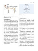
Fig. 6.15 Sequestration and lung malformations: (a) Schematic demonstration of the range
of foregut malformations, graded depending upon amount of tissue and/or vessel alteration.
(b) Typical sagittal US image of in this case an infradiaphragmatic echogenic sequestration in a
neonate with right-sided diaphragmatic hernia. NOTE: intrathoracic liver and slight effusion, as
well as preserved part of diaphragm covering sequestration. (c) CDS reveals the systemic vascular
supply, in this case deriving from a thoracic artery (chest wall vessel)
6.6.5
Sequestration
Definition: Lung tissue without function, usually without connecxion to tracheobronchial system. Part of spectrum of various congenital lung/foregut malformations (Fig. 6.15a).
Usually atypical vascular supply:
• Commonly from aorta, potentially draining into systemic vein
• Typically positioned in lower lobes, particularly left sided
US findings: Usually best seen from abdominal approach (Fig. 6.15b):
• More or less homogeneous, slightly echoic, space-occupying lesions
• May have complex inhomogeneous appearance with cysts (hybrid lesion)
• Large tubular structures often seen represent vessels
• Displaces lung, rarely also intraabdominal
6.6 Lung Pathology
207
Fig. 6.16 CCAM: CCAM type I (large macrocysts, + +) – difficult to differentiate from other
fluid filled/cystic mass (bronchogenic, cystic sequestration, etc.)
CDS: Commonly large-supplying artery deriving from abdominal aorta
(Fig. 6.15c):
• Draining vein depictable if drains into abdominal inferior cava; allows
differentiation of intra- versus extralobar sequestration
NOTE: Sequestration may also occur infradiaphragmatically.
Duplex Findings:
• If waveform resembles aorta/systemic veins, indicates systemic vascular supply
– extralobar sequestration
• If flow pattern resembles pulmonary artery/pulmonary vein, indicates vascular
supply from pulmonary circulation – intralobar sequestration
6.6.6
Congenital Cystic Adenomatoid Malformation (CCAM)
Definition: Part of spectrum of foregut malformations (see Fig. 6.15a). Typically
three types differentiated, depending on cyst size:
• Type 1 = large cyst(s) > 2 cm
• Type 2 = multiple medium-sized cysts around 1 cm
• Type 3 = pseudosolid mass with multiple microcysts not resolvable by US
US findings: More or less echoic, complex mass (Fig. 6.16):
• Varying number and size of cysts depending on type
• Between cysts there can be echogenic septae
CDS: No large vessels, some vascular supply sometimes seen
DDx: Atypical form of sequestration, hybrid lesion, bronchial atresia with
non-aerated malformed lung tissue, if more homogenous - thoracic kidney (usually
in the dorsal paramedian basal part of the chest)
208
6
a
b
Ultrasound of the Chest
c
Fig. 6.17 Cyst: (a) Fluid filled bronchial cyst - could be any kind of chest cyst adjacent to mediastinum. (b) Air-filled bronchogenic cyst. (c) Huge bronchial cyst connected to bronchial system
– air-fluid level
6.6.7
Cysts
Definition: Fluid- or (if connected to tracheobronchial system) air-containing
lesions:
• Seen only if positioned close to lung surface
US findings: Simple cyst(s) of varying size (Fig. 6.17a):
• Posterior acoustic enhancement, smooth surface
• Separation from other entities difficult
• If filled with air, bright reverberation echoes without change during respiratory
cycle, thus distinguishable from normal lung (Fig. 6.17b)
• Bronchial cysts – potentially air-fluid levels (may be difficult to depict)
(Fig. 6.17c)
DDx: Echinococcus/hydatid cyst (focus on typical wall appearance):
• Complicated cysts (echoes within lumen, aetiology not definable by US)
• Pericardial/pleural cysts
• Postinflammatory cysts, abscess, AV-malformation (use CDS)
6.6.8
Infarction
Definition: Pulmonary artery embolism PAE
US findings: Pneumonia-like subpleural triangular areas without perfusion
• Usually hypoechoic, liver-like appearance, often relatively homogeneous:
– Beginning – some ventilation possible
– Later stage – completely resemble pneumonia, but no pulmonary vessels
depicetd by (a) CDS
CDS: No central vascularisation/flow, no pulmonary vascular supply (Fig. 6.18):
• Some pleural vessels may be depicted.
6.6 Lung Pathology
209
a
b
c
d
e
Fig. 6.18 PAE and pulmonary perfusion deficit: (a) Axial view, infant after Glen procedure – with
peripheral pneumonia-like oedematous lung. (b) Same child as in (a): note severe postoperative
perfusion deficit of one lung. (c) Axial view through liver: Large infarction, no air sonobronchogram as would be seen with infection. (d) Dorsal scan through intercostals space: triangular subpleural pneumonia-like lung area, typical for infarction induced pneumonia. (e) CDS demonstrates
lack of perfusion in the peripheral triangular subpleural consolidation
NOTE: Perfusion deficit for other reasons (cardiac, postoperative, etc.) sometimes
difficult to distinguish, but tends to be more global, with less pronounced changes
of the lung echotexture.
210
6
Ultrasound of the Chest
Fig. 6.19 Lung tumour. Axial view: chest filled with partially cystic tumour that turned out to be
a pulmonary blastoma
6.6.9
Tumours and Space-Occupying Lesions
Definition: Lung tumours rare in children. US has limited role:
• One may visualise tumours if reaches lung surface
US findings: US findings vary depending on tumour and composition (Fig. 6.19):
• More or less echogenic, potentially necrotic areas or calcifications
• Sometimes origin depictable – allows speculation on aetiology
CDS: Evaluate vascularisation, depict necrosis, asses supplying/draining vessels:
• Pneumonia, atelectasis: CDS helpful for showing normal vascular supply
allowing differentiation from infarction or depiction of necrotic area before
typical abscess formations manifest
• Superficial/pleural/soft tissue arteriovenous malformation: CDS irreplaceable
for diagnosis
DDx: Any other cause of non-aerated lung, particularly CCAM, sequestration,
pneumonia, abscess, complicated cysts and hernia:
• In medial/mediastinal aspect: thymus versus lymphoma, etc.
• Extremely rare intrathoracic kidney (normal kidney in atypical location):
– Typical renal vascularisation pattern – undisputable diagnosis
6.7
Other Miscellaneous and Rare Applications
Many More Partially Rare Applications Reported: Most Relevant Ones
6.7.1
US for Interstitial Lung Disease
Increased extravascular lung water creates “B-line” on lung US; a few B-lines can be
found in a healthy population:
6.8
Additional Imaging
211
• B-lines are vertical, sharply defined hyperechoic lines and structures without
decrease in brightness with depth (ring-down artefact)
• Low-frequency convex transducer detects more B-lines than high-frequency linear transducers
• Multiple B-lines suggest interstitial lung syndrome
• Non-specific in terms of aetiology – wide variety of conditions (pulmonary
oedema, ARDS, pulmonary contusion, pneumonia, pulmonary fibrosis, etc.)
• B-lines correlate with CT abnormalities (e.g. thickened interlobular septa and
ground-glass opacities – closely packed B-lines)
6.7.2
US for Pneumothorax
Detection by US is possible and beneficial for patients in the emergency department
(ER) and on ventilation support (NICU, ICU, etc.)
US findings:
• To-and-fro motion during respiration in real-time US with B-mode (visceral
pleura or parietal pleura) “gliding” or “sliding” sign and M-mode “seashore” sign
• Additional findings: lung point, absence of B-lines and absence of lung
pulse
• Pneumothorax directly beneath the US transducer: no sliding sign in B-mode
and “stratosphere” sign on M-mode
NOTE: False-positive findings from hyperextended hyperinflated lung (COPD,
severe asthma, aspiration with focal emphysema/hyperinflation, etc.)
6.8
Additional Imaging
Plain film, CT, sometimes (and increasingly) MRI:
• Rarely angiography (vascular malformations) or fluoroscopy
• Role/value of US exquisite for follow-up of effusions or diaphragmatic
palsy:
– Image-guided interventions: diagnostic or therapeutic puncture (in effusions,
abscess, chylothorax, haematothorax, tumour biopsy, etc.)
NOTE: US has limitations in the chest – complementary imaging tool. Plain film
often initially compulsory. US often used in equivocal findings (e.g. white haemithorax) (Fig. 6.20). Always include assessment of lung base in upper abdominal US
and FAST examinations.
212
6
a
b
c
d
Ultrasound of the Chest
Fig. 6.20 Case examples where US was helpful to further define the cause of equivocal chest film
findings: (a, b) White haemithorax on chest film (a): US (b) reveals a partially atelectatic, partially
pneumonic lung with elevated position of the diaphragm. The pulmonary vessels are well
perfused on CDS, no sign of tumour, only slight effusion. (c) Atypical opacification of right
lower lung on plain film: US (d) demonstrates collapsed lung with secondary pneumonic changes
in a child after aspiration
7
Liver and Bile System
Michael Riccabona
Contents
7.1 Requisites and Investigation............................................................................................
7.1.1 Preparation.........................................................................................................
7.1.2 Positioning .........................................................................................................
7.1.3 Transducers........................................................................................................
7.1.4 Course of Investigation......................................................................................
7.1.5 Standard Planes .................................................................................................
7.2 Normal Findings .............................................................................................................
7.2.1 Structure ............................................................................................................
7.2.2 Ligaments ..........................................................................................................
7.2.3 Hepatic Veins (HV) ...........................................................................................
7.2.4 Portal Vein (PV) ................................................................................................
7.2.5 Hepatic Artery (HA)..........................................................................................
7.2.6 Gall Bladder ......................................................................................................
7.2.7 Common Bile Duct (Commonly Addressed as Hepato-Choledochal Duct) .....
7.2.8 Intrahepatic Bile Ducts ......................................................................................
7.2.9 Doppler Findings ...............................................................................................
7.2.10 Special Aspects of Newborns and Infants .........................................................
7.3 Pathology of the Liver .....................................................................................................
7.3.1 Congenital Changes and Normal Variance........................................................
7.3.2 Inflammatory Conditions...................................................................................
7.3.3 Other Parenchymal Liver Disease .....................................................................
7.3.4 Portal Hypertension and Vascular Problems .....................................................
7.3.5 Liver Trauma .....................................................................................................
7.3.6 Space-Occupying Liver Lesions........................................................................
214
214
214
214
215
215
216
216
217
217
217
218
218
218
219
219
220
222
222
223
225
230
234
237
M. Riccabona
Division of Pediatric Radiology, Department of Radiology,
University Hospital Graz, Auenbruggerplatz 3,
Graz 8036, Austria
e-mail:
M. Riccabona, Pediatric Ultrasound,
DOI 10.1007/978-3-642-39156-9_7, © Springer Berlin Heidelberg 2014
213