Ebook Introduction to sectional anatomy (3rd edition): Part 2
Bạn đang xem bản rút gọn của tài liệu. Xem và tải ngay bản đầy đủ của tài liệu tại đây (38.28 MB, 313 trang )
Madden_3e_CH06.qxd:Layout 1 8/3/12 2:59 AM Page 319
CHAPTER
ABDOMEN
6 Abdomen
OBJECTIVES
Upon completion of this chapter, the student should be able to do the following:
1. Describe the superior and inferior boundaries of the abdomen.
2. Describe the general location of the segments of the small and large intestines within the abdomen.
3. Identify and describe the location and lobes of the liver.
4. Describe the enclosing structures separating the abdomen.
5. Explain the location and general function of the gallbladder, pancreas, spleen, adrenal glands, and kidneys.
6. Describe the bile duct system.
7. Follow the course of blood as it passes through the portal system.
8. Describe the major arteries and veins located within the lower chest and abdomen.
9. Explain the relationships between structures located within the abdomen.
10. Correctly identify anatomic structures on patient computed tomography (CT) images of the abdomen.
common anomalies that may confuse the viewer when determining image location. Compared to the other vertebrae,
these can be distinguished by their large size and the
absence of costal facets and transverse foramina.
ANATOMIC OVERVIEW
The abdomen is generally considered as the region of the
body between the chest and pelvis. Although this seems
quite simple, the boundaries of the abdomen are often
defined differently by different texts because the abdominal
cavity extends well into each of the adjacent regions. The
most superior boundary of the abdominal cavity is the
dome-shaped diaphragm, which allows a considerable part
of the abdomen to lie within the bony thoracic cage. Inferiorly, the abdominal cavity extends into the pelvis and
occupies most of the false or greater pelvis, leading some
individuals to consider the pelvis as the lower part of the
abdomen. Because the abdomen and pelvis are often
imaged separately, the pelvis will be further described in
the next chapter.
Enclosing Structures
Diaphragm (DI¯ -a˘ -fram). The diaphragm is a broad, flat
muscle made up of skeletal muscle along the periphery
that converges on a broad flat tendon, the central tendon
(Fig. 6-1). It is often described as two hemidiaphragms
(the right and left) because the right side is usually more
superior because of the underlying liver. Its muscular portion originates from several sources: (1) the sternal
process, (2) the costal cartilages and bone of ribs 7
through 12, and (3) the upper lumbar vertebrae. Although
the diaphragm forms a septum between the thoracic and
abdominal cavities, several structures (inferior vena cava,
esophagus, and descending aorta) pass through openings
within the diaphragm to pass between the chest and
abdomen.
Crura (KRU˘-ra˘ ). The muscular parts of the diaphragm
that originate from the lumbar vertebrae and ascend to
Skeleton
Lumbar (LU˘M-bar) vertebrae. Typically, the vertebral
column contains five lumbar vertebrae, which form the posterior border of the abdominal cavity. Owing to the highly
variable division of lumbar vertebrae with adjacent thoracic
and sacral vertebrae, four and six lumbar vertebrae are
319
/>
/>
Madden_3e_CH06.qxd:Layout 1 8/3/12 2:59 AM Page 320
320
Introduction to Sectional Anatomy
Figure 6-1 The inferior surface of the diaphragm.
the central tendon. The right crus arises from the upper
three or four lumbar vertebrae, and the left crus originates from the upper two or three. The crura combine
with ligaments to form the openings for the aorta and
esophagus.
Peritoneum (PER-i-o˘-NE¯-um). Its structure and function
are similar to those of the pleura, described in Chapter 5
(Fig. 6-2). It is a smooth membrane lining the abdominal
cavity (parietal peritoneum) and the abdominal viscera (visceral peritoneum), creating the peritoneal cavity. Because
the organs within the abdominal cavity are closely arranged,
the peritoneal cavity is normally only a small space containing a thin film of serous fluid produced by the membranes.
Much like the pleura, the peritoneum minimizes friction
and acts as a barrier to the spread of infection within the
abdomen.
Mesentery (MES-en-ter-e¯). In addition to the parietal and visceral peritoneum, the mesentery is a double
layer of peritoneum that encloses the viscera and
attaches it to the abdominal wall. Because of constant
movement and changes in shape, much of the intestine
is described as having no fixed position, being only
loosely organized by the mesentery. The mesentery also
contains the arteries, veins and nerves that supply the
intestines and is a primary site for fat storage within
the body.
Retroperitoneal (RE-tro¯-PER-i-to¯-NE¯ -a˘ l). Behind the
peritoneal cavity, this space is adjacent to the posterior
abdominal wall and contains the following abdominal
organs: kidneys, pancreas, distal duodenum, and ascending
and descending portions of the colon.
Viscera (VIS-er-a˘)
Stomach. A mobile organ situated in the upper left side of
the abdominal cavity just below the left hemidiaphragm.
The esophagus descends through the esophageal hiatus in
the diaphragm to join the body of the stomach. Above the
gastroesophageal junction, the fundus is the part of the
stomach found next to the esophagus directly under the diaphragm. Below the body of the stomach, the pyloric part is
the narrowing region that is continuous with the duodenum
(Fig. 6-3). Although the location and shape of the stomach
will vary among individuals and can change over time within
a single individual, the relationship of the three segments
from superior to inferior will usually remain the same.
Small intestine. The site of the major part of digestion.
It extends from the termination of the stomach to the large
intestine, ranging from 5 to 8 m in length. It includes the
duodenum, jejunum, and ileum.
Duodenum (du¯-o¯-DE¯-nu˘m). The first segment of the
small intestine, extending from the pyloric part of the
/>
Madden_3e_CH06.qxd:Layout 1 8/3/12 2:59 AM Page 321
321
ABDOMEN
Chapter 6 / Abdomen
Figure 6-2 A median sagittal view of the abdomen demonstrating the
peritoneum and mesentery.
stomach to the jejunum. It is approximately 25-cm long.
Its C shape wraps around the head of the pancreas and the
superior mesenteric vessels (Fig. 6-4). Only the superior
part of the duodenum lies within the peritoneum; the
remaining three parts (descending, inferior, and ascending) are all retroperitoneal and are fixed in position.
Jejunum (je˘-JU¯-nu˘m). The second segment of the small
intestine is arranged in numerous coils or loops, is approximately 2.4-m long, and extends from the duodenum to the
ileum (Fig. 6-5). It is difficult to distinguish from the ileum,
even though it has a thicker wall, greater diameter, and
larger vascular supply. In the average patient, location typically provides a general means for distinguishing between
the jejunum and ileum; the jejunum usually lies in the umbilical region, whereas the ileum lies in the lower abdomen
and pelvis.
Ileum (IL-e¯-u˘m). The third segment is also arranged in
numerous coils or loops and is the longest segment of the
small intestine, averaging 3.6 m in length. As noted, the
ileum is difficult to distinguish from the jejunum, except for
its lower position in the abdominal cavity. It terminates in
the lower right quadrant of the abdominal cavity at the ileocecal valve and is continuous with the first part of the large
intestine. Helpful hint: The spelling of the ileum of the
intestine is often confused with the ilium of the bony pelvis.
If one notes that the shape of the coiled intestine resembles the letter e, then one should remember the proper
spelling for both anatomic structures.
Large intestine. The large intestine is approximately 1.5 m
in length and extends from the terminal ileum to the anus
(Figs. 6-5 and 6-6). The material passing from the terminal
ileum to the large intestine is about 90% water, most of
which is absorbed by the large intestine. Many individuals
will use the term colon synonymously with large intestine;
however, this is incorrect. The large intestine is made up of
two parts: the cecum and the colon.
/>
/>
Madden_3e_CH06.qxd:Layout 1 8/3/12 2:59 AM Page 322
322
Introduction to Sectional Anatomy
Figure 6-3 A sketch illustrating the three parts of the
stomach.
Cecum (SE¯-ku˘m). The first segment of the large intestine located in the lower right side of the abdomen posterior to the peritoneum. It is below the ileocecal valve
and forms a blind pouch that is continuous with the ascending colon. At 1 to 2 cm below the opening of the ileocecal valve within the cecum, a smaller opening leads into
the appendix. The appendix is a long narrow tube averaging about 8 cm in length with a highly variable position
that partially depends on the shape and contents of the
cecum.
Ascending colon. The segment originating above the
ileocecal valve that is continuous with the cecum and
extends upward to the hepatic flexure next to the liver on
the right side of the abdomen. Similar to the cecum, it is
retroperitoneal and relatively fixed in position along the
posterior wall of the abdomen. In the lower abdomen, it lies
adjacent to the musculature forming the posterior abdominal wall; in the upper abdomen, it lies anterior to the right
kidney.
Hepatic (he-PAT-ik) flexure of colon. The bend or right
flexure of the colon between the ascending and transverse
Figure 6-4 An anterior view of the structures within the upper abdominal cavity after removal of the stomach, jejunum, and
transverse colon.
/>
Madden_3e_CH06.qxd:Layout 1 8/3/12 2:59 AM Page 323
323
ABDOMEN
Chapter 6 / Abdomen
Figure 6-5 An anterior view of the contents within the abdomen following removal
and reflection of the anterior abdominal wall.
Figure 6-6 A sketch demonstrating the location of the large intestine as compared
to the selected structures adjacent to the posterior abdominal wall.
/>
/>
Madden_3e_CH06.qxd:Layout 1 8/3/12 2:59 AM Page 324
324
Introduction to Sectional Anatomy
segments of the colon. As the name implies, the flexure is
next to the liver on the upper right side of the abdomen.
Owing to the more anterior position of the transverse colon,
the hepatic flexure is best demonstrated in an oblique view
from the right anterior side.
Transverse colon. The segment of the colon traversing
across the abdomen between the hepatic and splenic
flexures. In contrast to the ascending colon, it is invested
with peritoneum and is suspended from the posterior abdominal wall by mesentery (the transverse mesocolon).
Although the ends have a fixed position, the location of the
middle region is highly variable and may be found from the
upper abdomen to the greater pelvis. Despite the level, the
middle region usually lies adjacent to the anterior abdominal wall.
Splenic (SPLEN-ik) flexure of colon. At the terminal
end of the transverse colon, the left flexure of the colon
redirects the colon downward to become the descending
colon. Unlike the hepatic flexure, this flexure is best
demonstrated in the oblique view from the left anterior
side and is usually more superiorly situated, adjacent to
the spleen.
Descending colon. The part of the large intestine originating at the splenic flexure that extends along the left
posterior wall to the level of the pelvic brim or inlet. Within
the greater pelvis, it travels downward to join the sigmoid
colon. Similar to the ascending colon, it is retroperitoneal
and is fixed in position by the musculature of the posterior
abdominal wall.
Liver. The largest gland in the body, found in the upper
abdominal cavity on the right side. For the most part, it lies
within the bony thoracic cage, and its superior surface is
covered by the diaphragm. The superior liver is dome
shaped, following the contour of the diaphragm, and the inferior or visceral surface is somewhat flattened, facing
downward toward the other viscera within the abdomen.
On the visceral surface, an H-shaped arrangement of fissures and fossae is found dividing the liver into four separate lobes (Fig. 6-7). The transverse part of the H is formed
by the porta hepatis, which includes the hepatic ducts, portal vein, and proper hepatic artery. The sides of the H are
formed by the gallbladder and the inferior vena cava on the
left side and the ligamentum teres (obliterated remains of
the left umbilical vein) and ligamentum venosum (the
Figure 6-7 The visceral surface of the liver as seen from below.
/>
Madden_3e_CH06.qxd:Layout 1 8/3/12 2:59 AM Page 325
fibrous remains of the embryologic ductus venosus) on the
right side.
Left lobe. The left part of the liver demarcated on the
diaphragmatic surface by the falciform ligament. On the
visceral side, the ligamentum teres in front and the ligamentum venosum in back form the boundary for the left lobe.
In the abdomen, the left lobe of the liver usually lies anterior to the body of the stomach.
Right lobe. The largest part of the liver opposite the left
lobe. On the visceral surface, the hepatic flexure of the
colon lies near the anterior part of the right lobe and lateral
to the gallbladder.
Caudate (KAW-da¯t) lobe. The small, posterior lobe located between the inferior vena cava and the ligamentum
venosum, posterior to the porta hepatis. Helpful hint: The
“c” in caudate can help you remember that it lies next to
the inferior vena cava (also starts with a “c”).
Quadrate (KWAH-dra˘t) lobe. The small, anterior lobe
located between the gallbladder and the ligamentum teres.
Helpful hint: The “q” in quadrate is shaped much like the
“g” in gallbladder.
325
Gallbladder. Lies just below the anterior liver within its
fossa on the visceral surface and acts as a reservoir for bile
produced by the liver.
Common bile duct. Transports bile from the gallbladder
(via the cystic duct) and the liver (via the hepatic duct)
to the duodenum (Fig. 6-8). In its course, it lies posterior to
the superior duodenum and beside the head of the pancreas. It is approximately 7.5 cm in length and ends at the
duodenal wall, where it joins with the main pancreatic duct.
Pancreas (PAN-kre¯-as). A collection of glandular tissue
with little connective tissue, it has both exocrine and endocrine functions (Figs. 6-8 and 6-9).
Head. The expanded part of the pancreas lying within
the curvature of the duodenum. Because the pancreas is
covered only on its anterior surface by peritoneum, it is
considered retroperitoneal similar to the adjacent parts of
the duodenum. The head of the pancreas is divided by the
superior mesenteric artery and vein that partially separate
the uncinate process, the part of the pancreas located inferior to the mesenteric vessels.
Figure 6-8 A drawing from an anterior view illustrating the bile duct
system and adjacent structures.
/>
/>
ABDOMEN
Chapter 6 / Abdomen
Madden_3e_CH06.qxd:Layout 1 8/3/12 2:59 AM Page 326
326
Introduction to Sectional Anatomy
Figure 6-9
A drawing from a posterior view illustrating the pancreas and adjacent structures.
Body. The central region of the pancreas primarily located posterior to the stomach and anterior to the left kidney.
Tail. The narrowed left end of the pancreas extending
toward the surface of the spleen.
Spleen (sple¯n). The soft, lymphatic organ that lies against
the diaphragm on the upper left side of the abdomen within
the thoracic cage (Fig. 6-4). Its size and shape vary considerably, depending somewhat on the adjacent structures. Its
anterior surface is next to the stomach, its posterior surface
is next to the left kidney, its superior surface is next to the
diaphragm, and its inferior surface is next to the left splenic
flexure of the colon.
Kidneys. The bean-shaped, retroperitoneal organs on either side of the vertebral column typically centered at the
level of the 1st lumbar vertebra. Anomalies in formation are
common during development, resulting in variations in the
shape and location of the kidneys. Within the kidney, fluid
and waste products are filtered from the blood to form
urine, which is collected in the renal pelvis and drains into
the ureters (Fig. 6-10).
Ureters (yu¯-RE¯ -terz). Retroperitoneal, originating from
the renal pelvis and extending downward to drain urine into
the bladder. Although most people have two ureters (one
for each kidney), common congenital anomalies include
duplication of part or all of the ureter.
Adrenal (a˘-DRE¯-na˘l) glands. Also referred to as the
suprarenal glands, these soft, glandular organs are located
on the top pole of the kidneys (Fig. 6-11). Roughly pyramidal in shape, their average dimensions in the adult are
approximately 5-cm long, 3-cm wide, and 1-cm thick.
Although these endocrine glands are relatively small, they
produce hormones with widespread effects, including epinephrine and norepinephrine, which are responsible for the
fight-or-flight response. In axial images, the glands are considerably thinner and are less dense than the underlying kidneys (which average 3-cm thick).
Arteries
Abdominal or descending aorta (a¯-O¯R-ta˘). The continuation of the thoracic aorta, it originates at the level of the
diaphragm and extends to the pelvis (Fig. 6-6). The
retroperitoneal artery lies on the left side of the vertebral
column and terminates at the origin of the right and left
common iliac arteries.
/>
Madden_3e_CH06.qxd:Layout 1 8/3/12 2:59 AM Page 327
327
ABDOMEN
Chapter 6 / Abdomen
Figure 6-10 A sketch illustrating the contents of the kidney.
Figure 6-11 The adrenal gland and kidney with adjoining
structures.
Celiac (SE¯-le¯-ak) trunk. The first branch off the abdominal aorta, it originates just below the diaphragm between
the lesser curvature of the stomach and the liver
(Fig. 6-12). The artery is relatively short (1 to 2 cm long)
and originates nearly perpendicular to the aorta. It gives rise
to the common hepatic artery, left gastric artery, and splenic
artery.
Common hepatic artery. The branch of the celiac trunk
that gives rise to the proper hepatic artery (supplies the liver
and gallbladder) and the gastroduodenal artery (supplies
the stomach, duodenum, and pancreas). Anomalies of the
artery are quite common. Approximately 41% of patients
have aberrant common hepatic arteries, including instances
in which the artery originates directly from the aorta or the
superior mesenteric artery.
Splenic artery. The largest branch of the celiac trunk,
it travels behind the stomach to end at the spleen. It usually
travels a tortuous path, giving it a distinctive appearance
and facilitating its identification in sectional images.
Superior mesenteric artery. It originates from the
abdominal aorta approximately 1 cm below the celiac
trunk. It extends downward to supply blood to the small
intestine and the first half of the large intestine, including
the cecum, the ascending colon, and the right half of the
transverse colon (Fig. 6-13). Originating posterior to the
pyloric part of the stomach, it extends at an oblique angle
from the aorta. Compared to the perpendicular origin of
/>
/>
Madden_3e_CH06.qxd:Layout 1 8/3/12 2:59 AM Page 328
328
Introduction to Sectional Anatomy
Figure 6-12
Branches of the celiac trunk as compared to the stomach and spleen.
the nearby celiac trunk, its oblique course can be a distinguishing characteristic in sectional images. As the artery
descends into the abdomen, it travels through the head of
the pancreas within the C loop of the duodenum to enter
the mesentery.
Renal (RE¯ -na˘ l) arteries. Two large trunks arising on
either side of the aorta just below the superior mesenteric
artery. Each artery forms a nearly right angle with the aorta
as it extends to the kidneys (Fig. 6-14). Because the right
renal artery passes behind the inferior vena cava and the
right renal vein, it is usually slightly longer than the left.
In approximately one in four cases, additional renal arteries
are present and are more frequently found on the left side.
Instead of entering the kidney at the hilum, additional renal
arteries usually join with either the upper or the lower poles
of the kidney.
Inferior mesenteric artery. Originating from the aorta
in the mid-lumbar region, it enters the mesentery to
supply blood to the left half of the transverse colon,
descending colon, sigmoid colon, and upper rectum
(Fig. 6-15).
Common iliac arteries. Bilateral arteries arising from the
abdominal aorta at the level of the 4th lumbar vertebra; they
diverge laterally as they enter the pelvis. Within the greater
pelvis, each artery bifurcates to give rise to the internal and
external iliac arteries.
Veins
Inferior vena cava (VE¯ -na˘ -KA¯ -va˘). The major route
for drainage of venous blood from the abdomen, pelvis,
and lower extremities (Fig. 6-16). It lies parallel to the
/>
Madden_3e_CH06.qxd:Layout 1 8/3/12 2:59 AM Page 329
329
ABDOMEN
Chapter 6 / Abdomen
Figure 6-13 Following superior reflection of the transverse colon, the branches of the superior mesenteric artery.
Figure 6-14 A sketch illustrating the renal arteries and veins.
/>
/>
Madden_3e_CH06.qxd:Layout 1 8/3/12 2:59 AM Page 330
330
Introduction to Sectional Anatomy
Figure 6-15 The lower abdominal aorta including the branches of the inferior
mesenteric artery and the common iliac arteries.
/>
Madden_3e_CH06.qxd:Layout 1 8/3/12 2:59 AM Page 331
331
ABDOMEN
Chapter 6 / Abdomen
Figure 6-16 The veins of the abdomen.
/>
/>
Madden_3e_CH06.qxd:Layout 1 8/3/12 2:59 AM Page 332
332
Introduction to Sectional Anatomy
Figure 6-17 The posterior abdominal wall and adjacent structures.
abdominal aorta, on the right side near the lumbar vertebral bodies. Originating from the joining of the common
iliac veins within the upper pelvis, it ascends through the
abdomen and thoracic cavity to drain into the right atrium
of the heart.
Hepatic veins. The right and left hepatic veins drain
the filtered blood from the liver into the inferior vena cava.
The vessels are short and are surrounded by liver tissue
molded around the inferior vena cava.
Portal (PO¯ R-ta˘l) vein. Originating from the veins
draining most of the gastrointestinal system, it carries
nutrient-rich blood to the middle of the visceral surface
of the liver. Lying adjacent to the hepatic bile ducts and
the hepatic artery proper, it forms part of the porta hepatis, the transverse part of the H on the visceral surface
of the liver.
Splenic vein. Found traversing the abdomen posterior to
the stomach and the pancreas, it drains nutrient-rich blood
from the spleen and the inferior mesenteric vein into the
portal vein. In contrast to the tortuous path of the splenic
artery, the course of the vein is nearly linear; this difference can be used to distinguish the two neighboring
vessels.
Inferior mesenteric vein. The vessel draining blood from
the rectum, sigmoid colon, and descending colon to the
splenic vein located posterior to the stomach and pancreas.
During its course, the vein lies within the mesentery, attaching the intestine to the posterior abdominal wall.
Superior mesenteric vein. Ending at the portal vein immediately posterior to the pancreas, the branches of this
vessel drain blood from the stomach, duodenum, jejunum,
ileum, cecum, appendix, ascending colon, transverse colon,
and pancreas. Like the other mesenteric veins, it lies within
the mesentery and carries nutrient-filled venous blood from
the intestine to the portal vein.
Renal veins. The right and left renal veins drain venous
blood from the kidneys to the inferior vena cava (Fig. 6-17).
Because the abdominal aorta is on the left side of the inferior vena cava, the longer left renal vein crosses anterior to
the abdominal aorta.
/>
Madden_3e_CH06.qxd:Layout 1 8/3/12 2:59 AM Page 333
Chapter 6 / Abdomen
Muscles
Psoas (SO¯-as). Originating from the transverse processes
of L1 to L5 and inserting on the lesser trochanter of the
femur on either side, these muscles form part of the posterior abdominal wall. In axial section, the large muscles are
round and readily identified lying on either side of the vertebral column and aid in the identification of the adjacent
ureters and vessels.
/>
/>
ABDOMEN
Common iliac veins. These two veins (the right and left)
drain venous blood from the lower limbs and pelvis into the
inferior vena cava. Arising at the juncture of the internal
and external iliac veins, they originate anterior to the L5 to
S1 joint space and extend only a short distance to join in
front of the L4 vertebral body. Unlike most regions of the
body, here the veins are more posteriorly and inferiorly situated than are the adjacent common iliac arteries.
333
Madden_3e_CH06.qxd:Layout 1 8/3/12 2:59 AM Page 334
334
Introduction to Sectional Anatomy
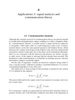
At the top of the abdomen, the liver is shown occupying most of the right side surrounded by the lower lobe of the right lung. On the left side, the lower lobe of the
lung forms a margin around the contents of the upper abdomen. Within the window, the upper pole of the dense spleen and the contrast-filled fundus of the stomach are both demonstrated. Within the mediastinum, the bottom of the heart is
sectioned, and the right ventricle is more anterior than the left ventricle. Behind
the heart, the esophagus is found extending downward to the stomach in front of
the descending aorta and the azygos vein. On the right side of the patient, the inferior vena cava is difficult to discern from the surrounding liver tissue.
1. Rt ventricle
2. Lt ventricle
11. Inf vena cava
3. Fundus
of stomach
10. Liver
4. Spleen
5. Lt lung
9. Rt lung
8. Esophagus
A
Figure 6-18
B
6. Descending aorta
7. Azygos V
(A,B) Axial computed tomography (CT) image 1.
/>
Madden_3e_CH06.qxd:Layout 1 8/3/12 2:59 AM Page 335
1. Heart
6. Abdominal aorta
5. Sup mesenteric A
4. Common iliac A
2. Internal iliac A
3. External iliac A
Figure 6-19
Anterior magnetic resonance angiogram (MRA) view of blood flow within the
abdominal aorta.
A
Figure 6-20
B
(A) Longitudinal sonogram through the proximal abdominal
aorta (a) demonstrates the origins of the celiac axis and superior mesenteric artery (sma) (^^^, anterior aspect of the vertebral bodies; L, liver) (B) Longitudinal sonogram through the
middle portion of the abdominal aorta (a). (C) Longitudinal
sonogram through the distal portion of the abdominal aorta
(a). Note tapering of the vessel at this point.
/>
C
/>
335
ABDOMEN
Chapter 6 / Abdomen
Madden_3e_CH06.qxd:Layout 1 8/3/12 2:59 AM Page 336
336
Introduction to Sectional Anatomy
The body of the liver fills most of the right side and is difficult to distinguish from
the base of the heart. Even though the diaphragm is not seen between the two organs, the interventricular septum can be seen separating the right and left ventricles of the heart. Next to the heart, the fundus of the stomach, filled with contrast,
is shown on the left side. Although the esophagus is still between the descending
aorta and inferior vena cava, the fundus of the stomach is also found within this
session. Posterior to the stomach, the spleen appears as a dense organ bordered
by the lower lobe of the left lung.
1. Rt ventricle
2. Lt ventricle
11. Liver
3. Fundus
of stomach
10. Inf
vena cava
4. Spleen
5. Lt lung
9. Rt lung
A
Figure 6-21
B
8. Azygos V
7. Esophagus
(A,B) Axial computed tomography (CT) image 2.
/>
6. Descending aorta
Madden_3e_CH06.qxd:Layout 1 8/3/12 2:59 AM Page 337
337
ABDOMEN
Chapter 6 / Abdomen
7. Celiac A
1. Abdominal aorta
6. Sup mesenteric A
2. Lt renal A
5. Common iliac A
3. Int iliac A
4. Ext iliac A
Figure 6-22
Oblique magnetic resonance angiogram (MRA) of blood flow within the abdominal
aorta image 1.
5. Pyloric part
of stomach
1. Fundus of stomach
4. Hepatic flexure
2. Splenic flexure
of colon
3. Ascending colon
Figure 6-23
Computed tomography (CT) abdomen coronal image 1.
/>
/>
Madden_3e_CH06.qxd:Layout 1 8/3/12 2:59 AM Page 338
338
Introduction to Sectional Anatomy
Unlike the previous image, the liver occupies most of the right side and extends
through the midline to lie beside the fundus of the stomach. The esophagus, no
longer between the inferior vena cava and descending aorta, is near the point
where it joins the stomach. On the left side, the costodiaphragmatic recess of the
lung forms a margin around the spleen. Between the lungs, the small azygos and
hemiazygos veins are cross-sectioned on either side of the descending aorta.
1. Fundus of stomach
9. Liver
2. Spleen
8. Inf
vena cava
3. Lt lung
7. Esophagus
A
Figure 6-24
B
4. Hemiazygos V
6. Azygos V 5. Descending aorta
(A,B) Axial computed tomography (CT) image 3.
/>
Madden_3e_CH06.qxd:Layout 1 8/3/12 2:59 AM Page 339
339
ABDOMEN
Chapter 6 / Abdomen
Figure 6-25
Transverse sonogram through the inferior vena cava (IVC ) at a point just
below the right atrium of the heart
demonstrating the hepatic veins (rhv,
right hepatic vein; mhv, middle hepatic
vein; lhv, left hepatic vein; L, liver).
7. Gallbladder
1. Liver
6. Hepatic flexure
of colon
5. Ascending colon
2. Kidney
4. Cecum
3. Ileum
Figure 6-26 Computed tomography (CT) abdomen sagittal image 1.
/>
/>
Madden_3e_CH06.qxd:Layout 1 8/3/12 2:59 AM Page 340
340
Introduction to Sectional Anatomy
Similar to the previous image, the liver occupies the majority of the abdominal
cavity. The right and left lobes of the liver can now be identified. In this section, the
esophagus joins the stomach, marking the middle portion of the stomach (the
body). The inferior vena cava cannot be clearly distinguished from the liver and is
separated from the descending aorta by the right crus of the diaphragm. On either side of the descending aorta, the hemiazygos and azygos veins are clearly
seen anterior to the vertebral body. Along the posterior wall of the thoracic cage,
the costodiaphragmatic recesses of the lungs form a narrow margin around the
liver and spleen.
10. Lt lobe of liver
1. Body of stomach
9. Rt lobe
of liver
2. Gastroesophageal
junction
8. Inf
vena cava
3. Spleen
7. Rt crus of
diaphragm
A
Figure 6-27
B
6. Azygos V 5. Descending aorta
4. Hemiazygos V
(A,B) Axial computed tomography (CT) image 4.
/>
Madden_3e_CH06.qxd:Layout 1 8/3/12 2:59 AM Page 341
341
ABDOMEN
Chapter 6 / Abdomen
L
Posterior
branch
LPv
RPv
IVC
Ao
K
A
A
B
Figure 6-28 Transverse view within the liver. The portal vein branches into the left and right portal veins. The right vein again
bifurcates the posterior branch supplying the posterior right lobe of the liver. (A) Diagram. (B) Ultrasound image.
1. Lt lobe of liver
7. Rt lobe of liver
2. Fundus
of stomach
6. Pyloric part
of stomach
3. Small bowel
5. Ascending colon
4. Bladder
Figure 6-29 Computed tomography (CT) abdomen coronal image 2.
/>
/>
Madden_3e_CH06.qxd:Layout 1 8/3/12 2:59 AM Page 342
342
Introduction to Sectional Anatomy
The liver is limited to the right side of the abdomen and is divided into right and left
lobes by the fossa for the ligamentum teres. The portal vein is within the porta hepatis, as described earlier, forming the transverse part of the H on the visceral surface of the liver. The caudate lobe of the liver is between the porta hepatis and the
inferior vena cava. As in the previous image, the inferior vena cava is separated
from the descending aorta by the right crus of the diaphragm. Behind the descending aorta, the azygos and hemiazygos veins traverse through the diaphragm
and are bordered by crural fibers. On the left side, an air–fluid level is shown in the
contrast-filled stomach. Lateral to the stomach, the splenic flexure of the colon is
now anterior to the spleen.
13. Lt lobe of liver
1. Air in stomach
12. Ligamentum
teres fossa
11. Portal V
2. Splenic
flexure of
colon
10. Caudate
lobe of liver
3. Spleen
9. Inf
vena cava
4. Lt crus of diaphragm
A
Figure 6-30
B
7. Azygos V
5. Hemiazygos V
6. Descending aorta
8. Rt crus of diaphragm
(A,B) Axial computed tomography (CT) image 5.
/>
Madden_3e_CH06.qxd:Layout 1 8/3/12 2:59 AM Page 343
343
7. Pyloric part
of stomach
1. Fundus of stomach
6. Gallbladder
2. Small bowel
5. Ascending colon
4. Cecum
3. Bladder
Figure 6-31 Computed tomography (CT) abdomen coronal image 3.
1. Liver
7. Gallbladder
2. Kidney
6. Jejunum
5. Cecum
4. Ileum
Figure 6-32 Computed tomography (CT) abdomen sagittal image 2.
/>
/>
3. Psoas M
ABDOMEN
Chapter 6 / Abdomen