Ebook Color atlas of ENT diagnosis (4/E): Part 2
Bạn đang xem bản rút gọn của tài liệu. Xem và tải ngay bản đầy đủ của tài liệu tại đây (31.19 MB, 128 trang )
131
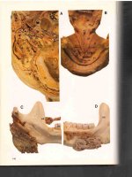
Inflammation: nasal vestibulitis
Fig. 3.47 Vestibulitis presents as crusting and irritation in the anterior nares with
resulting nasal obstruction. Examination shows excoriated vestibular skin and septal mucous membrane. Rubbing or over-diligent cleaning of the nose by the patient
usually causes vestibulitis, particularly if, as in this case, the septum is deviated
anteriorly and impinges on the lateral wall of the nose. Advice and the use of
antibiotic and corticosteroid ointment are effective in controlling vestibulitis. Correction of the septum may be necessary.
Fig. 3.48 Nasal vestibulitis
with squamous epithelium replacing the mucosa.
A deviation of the septum
has predisposed to a chronic vestibulitis. Digital irritation, or the use of cocaine,
which may also lead to a
septal perforation, may
underlie this problem.
Bull, Color Atlas of ENT Diagnosis © 2003 Thieme
All rights reserved. Usage subject to terms and conditions of license.
132 The Nose
Fig. 3.49 Vestibulitis in a
child overlying a grossly
deviated anterior septum.
Septal surgery is avoided in
children, but cases in which
the obstruction is gross
require a conservative septoplasty. Exessive cartilage
resection may retard nasal
growth, predisposing to
saddling or an infantile nose
(Fig 3.27a).
Fig. 3.50 Vestibulitis. Painful crusting of the nasal vestibule and anterior nares
may be a simple eczematous type of skin lesion which settles with a topical antibiotic and steroid ointment. There should, however, be an awareness that this
vestibulitis is a granuloma, or part of the manifestation of systemic disease such as
polyarteritis nodosa or systemic lupus erythematosus. A further possibility is an
“irritative” vestibulitis from cocaine snuff, or columellar carcinoma, as in this case
Fig. 3.51 Granular rhinitis.
Granulation tissues in the
nose requires biopsy. Sarcoidosis not infrequently
involves the upper respiratory tract mucosa of the
nasal fossae and larynx. In
the nose the granulations
are pale, but tuberculosis,
malignant granuloma, and
neoplasia are among the
differential diagnoses.
Bull, Color Atlas of ENT Diagnosis © 2003 Thieme
All rights reserved. Usage subject to terms and conditions of license.
Inflammation: nasal vestibulitis
133
Fig. 3.52 Nasal adhesion. Adhesion or synechiae may follow nasal trauma (including surgical trauma) and bridge the lateral wall of the nose, frequently from the
inferior turbinate to the septum, causing nasal obstruction. Recurrence follows surgical division of the larger adhesions unless an indwelling silastic splint is left in situ
until mucosa underlying the adhesion regenerates.
a
b
Fig. 3.53 Furuncles and cellulitis of the columella (a). These may spread to
involve the skin of the nose and face (b). Treatment is with systemic penicillin.
Bull, Color Atlas of ENT Diagnosis © 2003 Thieme
All rights reserved. Usage subject to terms and conditions of license.
134 The Nose
a
b
c
Fig. 3.54a-c Acute rhinitis. In the common cold, the nasal mucous membrane is
edematous, so the inferior turbinate abuts against the septum to result in obstruction and an excess of mucous which causes the running nose.
A similar appearance is seen in nasal allergy, either “seasonal hay fever” or
perennial allergy, but the edematous turbinate mucous membrane appears gray (c)
rather than red (b). A persistent purulent nasal discharge usually means that there
is a sinusitis. Corticosteroid nasal sprays for nasal allergy reduce the obstruction,
rhinorrhea, and sneezing that characterize both seasonal and perennial nasal allergy. Skin tests to detect specific allergens are of use with grass pollen and house
dust allergy related to the house dust mite.
Nasal sprays, along with allergen avoidance where possible, and oral antihistamines without sedative side effects are the first lines of treatment for nasal allergy.
This management of nasal allergy is preferable to desensitization, as there is an
increased awareness and concern regarding anaphylactic shock.
Bull, Color Atlas of ENT Diagnosis © 2003 Thieme
All rights reserved. Usage subject to terms and conditions of license.
Inflammation: nasal vestibulitis
135
Fig. 3.55 Chronic rhinitis. The turbinate mucous membrane frequently reacts to irritants, whether tobacco, excessive use of vasoconstrictor drops, or atmospheric irritants, by enlarging. Thickened red inferior turbinates are seen adjacent to the septum,
limiting the airway. Nasal obstruction, either intermittent or persistent, with a postnasal discharge of mucus (“postnasal drip”) are the symptoms of chronic rhinitis. This
is the condition most frequently labeled by the patient as “catarrh” or “sinus trouble.”
If the changes due to chronic rhinitis are irreversible, i.e., the nasal obstruction
persists when the irritants are removed, it is probable that minor surgery to reduce
the turbinates in size will be necessary.
A nasal corticosteroid spray and nonsedating oral antihistamines help, but vasoconstrictor drops have no place in the treatment of chronic rhinitis and their constant use is a cause of rhinitis medicamentosa.
Rhinitis frequently coexists with asthma (the upper and lower respiratory tract
sharing a common epithelium), and about 30% of those with rhinitis have asthma.
(About 80% of asthmatics have rhinitis.)
Fig. 3.56 Wegener’s granuloma. An
endoscopic view of the granulomatous
tissue seen on nasal endoscopy. Wegener’s granuloma is a rare autoimmune
inflammatory disease which often presents with nasal symptoms of obstruction, crusting, and epistaxis. Damage to
the septum may lead to a saddle deformity (Fig. 3.21-3.23).
The granulomas may be limited to
the nose, but the respiratory tract may
be involved along with a generalized
vasculitis and glomerulonephritis. The
condition is characterized by periods of
remission, and treatment with oral
steroids and cytotoxic drugs has dramatically improved the prognosis of a
previously fatal condition.
Bull, Color Atlas of ENT Diagnosis © 2003 Thieme
All rights reserved. Usage subject to terms and conditions of license.
136 The Nose
In most inflammatory conditions of the nasal mucous membrane, there is
an excess of mucus. An atrophy of the mucosa and mucous glands with
fetid crusting of wide nasal fossae, however, is seen with atrophic rhinitis.
This is uncommon and idiopathic. It may be an isolated nasal condition,
part of Wegener’s granuloma, or disseminated lupus erythematosus.
There is also a phase of atrophic nasal crusting in rhinoscleroma.
Nasal surgery in which there is excessive resection of nasal tissue and
mucosa also predisposes to atrophic crusting.
Acute Maxillary Sinusitis
a
b
Fig. 3.57a, b A CT scan showing total opacity of the left antrum and ethmoids
due to infection (arrows). Clearing and a return to a normal CT scan of an infected maxillary and ethmoidal sinuses following intranasal antrostomy (arrow). In this
instance the antrostomy (or opening into the maxillary antrum), has been made
through the inferior meatus. It is more commonly made through the middle meatus.
Bull, Color Atlas of ENT Diagnosis © 2003 Thieme
All rights reserved. Usage subject to terms and conditions of license.
Inflammation: nasal vestibulitis
137
Fig 3.58a, b Maxillary
sinusitis with pus (a,
arrow) adjacent to the middle turbinate issuing into
the middle meatus, seen
with the endoscope (b).
a
b
Acute Maxillary Sinusitis
This is a common complication of a head cold. If a head cold persists
beyond four to five days with continued nasal obstruction, purulent
rhinorrhea, and headache, the probable diagnosis is maxillary sinusitis.
Apical infection of the teeth related to the antrum or an oroantral fistula
following dental extraction also cause maxillary sinusitis, as may trauma
with bleeding into the antrum or barotrauma.
Frontal or facial pain may be referred to the upper teeth; nasal
obstruction and purulent rhinorrhea are the other symptoms. The antrum
is opaque on computed tomography (CT; Fig. 3.57a). There may be
tenderness over the sinus, but swelling is rare. Pus is seen issuing from
the middle meatus (Fig. 3.58a, arrow).
Acute infection may less commonly affect the ethmoid, frontal, and
sphenoid sinuses. Systemic antibiotics, a vasoconstrictor spray, or drops
Bull, Color Atlas of ENT Diagnosis © 2003 Thieme
All rights reserved. Usage subject to terms and conditions of license.
138 The Nose
and inhalations are usually curative for acute sinusitis. A persistent
maxillary sinusitis, however, requires surgery.
Although frontal headache, and less commonly pain over the cheek, are
characteristic of maxillary sinusitis, very severe pain suggests either a
complication of the sinusitis, or a neuralgic cause for the pain. Migrainous
neuralgia (cluster headaches) characterized by episodes of frontal pain
which increase in severity reaching the level of extremely severe pain,
which then regresses. Such a history, without nasal symptoms, suggests a
diagnosis of migrainous neuralgia and further investigation is needed.
a
b
Fig. 3.59a, b An antral washout may be
needed, albeit rarely today, for a persistent maxillary sinusitis. This involves
inserting a trocar and cannula under the
inferior turbinate, and puncturing the lateral wall of the nose through the maxillary process of the thin inferior turbinate
bone, to enter the antrum. Water is irrigated through the cannula, and the pus
emerges through the maxillary ostium.
An acutely infected maxillary sinus
must not be washed out until medical
treatment has controlled the acute
phase. Cavernous sinus thrombosis
remains a danger. The bad reputation
that antral washout has for pain is not
justified if a good local anesthetic and
gentle technique are used.
Bull, Color Atlas of ENT Diagnosis © 2003 Thieme
All rights reserved. Usage subject to terms and conditions of license.
Inflammation: nasal vestibulitis
139
Recurrent attacks of acute maxillary sinusitis may require operation. A
permanent intranasal opening into the antrum is made either in the
middle or inferior meatus (intranasal antrostomy). This operation is also
effective for those cases of acute sinusitis that fail to respond to
conservative treatment and antral washouts.
Fig. 3.60 Dental sinusitis. The apices
of the molar teeth may be extremely
close to the antral mucosal lining. The
upper wisdom tooth apparent on this
radiograph (arrow), if infected, would
be likely to cause maxillary sinusitis or, if
removed, would be clearly at risk for
causing an oroantral fistula.
Fig. 3.61 Orbital cellulitis. Complications of acute sinusitis confined to the
antrum are rare. A severe maxillary sinusitis, however, usually involves the ethmoid
and frontal sinuses. Infection spreading
via the lamina papyracea or floor of the
frontal sinus leads to an orbital cellulitis.
A CT scan is essential in these cases to
define the extent of infection and to
exclude frontal lobe involvement.
Fig. 3.62 An orbital abscess, requiring
external drainage, may form. Meningitis
or brain abscess may also follow the
spread of infection from the roof of the
ethmoid, frontal, or sphenoid sinus to
the anterior cranial fossa.
Infection associated with a rapidly
growing neoplasm, such as a rhabdomyosarcoma, is the differential diagnosis in this case.
Bull, Color Atlas of ENT Diagnosis © 2003 Thieme
All rights reserved. Usage subject to terms and conditions of license.
140 The Nose
Chronic Sinusitis
Chronic sinusitis may develop from incomplete resolution of an acute
infection. The onset, however, may be insidious and secondary to nasal
obstruction (e.g., due to a deviated septum, nasal polyps, or, in children,
to enlarged adenoids. Apical infection of the teeth related to the antra can
also cause chronic sinusitis.
Purulent rhinorrhea, nasal obstruction, and headache are the main
symptoms of chronic sinusitis. Pus in the middle meatus with
radiographic opacity of the sinus are confirmatory of infection. Pus
confined to the antrum rarely gives complications, but often there is a
spread of infection to the ethmoids and frontal sinuses. It is not common
for frontal and ethmoid sinusitis to occur without maxillary sinusitis. Pus
in the frontal and ethmoid sinus, as with acute infections, may spread to
involve the orbit and brain. Obstruction of the sinus ostium may lead to
encysted collection of mucus within the sinus—a mucocele.
Fig. 3.63 A mucocele. The
front sinus is commonly
affected, and erosion of the
roof of the orbit leads to
orbital displacement downwards and laterally.
Bull, Color Atlas of ENT Diagnosis © 2003 Thieme
All rights reserved. Usage subject to terms and conditions of license.
Inflammation: nasal vestibulitis
141
a
b
Fig. 3.64 A mucocele. Proptosis also occurs with mucoceles, and is best confirmed by examination from above (a, arrow). The frontal sinus wall may be eroded
both posteriorly and anteriorly. An eroded anterior wall results in a fluctuant
swelling on the forehead (b, arrow). In this case, there is also orbital displacement
and proptosis.
Fig. 3.65 Lateral displacement of the
orbit. This occurs with a mucocele arising in the ethmoid sinus, and is usually
accompanied by a swelling of the medial canthus. In this case, the mucocele is
infected—a pyocele.
Bull, Color Atlas of ENT Diagnosis © 2003 Thieme
All rights reserved. Usage subject to terms and conditions of license.
142 The Nose
a
b
Fig. 3.66a, b Maxillary sinus radiographs. In acute and chronic maxillary sinusitis,
a fluid level may be seen on radiography. A tilted view is taken to confirm the presence of fluid (b, arrows). A thickened or rather “straight” mucous membrane may
look like a fluid level, as may a bony shadow if the radiograph is wrongly angled.
a
b
Fig. 3.67 CT scans to show the sinuses. CT scans give a much more detailed picture of the maxillary, ethmoid, frontal, and sphenoid sinuses. They are routine
when endoscopic sinus surgery is anticipated, and are also of additional help to the
plain sinus radiograph for diagnosis. CT scans, however, involve considerably more
radiation to the orbit and are expensive. Opacity of the ethmoid sinuses characteristic of infection is seen (a, arrow). Also seen is an air cell in the middle turbinate
(concha bullosa; b, upper arrow) and a right intranasal antrostomy into the maxillary sinus (b, lower arrow).
Bull, Color Atlas of ENT Diagnosis © 2003 Thieme
All rights reserved. Usage subject to terms and conditions of license.
Inflammation: nasal vestibulitis
143
Mucopus
Middle turbinate
Nasal septum
Fig. 3.68 Endoscopic sinus surgery. In
cases of persistent sinusitis that do not
respond to medical treatment, endoscopic sinus surgery is now successful in
curing most cases. The improvement of
instruments and techniques for nasal
and sinus surgery enable biopsies of
antral mucosa, excision of nasal cysts
and foreign bodies in the antrum, e.g., a
misplaced apical dental filling, to be
dealt with via the sinus endoscope.
The Caldwell–Luc operation (Fig. 3.68)
and radical or “open” surgery for chronic
frontal sinus infections are now a rarity.
Fig. 3.69 View through the sinus
endoscope.
Fig. 3.70 The Caldwell–Luc operation
in which the antrum is opened with a
sublabial antrostomy, the antral mucous
membrane removed, and an intranasal
antrostomy is made. The Caldwell–Luc
operation, previously commonly carried
out, is rare. Antibiotics, endoscopic
sinus surgery, and a possible change in
the nature of the sinus disease account
for this.
Bull, Color Atlas of ENT Diagnosis © 2003 Thieme
All rights reserved. Usage subject to terms and conditions of license.
144 The Nose
Polyps
Nasal polyps are a common cause of nasal obstruction, and may cause
anosmia. They are benign and do not present with bleeding. Examination
shows a gray pendulous opalescent swelling arising from the ethmoid. A
polyp is very different in appearance from the red inferior turbinate
adjacent to it.
Polyps may be solitary or multiple, often extending from the nasal
vestibule to the posterior choana. They are usually bilateral. Nasal polyps
may become extremely large, causing expansion of the nasal bones and
alae nasi. A nasal polyp which is ulcerated and bleeds is probably
malignant.
Nasal polyps result from a distension of an area of nasal mucous
membrane with intercellular fluid.������������������������������������������������������������������������������������������������������������������������������������������������������������������������������������������������������������������������������������������������������������������������������������������������������������������������������������������������������������������������������������������������������������������������������������������������������������������������������������������������������������������������������������������������������������������������������������������������������������������������������������������������������������������������������������������������������������������������������������������������������������������������������������������������������������������������������������������������������������������������������������������������������������������������������������������������������������������������������������������������������������������������������������������������������������������������������������������������������������������������������������������������������������������������������������������������������������������������������������������������������������������������������������������������������������������������������������������������������������������������������������������������������������������������������������������������������������������������������������������������������������������������������������������������������������������������������������������������������������������������������������������������������������������������������������������������������������������������������������������������������������������������������������������������������������������������������������������������������������������������������������������������������������������������������������������������������������������������������������������������������������������������������������������������������������������������������������������������������������������������������������������������������������������������������������������������������������������������������������������������������������������������������������������������������������������������������������������������������������������������������������������������������������������������������������������������������������������������������������������������������������������������������������������������������������������������������������������������������������������������������������������������������������������������������������������������������������������������������������������������������������������������������������������������������������������������������������������������������������������������������������������������������������������������������������������������������������������������������������������������������������������������������������������������������������������������������������������������������������������������������������������������������������������������������������������������������������������������������������������������������������������������������������������������������������������������������������������������������������������������������������������������������������������������������������������������������������������������������������������������������������������������������������������������������������������������������������������������������������������������������������������������������������������������������������������������������������������������������������������������������������������������������������������������������������������������������������������������������������������������������������������������������������������������������������������������������������������������������������������������������������������������������������������������������������������������������������������������������������������������������������������������������������������������������������������������������������������������������������������������������������������������������������������������������������������������������������������������������������������������������������������������������������������������������������������������������������������������������������������������������������������������������������������������������������������������������������������������������������������������������������������������������������������������������������������������������������������������������������������������������������������������������������������������������������������������������������������������������������������������������������������������������������������������������������������������������������������������������������������������������������������������������������������������������������������������������������������������������������������������������������������������������������������������������������������������������������������������������������������������������������������������������������������������������������������������������������������������������������������������������������������������������������������������������������������������������������������������������������������������������������������������������������������������������������������������������������������������������������������������������������������������������������������������������������������������������������������������������������������������������������������������������������������������������������������������������������������������������������������������������������������������������������������������������������������������������������������������������������������������������������������������������������������������������������������������������������������������������������������������������������������������������������������������������������������������������������������������������������������������������������������������������������������������������������������������������������������������������������������������������������������������������������������������������������������������������������������������������������������������������������������������������������������������������������������������������������������������������������������������������������������������������������������������������������������������������������������������������������������������������������������������������������������������������������������������������������������������������������������������������������������������������������������������������������������������������������������������������������������������������������������������������������������������������������������������������������������������������������������������������������������������������������������������������������������������������������������������������������������������������������������������������������������������������������������������������������������������������������������������������������������������������������������������������������������������������������������������������������������������������������������������������������������������������������������������������������������������������������������������������������������������������������������������������������������������������������������������������������������������������������������������������������������������������������������������������������������������������������������������������������������������������������������������������������������������������������������������������������������������������������������������������������������������������������������������������������������������������������������������������������������������������������������������������������������������������������������������������������������������������������������������������������������������������������������������������������������������������������������������������������������������������������������������������������������������������������������������������������������������������������������������������������������������������������������������������������������������������������������������������������������������������������������������������������������������������������������������������������������������������������������������������������������������������������������������������������������������������������������������������������������������������������������������������������������������������������������������������������������������������������������������������������������������������������������������������������������������������������������������������������������������������������������������������������������������������������������������������������������������������������������������������������������������������������������������������������������������������������������������������������������������������������������������������������������������������������������������������������������������������������������������������������������������������������������������������������������������������������������������������������������������������������������������������������������������������������������������������������������������������������������������������������������������������������������������������������������������������������������������������������������������������������������������������������������������������������������������������������������������������������������������������������������������������������������������������������������������������������������������������������������������������������������������������������������������������������������������������������������������������������������������������������������������������������������������������������������������������������������������������������������������������������������������������������������������������������������������������������������������������������������������������������������������������������������������������������������������������������������������������������������������������������������������������������������������������������������������������������������������������������������������������������������������������������������������������������������������������������������������������������������������������������������������������������������������������������������������������������������������������������������������������������������������������������������������������������������������������������������������������������������������������������������������������������������������������������������������������������������������������������������������������������������������������������������������������������������������������������������������������������������������������������������������������������������������������������������������������������������������������������������������������������������������������������������������������������������������������������������������������������������������������������������������������������������������������������������������������������������������������������������������������������������������������������������������������������������������������������������������������������������������������������������������������������������������������������������������������������������������������������������������������������������������������������������������������������������������������������������������������������������������������������������������������������������������������������������������������������������������������������������������������������������������������������������������������������������������������������������������������������������������������������������������������������������������������������������������������������������������������������������������������������������������������������������������������������������������������������������������������������������������������������������������������������������������������������������������������������������������������������������������������������������������������������������������������������������������������������������������������������������������������������������������������������������������������������������������������������������������������������������������������������������������������������������������������������������������������������������������������������������������������������������������������������������������������������������������������������������������������������������������������������������������������������������������������������������������������������������������������������������������������������������������������������������������������������������������������������������������������������������������������������������������������������������������������������������������������������������������������������������������������������������������������������������������������������������������������������������������������������������������������������������������������������������������������������������������������������������������������������������������������������������������������������������������������������������������������������������������������������������������������������������������������������������������������������������������������������������������������������������������������������������������������������������������������������������������������������������������������������������������������������������������������������������������������������������������������������������������������������������������������������������������������������������������������������������������������������������������������������������������������������������������������������������������������������������������������������������������������������������������������������������������������������������������������������������������������������������������������������������������������������������������������������������������������������������������������������������������������������������������������������������������������������������������������������������������������������������������������������������������������������������������������������������������������������������������������������������������������������������������������������������������������������������������������������������������������������������������������������������������������������������������������������������������������������������������������������������������������������������������������������������������������������������������������������������������������������������������������������������������������������������������������������������������������������������������������������������������������������������������������������������������������������������������������������������������������������������������������������������������������������������������������������������������������������������������������������������������������������������������������������������������������������������������������������������������������������������������������������������������������������������������������������������������������������������������������������������������������������������������������������������������������������������������������������������������������������������������������������������������������������������������������������������������������������������������������������������������������������������������������������������������������������������������������������������������������������������������������������������������������������������������������������������������������������������������������������������������������������������������������������������������������������������������������������������������������������������������������������������������������������������������������������������������������������������������������������������������������������������������������������������������������������������������������������������������������������������������������������������������������������������������������������������������������������������������������������������������������������������������������������������������������������������������������������������������������������������������������������������������������������������������������������������������������������������������������������������������������������������������������������������������������������������������������������������������������������������������������������������������������������������������������������������������������������������������������������������������������������������������������������������������������������������������������������������������������������������������������������������������������������������������������������������������������������������������������������������������������������������������������������������������������������������������������������������������������������������������������������������������������������������������������������������������������������������������������������������������������������������������������������������������������������������������������������������������������������������������������������������������������������������������������������������������������������������������������������������������������������������������������������������������������������������������������������������������������������������������������������������������������������������������������������������������������������������������������������������������������������������������������������������������������������������������������������������������������������������������������������������������������������������������������������������������������������������������������������������������������������������������������������������������������������������������������������������������������������������������������������������������������������������������������������������������������������������������������������������������������������������������������������������������������������������������������������������������������������������������������������������������������������������������������������������������������������������������������������������������������������������������������������������������������������������������������������������������������������������������������������������������������������������������������������������������������������������������������������������������������������������������������������������������������������������������������������������������������������������������������������������������������������������������������������������������������������������������������������������������������������������������������������������������������������������������������������������������������������������������������������������������������������������������������������������������������������������������������������������������������������������������������������������������������������������������������������������������������������������������������������������������������������������������������������������������������������������������������������������������������������������������������������������������������������������������������������������������������������������������������������������������������������������������������������������������������������������������������������������������������������������������������������������������������������������������������������������������������������������������������������������������������������������������������������������������������������������������������������������������������������������������������������������������������������������������������������������������������������������������������������������������������������������������������������������������������������������������������������������������������������������������������������������������������������������������������������������������������������������������������������������������������������������������������������������������������������������������������������������������������������������������������������������������������������������������������������������������������������������������������������������������������������������������������������������������������������������������������������������������������������������������������������������������������������������������������������������������������������������������������������������������������������������������������������������������������������������������������������������������������������������������������������������������������������������������������������������������������������������������������������������������������������������������������������������������������������������������������������������������������������������������������������������������������������������������������������������������������������������������������������������������������������������������������������������������������������������������������������������������������������������������������������������������������������������������������������������������������������������������������������������������������������������������������������������������������������������������������������������������������������������������������������������������������������������������������������������������������������������������������������������������������������������������������������������������������������������������������������������������������������������������������������������������������������������������������������������������������������������������������������������������������������������������������������������������������������������������������������������������������������������������������������������������������������������������������������������������������������������������������������������������������������������������������������������������������������������������������������������������������������������������������������������������������������������������������������������������������������������������������������������������������������������������������������������������������������������������������������������������������������������������������������������������������������������������������������������������������������������������������������������������������������������������������������������������������������������������������������������������������������������������������������������������������������������������������������������������������������������������������������������������������������������������������������������������������������������������������������������������������������������������������������������������������������������������������������������������������������������������������������������������������������������������������������������������������������������������������������������������������������������������������������������������������������������������������������������������������������������������������������������������������������������������������������������������������������������������������������������������������������������������������������������������������������������������������������������������������������������������������������������������������������������������������������������������������������������������������������������������������������������������������������������������������������������������������������������������������������������������������������������������������������������������������������������������������������������������������������������������������������������������������������������������������������������������������������������������������������������������������������������������������������������������������������������������������������������������������������������������������������������������������������������������������������������������������������������������������������������������������������������������������������������������������������������������������������������������������������������������������������������������������������������������������������������������������������������������������������������������������������������������������������������������������������������������������������������������������������������������������������������������������������������������������������������������������������������������������������������������������������������������������������������������������������������������������������������������������������������������������������������������������������������������������������������������������������������������������������������������������������������������������������������������������������������������������������������������������������������������������������������������������������������������������������������������������������������������������������������������������������������������������������������������������������������������������������������������������������������������������������������������������������������������������������������������������������������������������������������������������������������������������������������������������������������������������������������������������������������������������������������������������������������������������������������������������������������������������������������������������������������������������������������������������������������������������������������������������������������������������������������������������������������������������������������������������������������������������������������������������������������������������������������������������������������������������������������������������������������������������������������������������������������������������������������������������������������������������������������������������������������������������������������������������������������������������������������������������������������������������������������������������������������������������������������������������������������������������������������������������������������������������������������������������������������������������������������������������������������������������������������������������������������������������������������������������������������������������������������������������������������������������������������������������������������������������������������������������������������������������������������������������������������������������������������������������������������������������������������������������������������������������������������������������������������������������������������������������������������������������������������������������������������������������������������������������������������������������������������������������������������������������������������������������������������������������������������������������������������������������������������������������������������������������������������������������������������������������������������������������������������������������������������������������������������������������������������������������������������������������������������������������������������������������������������������������������������������������������������������������������������������������������������������������������������������������������������������������������������������������������������������������������������������������������������������������������������������������������������������������������������������������������������������������������������������������������������������������������������������������������������������������������������������������������������������������������������������������������������������������������������������������������������������������������������������������������������������������������������������������������������������������������������������������������������������������������������������������������������������������������������������������������������������������������������������������������������������������������������������������������������������������������������������������������������������������������������������������������������������������������������������������������������������������������������������������������������������������������������������������������������������������������������������������������������������������������������������������������������������������������������������������������������������������������������������������������������������������������������������������������������������������������������������������������������������������������������������������������������������������������������������������������������������������������������������������������������������������������������������������������������������������������������������������������������������������������������������������������������������������������������������������������������������������������������������������������������������������������������������������������������������������������������������������������������������������������������������������������������������������������������������������������������������������������������������������������������������������������������������������������������������������������������������������������������������������������������������������������������������������������������������������������������������������������������������������������������������������������������������������������������������������������������������������������������������������������������������������������������������������������������������������������������������������������������������������������������������������������������������������������������������������������������������������������������������������������������������������������������������������������������������������������������������������������������������������������������������������������������������������������������������������������������������������������������������������������������������������������������������������������������������������������������������������������������������������������������������������������������������������������������������������������������������������������������������������������������������������������������������������������������������������������������������������������������������������������������������������������������������������������������������������������������������������������������������������������������������������������������������������������������������������������������������������������������������������������������������������������������������������������������������������������������������������������������������������������������������������������������������������������������������������������������������������������������������������������������������������������������������������������������������������������������������������������������������������������������������������������������������������������������������������������������������������������������������������������������������������������������������������������������������������������������������������������������������������������������������������������������������������������������������������������������������������������������������������������������������������������������������������������������������������������������������������������������������������������������������������������������������������������������������������������������������������������������������������������������������������������������������������������������������������������������������������������������������������������������������������������������������������������������������������������������������������������������������������������������������������������������������������������������������������������������������������������������������������������������������������������������������������������������������������������������������������������������������������������������������������������������������������������������������������������������������������������������������������������������������������������������������������������������������������������������������������������������������������������������������������������������������������������������������������������������������������������������������������������������������������������������������������������������������������������������������������������������������������������������������������������������������������������������������������������������������������������������������������������������������������������������������������������������������������������������������������������������������������������������������������������������������������������������������������������������������������������������������������������������������������������������������������������������������������������������������������������������������������������������������������������������������������������������������������������������������������������������������������������������������������������������������������������������������������������������������������������������������������������������������������������������������������������������������������������������������������������������������������������������������������������������������������������������������������������������������������������������������������������������������������������������������������������������������������������������������������������������������������������������������������������������������������������������������������������������������������������������������������������������������������������������������������������������������������������������������������������������������������������������������������������������������������������������������������������������������������������������������������������������������������������������������������������������������������������������������������������������������������������������������������������������������������������������������������������������������������������������������������������������������������������������������������������������������������������������������������������������������������������������������������������������������������������������������������������������������������������������������������������������������������������������������������������������������������������������������������������������������������������������������������������������������������������������������������������������������������������������������������������������������������������������������������������������������������������������������������������������������������������������������������������������������������������������������������������������������������������������������������������������������������������������������������������������������������������������������������������������������������������������������������������������������������������������������������������������������������������������������������������������������������������������������������������������������������������������������������������������������������������������������������������������������������������������������������������������������������������������������������������������������������������������������������������������������������������������������������������������������������������������������������������������������������������������������������������������������������������������������������������������������������������������������������������������������������������������������������������������������������������������������������������������������������������������������������������������������������������������������������������������������������������������������������������������������������������������������������������������������������������������������������������������������������������������������������������������������������������������������������������������������������������������������������������������������������������������������������������������������������������������������������������������������������������������������������������������������������������������������������������������������������������������������������������������������������������������������������������������������������������������������������������������������������������������������������������������������������������������������������������������������������������������������������������������������������������������������������������������������������������������������������������������������������������������������������������������������������������������������������������������������������������������������������������������������������������������������������������������������������������������������������������������������������������������������������������������������������������������������������������������������������������������������������������������������������������������������������������������������������������������������������������������������������������������������������������������������������������������������������������������������������������������������������������������������������������������������������������������������������������������������������������������������������������������������������������������������������������������������������������������������������������������������������������������������������������������������������������������������������������������������������������������������������������������������������������������������������������������������������������������������������������������������������������������������������������������������������������������������������������������������������������������������������������������������������������������������������������������������������������������������������������������������������������������������������������������������������������������������������������������������������������������������������������������������������������������������������������������������������������������������������������������������������������������������������������������������������������������������������������������������������������������������������������������������������������������������������������������������������������������������������������������������������������������������������������������������������������������������������������������������������������������������������������������������������������������������������������������������������������������������������������������������������������������������������������������������������������������������������������������������������������������������������������������������������������������������������������������������������������������������������������������������������������������������������������������������������������������������������������������������������������������������������������������������������������������������������������������������������������������������������������������������������������������������������������������������������������������������������������������������������������������������������������������������������������������������������������������������������������������������������������������������������������������������������������������������������������������������������������������������������������������������������������������������������������������������������������������������������������������������������������������������������������������������������������������������������������������������������������������������������������������������������������������������������������������������������������������������������������������������������������������������������������������������������������������������������������������������������������������������������������������������������������������������������������������������������������������������������������������������������������������������������������������������������������������������������������������������������������������������������������������������������������������������������������������������������������������������������������������������������������������������������������������������������������������������������������������������������������������������������������������������������������������������������������������������������������������������������������������������������������������������������������������������������������������������������������������������������������������������������������������������������������������������������������������������������������������������������������������������������������������������������������������������������������������������������������������������������������������������������������������������������������������������������������������������������������������������������������������������������������������������������������������������������������������������������������������������������������������������������������������������������������������������������������������������������������������������������������������������������������������������������������������������������������������������������������������������������������������������������������������������������������������������������������������������������������������������������������������������������������������������������������������������������������������������������������������������������������������������������������������������������������������������������������������������������������������������������������������������������������������������������������������������������������������������������������������������������������������������������������������������������������������������������������������������������������������������������������������������������������������������������������������������������������������������������������������������������������������������������������������������������������������������������������������������������������������������������������������������������������������������������������������������������������������������������������������������������������������������������������������������������������������������������������������������������������������������������������������������������������������������������������������������������������������������������������������������������������������������������������������������������������������������������������������������������������������������������������������������������������������������������������������������������������������������������������������������������������������������������������������������������������������������������������������������������������������������������������������������������������������������������������������������������������������������������������������������������������������������������������������������������������������������������������������������������������������������������������������������������������������������������������������������������������������������������������������������������������������������������������������������������������������������������������������������������������������������������������������������������������������������������������������������������������������������������������������������������������������������������������������������������������������������������������������������������������������������������������������������������������������������������������������������������������������������������������������������������������������������������������������������������������������������������������������������������������������������������������������������������������������������������������������������������������������������������������������������������������������������������������������������������������������������������������������������������������������������������������������������������������������������������������������������������������������������������������������������������������������������������������������������������������������������������������������������������������������������������������������������������������������������������������������������������������������������������������������������������������������������������������������������������������������������������������������������������������������������������������������������������������������������������������������������������������������������������������������������������������������������������������������������������������������������������������������������������������������������������������������������������������������������������������������������������������������������������������������������������������������������������������������������������������������������������������������������������������������������������������������������������������������������������������������������������������������������������������������������������������������������������������������������������������������������������������������������������������������������������������������������������������������������������������������������������������������������������������������������������������������������������������������������������������������������������������������������������������������������������������������������������������������������������������������������������������������������������������������������������������������������������������������������������������������������������������������������������������������������������������������������������������������������������������������������������������������������������������������������������������������������������������������������������������������������������������������������������������������������������������������������������������������������������������������������������������������������������������������������������������������������������������������������������������������������������������������������������������������������������������������������������������������������������������������������������������������������������������������������������������������������������������������������������������������������������������������������������������������������������������������������������������������������������������������������������������������������������������������������������������������������������������������������������������������������������������������������������������������������������������������������������������������������������������������������������������������������������������������������������������������������������������������������������������������������������������������������������������������������������������������������������������������������������������������������������������������������������������������������������������������������������������������������������������������������������������������������������������������������������������������������������������������������������������������������������������������������������������������������������������������������������������������������������������������������������������������������������������������������������������������������������������������������������������������������������������������������������������������������������������������������������������������������������������������������������������������������������������������������������������������������������������������������������������������������������������������������������������������������������������������������������������������������������������������������������������������������������������������������������������������������������������������������������������������������������������������������������������������������������������������������������������������������������������������������������������������������������������������������������������������������������������������������������������������������������������������������������������������������������������������������������������������������������������������������������������������������������������������������������������������������������������������������������������������������������������������������������������������������������������������������������������������������������������������������������������������������������������������������������������������������������������������������������������������������������������������������������������������������������������������������������������������������������������������������������������������������������������������������������������������������������������������������������������������������������������������������������������������������������������������������������������������������������������������������������������������������������������������������������������������������������������������������������������������������������������������������������������������������������������������������������������������������������������������������������������������������������������������������������������������������������������������������������������������������������������������������������������������������������������������������������������������������������������������������������������������������������������������������������������������������������������������������������������������������������������������������������������������������������������������������������������������������������������������������������������������������������������������������������������������������������������������������������������������������������������������������������������������������������������������������������������������������������������������������������������������������������������������������������������������������������������������������������������������������������������������������������������������������������������������������������������������������������������������������������������������������������������������������������������������������������������������������������������������������������������������������������������������������������������������������������������������������������������������������������������������������������������������������������������������������������������������������������������������������������������������������������������������������������������������������������������������������������������������������������������������������������������������������������������������������������������������������������������������������������������������������������������������������������������������������������������������������������������������������������������������������������������������������������������������������������������������������������������������������������������������������������������������������������������������������������������������������������������������������������������������������������������������������������������������������������������������������������������������������������������������������������������������������������������������������������������������������������������������������������������������������������������������������������������������������������������������������������������������������������������������������������������������������������������������������������������������������������������������������������������������������������������������������������������������������������������������������������������������������������������������������������������������������������������������������������������������������������������������������������������������������������������������������������������������������������������������������������������������������������������������������������������������������������������������������������������������������������������������������������������������������������������������������������������������������������������������������������������������������������������������������������������������������������������������������������������������������������������������������������������������������������������������������������������������������������������������������������������������������������������������������������������������������������������������������������������������������������������������������������������������������������������������������������������������������������������������������������������������������������������������������������������������������������������������������������������������������������������������������������������������������������������������������������������������������������������������������������������������������������������������������������������������������������������������������������������������������������������������������������������������������������������������������������������������������������������������������������������������������������������������������������������������������������������������������������������������������������������������������������������������������������������������������������������������������������������������������������������������������������������������������������������������������������������������������������������������������������������������������������������������������������������������������������������������������������������������������������������������������������������������������������������������������������������������������������������������������������������������������������������������������������������������������������������������������������������������������������������������������������������������������������������������������������������������������������������������������������������������������������������������������������������������������������������������������������������������������������������������������������������������������������������������������������������������������������������������������������������������������������������������������������������������������������������������������������������������������������������������������������������������������������������������������������������������������������������������������������������������������������������������������������������������������������������������������������������������������������������������������������������������������������������������������������������������������������������������������������������������������������������������������������������������������������������������������������������������������������������������������������������������������������������������������������������������������������������������������������������������������������������������������������������������������������������������������������������������������������������������������������������������������������������������������������������������������������������������������������������������������������������������������������������������������������������������������������������������������������������������������������������������������������������������������������������������������������������������������������������������������������������������������������������������������������������������������������������������������������������������������������������������������������������������������������������������������������������������������������������������������������������������������������������������������������������������������������������������������������������������������������������������������������������������������������������������������������������������������������������������������������������������������������������������������������������������������������������������������������������������������������������������������������������������������������������������������������������������������������������������������������������������������������������������������������������������������������������������������������������������������������������������������������������������������������������������������������������������������������������������������������������������������������������������������������������������������������������������������������������������������������������������������������������������������������������������������������������������������������������������������������������������������������������������������������������������������������������������������������������������������������������������������������������������������������������������������������������������������������������������������������������������������������������������������������������������������������������������������������������������������������������������������������������������������������������������������������������������������������������������������������������������������������������������������������������������������������������������������������������������������������������������������������������������������������������������������������������������������������������������������������������������������������������������������������������������������������������������������������������������������������������������������������������������������������������������������������������������������������������������������������������������������������������������������������������������������������������������������������������������������������������������������������������������������������������������������������������������������������������������������������������������������������������������������������������������������������������������������������������������������������������������������������������������������������������������������������������������������������������������������������������������������������������������������������������������������������������������������������������������������������������������������������������������������������������������������������������������������������������������������������������������������������������������������������������������������������������������������������������������������������������������������������������������������������������������������������������������������������������������������������������������������������������������������������������������������������������������������������������������������������������������������������������������������������������������������������������������������������������������������������������������������������������������������������������������������������������������������������������������������������������������������������������������������������������������������������������������������������������������������������������������������������������������������������������������������������������������������������������������������������������������������������������������������������������������������������������������������������������������������������������������������������������������������������������������������������������������������������������������������������������������������������������������������������������������������������������������������������������������������������������������������������������������������������������������������������������������������������������������������������������������������������������������������������������������������������������������������������������������������������������������������������������������������������������������������������������������������������������������������������������������������������������������������������������������������������������������������������������������������������������������������������������������������������������������������������������������������������������������������������������������������������������������������������������������������������������������������������������������������������������������������������������������������������������������������������������������������������������������������������������������������������������������������������������������������������������������������������������������������������������������������������������������������������������������������������������������������������������������������������������������������������������������������������������������������������������������������������������������������������������������������������������������������������������������������������������������������������������������������������������������������������������������������������������������������������������������������������������������������������������������������������������������������������������������������������������������������������������������������������������������������������������������������������������������������������������������������������������������������������������������������������������������������������������������������������������������������������������������������������������������������������������������������������������������������������������������������������������������������������������������������������������������������������������������������������������������������������������������������������������������������������������������������������������������������������������������������������������������������������������������������������������������������������������������������������������������������������������������������������������������������������������������������������������������������������������������������������������������������������������������������������������������������������������������������������������������������������������������������������������������������������������������������������������������������������������������������������������������������������������������������������������������������������������������������������������������������������������������������������������������������������������������������������������������������������������������������������������������������������������������������������������������������������������������������������������������������������������������������������������������������������������������������������������������������������������������������������������������������������������������������������������������������������������������������������������������������������������������������������������������������������������������������������������������������������������������������������������������������������������������������������������������������������������������������������������������������������������������������������������������������������������������������������������������������������������������������������������������������������������������������������������������������������������������������������������������������������������������������������������������������������������������������������������������������������������������������������������������������������������������������������������������������������������������������������������������������������������������������������������������������������������������������������������������������������������������������������������������������������������������������������������������������������������������������������������������������������������������������������������������������������������������������������������������������������������������������������������������������������������������������������������������������������������������������������������������������������������������������������������������������������������������������������������������������������������������������������������������������������������������������������������������������������������������������������������������������������������������������������������������������������������������������������������������������������������������������������������������������������������������������������������������������������������������������������������������������������������������������������������������������������������������������������������������������������������������������������������������������������������������������������������������������������������������������������������������������������������������������������������������������������������������������������������������������������������������������������������������������������������������������������������������������������������������������������������������������������������������������������������������������������������������������������������������������������������������������������������������������������������������������������������������������������������������������������������������������������������������������������������������������������������������������������������������������������������������������������������������������������������������������������������������������������������������������������������������������������������������������������������������������������������������������������������������������������������������������������������������������������������������������������������������������������������������������������������������������������������������������������������������������������������������������������������������������������������������������������������������������������������������������������������������������������������������������������������������������������������������������������������������������������������������������������������������������������������������������������������������������������������������������������������������������������������������������������������������������������������������������������������������������������������������������������������������������������������������������������������������������������������������������������������������������������������������������������������������������������������������������������������������������������������������������������������������������������������������������������������������������������������������������������������������������������������������������������������������������������������������������������������������������������������������������������������������������������������������������������������������������������������������������������������������������������������������������������������������������������������������������������������������������������������������������������������������������������������������������������������������������������������������������������������������������������������������������������������������������������������������������������������������������������������������������������������������������������������������������������������������������������������������������������������������������������������������������������������������������������������������������������������������������������������������������������������������������������������������������������������������������������������������������������������������������������������������������������������������������������������������������������������������������������������������������������������������������������������������������������������������������������������������������������������������������������������������������������������������������������������������������������������������������������������������������������������������������������������������������������������������������������������������������������������������������������������������������������������������������������������������������������������������������������������������������������������������������������������������������������������������������������������������������������������������������������������������������������������������������������������������������������������������������������������������������������������������������������������������������������������������������������������������������������������������������������������������������������������������������������������������������������������������������������������������������������������������������������������������������������������������������������������������������������������������������������������������������������������������������������������������������������������������������������������������������������������������������������������������������������������������������������������������������������������������������������������������������������������������������������������������������������������������������������������������������������������������������������������������������������������������������������������������������������������������������������������������������������������������������������������������������������������������������������������������������������������������������������������������������������������������������������������������������������������������������������������������������������������������������������������������������������������������������������������������������������������������������������������������������������������������������������������������������������������������������������������������������������������������������������������������������������������������������������������������������������������������������������������������������������������������������������������������������������������������������������������������������������������������������������������������������������������������������������������������������������������������������������������������������������������������������������������������������������������������������������������������������������������������������������������������������������������������������������������������������������������������������������������������������������������������������������������������������������������������������������������������������������������������������������������������������������������������������������������������������������������������������������������������������������������������������������������������������������������������������������������������������������������������������������������������������������������������������������������������������������������������������������������������������������������������������������������������������������������������������������������������������������������������������������������������������������������������������������������������������������������������������������������������������������������������������������������������������������������������������������������������������������������������������������������������������������������������������������������������������������������������������������������������������������������������������������������������������������������������������������������������������������������������������������������������������������������������������������������������������������������������������������������������������������������������������������������������������������������������������������������������������������������������������������������������������������������������������������������������������������������������������������������������������������������������������������������������������������������������������������������������������������������������������������������������������������������������������������������������������������������������������������������������������������������������������������������������������������������������������������������������������������������������������������������������������������������������������������������������������������������������������������������������������������������������������������������������������������������������������������������������������������������������������������������������������������������������������������������������������������������������������������������������������������������������������������������������������������������������������������������������������������������������������������������������������������������������������������������������������������������������������������������������������������������������������������������������������������������������������������������������������������������������������������������������������������������������������������������������������������������������������������������������������������������������������������������������������������������������������������������������������������������������������������������������������������������������������������������������������������������������������������������������������������������������������������������������������������������������������������������������������������������������������������������������������������������������������������������������������������������������������������������������������������������������������������������������������������������������������������������������������������������������������������������������������������������������������������������������������������������������������������������������������������������������������������������������������������������������������������������������������������������������������������������������������������������������������������������������������������������������������������������������������������������������������������������������������������������������������������������������������������������������������������������������������������������������������������������������������������������������������������������������������������������������������������������������������������������������������������������������������������������������������������������������������������������������������������������������������������������������������������������������������������������������������������������������������������������������������������������������������������������������������������������������������������������������������������������������������������������������������������������������������������������������������������������������������������������������������������������������������������������������������������������������������������������������������������������������������������������������������������������������������������������������������������������������������������������������������������������������������������������������������������������������������������������������������������������������������������������������������������������������������������������������������������������������������������������������������������������������������������������������������������������������������������������������������������������������������������������������������������������������������������������������������������������������������������������������������������������������������������������������������������������������������������������������������������������������������������������������������������������������������������������������������������������������������������������������������������������������������������������������������������������������������������������������������������������������������������������������������������������������������������������������������������������������������������������������������������������������������������������������������������������������������������������������������������������������������������������������������������������������������������������������������������������������������������������������������������������������������������������������������������������������������������������������������������������������������������������������������������������������������������������������������������������������������������������������������������������������������������������������������������������������������������������������������������������������������������������������������������������������������������������������������������������������������������������������������������������������������������������������������������������������������������������������������������������������������������������������������������������������������������������������������������������������������������������������������������������������������������������������������������������������������������������������������������������������������������������������������������������������������������������������������������������������������������������������������������������������������������������������������������������������������������������������������������������������������������������������������������������������������������������������������������������������������������������������������������������������������������������������������������������������������������������������������������������������������������������������������������������������������������������������������������������������������������������������������������������������������������������������������������������������������������������������������������������������������������������������������������������������������������������������������������������������������������������������������������������������������������������������������������������������������������������������������������������������������������������������������������������������������������������������������������������������������������������������������������������������������������������������������������������������������������������������������������������������������������������������������������������������������������������������������������������������������������������������������������������������������������������������������������������������������������������������������������������������������������������������������������������������������������������������������������������������������������������������������������������������������������������������������������������������������������������������������������������������������������������������������������������������������������������������������������������������������������������������������������������������������������������������������������������������������������������������������������������������������������������������������������������������������������������������������������������������������������������������������������������������������������������������������������������������������������������������������������������������������������������������������������������������������������������������������������������������������������������������������������������������������������������������������������������������������������������������������������������������������������������������������������������������������������������������������������������������������������������������������������������������������������������������������������������������������������������������������������������������������������������������������������������������������������������������������������������������������������������������������������������������������������������������������������������������������������������������������������������������������������������������������������������������������������������������������������������������������������������������������������������������������������������������������������������������������������������������������������������������������������������������������������������������������������������������������������������������������������������������������������������������������������������������������������������������������������������������������������������������������������������������������������������������������������������������������������������������������������������������������������������������������������������������������������������������������������������������������������������������������������������������������������������������������������������������������������������������������������������������������������������������������������������������������������������������������������������������������������������������������������������������������������������������������������������������������������������������������������������������������������������������������������������������������������������������������������������������������������������������������������������������������������������������������������������������������������������������������������������������������������������������������������������������������������������������������������������������������������������������������������������������������������������������������������������������������������������������������������������������������������������������������������������������������������������������������������������������������������������������������������������������������������������������������������������������������������������������������������������������������������������������������������������������������������������������������������������������������������������������������������������������������������������������������������������������������������������������������������������������������������������������������������������������������������������������������������������������������������������������������������������������������������������������������������������������������������������������������������������������������������������������������������������������������������������������������������������������������������������������������������������������������������������������������������������������������������������������������������������������������������������������������������������������������������������������������������������������������������������������������������������������������������������������������������������������������������������������������������������������������������������������������������������������������������������������������������������������������������������������������������������������������������������������������������������������������������������������������������������������������������������������������������������������������������������������������������������������������������������������������������������������������������������������������������������������������������������������������������������������������������������������������������������������������������������������������������������������������������������������������������������������������������������������������������������������������������������������������������������������������������������������������������������������������������������������������������������������������������������������������������������������������������������������������������������������������������������������������������������������������������������������������������������������������������������������������������������������������������������������������������������������������������������������������������������������������������������������������������������������������������������������������������������������������������������������������������������������������������������������������������������������������������������������������������������������������������������������������������������������������������������������������������������������������������������������������������������������������������������������������������������������������������������������������������������������������������������������������������������������������������������������������������������������������������������������������������������������������������������������������������������������������������������������������������������������������������������������������������������������������������������������������������������������������������������������������������������������������������������������������������������������������������������������������������������������������������������������������������������������������������������������������������������������������������������������������������������������������������������������������������������������������������������������������������������������������������������������������������������������������������������������������rotitis is a common infection, and the diagnosis is
usually obvious. Well-defined tender swelling of the parotid gland, first on one side
and shortly after on the other, with associated trismus and malaise, are characteristic. However, mumps can be deceptive when it remains unilateral and the swelling
is not strictly confined to the parotid. In this case of mumps (a, b), the swelling
involved the side of the face, causing lid and facial edema. Unilateral total deafness
is a complication of mumps.
Bull, Color Atlas of ENT Diagnosis © 2003 Thieme
All rights reserved. Usage subject to terms and conditions of license.
Salivary Glands
243
Fig. 5.13 Sebaceous cyst. A swelling in
the parotid region (arrow), but on the
face suggests another diagnosis. There
is a small punctum on the swelling in
this picture, diagnostic of a sebaceous
cyst.
Fig. 5.14 Sebaceous cyst
on the face. Minor lesions
such as sebaceous cysts
present a problem on the
face when excision is needed. Particular care is needed
to enucleate these cysts
meticulously, through incisions made within the
relaxed skin tension lines.
It may also be necessary to
“break-up” the straight incision line so that it is less
obvious.
A keloid is a further concern, particularly in black
skin. This followed excision
of a sebaceous cyst in the
upper neck.
Bull, Color Atlas of ENT Diagnosis © 2003 Thieme
All rights reserved. Usage subject to terms and conditions of license.
244 The Head and Neck
Fig. 5.15 Sialectasis of the parotid
gland. This presents as intermittent
episodes of painful swelling. Calculi in
the parotid duct are uncommon, and
are not easily demonstrated on radiograph. An intraoral view is necessary.
A sialogram confirms sialectasis,
and the punctate dilations of the
parotid ducts are similar in appearance
to bronchiectasis. The parotid swelling
with sialectasis is often infrequent and
mild, and triggered by certain foods.
There is no simple treatment; superficial
parotidectomy is reserved for the rare,
severe cases.
Fig. 5.16 Normal submandibular sialogram. The
pattern of ducts not involved
with sialectasis is demonstrated. A parotid sialogram
is not difficult to perform,
since the duct orifice opposite the second upper molar
tooth is obvious and can be
made more apparent by
massaging over the parotid
gland to cause a visible flow
of saliva. The submandibular
duct orifice anteriorly in the
floor of the mouth is not
obvious; cannulation for
sialography may be difficult.
Bull, Color Atlas of ENT Diagnosis © 2003 Thieme
All rights reserved. Usage subject to terms and conditions of license.
245
Swelling of the Neck
Inflammatory Neck Swellings
The spread of dental infection must be remembered as a possible cause of
inflammatory neck swelling.
Fig. 5.17 Ludwig’s angina. An indurated, tender, mid-line inflammation is
characteristic of Ludwig’s angina. Bimanual palpation reveals a characteristic
woody firmness of the normally soft tissues of the floor of the mouth, which is
an early sign. This acute infection may
spread from the apices of the lower incisors, in this case following extraction.
In the preantibiotic era this condition was serious, because spread of
infection involved the larynx and caused
the acute onset of stridor. This complication is still to be remembered,
although extensive neck incisions to
relieve pus under pressure are rarely
necessary, and the response to intramuscular penicillin is good.
Fig. 5.18 Cervical cellulitis may develop from a dental abscess in the lower
molars and involve the neck laterally.
Bull, Color Atlas of ENT Diagnosis © 2003 Thieme
All rights reserved. Usage subject to terms and conditions of license.
246 The Head and Neck
Fig. 5.19 Submental sinus. A chronic,
localized, mid-line infection under the
chin is probably a submental sinus. This
recurrent mass of granulation tissue
formed at the opening of a sinus, leading to apical infection in a lower incisor
tooth.
Fig. 5.20 Tuberculous cervical
abscesses. These are uncommon in
countries where cattle are tuberculintested, as intake of infected milk is the
usual cause. A chronic, discharging neck
abscess in the posterior triangle is characteristic of tuberculosis. Firm, nontender nodes without sinus formation in
the same site are also suggestive of
tuberculosis. Chemotherapy alone usually fails to control this condition, and
excision of the nodes or chronic
abscesses is required.
Bull, Color Atlas of ENT Diagnosis © 2003 Thieme
All rights reserved. Usage subject to terms and conditions of license.
Swelling of the Neck
247
Mid-line Neck Swellings
a
c
b
Fig. 5.21 Thyroglossal cyst. This is a
mid-line neck swelling forming in the
remnant of the thyroglossal tract (a).
The swelling is commonly between the
thyroid and hyoid, but suprahyroid cysts
also occur. The convexity of the hyoid
bone and thyroid cartilage push the cyst
to one side, so it may not be strictly
mid-line. The cyst moves on swallowing
and on protrusion of the tongue (b, c,
arrows). It is usually nontender but may
present with recurrent episodes of acute
swelling and tenderness.
Treatment is excision with removal of
the body of the hyoid bone tract. Failure
to excise the body of the hyoid predisposes to recurrence for the thyroglossal
tract extends in a loop deep to the
hyoid bone.
Bull, Color Atlas of ENT Diagnosis © 2003 Thieme
All rights reserved. Usage subject to terms and conditions of license.
248 The Head and Neck
Fig. 5.22 Thyroglossal cyst. Excision of
the cyst alone, without the tract and
body of the hyoid bone, leads to recurrence. The cyst remnant causes inflammation and discharge at the scar. This
appearance is characteristic of an inadequately excised thyroglossal cyst.
a
b
Fig. 5.23 Dermoids. Mid-line neck swellings in the submandibular region (a) or
suprasternal region (b) are commonly dermoids.
Bull, Color Atlas of ENT Diagnosis © 2003 Thieme
All rights reserved. Usage subject to terms and conditions of license.
Swelling of the Neck
249
Lateral Neck Swellings
a
c
b
Fig. 5.24 Branchial cyst (a, arrow; b).
This has a consistent site, is smooth,
and, if there is no secondary infection,
nontender. It lies between the upper
one-third and lower two-thirds of the
anterior border of the sternomastoid,
and is deep to and partly concealed by
this muscle (c). It can be large by the
time it presents. When excised, the deep
surface is found to be closely related to
the internal jugular vein.
A metastatic lymph node from the
thyroid, upper respiratory tract (e.g.,
nasopharynx) or postcricoid region, and
swellings of neurogenous origin
(chemodectomas, neurofibromas, neuroblastomas) are among the important
differential diagnoses of a lateral neck
swelling. The ubiquitous lipoma is also
not uncommon in the neck, and in children the cystic hygroma is to be remembered. Hodgkin’s disease also frequently
presents with an enlarged cervical lymph
node.
Bull, Color Atlas of ENT Diagnosis © 2003 Thieme
All rights reserved. Usage subject to terms and conditions of license.
250 The Head and Neck
Fig. 5.25 Laryngocele. This is an
unusual neck swelling that the patient
can inflate with the Valsalva maneuver.
It is an enlargement of the laryngeal saccule into the neck between hyoid and
thyroid cartilage. It tends to occur in
musicians who play wind instruments,
or in glass blowers. Infection may develop in laryngoceles (a pyolaryngocele),
and presents as an acute neck swelling
often with hoarseness and stridor.
a
b
Fig. 5.26 Test for accessory cranial nerve (XI) function. The sternomastoid muscle is supplied by the accessory nerve. If the patient is asked to press the forehead
against the examiner’s hand (a), the sternal attachments of the muscle stand out
(b, arrow). When cranial nerve X is inactive, the sternal head on the side of the
lesion remains flat (c, arrow).
Bull, Color Atlas of ENT Diagnosis © 2003 Thieme
All rights reserved. Usage subject to terms and conditions of license.
c
Swelling of the Neck
251
Fig. 5.27 Horner’s syndrome. Pressure on the sympathetic nerve trunk in the
neck, particularly by malignant disease, causes changes in the eye. Ptosis, with a
small pupil, is apparent in the patient’s left eye; this is also associated with an
enophthalmos and a lack of sweating. With a cervical swelling, examination should
exclude Horner’s syndrome.
Bull, Color Atlas of ENT Diagnosis © 2003 Thieme
All rights reserved. Usage subject to terms and conditions of license.
252
Index
A
Abscess
brain 139
cervical, tuberculous 246
dental 172
lymph node 239
mastoid 80
orbital 139
peritonsillar 197
pinna 54
septal 118
Accessory cranial nerve
function test 250
Acoustic neuroma 12, 16, 18, 24
Adenoid cystic carcinoma 171,
238, 241
Adenoidectomy 109
Adenoids 109–110, 189, 205
glue ear and 84, 89
Adenolymphoma 241
Adenoma, pleomorphic 171,
238, 240, 241
AIDS
candidiasis 200, 202
hairy leukoplakia 202
Amphotericin 200
Angiofibroma of male puberty
147–148
Angular stomatitis 168
Anosmia 40, 144
testing 39, 40
Antrochoanal polyp 147, 149
Antroscopy 34–35
Antrostomy 136, 142, 143
Antrum
carcinoma 157
washout 138
Aortic aneurysm 226
Aphthous ulcers 171–175
tongue 173, 180
Aspergillus niger 69, 176
Aspirin hypersensitivity 144
Asthma 135, 144
Atherosclerosis 24
Audiogram 17
Audiometry 17–19
children 18
impedance 19, 82
pure-tone 17
speech discrimination 18
Auditory brain stem response
(ABR) 18
Auriscope 2, 5, 7, 81
B
squamous cell 106, 160,
182, 220
tongue 180, 182
tonsil 190
Catarrh 135
Cellulitis
cervical 235, 245
columella 133
orbital 139
Cerebral ischaemia 20
Cervical cellulitis 235, 245
Cervical osteoarthritis 24, 233
Chalk patches 72, 83
Chancre 181
Chemodectoma 191, 249
Children
hearing assessment
audiometry 18, 19
otoacoustic
emissions 20
laryngeal examination 37
nasal examination 29
nasal glioma 102
snoring in 109, 186
C
see also Specific
conditions
Calculus
Chloramphenicol 66
parotid gland 175, 244
Choanal atresia 100, 101
submandibular gland
Cholesteatoma 6, 76, 77–78,
238–240
97
Caldwell-Luc operation 143
Chondrodermatitis nodularis
Caloric test 24
helicis chronicis 61
Canal paresis 24
Chorda tympani nerve 7, 8
Candida albicans 69, 200
Chowes 58
Candidiasis, oral 200–202
Ciliary dyskinesia 144
AIDS-related 200, 202
Circumvallate papillae 36
Carbenoxolone 174
Cocaine-related vestibulitis
Carcinoma 97
131, 132
adenoid cystic 171, 238, 241
basal cell 60, 106, 107, 158 Cochlear implant 15
Columella
treatment 107, 158
carcinoma 132, 160
esophageal 236
cellulitis 133
external auditory meatus 70
furuncle 133
laryngeal 220–221, 223
retraction 118
subglottic 220
Common cold 134
supraglottic 220
Conductive hearing loss see
vocal cord 220, 221
Hearing loss
nasal 106, 156–160
Cor pulmonale 189
antrum 157
Cranio-facial resection 156
columella 132, 160
Cricopharyngeal spasm 232,
septum 160
233
vestibule 160
Cricothyrotomy
palate 190
cannula with trocar 229
pinna 60
site of 230
postnasal space 162–163
Cystic fibrosis 144
pyriform fossa 236
Cystic hygroma 249
sinuses
Cysts
ethmoid 157
branchial 249
maxillary 156
Balance tests 20–24
Barany box 12
Barium swallow 232, 233, 234
Barotrauma 7, 91, 137
Basal cell carcinoma see
Carcinoma
Bat ears 49–51
Behçet's syndrome 175
Bell's palsy 8, 97, 98
Benign migratory glossitis 176
Benign paroxysmal positional
vertigo 22–23
Bing (occlusion) test 12
Blom–Singer valve 222–223
Blue drum 84, 91
Blue sclerae 92
Brain abscess 139
Branchial cyst 249
Bullae, hemorrhagic 175
Burn scars 58
Bull, Color Atlas of ENT Diagnosis © 2003 Thieme
All rights reserved. Usage subject to terms and conditions of license.
Index
dental 34
dermoid 104, 248
nasoalveolar 103
postnasal (Thornvaldts)
35
retention 194
sebaceous 56, 243
thyroglossal 247–248
D
Darwin's tubercle 44
Dental cysts 34
Dental sinusitis 139
Dermoid 104, 248
Diabetes mellitus 66
Diplacusis 25
Disseminated lupus
erythematosus 136
Down's syndrome 189
Drop attacks 24
E
Ear
bleeding from 91
cauliflower ear 56
examination 4–9
auriscopy 5, 7, 81
microscopy 9
pinna retraction 4
pneumatic otoscopy
6–7
foreign body 65
ossicles, injury to 91
prominent 49–51
prosthetic 46
referred pain 8, 190
syringing 64–65
after mastoidectomy
80
see also External auditory
meatus; Middle ear;
Pinna;
Tympanic membrane
Eardrum see Tympanic
membrane
Earrings 52–54
Eczema 53, 66, 132
see also Nasal vestibulitis
Electrocochleography (ECoG)
18
Electrogustometry 41, 98
Electronystagmography 24
Endaural incision 81
Endoscopy
ear 7
flexible 3
laryngeal 38, 211
nasal 30, 31, 126
postnasal space 31, 35, 163
rigid 3
sinuses 34–35
surgery 143
Epiglottis 37
laryngomalacia and 214
Epiglottitis 209
Epistaxis 150–155
control of 151
cautery 152
with enlarged adenoids
109
with hereditary nasal
telangiectasia
153–154
with nasal carcinoma 159
with septal hemangioma
155
with septal perforation
127
Epley maneuver 22, 23
Epstein–Barr virus 202
Erysipelas 58
Erythema multiforme 175
Esophageal reflux 233
Esophagus 38
carcinoma 236
foreign body 235
perforation 235
Ethmoid sinus 139
carcinoma 157
CT scan 33, 142
sinusitis 136, 137, 139,
140
Eustachian tube
dysfunction 78, 84
obstruction 82, 87
patulous 87
Examination
ear 4–9
instruments 2, 3
larynx 37–38
lighting 3
nose 29–35
pharynx 36
postnasal space 31, 35
sinuses 33–35
External auditory meatus
enlargement after
mastoidectomy 80
examination 4
furunculosis 67
osteoma 71
otitis externa 62, 66–70
skin migration 62–63
stenosis 67
syringing 64–65
wax impaction 62, 64
253
F
Facial palsy 59, 97–98
bilateral 97
tests of facial nerve
involvement 98
Fine needle aspiration (FNA)
242
Fistula
oroantral 137, 139
perilymph 6, 91
Fistula test 6
Fluconazole 202
Foliate linguae 36
Forceps, angled 2
Foreign body
ear 65
esophagus 235
nose 129–130
removal 130
Frenzel glasses 20
Frontal sinus 139
CT scan 142
mucocele 140–141
sinusitis 137, 139, 140
Fungiform papillae 41
Furuncle 4, 47–48, 66
columella 133
Furunculosis 4, 67
G
Gait abnormalities 21
Gentamycin 25
Geographic tongue 176
Glioma, nasal 102
Globus pharyngeus 232–233
Glomus jugulare tumor 28, 90,
183
Glossitis 168
benign migratory 176
median rhomboid 181
Glue ear 82–86
impedance testing 19, 82
see also Otitis media
Gonococcus 195
Gouty tophi 61
Granulation tissue
aural 79
laryngeal 220
malignant otitis externa
69
nasal 132, 156
Granuloma 53, 88, 132, 156
larynx 216
intubation 216, 221
pyogenic 166
Wegener's 135, 136
Grommets 86, 88
insertion 85–86
occlusion 88–89
Gumma 180
Bull, Color Atlas of ENT Diagnosis © 2003 Thieme
All rights reserved. Usage subject to terms and conditions of license.