Ebook Atlas of office based andrology procedures: Part 1
Bạn đang xem bản rút gọn của tài liệu. Xem và tải ngay bản đầy đủ của tài liệu tại đây (3.7 MB, 69 trang )
Atlas of Office
Based Andrology
Procedures
John P. Mulhall
Lawrence C. Jenkins
Editors
123
Atlas of Office Based Andrology Procedures
John P. Mulhall • Lawrence C. Jenkins
Editors
Atlas of Office Based
Andrology Procedures
Editors
John P. Mulhall, MD, MSc, FECSM, FACS
Director, Sexual & Reproductive Medicine
Program
Section of Urology
Department of Surgery
Memorial Sloan Kettering Cancer Center
New York, NY, USA
Lawrence C. Jenkins, MD, MBA
Fellow, Sexual & Reproductive Medicine
Program
Section of Urology
Department of Surgery
Memorial Sloan Kettering Cancer Center
New York, NY, USA
ISBN 978-3-319-42176-6
ISBN 978-3-319-42178-0
DOI 10.1007/978-3-319-42178-0
(eBook)
Library of Congress Control Number: 2016951719
© Springer International Publishing Switzerland 2017
This work is subject to copyright. All rights are reserved by the Publisher, whether the whole or part of
the material is concerned, specifically the rights of translation, reprinting, reuse of illustrations, recitation,
broadcasting, reproduction on microfilms or in any other physical way, and transmission or information
storage and retrieval, electronic adaptation, computer software, or by similar or dissimilar methodology
now known or hereafter developed.
The use of general descriptive names, registered names, trademarks, service marks, etc. in this publication
does not imply, even in the absence of a specific statement, that such names are exempt from the relevant
protective laws and regulations and therefore free for general use.
The publisher, the authors and the editors are safe to assume that the advice and information in this book
are believed to be true and accurate at the date of publication. Neither the publisher nor the authors or the
editors give a warranty, express or implied, with respect to the material contained herein or for any errors
or omissions that may have been made.
Printed on acid-free paper
This Springer imprint is published by Springer Nature
The registered company is Springer International Publishing AG Switzerland
Preface
Andrology is the medical specialty that deals with male health, especially as it pertains to problems of the male sexual and reproductive system. Andrological issues
in urologic practice and indeed in general medical practice are commonly encountered, yet perplexing for many clinicians. Sexual dysfunction is gaining increased
attention in the media as it becomes more acceptable to discuss previously taboo
topics. These are often topics that men suffered from but either did not know to ask
or were too uncomfortable to ask with their physician. Sexual dysfunction is a common problem that can have a major impact on a patient’s quality of life, including
their relationship and treatment satisfaction.
There is an increasing trend towards more medical care being delivered in the
office setting rather than in an operating room. Office-based andrology procedures
are more common than other areas of urology but yet not as well trained during
urology residency training. The increasing pressure of duty hours on urology residency training leaves residents often lacking comfort with these procedures.
The purpose of this text is to act as a resource to aid andrology practitioners,
including physicians, nurse practitioners/physician assistants, clinical trainees,
nurses, medical assistants, and others, who perform or assist in-office andrology procedures. We tried to cover the most common procedures within a typical andrology
office practice. However, some procedures were excluded because they are not very
common in an office setting. We hope this book will be found useful to those who
have had no specific andrology training and to those who are simply out of practice.
New York, NY
New York, NY
John P. Mulhall
Lawrence C. Jenkins
v
Contents
1
Focused Genital Exam ............................................................................
John P. Mulhall and Lawrence C. Jenkins
1
2
Biothesiometry.........................................................................................
John P. Mulhall and Lawrence C. Jenkins
9
3
Nocturnal Penile Tumescence ................................................................
John P. Mulhall and Lawrence C. Jenkins
15
4
Penile Block .............................................................................................
John P. Mulhall and Lawrence C. Jenkins
19
5
Spermatic Cord Block ............................................................................
John P. Mulhall and Lawrence C. Jenkins
27
6
Penile Duplex Doppler Ultrasonography ..............................................
John P. Mulhall and Lawrence C. Jenkins
31
7
Penile Deformity Assessment .................................................................
John P. Mulhall and Lawrence C. Jenkins
47
8
No-Scalpel Vasectomy .............................................................................
Kelly A. Chiles and Marc Goldstein
55
9
Nonsurgical Sperm Retrieval .................................................................
John P. Mulhall and Lawrence C. Jenkins
63
10
Subcutaneous Testosterone Pellet Insertion .........................................
David Ray Garcia
67
11
Intralesional Collagenase Injection .......................................................
John P. Mulhall and Lawrence C. Jenkins
79
12
Intralesional Verapamil ..........................................................................
John P. Mulhall and Lawrence C. Jenkins
87
vii
viii
Contents
13
Intramuscular Testosterone Training ....................................................
Natalie C. Wolchasty
97
14
Vacuum Erection Device Training ......................................................... 103
John P. Mulhall and Lawrence C. Jenkins
15
Penile Traction Device Training............................................................. 109
John P. Mulhall and Lawrence C. Jenkins
16
Intraurethral Alprostadil Training ........................................................ 113
John P. Mulhall and Lawrence C. Jenkins
17
Intracavernosal Injection Training ....................................................... 117
Joseph B. Narus
18
Office Management of Prolonged Erection/Priapism.......................... 129
John P. Mulhall and Lawrence C. Jenkins
Index ................................................................................................................. 135
Contributors
Kelly A. Chiles, MD, MSc George Washington University, Washington, DC, USA
David Ray Garcia Male Sexual and Reproductive Medicine Program, Memorial
Sloan Kettering Cancer Center, New York, NY, USA
Marc Goldstein, MD, DSc (Hon.), FACS Cornell Institute for Reproductive
Medicine, Weill Cornell Medical College of Cornell University, New YorkPresbyterian Hospital/Weill Cornell Medical Center, New York, NY, USA
Lawrence C. Jenkins, MD, MBA Department of Surgery, Section of Urology,
Memorial Sloan Kettering Cancer Center, New York, NY, USA
John P. Mulhall, MD, MSc, FECSM, FACS Department of Surgery, Section of
Urology, Memorial Sloan Kettering Cancer Center, New York, NY, USA
Joseph B. Narus, DNP, GNP-BC, ANP Male Sexual and Reproductive Medicine
Program, Memorial Sloan Kettering Cancer Center, New York, NY, USA
Natalie C. Wolchasty, MS, AGACNP-BC Male Sexual and Reproductive
Medicine Program, Memorial Sloan Kettering Cancer Center, New York, NY, USA
ix
Chapter 1
Focused Genital Exam
John P. Mulhall and Lawrence C. Jenkins
Introduction
The patient’s medical history can often lead to a diagnosis before an examination or
adjuvant testing has been performed. A thorough medical, sexual, and fertility
history will help identify (1) the nature of the problem(s), (2) the chronology of the
complaints, (3) the interaction between multiple sexual complaints, (4) potential
etiological (risk) factors, and (5) the impact of the problem on the patient, his partner
(where one exist), and their relationship, sexual and otherwise (Table 1.1).
Focused Genital Examination
The physical examination complements the history and, while sometimes noncontributory, is an essential component to confirm a suspected diagnosis or pick up an
otherwise unsuspected etiology to the patients problem (Table 1.2). Useful anatomical images can be found in figures 1.1, 1.2, and 1.3.
The Penis
For the man with sexual problems, penile examination is essential and, while often
unremarkable, for example, in a young man with premature ejaculation, may shed
light on the patient’s complaint(s) (micropenis, Peyronie’s disease plaque,
J.P. Mulhall, MD, MSc, FECSM, FACS (*) • L.C. Jenkins, MD, MBA
Department of Surgery, Section of Urology, Memorial Sloan Kettering Cancer Center,
16 East 60th Street, Suite 402, New York, NY 10022, USA
e-mail: ;
© Springer International Publishing Switzerland 2017
J.P. Mulhall, L.C. Jenkins (eds.), Atlas of Office Based Andrology Procedures,
DOI 10.1007/978-3-319-42178-0_1
1
2
Table 1.1 Key history points
Table 1.2 Key exam points
J.P. Mulhall and L.C. Jenkins
History of medical comorbidities (especially vascular
risk factors)
Congenital or childhood diseases
Psychological disorders (anxiety, depression)
Prior surgeries (especially pelvic or genital)
Medications
Social (smoking, alcohol, recreational drugs, occupational
exposures)
Exercise capacity
Duration of sexual dysfunction or infertility
Onset (sudden, gradual) and chronology of complaint(s)
Situational factors
History with partner(s)
Aggravating/alleviating factors
Current and prior sexual function
Penile pain (characterize)
Discuss ejaculation (presence/absence, normal/premature)
Discuss orgasm (presence/absence, normal/delayed)
Assess for sexual incontinence
Reproductive history (prior pregnancies/children, duration
trying to conceive)
Prior evaluation(s)
Prior treatments
General appearance
Gynecomastia
Hair distribution
Pre-pubic fat pad
Scars from prior surgery
Penile skin assessment
Penile meatus assessment
Penile stretch and length
Penile plaques (tenderness)
Testicular volumes
Epididymal presence and consistency
Vasa deferentia
Varicocele
diminished penile stretch) and/or may lead to the discovery of an unsuspected
problem (hypospadias, phimosis, skin abrasions, sexually transmitted infectionrelated lesions). Assessing general penile stretch is a good start. As resting penile
smooth muscle tone is under adrenaline control, anxious patients will often have a
contracted penis, which on stretch will elongate significantly. In some highly
1
3
Focused Genital Exam
Urinary bladder
Ampulla
Symphysis pubis
Seminal vesicle
Ductus deferens
Ejaculatory duct
Prostate gland
Corpora cavernosa
Corpus spongiosum
Urethra
Penis
Glans penis
Bulbourethtal gland
Anus
Ductus deferens
Epididymis
Testis
Prepuce
Scrotum
Fig. 1.1 Anatomy of the male pelvis
Glans of penis
Corona of glans
Frenulum
of prepuce
Skin
Deep dorsal vein
Tunica albuginea
Penile raphe
Corpus cavernosum:
Cavernosal artery
Trabeculae
Cavernosal spaces
Septum
Urethra
Corpus spongiosum
Scrotal
raphe
Perineal
raphe
Anus
Fig. 1.2 Penile anatomy
4
J.P. Mulhall and L.C. Jenkins
Superficial
inguinal ring
Internal oblique muscle
Spermatic cord
Cross section of penis
Vas deferens
Cremaster muscle
Pampiniform (venous) plexus
Testiculary artery
Vas deferens
Epididymis
Testis covered by visceral
layer of tunica vaginalis
Epididymis
Parietal layer of tunica vaginalis
Rete testis
Seminiferous
tubules
Internal spermatic fascia
External spermatic fascia
Dartos fascia
Raphe of scrotum
Scrotal skin
Gubernaculum
Fig. 1.3 Scrotal anatomy
anxious patients, the resting smooth muscle tone may be high enough that the penis
will feel woody throughout, but on gentle stretch (with distraction of the patient),
this generally disappears.
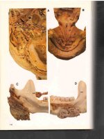
Careful palpation of the penile shaft should be performed, from pubic bone to
coronal sulcus, to elucidate any plaque (Fig. 1.4). The patient may have more than
one plaque present and may have plaques on both the dorsum and ventrum. The
latter group of patients will often have no significant deformity, as such plaques
counteract each other, but may complain of significant penile length loss. Palpation,
applying side-to-side, and dorsoventral pressure are the optimal means of outlining
plaque and septal anatomy. Side-to-side compression beginning at the 3 and 9
o’clock positions on the shaft and rolling firmly upward (for dorsal plaque) and
downward (for ventral plaque) should be conducted meticulously along the entire
shaft. The plaque location, morphology, and approximate size (where possible)
should be documented, and measurement of the stretched flaccid length is recommended (Fig. 1.5). We suggest that such measurement be conducted between two
fixed points, the pubic bone and the coronal sulcus. Measuring plaque size is surprisingly challenging and difficult to replicate because all methods are subject to
intra- and inter-observer variability (Fig. 1.6). Further confounding the problem is
the lack of universal agreement regarding the optimal method of measurement.
Despite this, the frequency of plaque size being reported in the literature mandates
an understanding of the methodology in addition to the limitations. Options include
using calipers and rulers during physical exam or utilizing imaging modalities.
Plaque size can be documented as either area or less appealingly as the longest
plaque dimension.
1
Focused Genital Exam
5
Fig. 1.4 Penile stretch
Scrotal Contents
Examination of the scrotal contents, while important for all men presenting to the
andrologist, is critical for the infertility patient. It is surprising to me how often a
urology resident/fellow has a poor appreciation for the normal content anatomy, and
this is especially true for the vasa deferentia. All urologists are encouraged to feel
for the vasa every time a scrotal exam is being conducted.
Each testis should be examined individually. The testis should be palpated along
its entire surface. Its regularity as well as consistency should be recorded. Assessment
of testicular volume is ideally performed using ultrasound; however, this is not practical in everyday andrology practice for every single patient. In the early stage of the
andrologist’s career, an orchidometer should be used to assist in the clinician
becoming familiar with testicular volumes. Following testis examination, attention
should next be focused on the epididymis. This structure lies posterolateral to the
testis, and examination on the epididymis should include its caput, corpus, and
cauda. Its presence and consistency as well as the presence of any induration should
be recorded. The vas deferens should be examined next. This is best examined by
rolling this fibrous cord between two fingers, placed anterior and posterior to the
structure. In heavy-set men, in men with varicoceles, or a spermatic core lipoma,
palpation of the vas deferens can be challenging. Of course, the absence of the vas
deferens on either side should lead one to suspect one of the congenital absence of
the vas deferens syndromes. Following this, an assessment for the presence of the
varicocele should be performed. Grade III varicoceles are routinely visible, grade II
varicoceles are using palpable within the spermatic cord, and Grade I varicoceles
require the conduct of a Valsalva maneuver. It is important when conducting the
Valsalva maneuver to be aware as to whether what is being felt as a venous thrill
may actually be contraction on this spermatic cord musculature. Thus, caution
should be exercised when diagnosing a Grade I varicocele.
J.P. Mulhall and L.C. Jenkins
6
Deep penile fascia
Fibrous plaque
Tunica albuginea
Corpus cavernosum
Corpus spongiosum
Corpus cavernosum
Fibrous plaque
Corpus spongiosum
Fig. 1.5 Peyronie’s plaques
Rectal Examination
Urologists are very familiar with digital rectal examination for the purposes of
prostate evaluation. While assessment of the prostatic anatomy, its size, shape,
consistency, and level of induration is important in routine clinical practice, the
andrologist should also be aware of any other generally subtle structural changes
on digital rectal examination. This is particularly important in patients with a history of hematospermia and ejaculatory duct obstruction. Even before assessing
1
Focused Genital Exam
7
Fig. 1.6 Peyronie’s plaque exam
prostatic anatomy, an external examination of the perianal area is a valuable step in
the evaluation of patients. While not uniformly accurate, it is worthwhile recording
the presence of a bulbocavernosus reflex and anal tone as part of the routine andrology examination.
Suggested Reading
Althof SE, Rosen RC, Perelman MA, Rubio-Aurioles E. Standard operating procedures for taking
a sexual history. J Sex Med. 2013;10(1):26–35.
Ghanem HM, Salonia A, Martin-Morales A. SOP: physical examination and laboratory testing for
men with erectile dysfunction. J Sex Med. 2013;10(1):108–10.
Gratzke C, Angulo J, Chitaley K, Dai YT, Kim NN, Paick JS, et al. Anatomy, physiology, and
pathophysiology of erectile dysfunction. J Sex Med. 2010;7(1 Pt 2):445–75.
Hatzichristou D, Rosen RC, Derogatis LR, Low WY, Meuleman EJ, Sadovsky R, et al.
Recommendations for the clinical evaluation of men and women with sexual dysfunction.
J Sex Med. 2010;7(1 Pt 2):337–48.
Montorsi F, Adaikan G, Becher E, Giuliano F, Khoury S, Lue TF, et al. Summary of the recommendations on sexual dysfunctions in men. J Sex Med. 2010;7(11):3572–88.
Mulhall JP, Stahl PJ, Stember DS. Clinical care pathways in andrology. New York: Springer; 2014.
p. 188. viii.
The process of care model for evaluation and treatment of erectile dysfunction. The process of care
consensus panel. Int J Impot Res. 1999;11(2):59–70; discussion −4.
Wein AJ, Kavoussi LR, Partin AW, Peters C. Campbell-Walsh urology. 11th ed. Philadelphia, PA:
Elsevier; 2016.
Chapter 2
Biothesiometry
John P. Mulhall and Lawrence C. Jenkins
Background
The biothesiometry device is used to measure the threshold of appreciation of vibration in patients. A decreased sensitivity to these vibrations may indicate the presence of a penile sensory neuropathy. This is a quantitative measure of the vibratory
sense of the penis. The biothesiometer vibrates at a known frequency, and it is compared to other parts of the body with known vibration thresholds. The effectiveness
of this test in documenting sensory neuropathy of the penis has not been established
but is considered as a useful screening test. It is worth noting two important points:
(1) this is only assessing sensory nerve function (and not motor or autonomic nerve
integrity) and (2) this test may not be covered by the patient’s insurance plan.
When patients complain of penile sensation loss, the andrology practitioner can
utilize biothesiometry to screen for a sensory neuropathy. As the symptom of sensation loss may be organically based (penile sensory neuropathy) or perceptual in
nature (psychogenic), biothesiometry is a useful diagnostic tool. Biothesiometry
cannot locate the focus of the lesion nor its severity. For this we refer the patient for
dorsal penile nerve somatosensory evoked potential (SSEP) analysis. Generally, any
etiology of neuropathy can lead to penile sensation loss, but the most commonly
encountered in routine andrology practice is diabetes mellitus.
While not well appreciated, reduced penile sensation may be a secondary psychosexual dysfunction. Typically anxiety-prone men, especially young men,
develop multiple sexual dysfunctions, become increasingly focused on the penis,
and may complain of reduced penile sensation. This increased focus on the penis, or
penocentricity, may lead to a form of genital dysmorphophobia.
J.P. Mulhall, MD, MSc, FECSM, FACS (*) • L.C. Jenkins, MD, MBA
Department of Surgery, Section of Urology, Memorial Sloan Kettering Cancer Center,
16 East 60th Street, Suite 402, New York, NY 10022, USA
e-mail: ;
© Springer International Publishing Switzerland 2017
J.P. Mulhall, L.C. Jenkins (eds.), Atlas of Office Based Andrology Procedures,
DOI 10.1007/978-3-319-42178-0_2
9
10
Table 2.1 Nomogram
J.P. Mulhall and L.C. Jenkins
Nomogram for penile biothesiometry
Age
Fingertip
Shaft
Glans
18–29
3
3
3
30–39
4
4
4
40–49
4
5
5
50–59
5
6
7
60–69
5
7
7
Biothesiometry is a useful way to detect neurologic disease in at-risk men (i.e.,
spinal cord injury or diabetes). It is also used to establish a baseline level of function
prior to any surgical procedure that may compromise sensation to the head of the penis.
Normalcy is determined by referring to the nomogram as shown in Table 2.1.
Indications
1. Patient complaining of penile numbness
2. Delayed orgasm
3. Prior to penile reconstructive surgery
Pre-procedural Considerations
Familiarize yourself with the device prior to starting the procedure (Figs. 2.1, 2.2,
and 2.3). The patient should be supine on the examination table in a calm and
relaxed state. The patient should be undressed from the waist down with a sheet
covering their lower body prior to starting. The examiner should check finger and
penile positions to be examined (Fig. 2.4). A standardized report should be ready to
document findings (a sample report is shown in Fig. 2.5).
Procedure
With the patient undressed and ready, place the device on a stand or table near the
patient (examination table). You should start by placing the probe on the tip of the
patient’s index finger (examine both the right and left fingers) and slowly increase
the intensity of the vibration until the patient declares they are feeling the vibration. This is then used as a baseline so the patient can compare to the vibration
sense of the penis.
The device is placed on the left and right mid-shaft of the penis, the left and right
mid-glans, and finally the frenulum. The probe should be held perpendicular to the
2 Biothesiometry
Fig. 2.1 Main unit
Fig. 2.2 Dial
11
12
J.P. Mulhall and L.C. Jenkins
Fig. 2.3 Handheld probe
Fig. 2.4 Locations for assessment
skin surface to assure the probe tip has full and even contact with the skin. At all
locations, intensity of vibration is slowly increased until the vibration is felt.
This location is repeated twice and the average result should be documented on the
report sheet.
2 Biothesiometry
13
BIOTHESIOMETRY
Date:
Patient:
MRN:
Biothesiometry
NOMOGRAM FOR BIOTHESIOMETRY
AGE
Pulp
Shaft
Glans
18-29
3
3
3
30-39
4
4
4
40-49
4
5
5
50-59
5
6
7
60-69
5
7
7
DIAGNOSIS:
□ Consistent with neuropathy
□ Normal
TREATMENT:
□ Neurology consult
□ SSEP
□ Monitor
Comments:
Provider’s Signature
Fig. 2.5 Report sheet
Complications
None.
14
J.P. Mulhall and L.C. Jenkins
Post-procedural Instructions and Management
If abnormal we suggest to all of our patients to consider referral for pudendal
nerve SSEP testing for confirmation as well to define the severity of the sensory
neuropathy as well as the location of the lesion within the neural arc. If the SSEP
is abnormal, we would recommend a full consultation with a neurologist to investigate further.
Suggested Reading
Bemelmans BL, Hendrikx LB, Koldewijn EL, Lemmens WA, Debruyne FM, Meuleman EJ.
Comparison of biothesiometry and neuro-urophysiological investigations for the clinical evaluation of patients with erectile dysfunction. J Urol. 1995;153(5):1483–6.
Breda G, Xausa D, Giunta A, Tamai A, Silvestre P, Gherardi L. Nomogram for penile biothesiometry. Eur Urol. 1991;20(1):67–9.
McMahon CG, Jannini E, Waldinger M, Rowland D. Standard operating procedures in the disorders of orgasm and ejaculation. J Sex Med. 2013;10(1):204–29.
Mulhall JP, Stahl PJ, Stember DS. Clinical care pathways in andrology. New York: Springer; 2014.
p. 188. viii.
Padma-Nathan H. Neurologic evaluation of erectile dysfunction. Urol Clin North Am.
1988;15(1):77–80.
Padma-Nathan H. Medical management of erectile dysfunction: a primary care manual. 1st ed.
Caddo, OK: Professional Communications Inc.; 1999. p. 160.
Vardi Y, Gruenwald I. Neurophysiologic testing in erectile dysfunction. In: Carson CC, Kirby R,
Goldstein I, editors. Textbook of erectile dysfunction. 2nd ed. New York: Informa Healthcare;
2009. p. 168–75.
Chapter 3
Nocturnal Penile Tumescence
John P. Mulhall and Lawrence C. Jenkins
Introduction
The test was developed to study a man’s erectile function while sleeping. The normal
male has 3–5 erections while sleeping and a lack of these erections may be a sign of
a larger problem. There have been several methods to assess rigidity in the past
including the postage stamp test, the snap gauge, and the strain gauge. The snap
gauge method uses bands of varying strengths wrapped around the penis. When an
erection occurred, bands would break based on the degree of rigidity. The postage
stamp test is a rudimentary form of the snap gauge. A roll of postage stamps was
placed around the flaccid penis, and if a rigid erection occurred, the postage stamp
roll would break. The strain gauge works by placing bands around the penis, and if
an erection occurred, they would become stretched and retain their distension based
on the change in penile circumference.
Indications
Psychogenic erectile dysfunction
Patients complaining of painful (and potentially prolonged) nocturnal erections
Medical-legal cases involving erectile function
J.P. Mulhall, MD, MSc, FECSM, FACS (*) • L.C. Jenkins, MD, MBA
Department of Surgery, Section of Urology, Memorial Sloan Kettering Cancer Center,
16 East 60th Street, Suite 402, New York, NY 10022, USA
e-mail: ;
© Springer International Publishing Switzerland 2017
J.P. Mulhall, L.C. Jenkins (eds.), Atlas of Office Based Andrology Procedures,
DOI 10.1007/978-3-319-42178-0_3
15
16
J.P. Mulhall and L.C. Jenkins
Fig. 3.1 Rigiscan® Plus device (GOTOP Medical, Inc., St. Paul, MN, USA)
Pre-procedural Considerations
The Rigiscan system (Rigiscan® Plus device—GOTOP Medical, Inc., St. Paul, MN,
USA) is composed of two parts: (1) a portable nocturnal penile tumescence and
rigidity data-logging unit and (2) a computer with printer for processing and printing the data (Fig. 3.1). It is important for the clinician to take note of conditions that
may interfere with the Rigiscan analysis (sleep apnea syndrome, use of psychotropic medications, insomnia). Penile rigidity is measured as a percent displacement
by the tension loops; thus, 100 % rigidity reflects zero tissue compression. Normal
has been defined as rigidity ≥60–70 % (represents penile rigidity adequate for vaginal penetration), and rigidity <40 % equates to a flaccid penis. Some authorities
have suggested using 55 % rigidity as a normal cutoff to yield the best sensitivityspecificity relationship.
Procedure
Prior to sleeping, the unit is secured to the patient’s thigh in a pouch worn on the leg,
and the penile loops are fitted to the penis, one toward the base and the other toward
the tip (Fig. 3.2). We typically place these at the junction of the mid-proximal one
third of the penis (base loop) and at the junction of the mid and distal one thirds of