Ebook Oral and maxillofacial surgery cliniscs: Part 2
Bạn đang xem bản rút gọn của tài liệu. Xem và tải ngay bản đầy đủ của tài liệu tại đây (30.18 MB, 99 trang )
Oral Maxillofacial Surg Clin N Am 20 (2008) 431–443
Thyroid Disorders: Evaluation and Management
of Thyroid Nodules
James I. Cohen, MD, PhDa,*, Kelli D. Salter, MD, PhDb
a
Department of Otolaryngology/Head and Neck Surgery, Oregon Health & Science University,
3181 SW Sam Jackson Park Road, PV-01, Portland, OR 97239-3098, USA
b
Department of General Surgery, Oregon Health & Science University,
3181 SW Sam Jackson Park Road, L223, Portland, OR 97239-3098, USA
Although it is well documented that thyroid
nodules are a common clinical disorder, significant controversy persists as to ideal management
strategies. Population studies suggest that approximately 3% to 7% of adults have asymptomatic
palpable thyroid nodules, and that the number of
nodules, including asymptomatic and symptomatic, increases with age [1–6]. However, the advent
and implementation of high-resolution radiographic imaging has significantly impacted the
discrepancy between clinically evident disease
and incidentally discovered disease. High-resolution ultrasound (US) can detect thyroid nodules
in 20% to 67% of randomly selected individuals,
with a higher frequency in women and the elderly
[3–8]. Moreover, 20% to 48% of patients who
have a single palpable nodule have additional
nodules identified on US. This discrepancy is further supported by data from autopsies conducted
for medical reasons unrelated to thyroid disorders. Such data suggest that the prevalence of
thyroid nodules in clinically normal glands is
approximately 50% to 70% [3–6,9]. Therefore,
the true prevalence of nodular thyroid disease in
the general population remains unknown.
As the incidence of thyroid nodules has exhibited a steady rise over the past decade, so too
has the incidence of thyroid cancer. The National
Cancer Institute estimates the number of new
cases and deaths from thyroid cancer in the
United States in 2007 to be 33,550 and 1,530,
* Corresponding author.
E-mail address: (J.I. Cohen)
respectively [10]. These numbers have steadily
increased from the reported 13,000 number of
new cases and 1000 thyroid cancer–associated
deaths in 1994 [10–12]. However, despite the notable increase in the number of new cases, mortality
rates have remained constant [10–12]. Most experts in the field of cancer agree that the increasing incidence of thyroid cancer likely reflects the
implementation of technology with increased
sensitivity and specificity for detecting thyroid
nodules. Such technology increases the need for
physicians to improve their ability to differentiate
benign from malignant thyroid lesions, because
the clinical importance of thyroid nodules rests
on the need to exclude thyroid cancer.
Incidentally discovered nodules present the
same risk for malignancy (w10%) as palpable
nodules if they are equivalent in size [3–6,13].
Therefore, the physician who finds an incidental
thyroid nodule is faced with the challenge of
determining the clinical significance of the lesion.
Differentiating a benign nodule, which may require observation only and no specific treatment,
from a malignant nodule, which requires more aggressive treatment, presents a diagnostic dilemma.
Because of the high prevalence of incidental disease, it is neither economically feasible nor necessary to surgically excise all, or even most, thyroid
nodules. It is essential that the physician develop
and follow a reliable, cost-effective strategy for
diagnosis and treatment of incidentally found thyroid nodules. This article provides practical guidelines, algorithms, and current recommendations
for the effective diagnosis and management of thyroid nodules incidentally discovered by physicians
1042-3699/08/$ - see front matter Ó 2008 Elsevier Inc. All rights reserved.
doi:10.1016/j.coms.2008.02.003
oralmaxsurgery.theclinics.com
432
COHEN & SALTER
has a thyroid nodule. Unfortunately, neither the
history nor the physical examination is highly
sensitive or specific for detecting malignancy. However, several well-documented factors are associated with an increased risk for malignancy and,
therefore, warrant further discussion [3–6]. Factors
that present a high risk for thyroid cancer include:
history of head and neck or total body radiation;
family history; rapid growth; hard, fixed nodule;
and/or regional, cervical lymphadenopathy. Factors that present a moderate risk include: male gender; age younger than 30 or older than 60 years;
and/or persistent local symptoms (hoarseness, dysphagia, dysphonia, dyspnea).
A history of head and neck or total body
irradiation is a well-known risk factor for
subsequent development of thyroid cancer. The
incidence of thyroid malignancy in a patient who
has a nodule and a previous history of radiation
has been reported to range from 20% to 50%
[2–6,14–18]. Therefore, the incidental finding of
a thyroid nodule in a patient who has had prior
radiation exposure requires careful and complete
evaluation, although by itself it does not justify
removal if the workup should prove negative.
Despite high levels of intraobserver and
interobserver variations, careful inspection and
palpation of the thyroid, the anterior neck compartments, and the lateral neck compartments
should always be performed. Texture and size of
the nodule should be documented. A firm or hard,
solitary or dominant nodule with an increased
managing patients for other medical reasons.
Important elements of the history and physical
examination, laboratory evaluation, and imaging
modalities are reviewed, and a suggested management strategy is presented. This outline is not
intended to be all inclusive, nor does it preclude
additional evaluation, according to the specific
clinical situation. Furthermore, the specific management of hypothyroidism, hyperthyroidism, or
thyroid malignancies is beyond the scope of this
article. These lesions should be specifically managed by a multidisciplinary team, including, at
a minimum, an endocrinologist and surgeon who
specialize in the treatment of such disorders.
Diagnosis
No reliable noninvasive way exists to distinguish a benign thyroid nodule from a thyroid
carcinoma. Multiple diagnostic methods must be
used to increase the accuracy of the diagnosis.
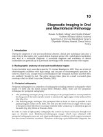
Fig. 1 provides a basic algorithm of diagnostic
modalities typically used in the initial evaluation
of a thyroid nodule. Generally, the inability to
accurately differentiate benign from malignant
nodules warrants operative removal of the lesion.
History and physical examination
The history and physical examination, including
that of adjacent cervical lymph nodes, remain the
diagnostic cornerstone in evaluating a patient who
Thyroid Nodule
History and Physical Examination
High
TSH
Normal
Low
Free T3 + Free T4
TPOAb+ Free T4
Ultrasound
Scintigraphy
No Suspicious
Features
Suspicious
Features
Cold
Hot
FNA
Asymptomatic
Symptomatic
Endocrinology
Consult
Fig. 1. Diagnosis and management of thyroid nodules. FNA, fine-needle aspiration; T3, triiodothyronine; T4, thyroxine;
TPOAb, thyroid peroxidase antibody; TSH, thyroid-stimulating hormone (thyrotropin).
EVALUATION AND MANAGEMENT OF THYROID NODULES
rate of growth that clearly differs from the rest of
the gland suggests an increased risk for malignancy [2,4,6]. The presence of multiple nodules
(symptomatic or asymptomatic) does not decrease
the likelihood that any one of them is a carcinoma,
as was once thought, although the overall incidence of malignancy in a multinodular gland is
the same as that for any given nodule (w10%)
[3–6,19,20]. Each nodule should be evaluated on
its own merit regardless of the number of nodules
present. Finally, ipsilateral or contralateral cervical lymphadenopathy is worrisome in the setting
of a thyroid nodule and significantly increases
the risk for malignancy.
Thyroid cancer may present as a familial trait
or syndrome [21–24]. Although medullary thyroid
carcinoma (MTC) accounts for only approximately 10% of all thyroid carcinomas, 25% of
MTCs occur secondary to an inherited cancer
risk, namely familial MTC (!2%) and multiple
endocrine neoplasia (MEN 2A, w25% or MEN
2B, !2%) [23–25]. Mutations in the RET protooncogene are responsible for all three conditions
[23–25]. Patients diagnosed with MTC should
undergo genetic testing to determine if mutations
in the RET proto-oncogene are present.
Papillary and follicular carcinomas, the two
most common forms of thyroid cancer, may also
present as a family trait or syndrome [21,22,25].
Patients who have familial adenomatous polyposis (FAP) syndrome or Gardner syndrome
(a variant of FAP), Cowden syndrome, and
Werner (adult progeroid) syndrome are at increased risk for development of thyroid cancer
[21,22,25]. Families with adenomatous polyposis
(FAP or Gardner syndrome) show an increased
incidence (2%) of papillary thyroid cancers, which
tend to be multicentric (65%), exhibit a higher female-to-male ratio (6:1), and develop at a younger
age (third decade) [21,22,25]. Patients who have
Cowden syndrome have up to a 10% lifetime
risk for follicular or papillary thyroid cancer,
with follicular being the most common
[21,22,25]. Approximately 70% to 85% of people
with Cowden syndrome will have benign thyroid
changes, including multinodular goiter, adenomatous nodules, and follicular nodules [21,22,25].
Thyroid cancer associated with Werner syndrome,
an autosomal connective tissue disorder, occurs
a decade earlier than in the general population,
with a mean age of 34 years. Variability in the
type of non-MTC occurring in patients who
have Werner syndrome has been observed among
ethnic groups. Although papillary (84%),
433
follicular (14%), and anaplastic (2%) forms have
been observed in Japanese patients, only papillary
appears to occur in Caucasian patients [25]. Finally, papillary thyroid carcinoma can occur in
families independent of syndromes such as FAP,
Cowden, or Werner [21,22,25]. This form of thyroid cancer is believed to be inherited as an autosomal dominant condition. However, a specific
genetic mutation has not been identified. Therefore, genetic testing is not currently available for
these families.
Extremes of age (!30 or O60) and male
gender are associated with an increased risk for
thyroid cancer if a nodule is present [2–6]. Thyroid
nodules during childhood and adolescence should
induce caution, because the rate of malignancy is
twofold higher in children than in adult patients
[2–6]. Furthermore, although thyroid nodules
are four times more common in women and
increase with age, men are at greater risk for
malignancy than women [2–6].
Most patients who have thyroid nodules have
few or no symptoms. When present, symptoms
are generally nonspecific. No defined relationship
exists between nodule histology or size and the
reported symptoms. However, persistent local
symptoms of hoarseness, dysphagia, dysphonia,
dyspnea, or cough should raise the suspicion of
malignancy and warrant further investigation,
including an evaluation for thyroid cancer [2–6].
Finally, iodine deficiency and socioeconomic
status have been proposed as independent risk
factors for thyroid carcinoma [6,26–29]. Population-based studies conducted from the 1960s to
the 1990s on residents living in areas of endemic
goiter indicated that iodine deficiency was an associated risk factor for thyroid cancer, primarily
of the follicular and papillary subtypes [26–29].
Lower socioeconomic status additionally was
identified as an independent risk factor for more
advanced disease secondary to limited access to
appropriate health care [26–29].
Laboratory evaluation
Because clinical evaluation is not sensitive for
thyroid gland disease, laboratory examination is
necessary. Measurement of the serum thyrotropin
or thyroid-stimulating hormone (TSH) concentration is the single most useful test, and may be the
only one warranted, in the initial evaluation of
thyroid nodules [2–6]. The TSH assay has a high
sensitivity in detecting even subtle thyroid dysfunction [30]. If the serum TSH level is within
434
COHEN & SALTER
the normal range, the measurement of free thyroid
hormones adds no further relevant information.
Abnormal serum TSH levels, however, generally
warrant further laboratory testing (see Fig. 1). If
the serum TSH level is high, a free thyroxine
(T4) and thyroglobulin or thyroid peroxidase antibody (TPOAb) should be obtained to evaluate for
hypothyroidism or thyroiditis [2–6]. In both these
situations, the thyroid gland can be enlarged or
nodular. By contrast, if the serum TSH level is
low, a free T4 and free triiodothyronine (T3) level
should be obtained to evaluate for hyperthyroidism, such as an autonomic functioning gland or
thyrotoxicosis [2–6].
Serum thyroglobulin, a protein normally produced by the thyroid gland, correlates with the
iodine status and the size of the thyroid gland
rather than the nature (malignant versus benign)
of a thyroid nodule. Many factors, including the
degree of thyrotropin receptor stimulation, the
volume of the gland, inflammation, radiation,
multinodular goiter, biopsy, or surgery, may
falsely elevate or decrease levels of thyroglobulin
[4–6,31]. Furthermore, the presence of TPOAb,
which attack the thyroglobulin protein, may decrease the reliability of the thyroglobulin assay
[4–6,31,32]. Such antibodies may be present in
10% of normal subjects, 15% to 30% of patients
who have differentiated thyroid cancer, 89% to
98% of patients who have Grave’s disease, and
100% of patients who have Hashimoto’s thyroiditis [4–6,31,32]. Additionally, autoimmune thyroid
diseases are associated with several other organspecific and systemic autoimmune disorders [32].
Therefore, a preoperative assay cannot be used
to diagnose or exclude cancerous lesions. Although commonly implemented as a means of
monitoring for recurrence of thyroid cancer in patients following thyroidectomy, measurement of
serum thyroglobulin should not be used in the
routine assessment of thyroid nodules.
Routine measurement of calcitonin, a useful
serum marker of MTC, in all patients is not costeffective [4–6]. However, the incidence of sporadic
MTC in patients who have nodular thyroid
glands can be as high as 1.5% [23,25]. Furthermore, unlike familial MTC which often is diagnosed early secondary to family history and
genetic testing, sporadic MTC usually presents
at a later stage with regional metastasis because
of increased difficulty in diagnosis due to various
morphologies [23,25]. Therefore, although not
recommended in routine assessment of thyroid
nodules, a calcitonin level should be considered
in patients who have factors suspicious for sporadic MTC and is imperative in those patients
who have a suspected familial MTC or a familial
MEN syndrome.
Imaging modalities
High-resolution ultrasound
High resolution ultrasonography (US) is the
cornerstone of imaging for assessment of thyroid
nodules. To date, it is the most accurate test
available to evaluate such lesions, measure their
dimensions, identify their structure, and evaluate
diffuse changes in the thyroid gland [4–6]. However, because of the high prevalence of clinically
inapparent, small thyroid nodules, routine US is
not recommended as a screening test in the general population unless well-known risk factors
are present.
Many studies have been published debating the
ability of US to distinguish between benign and
cancerous lesions [13,32–38]. In 2005, the Society
of Radiologists in Ultrasound convened a panel
of specialists from a variety of medical disciplines
to formulate a consensus regarding management
of thyroid nodules identified by ultrasonography
in adult patients [39]. The likelihood of cancer in
a thyroid nodule was shown to be the same
regardless of the size measured at US [13,32–39].
Furthermore, sonographic features suggestive of
malignancy were found to vary between types of
thyroid carcinomas [13,32–39]. Despite these
discrepancies, several sonographic features were
found to be suggestive of an increased risk for
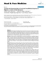
malignancy (Fig. 2, Table 1), including microcalcifications, hypoechogenicity, irregular margins,
absence of nodule halo, predominant solid composition, and intranodular vascularity [13,32–39].
However, the sensitivities, specificities, positive
predictive values and negative predictive values
for these criteria were variable between studies
[13,32–39]. No US feature was found to have
both a high sensitivity and positive predictive
value but the combination of factors was shown
to improve the positive predictive value of US to
some degree. Therefore, patients who have palpable thyroid nodules or incidentally discovered
nodules with concerning patient demographics or
risk factors should undergo US to evaluate for
sonographic features suggestive of malignancy,
baseline characteristics and volume of the nodule, coincidental thyroid nodules, and baseline
characteristics and volume of the remaining thyroid gland. In addition the cervical lymph nodes
435
EVALUATION AND MANAGEMENT OF THYROID NODULES
Fig. 2. Ultrasound images of thyroid nodules of varying parenchymal composition (cystic to solid) and vascularity. (A)
Gray-scale image of predominately cystic nodule (calipers) that proved to be benign at cytologic examination (fine-needle
aspiration [FNA]). (B) Gray-scale image of mixed solid and cystic nodule (calipers) with septate (arrow). (C) Addition of
color Doppler mode did not demonstrate marked internal vascularity. The lesion was benign at cytologic examination
(FNA). (D) Gray-scale image of predominately solid nodule (calipers) with surrounding halo (arrows) that proved to be
benign at cytologic examination (FNA) and surgery. (E) Gray-scale image of predominately solid nodule (calipers) with
irregular margins (arrows) and multiple fine echogenicities (arrowheads). (F) Addition of color Doppler mode demonstrated marked internal vascularity indicating increased likelihood that nodule is malignant. FNA and surgery confirmed
papillary carcinoma.
beds should be evaluated by ultrasonography as
warranted.
Despite recommendations from the Society of
Radiologists in Ultrasound Consensus Conference
Statement, ultrasonography cannot reliably distinguish between benign and cancerous lesions without
adjunct testing. Therefore, patients who have risk
factors and ultrasonographic characteristics concerning for malignancy should undergo cytohistologic analysis of a representative tissue sample
obtained by way of either fine-needle aspiration
(FNA) or coarse-needle biopsy (CNB) [39]. In general, FNA is preferred over CNB because it is extremely accurate and less invasive and allows for
Table 1
Sonographic features associated with thyroid cancer
US feature
Sensitivity (%)
Specificity (%)
PPV (%)
NPV (%)
Microcalcifications
Hypoechogenecity
Irregular margins or halo absence
Solid composition
Intranodular vascularity
26–59
27–87
17–78
69–75
54–74
86–95
43–94
39–85
53–56
79–81
24–71
11–68
9–60
16–27
24–42
42–94
74–94
39–98
88–92
86–97
Abbreviations: NPV, negative predictive value; PPV, positive predictive value.
Modified from Frates MC, Benson CB, Charboneau JW, et al. Management of thyroid nodules detected at US: Society
of Radiologists in Ultrasound consensus conference statement. Radiology 2005;237:794–800.
436
COHEN & SALTER
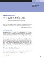
Fig. 3. Methods for obtaining thyroid tissue for cytohistologic analysis. A CNB uses a larger needle (16 or 18 gauge) and
requires that the thyroid nodule be at least 2 cm in size. By contrast, an FNA uses a smaller needle (25 or 27 gauge) and
allows for more complete sampling of the nodule because of the multiple passes taken through the nodule.
more complete sampling of the nodule because of
the multiple passes taken through the nodule
(Fig. 3) [4–6]. Additionally, US should be performed
in all patients who have a history of familial thyroid
cancer, MEN II, or childhood cervical irradiation,
even if palpation yields normal findings [39]. Furthermore, the physical finding of adenopathy suspicious for malignant involvement in the anterior or
lateral neck compartments warrants US examination of the lymph nodes and thyroid gland because
of the risk for a lymph node metastatic lesion from
an unrecognized thyroid carcinoma [39].
Radionuclide scintigraphy
Radionuclide scintigraphy (iodine 123 [123I]
or technetium-99m pertechnetate), once the cornerstone for thyroid imaging, has now been replaced by high-resolution ultrasonography as the
imaging modality of choice for evaluating thyroid
nodules [4–6]. Such scans, in the current status
of thyroid imaging, are used primarily as adjuncts to ultrasonography for differentiating
hyperfunctioning (‘‘hot’’) from hypofunctioning
(‘‘cold’’) nodules (Fig. 4) [4–6,40,41]. Hyperfunctioning nodules represent approximately 5% of
thyroid nodules and present a low risk for malignancy (%1%) [4–6]. Hypofunctioning nodules
have a reported malignant risk of 5% to 25%
and represent approximately 75% to 95% of thyroid nodules [4–6]. The remaining 10% to 15% of
nodules are indeterminate, with a variable risk for
malignancy [4–6]. Because most thyroid lesions
are ‘‘cold’’ and few of these lesions are malignant,
the predictive value of hypofunctioning nodules
for the presence of malignant involvement is
low. The diagnostic specificity is further reduced
in small lesions (!1 cm), which may not be identified by scintigraphy. For these reasons, thyroid
scintigraphy is not usually useful as a first-step
diagnostic study in the evaluation of thyroid
nodules. Indications that may warrant use of
thyroid scintigraphy include identification of a
solitary thyroid nodule in the setting of decreased
serum thyrotropin, an indeterminate FNA or
EVALUATION AND MANAGEMENT OF THYROID NODULES
437
Fig. 4. Iodine 123 (123I) thyroid scintigraphy patterns in thyroid glands (dashed lines) with ‘‘cold’’ and ‘‘hot’’ nodules. (A)
Nonfunctioning ‘‘cold’’ nodule in the lower left thyroid lobe (solid line). (B) Hyperfunctioning ‘‘hot’’ right thyroid nodule
(solid line), with suppressed serum TSH level and suppressed uptake of 123I in the remainder of the thyroid gland.
CNB of a thyroid nodule, and for the detection
of nonspecific neck masses or lymphadenopathy
[4–6,40,41].
CT and MRI
CT and MRI, like other imaging modalities,
cannot reliably differentiate between malignant
and benign nodules [4–6,42]. Therefore, these tests
are rarely indicated in the initial evaluation of
a thyroid nodule. However, such imaging modalities may be used as secondary adjuncts if
warranted. A CT scan can be used to evaluate
nodules in a difficult-to-palpate, diffusely enlarged
gland, to assist in detection of mediastinal thyroid
tissue, and to assess for extrathyroidal invasion
and cervical lymphadenopathy (Fig. 5). By contrast, MRI demonstrates exquisite soft tissue
details and vascular anatomy, and thus, allows
for identification of extraglandular invasion and
involvement of the great vessels, respectively.
Therefore, either of these imaging modalities
may be implemented in preoperative staging. CT
contrast medium contains iodine which reduces
subsequent uptake of iodine molecules and thus
may interfere with nuclear scintigraphy (123I) or
postoperative radioiodine ablation therapy (131I)
for malignant nodules. MRI uses contrast medium (gadolinium) that does not interfere with
nuclear scintigraphy.
Incidental clinically silent thyroid nodules are
commonly discovered in patients undergoing CT
or MRI for medical reasons unrelated to thyroid
disorders. The decision to pursue further workup
of such nodules depends on several factors already
discussed, including history and physical, laboratory analysis, and associated known risk factors.
Although abnormalities of the thyroid gland can
be detected on CT and MRI, sonography provides
important additional information that may be
useful in guiding further clinical management.
Therefore, patients who have an incidentally
discovered thyroid nodule on CT or MRI and
Fig. 5. CT scan of the neck demonstrating a metastatic
right thyroid lobe carcinoma. The anterior aspect of the
right thyroid lobe has a nodular exophytic mass (long
arrow) near the junction with the isthmus. On the right
side is a heterogeneous low-density enlarged lymph
node (short arrow) that contains septations and nodules
of high density. Fine-needle aspiration and surgery of
the mass demonstrated papillary carcinoma with metastasis to the right paratracheal and lateral neck lymph
nodes.
438
COHEN & SALTER
concerning clinical features should undergo ultrasonography to determine the need for biopsy and
further analysis.
Cytohistochemistry analysis
A cytohistochemistry analysis should be performed on thyroid nodules with associated features concerning for malignancy. Tissue for such
analysis is obtained by way of either FNA or
CNB (see Fig. 3). Detailed reviews of aspiration
biopsy of thyroid nodules have been published
previously [4–6,43–45]. In general, FNA is the removal of a few clusters of individual thyroid cells
by means of a small needle (usually a 25- or
27-gauge 1.5-in needle). By contrast, CNB uses
a larger needle (usually a 16- or 18-gauge needle)
and is more difficult to perform, and fewer physicians have experience in this procedure. In
addition, the large size of the needle may cause
a small amount of bleeding (%1%), injury to
the trachea, or injury to the recurrent laryngeal
nerves. Furthermore, unlike FNA, which can be
performed on all types of nodules, the nodule
must be at least 2 cm in size to perform a CNB
successfully. Finally, although CNB provides
a larger tissue sample that retains it cellular architecture, it rarely provides a more precise histologic
diagnosis than FNA. Therefore, because of its
minimal invasiveness, accuracy (w95%) and
cost effectiveness, US-guided FNA has now
become the diagnostic technique of choice for
evaluating thyroid nodules [4–6]. For these reasons, only the role of FNA in the evaluation of
thyroid nodules will be discussed in this article.
The accuracy of FNA or CNB is only as good
as the person performing the procedure and the
person who analyzes and reports the cytologic
findings. However, when performed by experienced personnel, the sensitivity and specificity
(Table 2) of thyroid FNA are excellent [4–6].
Fine-needle aspiration
Not every patient who has a thyroid nodule
should undergo FNA. Which thyroid nodule
should be aspirated is a topic of intense current
debate among multiple medical specialties. As
stated in the 2005 Society of Radiologists in
Ultrasound Consensus Conference Statement,
the decision to perform or defer FNA in a given
patient should be made according to the individual circumstances [39]. Several recommendations
(Table 3) based on current literature and common
practice strategies were made by the committee to
assist physicians in their decision-making process
Table 2
Statistical features of thyroid fine-needle aspiration
Statistical feature
Mean (%)
Range (%)
Sensitivity
Specificity
PPV
False-negative rate
False-positive rate
83
92
75
5
5
65–98
72–100
50–96
1–11
0–7
Abbreviation: PPV, positive predictive value.
Modified from AACE/AME Task Force on Thyroid
Nodules. American Association of Clinical Endocrinologists and Associazione Medici Endocrinologi
medical guidelines for clinical practice for the diagnosis
and management of thyroid nodules. Endocr Prac
2006;12(1):63–102; Gharib H, Papini E. Thyroid nodules:
clinical importance, assessment, and treatment. Endocrinol Metab Clin N Am 2007;36:707–35; with permission.
[4–6,39]. As a general rule, a solitary thyroid nodule larger than 1 cm in diameter with microcalcifications should be biopsied [4–6,39]. A solitary
thyroid nodule that is at least 1.5 cm in diameter
and solid, or almost entirely solid, or with coarse
calcifications should be biopsied [4–6,39]. Management of mixed solid and cystic (or almost entirely cystic) nodules is more controversial than
that of solid nodules. FNA is likely unnecessary
if the nodule is almost entirely cystic and
without US features concerning for malignancy
(see Table 1) [4–6,39]. However, it is generally
recommended that FNA be performed on a mixed
Table 3
Recommendations for thyroid nodules greater than or
equal to 1 cm in maximum diameter
Ultrasound features
Solitary nodule
Microcalcifications
Solid (or mostly solid)
Mixed
None of the above
but substantial
growth
Mostly cystic and
none of the above
Multiple nodules
Recommendation [4–6,39]
R1.0 cm: US-guided FNA
R1.5 cm: US-guided FNA
R2.0 cm: US-guided FNA
Consider US-guided FNA
FNA probably
not warranted
Consider US-guided FNA
of one or more nodules
based on above criteria;
sampling should be
focused on lesions with
suspicious US features
rather than size
439
EVALUATION AND MANAGEMENT OF THYROID NODULES
or almost entirely cystic nodule with a solid mural
component of at least 2 cm in size [4–6,39].
Finally, any nodule that exhibits substantial
growth should be biopsied [4–6,39].
Controversy remains regarding the optimal
management of patients who have multiple
thyroid nodules. Some advocate routine FNA of
all nodules larger than 10 mm, whereas others
recommended FNA of only the largest nodule.
The American Thyroid Association Guidelines
Taskforce currently recommended that in the
presence of two or more thyroid nodules larger
than 1 to 1.5 cm, those with suspicious sonographic appearance should be aspirated preferentially [5]. If none of the nodules exhibits suspicious
sonographic appearance and multiple sonographically similar coalescent nodules are present, only
the largest nodule should be aspirated [5]. This
lack of a consistent recommendation stems in
part from the absence of studies investigating
the prevalence and location of thyroid cancer in
patients who have multiple thyroid nodules.
Recently, a retrospective observational cohort
study conducted from 1995 to 2003 investigated
the prevalence and distribution of carcinoma in
patients who have solitary and multiple thyroid
nodules on sonography [20]. A total of 1985
patients underwent FNA of 3483 nodules. The
prevalence of thyroid cancer was similar between
patients who had a solitary nodule (14.8%) and
patients who had multiple nodules (14.9%) [20].
Sonographic characteristics were unable to distinguish benign from malignant disease accurately.
Consistent with previous evidence, solitary
nodules were found to have a higher likelihood
of malignancy than nonsolitary (cystic or mixed)
nodules [20]. Cancer was multifocal in 46% of
patients who had multiple nodules larger than
10 mm [20]. Seventy-two percent of cancers
occurred in the largest nodule [20]. However, as
the number of nodules increased, the frequency
of cancer in the largest nodule decreased, and
thus reduced the predictive value of FNA of the
largest nodule. A strategy of biopsying the largest
nodule detected only 86% of patients who had
two nodules, one of which contained cancer, and
only approximately 50% of patients who had
three or more nodules, one of which contained
cancer [20]. Thus, for confident exclusion of thyroid cancer in a gland with multiple nodules larger
than 10 mm, it was recommended that FNA be
performed in up to three or four nodules larger
than 10 mm [20].
Management of thyroid nodules following
biopsy depends on the cytohistologic diagnosis
(Fig. 6). However, before making a cytohistologic
diagnosis, the FNA specimen first must be evaluated for adequacy and classified as either adequate
or inadequate (or unsatisfactory) [46–48]. If the
specimen
is
considered
inadequate
or
FNA
Inadequate
Adequate
Repeat
FNA
Benign
Malignant
Follicular
Neoplasm
Suspicious
X1
Indeterminate
Inadequate
Endocrinology
and
Surgery Consult
Observe;
Endocrinology
Consult
Surgery
Consult
Repeat FNA
and/or
Surgery Consult
Surgery
Consult
Fig. 6. Recommended management of thyroid nodules based on cytohistologic diagnosis. Tissue samples must first be
evaluated for adequacy. If the specimen is considered inadequate or unsatisfactory, the FNA should be repeated with
ultrasound guidance. A second indeterminate classification generally warrants surgical excision for accurate tissue
analysis if the nodule has any features that are worrisome for malignancy.
440
COHEN & SALTER
unsatisfactory, the FNA should be repeated with
US guidance, because the risk for malignancy in
such samples reportedly ranges from 2% to 37%,
depending on patient demographics and preoperative analysis [49–53]. A second inadequate classification generally warrants surgical excision for
accurate tissue analysis if the nodule has any
features that are worrisome for malignancy. Once
the FNA specimen is considered adequate, it can
be evaluated further by the pathologists and categorized into one of five cytohistologic diagnostic
categories (Fig. 7) [4–6,46–48]: (1) benign or
nonneoplastic, (2) malignant (usually papillary
carcinoma), (3) suspicious for cancer, (4) follicular
neoplasm, or (5) indeterminate. Approximately
70% of FNA specimens are classified as benign,
10% as suspicious, 5% as malignant, and 10% to
15% as indeterminate [4–6,46–48].
Benign nodules, usually of macrofollicular
pattern, are characterized by abundant colloid,
including watery colloid, which leads to red blood
cell rouleau formation, and variably sized groups
of cytologically bland follicular epithelial cells.
They often have a cystic component, defined as
cyst fluid (absence of rouleau formation) with
conspicuous histiocytes. Cytopuncture of cyst
fluid is a source of scant biopsies, leading to
false-negative diagnosis. In general, benign
Fig. 7. Common thyroid cytology based on FNA analysis. (A) Benign thyroid nodule with abundant colloid, including
watery colloid (shown here), and variably sized groups of cytologically bland follicular epithelial cells. (B) Cystic
component of benign thyroid nodule with conspicuous histiocytes (arrow). (C) Papillary carcinoma with intranuclear
cytoplasmic pseudoinclusions (arrow) and dense squamoid cytoplasm. (D) Bizarre multinucleated giant cells (arrow)
of papillary carcinoma (compare with histiocyte in A). (E) Suspicious for papillary carcinoma lesion with many features
of papillary carcinoma, including enlarged follicular cells with enlarged and prominent nuclei, powdery chromatin,
nuclear grooves (arrow), and intranuclear cytoplasmic inclusions. (F) Follicular neoplasm with repetitive microfollicular
groups and minimal amount of colloid, as would be expected given the cellular neoplasm with scant colloid seen in the
accompanying histologic section of the follicular adenoma (G). (H) Indeterminate lesion exhibiting suboptimal
cellularity but with features suggestive of papillary carcinoma.
EVALUATION AND MANAGEMENT OF THYROID NODULES
thyroid nodules can be followed by an endocrinologist with clinical examination and ultrasonography [4–6].
Malignant lesions or those suspicious for
cancer (usually papillary carcinomas or follicular
neoplasms) warrant surgical excision [4–6]. Papillary thyroid carcinoma on cytohistologic examination may have moderate amounts of colloid
and a cystic component similar to benign nodules
but it is characterized by the combination of
intranuclear cytoplasmic pseudoinclusions, dense
squamoid cytoplasm, and papillary architecture.
Other minor criteria that may support the
diagnosis of papillary carcinoma include bizarre,
multinucleated giant cells, psammoma bodies,
thick ‘‘bubble-gum’’ colloid, nuclear membrane
irregularities (so-called nuclear grooves), and
nuclear enlargement. By contrast, follicular
neoplasms, including follicular adenoma, follicular carcinoma, follicular variant of papillary
carcinoma, and Hurthle cell neoplasm, are
characterized by a cellular aspirate with repetitive
microfollicular groups and minimal amount of
colloid. Currently, no noninvasive methods
reliably differentiate between follicular adenoma
and follicular carcinoma.
Indeterminate lesions exhibit cellularity
suboptimal for making a definitive diagnosis but
generally show features suggestive of one of the
above categories. Patients who have such lesions
may undergo a second FNA or be directly triaged
to surgery. The decision to repeat the FNA or
surgically excise the lesion must be based on
a combination of factors, including patient preference, physician recommendations, and clinical
history of the lesion [4–6,46–48].
Summary
Thyroid nodules are a common clinical entity.
Most nodules are discovered incidentally in patients undergoing surveillance for medical reasons
unrelated to thyroid disorders. The physician who
identifies an incidental thyroid nodule is faced
with the challenge of determining the clinical
significance of the lesion, with the primary objective being to evaluate the nodule for malignancy.
Using a reliable, cost-effective strategy for diagnosis and treatment of incidentally discovered
thyroid nodules improves the ability to differentiate benign from malignant nodules. This article
provides practical guidelines and a suggested
management strategy for the effective diagnosis
441
and management of incidentally discovered
thyroid nodules.
Appendix 1 contains a summary of key aspects
for examination of thyroid nodules, as recommended by the American Thyroid Association [5], the
American Association of Clinical Endocrinologists [6], the Associazione Medici Endocrinologi
[6], and the Society of Radiologists in Ultrasound
[39].
Appendix 1
Summary of key factors and recommendations
regarding thyroid nodule examination
History and physical examination
About 90% to 95% of thyroid nodules are
benign.
Risk for cancer is similar in solitary nodules
and multinodular goiter.
Absence of symptoms does not exclude
malignancy.
Pertinent patient demographics and physical
examination factors should be assessed:
History of head and neck or total body
irradiation
Family history of thyroid carcinoma in firstdegree relative
Rapid growth and hoarseness
Ipsilateral cervical lymphadenopathy
Fixation of nodule to surrounding tissue
Vocal cord paralysis
TSH level should be obtained.
Diagnostic imaging
US of thyroid nodules should be performed in
high-risk patients who have pertinent patient
demographics or physical examination
factors.
Nodules should be identified for FNA biopsy.
Cytohistochemistry analysis
Biopsy should be obtained from all solitary,
firm, or hard nodules.
FNA should be performed:
Nodules of any size in patients who have
concerning patient demographics or physical examination findings suggestive of
malignancy
All hypoechoic nodules greater than or equal
to 1 cm with microcalcifications, irregular
margins, intranodular vascularity, absence
of halo, or predominately solid consistency
442
COHEN & SALTER
Solid (or mostly solid) nodules (independent
of size) with substantial or extracapsular
growth or metastatic cervical lymph nodes
References
[1] Vander JB, Gaston EA, Dawber TR. The significance of nontoxic thyroid nodules: final report of
a 15-year study of the incidence of thyroid malignancy. Ann Intern Med 1968;69:537–40.
[2] Hegedus L. Clinical practice: the thyroid nodule.
N Engl J Med 2004;351(17):1764–71.
[3] Datta RV, Petrelli NJ, Ramzy J. Evaluation and
management of incidentally discovered thyroid
nodules. Surg Oncol 2006;15:33–42.
[4] Gharib H, Papini E. Thyroid nodules: clinical
importance, assessment, and treatment. Endocrinol
Metab Clin North Am 2007;36:707–35.
[5] Cooper DS, Doherty GM, Haugen BR, et al. The
American Thyroid Association Guidelines Taskforce. Management guidelines for patients with
thyroid nodules and differentiated thyroid cancer.
Thyroid 2006;16(2):109–42.
[6] AACE/AME Task Force on Thyroid Nodules.
American Association of Clinical Endocrinologists
and Associazione Medici Endocrinologi medical
guidelines for clinical practice for the diagnosis and
management of thyroid nodules. Endocr Pract
2006;12(1):63–102.
[7] Tan GH, Gharib H. Thyroid incidentalomas:
management approaches to nonpalpable nodules
discovered incidentally on thyroid imaging. Ann
Intern Med 1997;126:226–31.
[8] Ezzat S, Sarti DA, Cain DR, et al. Thyroid incidentalomas: prevalence by palpation and ultrasonography. Arch Intern Med 1994;154:1838–40.
[9] Mortensen JD, Woolner LB, Bennett WA. Gross
and microscopic findings in clinically normal thyroid
glands. J Clin Endocrinol Metab 1955;15:1270–80.
[10] Jemal A, Siegal R, Ward E, et al. Cancer statistics,
2007. CA Cancer J Clin 2007;57:43–66.
[11] Hundahl SA, Fleming ID, Fremgen AM, et al. A
national cancer data base report on 53,856 cases of
thyroid carcinoma treated in the U.S., 1985–1995.
Cancer 1998;83:2638–48.
[12] Howe HL, Wingo PA, Thun MJ, et al. Annual
report to the nation on the status of cancer (1973
through 1998) featuring cancers with recent increasing trends. J Natl Cancer Inst 2001;93:824–42.
[13] Papini E, Guglielmi R, Bianchini A, et al. Risk of
malignancy in nonpalpable thyroid nodules:
predictive value of ultrasound and color-Doppler
features. J Clin Endocrinol Metab 2002;87(5):
1941–6.
[14] Racini F, Vorontsova T, Demidchik E, et al. PostChernobyl thyroid carcinoma in Belarus children
and adolescents: comparison with naturally
[15]
[16]
[17]
[18]
[19]
[20]
[21]
[22]
[23]
[24]
[25]
[26]
[27]
[28]
[29]
occurring thyroid carcinoma in Italy and France.
J Clin Endocrinol Metab 1997;81:3563–9.
Acharya S, Sarafoglou K, LaQuaglia M, et al.
Thyroid neoplasms after therapeutic radiation for
malignancies during childhood or adolescence.
Cancer 2003;97:2397–403.
Tronko MD, Howe GR, Bogdanova TI, et al. A
cohort study of thyroid cancer and other thyroid
diseases after the Chornobyl accident: thyroid cancer in Ukraine detected during first screening.
J Natl Cancer Inst 2006;98:897–903.
Sigurdson AJ, Ronckers CM, Mertens AC, et al. Primary thyroid cancer after a first tumour in childhood
(the childhood cancer survivor study): a nested
case-control study. Lancet 2005;365:2014–23.
Ronckers CM, Sigurdson AJ, Stovall M, et al. Thyroid cancer in childhood cancer survivors: a detailed
evaluation of radiation dose response and its
modifiers. Radiat Res 2006;166:618–28.
Kim WB, Han SM, Kim Ty, et al. Ultrasonographic
screening for detection of thyroid cancer in patients
with Graves’ disease. Clin Endocrinol 2004;60:
719–25.
Frates MC, Benson CB, Doubilet PM, et al. Prevalence and distribution of carcinoma in patients
with solitary and multiple thyroid nodules on sonography. J Clin Endocrinol Metab 2006;91:3411–7.
Houlston RS. Genetic predisposition to nonmedullary thyroid cancer. Nucl Med Commun
1998;19:911–3.
Sippel RS, Caron NR, Clark OH. An evidencebased approach to familial nonmedullary thyroid
cancer: screening, clinical management, and followup. World J Surg 2007;31:924–33.
Moley JF, Fialkowski EA. Evidence-based
approach to the management of sporadic medullary
thyroid carcinoma. World J Surg 2007;31:946–56.
Machens A, Dralle H. Genotype-phenotype based
surgical concept of hereditary medullary thyroid
carcinoma. World J Surg 2007;31:957–68.
Alsanea O, Clark OH. Familial thyroid cancer. Curr
Opin Oncol 2001;13:44–51.
Pettersson B, Coleman MP, Ron E, et al. Iodine
supplementation in Sweden and regional trends in
thyroid cancer incidence by histopathologic type.
Int J Cancer 1996;65:13–9.
Szybinski Z, Huszno B, Zemla B, et al. Incidence of
thyroid cancer in the selected areas of iodine
deficiency in Poland. J Endocrinol Invest 2003;
26(Suppl 2):63–70.
Shakhtarin VV, Tsyb AF, Stepanenko VF, et al.
Iodine deficiency, radiation dose, and the risk of
thyroid cancer among children and adolescents in
the Bryansk region of Russia following the Chernobyl
power station accident. Int J Epidemiol 2003;32:
584–91.
Sehestedt T, Knudsen N, Perrild H, et al. Iodine
intake and incidence of thyroid cancer in Denmark.
Clin Endocrinol 2006;65:229–33.
EVALUATION AND MANAGEMENT OF THYROID NODULES
[30] Christ-Crain M, Meier C, Roth CB, et al. Basal TSH
levels compared with TRH-stimulated TSH levels to
diagnose different degrees of TSH suppression: diagnostic and therapeutic impact of assay performance.
Eur J Clin Invest 2002;32(12):931–7.
[31] Mazzaferri EL, Robbins RJ, Spencer CA, et al. A
consensus report on the role of serum thyroglobulin
as a monitoring method for low-risk patients with
papillary thyroid carcinoma. J Clin Endocrinol
Metab 2003;88:1433–41.
[32] Sinclair D. Clinical and laboratory aspects of thyroid
autoantibodies. Ann Clin Biochem 2006;43:173–83.
[33] Khoo JL, Asa LS, Witterick IJ, et al. Thyroid calcification and its association with thyroid carcinoma.
Head Neck 2002;24:651–5.
[34] Kim EK, Park CS, Chung Wy, et al. New
sonographic criteria for recommending fine-needle
aspiration biopsy of nonpalpable solid nodules of
the thyroid. Am J Roentgenol 2002;178:687–91.
[35] Peccin S, de Castro JA, Furlanetto TW, et al.
Ultrasonography: is it useful in the diagnosis of
cancer in thyroid nodules? J Endocrinol Invest
2002;25:39–43.
[36] Frates MC, Benson CB, Doubilet PM, et al. Can
color Doppler sonography aid in the prediction of
malignancy of thyroid nodules? J Ultrasound Med
2003;22:127–31.
[37] Alexander EK, Marqusee E, Orcutt J, et al. Thyroid
nodule shape and prediction of malignancy. Thyroid
2004;14:953–8.
[38] Iannuccilli JD, Cronan JJ, Monchik JM. Risk for
malignancy of thyroid nodules as assessed by sonographic criteria. J Ultrasound Med 2004;23:1455–64.
[39] Frates MC, Benson CB, Charboneau JW, et al.
Management of thyroid nodules detected at US:
Society of Radiologist in Ultrasound consensus
conference statement. Radiology 2005;237:794–800.
[40] McHenry CR, Slusarczyk SJ, Askari AT, et al.
Refined use of scintigraphy in the evaluation of
nodular thyroid disease. Surgery 1998;124:656–61.
[41] Meller J, Becker W. The continuing importance of
thyroid scintigraphy in the era of high-resolution
[42]
[43]
[44]
[45]
[46]
[47]
[48]
[49]
[50]
[51]
[52]
[53]
443
ultrasound. Eur J Nucl Med Mol Imaging 2002;
29(Suppl 2):S425–38.
Shetty SK, Maher MM, Hahn PF, et al. Significance
of incidental thyroid lesions detected on CT:
correlation among CT, sonography and pathology.
Am J Roentgenol 2006;187(5):1349–56.
Gharib H. Fine-needle aspiration biopsy of thyroid
nodules: advantages, limitations, and effect. Mayo
Clin Proc 1994;69(1):44–9.
Castro MR, Gharib H. Thyroid fine-needle aspiration biopsy: progress, practice, and pitfalls. Endocr
Pract 2003;9:128–36.
Baskin
HJ.
Ultrasound-guided
fine-needle
aspiration biopsy of thyroid nodules and multinodular goiter. Endocr Pract 2004;10(3):242–5.
Baloch ZW, LiVolsi VA. Fine-needle aspiration of
thyroid nodules: past, present and future. Endocr
Pract 2004;10(3):234–41.
Oertel YC. Cytopathology reports from fine needle
aspirations of the thyroid gland: can they be
improved? Thyroid 2007;17(1):33–5.
Oertel YC. Fine-needle aspiration of the thyroid:
technique and terminology. Endocrinol Metab Clin
North Am 2007;36:737–51.
McHenry CR, Walfish PG, Rosen IB. Nondiagnostic fine needle aspiration biopsy: a dilemma
in management of nodular thyroid disease. Am
Surg 1993;59:415–9.
Burch HB, Burman KD, Reed HL, et al. Fine needle
aspiration of thyroid nodules. Determinants of
insufficiency rate and malignancy yield at thyroidectomy. Acta Cytol 1996;40:1176–83.
MacDonald L, Yazdi HM. Nondiagnostic fine
needle aspiration biopsy of the thyroid gland:
a diagnostic dilemma. Acta Cytol 1996;40:423–8.
Schmidt T, Riggs MW, Speights VO Jr. Significance
of nondiagnostic fine-needle aspiration biopsy of
thyroid nodules. South Med J 1997;90:1183–6.
Chow LS, Gharib H, Goellner JR, et al. Nondiagnostic thyroid fine-needle aspiration cytology:
management dilemmas. Thyroid 2001;11(12):
1147–51.
Oral Maxillofacial Surg Clin N Am 20 (2008) 445–458
Clinical Implications of the Neck
in Salivary Gland Disease
Andrew R. Salama, DDS, MD*,
Robert A. Ord, DDS, MD, FRCS, FACS
Department of Oral and Maxillofacial Surgery, University of Maryland Medical Center, Baltimore
College of Dental Surgery, 650 West Baltimore Street, Suite 1401, Baltimore, MD 21201, USA
Few regions of the human body are as anatomically and functionally complex as the neck.
The proximity of the salivary glands to the neck
compels clinicians to comprehensively understand
the multitude of disease processes in the neck that
relate to salivary tissue. Because the embryogenesis of the major salivary glands is intrinsically
related to the development of the neck, it is not
surprising that salivary tissue can occasionally be
found within the neck distinct from the major
salivary glands. The submandibular gland and
parotid tail are confined to the anatomic boundaries of the neck and serve as the source of
neoplastic and nonneoplastic processes. The
neck also serves as a primary lymphatic drainage
basin for the major and minor salivary glands.
This article reviews the clinical spectrum of benign
and malignant processes related to salivary gland
tissues in the neck.
Heterotopic salivary gland tissue
The developmental complexity of the head and
neck, particularly the propinquity to major salivary glands, makes them common sites for
aberrant tissue growth. Among the major salivary
glands, the parenchyma of the parotid gland,
which is derived from oral epithelium, typically
develops first. Encapsulation of glandular tissues,
however, is a late embryologic event and occurs
last in the parotid gland.
* Corresponding author.
E-mail address:
(A.R. Salama)
This temporal sequence gives rise to the unique
phenomenon of intraglandular lymph nodes and
extracapsular salivary tissue. Heterotopic salivary
gland tissue (HSGT) is defined as salivary tissue
not contained in either major or minor salivary
glands. Although rare, this phenomenon has been
reported in a multitude of head and neck sites and
even distantly in the digestive tract [1]. Most
heterotopic implantations occur along lines of
embryologic fusion, commonly along the sternocleidomastoid muscle and the sternoclavicular
joint and may even be bilateral [2].
Daniel and McGuirt [3], however, found
HSGT to be more common in the periparotid
region. A slight right-sided predilection seems to
occur. The most commonly supported hypothesis
is that HSGT develops from vestigial portions or
ectodermal heteroplasia of the precervical sinus of
His. Other proposed mechanisms are the developmental entrapment of salivary gland tissue in cervical lymph nodes, or embryologic migration of
salivary tissue.
An underlying genetic basis is suggested by the
association of HSGT with branchio-oto-renal
syndrome [4]. Lesions typically appear in infancy
and manifest as cervical cysts, masses, or productive sinuses that drain serous and mucoid secretions. Some disagreement exists regarding their
association with branchial cleft cysts. Although
salivary gland tissue may be found in branchial
cleft cysts, HSGT lacks lining epithelium typically
found in branchial cleft cysts.
Clinical features that distinguish HSGT from
developmental cysts include absence of infection,
drainage of clear fluid associated with eating, and
absence of communication into the pharynx [5].
1042-3699/08/$ - see front matter Ó 2008 Elsevier Inc. All rights reserved.
doi:10.1016/j.coms.2008.03.002
oralmaxsurgery.theclinics.com
446
SALAMA & ORD
Absolute distinction is only possible with histologic examination.
Histologically, HSGT largely resembles normal salivary gland tissue, but has a marked
absence of excretory ducts. HSGT without its
own duct system is called aberrant glands, and
accessory glands when a duct system is present.
This distinction has treatment implications,
because surgery for HSGT is simple compared
with the potential complexity of branchial clefts
cysts. The differential diagnosis should include
branchial cyst anomalies, accessory salivary
glands, and neoplasia.
Neoplastic transformation in HSGT is uncommon, but the pathologic diversity is the same as
that of orthotopic salivary glands [3,6]. Nearly
80% of neoplasms arising in HSGT are benign;
the most common is Warthin’s tumor, although
various benign and malignant tumors have been
reported [7]. HSGT can be simply excised;
however, with neoplasia, the surgical treatment
depends on the histologic nature of the underlying
tissue.
The plunging ranula
A ranula is simply a mucocele in the floor of
the mouth, notably in the lingual gutter. The
term’s origin is Latin, ranula (frog), because the
clinical presentation resembles the bulging underbelly of a frog [8].
Ranula commonly arise from the sublingual
gland and represent a mucus extravasation after
trauma or obstruction of the sublingual duct. A
limited number of patients actually report a history of surgery or trauma in the affected area.
The swelling or extravasation typically expands the surrounding tissue, which may be
confined within the oral cavity, occur simultaneously in the oral cavity and neck, and occasionally be present in the neck without an intraoral
component [9]. Plunging or diving ranula describes
the extension of the swelling to involve the submandibular or parapharyngeal spaces [10].
Clinically, ranula manifest as painless, fluctuant lateral neck swellings that do not change
shape or size with swallowing or eating (Fig. 1A,
B). Average size is 4 to 10 cm, but they can extend
to the skull base or the retropharyngeal space or
toward the supraclavicular region [11]. Approximately 80% are associated with an intraoral
component.
Extension into the neck occurs through two
mechanisms. Extravasated secretions may dissect
along the deep lobe of the submandibular gland
between the mylohyoid and hyoglossus muscles.
Alternatively, a dehiscence in the mylohyoid
muscle allows for unimpeded flow from the
sublingual to the submandibular space [12]. One
study showed fenestrations in the mylohyoid in
36% to 45% of cadaver dissections [13].
The diagnosis is clinical and fairly straightforward when a cystic swelling in the lateral portion
of the neck is accompanied by the prototypical
swelling of the floor of the mouth. Diagnosis can
be more difficult in the absence of an intraoral
component. Fine needle aspiration cytology
(FNAC) may be helpful. Analysis of the fluid
shows high levels of amylase and may also show
histiocytes, which are common in the wall of the
pseudocyst [14].
The differential diagnosis should include
epidermoid cyst, dermoid cyst, cystic branchial
anomalies, cervical lymphangiomas, and malignancy. Cervical metastases, particularly from
oropharyngeal cancer, may present as a cystic
neck mass, which in patients older than 40 years
should be considered malignant until proven
otherwise.
CT is valuable diagnostic tool. Cystic swellings
in the submandibular or parapharyngeal space
that abut or extend into the sublingual space
suggest a plunging ranula [10]. The ‘‘tail sign’’ is
a radiographic description of a radiolucent ductlike extension between the cervical component
and sublingual gland, and is usually located at
the posterior margin or through the mylohyoid
(Fig. 1C, D) [15].
The most commonly advocated surgical
approach is excision of the sublingual gland.
Removing the source of the extravasation has
lower recurrence rates than other methods. Incision, drainage, and marsupialization generally
do not have high rates of success. Recurrence
rates reported by Crysdale and colleagues [16]
were 61% with simple marsupialization, 100%
with incision and drainage, and 0% with sublingual gland excision.
Treating the neck component of the plunging
ranula does not require a cervical approach in
most cases, and remains somewhat controversial.
Drainage rather than excision of the neck
component has yielded comparably low rates of
recurrence when combined with excision of the
sublingual gland [17]. Intraoral sublingual gland
removal should be performed, followed by drainage of the neck pseudocyst, which may be
approached intraorally using suction catheters,
SALIVARY GLAND DISEASE
447
Fig. 1. (A, B) A 23-year-old African American man who has a recurrent ranula after experiencing a low-velocity gunshot
wound 2 years prior. Progression of the ranula manifested as a fluctuant submental swelling. (C, D) Axial and coronal
CT showing an in-continuity cystic lesion extending from the floor of the mouth into the neck with a dehiscence of the
mylohyoid muscle. He underwent a right sublingual gland excision and transoral decompression of the neck component.
or transcervically with needle decompression.
Compression dressings or surgical suction drains
are helpful in preventing fluid reaccumulation in
the neck. Closure of the mylohyoid dehiscence is
not necessary, but may help eliminate neck recurrence [18].
Rho and colleagues [19] showed complete
shrinkage and resolution of plunging ranulae in
33% of patients after one treatment with OK432, a sclerosant used to treat cervical lymphangiomas. The described technique required
multiple reinjections yielding a final recurrence
rate of 14%. Fukase and colleagues [20] showed
disappearance or marked reduction in 97% of
patients treated with OK-432 injections. Another
nonsurgical approach is to use Botulinum toxin,
which has shown some efficacy in treating floor
of mouth ranulae [21].
Extraparotid Warthin’s tumor
Warthin’s tumor (papillary cystadenoma lymphomatosum) is a slow-growing tumor arising
almost exclusively in the parotid, typically in the
tail [14]. It comprises 6% to 10% of all benign
salivary glands tumors and is most common in
white men in their 50s and 60s. The gender distribution has changed over time with a near-equal
distribution among men and women [22].
A strong statistical relationship exists between
Warthin’s tumor and tobacco smoke. Klussmann
and colleagues [23] report that 89% of 185 patients in their series were smokers and that
smoking was a statistically significant factor in
the development of bilateral lesions. It has a broad
spectrum of clinical presentation, including bilateralism, multicentricity, and extraparenchymal
448
SALAMA & ORD
tumor implantation [24]. Several etiopathogenic
theories have been suggested.
One explanation is that salivary gland tissue
becomes entrapped in the periparotid or intraparotid lymph nodes and develops into tumors.
Theoretically, this phenomenon stems from the
late developmental encapsulation of the parotid
gland, which allows intermingling of lymphoid
and salivary tissue. Another possible mechanism
purports that Warthin’s tumors arise as a reactive
process to degenerated oncocytes.
Extraparotid Warthin’s tumor (EPWT) is
a rare event and is commonly seen in the
periparotid lymph nodes. Among 14 cases of
EPWT, Snyderman and colleagues [25] reported
that nearly half were incidental pathologic findings in neck dissection specimens performed for
malignancy, and one third presented as solitary
neck masses. EPWTs not associated with synchronous lesions of the parotid appear as solitary
cystic masses along the jugular lymph node chain
(levels II and III) [24]. A parotid tail mass may be
difficult to distinguish from one located in level II
of the neck through clinical examination alone.
CT or MRI may be used to localize a mass and
define tumor architecture (cystic versus solid)
(Fig. 2). Technetium 99m pertechnetate scintigraphy is particular sensitive in detecting Warthin’s
tumor and even distinguishing between benign
and malignant salivary gland neoplasms. The
epithelial cells in Warthin’s have the ability to
concentrate large anions (pertechnetate). When
large cystic spaces are present, the value of technetium 99m scintigraphy is diminished. Diffusionweighted and dynamic contrast-enhanced MRI
have been shown to be more predictive of Warthin’s than technetium 99m scintigraphy [26].
EPWT should be included in the differential
diagnosis of a cystic neck mass, particularly
when found in conjunction with a synchronous
parotid mass. A fine-needle aspiration biopsy
(FNAB) may help evaluate an EPWT, because
its sensitivity approaches 90% [27]. A review of
97 cases reported the accuracy to be 74% because
of confounding variables in the specimen
(squamous metaplasia/atypia, mucoid/mucinous
background, spindle-shaped cells, and cystic/
inflammatory debris) [28].
Warthin’s tumors and EPWT are slow-growing and typically treated surgically. An extracapsular dissection is recommended for surgical
management of EPWT. With multifocal intraparotid lesions, a superficial parotidectomy is
advocated. Alternatively, an extracapsular dissection may be performed for a single tumor focus
within the parotid gland. An evaluation of the role
of extracapsular dissection for parotid tumors
showed nearly equivalent 5- and 10-year survival
rates, with decreased morbidity, compared to
superficial parotidectomy [29].
Because most parotid lymph nodes are found
in the tail, Warthin’s tumors often occur in this
region and may be mistaken for a neck mass [30].
The preferred treatment for these tumors is partial
Fig. 2. (A, B) A CT and PET/CT showing a well-defined mass of the parotid tail. The standard uptake value of the mass
was 22. Fine needle aspiration showed atypia without overt malignancy. The final pathology after superficial parotidectomy was Warthin’s tumor. (Courtesy of Steven Engroff, DDS, MD, State College, PA).
SALIVARY GLAND DISEASE
449
parotidectomy with dissection of the cervical and
mandibular branches of the facial nerve. Malignant transformation within Warthin’s tumors is
reported to be extremely rare; management
should be based on the nature of the underlying
malignancy.
Pleomorphic adenoma
Pleomorphic adenoma (PA), which is a benign
mixed tumor, is the most common salivary gland
tumor and accounts for approximately 80% of
parotid tumors. These occur over a wide age
range, although are most common in the 30s
and 40s [14]. PA has been reported in various
anatomic locations within the maxillofacial
region, including the neck. In a review from the
archives of the Armed Forces Institute of Pathology (AFIP) that included 6880 cases of PA, 89
(1.3%) were localized to the cervical lymph nodes.
PA may be found in the neck in several clinical
scenarios. PAs of the parotid tail may encroach on
level II of the neck. The origin of a mass in this
location may be difficult to clinically distinguish as
a parotid tail mass, submandibular gland mass, or
cervical lymph node. Pedunculated masses arising
from the inferior pole of the parotid have been
referred to as ‘‘earring lesions.’’ No anatomic
divisions exist between the parotid tail and the
main body of the parotid gland.
Hamilton and colleagues [31] consider the tail
to be the inferior 2.0 cm of the gland. Nearly three
quarters of parotid tail tumors are benign, with
a near-equal distribution between PA and Warthin’s tumors. Localizing lesions to the parotid
gland in these instances is important to avoid
a surgical approach that would injure the marginal mandibular branch of the facial nerve.
A superficial parotidectomy is nearly universally accepted in the surgical management of
benign parotid tumors. When used in the management of small (!4 cm) mobile PAs confined to
the superficial lobe, recurrence rates range from
1% to 4% [32]. A more conservative surgical
approach is a subtotal resection of the superficial
lobe, which does not dissect all branches of the
facial nerve and removes less nontumorous tissue.
The primary difference between a partial
superficial parotid resection and extracapsular
dissection is the identification and dissection of
the facial nerve and the removal of a margin
of uninvolved glandular tissue (Fig. 3). Several
authors have shown that partial parotidectomy
and extracapsular dissection of a benign PA can
Fig. 3. 44-year-old African American woman who has
a parotid tail mass. The cervical and marginal mandibular branches of the facial nerve have been dissected to
perform a partial superficial parotidectomy.
be performed with comparable rates of local
recurrence. A meta-analysis by Witt [32] did not
show a difference in rates of recurrence between
superficial parotidectomy and extracapsular
dissection.
Although PA displays extracapsular tumor
extension, the value of margins has been questioned in relation to local recurrence. Natvig and
Soberg [33] did not find a difference in recurrence
based on histologic margin status.
Metastasizing pleomorphic adenoma
Although benign, PAs have been reported to
metastasize regionally and distantly. Metastasizing pleomorphic adenoma (MPA) displays identical histologic features to their primary site
counterparts. El-Naggar and colleagues [34] question the true benign nature of MPA and draw
attention to the atypia found in reviewed cases.
They believe that the histologic diversity of PA
increases chances for sampling errors and misinterpretation and suggest that MPA may represent
an unclassified malignant neoplasm.
An overwhelming association exists between
incomplete excision of the primary tumor and
repeated surgical procedures in the development
of MPA [35]. Local recurrence is notably associated with enucleation and capsular rupture during
surgery. Most reported cases occur after surgery
for a primary tumor, typically in the parotid,
minor salivary, or submandibular glands. Experts
450
SALAMA & ORD
have suggested that surgical manipulation of
tumors allows tumor cells to enter the bloodstream and spread hematogenously [36].
Up to 90% of patients who have MPA have
concomitant local recurrence [37]. Metastases
typically present several years after the primary
is diagnosed. Nouraei and colleagues [38] reported
the mean time of metastasis to be 16 years in
patients who had a history of local recurrence.
The median age of patients who have MPA is
approximately 60 years, and no sex predilection
is apparent.
Hematogenous metastasis to distant sites is
more common than regional cervical metastasis.
The most common sites of metastases are bone,
head and neck, lungs, and abdomen. Metastatic
sites within the head and neck are nearly equally
distributed among the cervical lymph nodes and
nonlymphatic sites. Metastases at multiple sites
and those that occur within 10 years of the primary
tumor are associated with a poor prognosis.
Despite the benignity of the tumor, patients
who have MPA have 5-year disease-specific survival rates of 58%. Surgical treatment of metastases generally offers the most favorable degree of
disease-free survival [35,39]. The value of a therapeutic neck dissection in the presence of cervical
metastasis is unclear.
Malignant mixed tumors
Carcinoma ex pleomorphic adenoma (CExPA)
is a rare, epithelial malignancy of salivary gland
origin that accounts for 3.6% of all salivary
neoplasms, 6.2% of all mixed tumors, and
11.6% of malignant salivary neoplasms [40].
Unlike carcinosarcomas of the salivary glands,
only the epithelial component is malignant. This
malignant component is most commonly adenocarcinoma not otherwise specified, and is recognized as an aggressive clinical entity with
propensity for metastasis.
Whether CExPA represents a de novo malignancy or stems from transformation of a benign
PA is unclear. Diagnostic criteria include the
presence of some histologically benign tissue or
history of an excised benign mixed tumor.
Diagnosis can be difficult because of the variable
size of the malignant component, which may
result in biopsy sampling errors. CExPAs are
most common in the parotid, followed by the
submandibular gland and minor salivary glands.
Malignant transformation is related to the
duration of the preexisting benign tumor (Fig. 4).
The incidence of transformation is nearly 10% in
untreated tumors present for 15 or more years.
Among cases reviewed at the AFIP, CExPA
occurred an average of 13 years later than their
benign counterpart (60 versus 47 years) [41].
Malignant transformation is also seen in with
recurrent PAs, with rates ranging from 5% to 7%.
Clinical behavior largely depends on the
underlying nature of the malignant component
of the tumor; high-grade tumors (adenocarcinoma
and ductal carcinoma) are associated with
higher rates of regional metastasis. The presence
of regional metastasis portends a poor clinical
outcome; 5-year survival decreased from 67% to
16% in one study [42]. In a review of 73 patients
who had CExPA, Olsen and Lewis [43] reported
that 33% had clinically evidence of cervical metastasis at presentation and 16% had occult metastasis after neck dissection.
In a comprehensive review of malignant
parotid tumors by Lima and colleagues [44], all
cases of CExPA were high-grade tumors. Moreover, grade was a factor in development of metastases and survival.
Cervical lymphadenopathy in the setting of
a biopsy-proven CExPA should mandate a neck
dissection. Neck dissection confers a survival
benefit when performed therapeutically. The value
of an elective neck dissection is still debated,
although it seems prudent for staging purposes
and clearance of occult metastasis. The type of neck
dissection for Nþ disease (selective versus comprehensive/radical) has not been determined because
of the limited number of cases in the literature [45].
Carcinosarcomas are biphasic tumors, with the
malignant component comprised of epithelial and
mesenchymal tissues. They are rarer than CExPA,
representing less than 0.1% of salivary gland
tumors. The limited number of cases (8) in the
AFIP files confirms their rarity [41].
The major salivary glands are the most common site for carcinosarcomas (80%), although
they have been reported in minor salivary glands.
Whether they arise de novo or from a preexisting
PA, or whether the epithelial and mesenchymal
components simultaneously transform is currently
debated. Approximately 30% occur in the setting
of an existing PA [46]; some experts believe they
represent variants of carcinomas.
The prognosis of patients who have salivary
carcinosarcomas is extremely poor. A correlation
exists between the most abundant malignant
histologic component and clinical behavior.
The carcinomatous component is typically
SALIVARY GLAND DISEASE
451
Fig. 4. (A, B) 57-year-old man who has a 10-year history of progressive preauricular swelling. He presented with a complete ipsilateral facial nerve palsy and pain. (C, D) CT scan showing extensive tumor infiltration with a central cystic
space; the borders of the tumor are ill-defined.
adenocarcinoma, undifferentiated carcinoma, or
squamous cell carcinoma, whereas the sarcomatous tissue is predominantly chondrosarcoma and
osteosarcoma [46].
Regional metastasis is uncommon and most
metastases are hematogenous rather than lymphatic. The lung is the most common site of
metastasis [41]. Regional metastasis mandates
a radical neck dissection.
Submandibular gland tumors
The submandibular triangle of the neck (level
I) contains the submandibular gland and several
first-echelon lymph nodes that drain the oral
cavity. Any swelling in this region may indicate
a possible neoplasm. Most pathologic processes in
the submandibular triangle, however, are nonneoplastic. Approximately three quarters of patients
in a survey review of submandibular triangle
pathology had either sialadenitis or sialolithiasis.
The remainder of the cases were neoplasms; 12%
benign and 11% malignant [47].
An estimated 10% to 15% of salivary gland
tumors occur in the submandibular gland. The
distribution of benign and malignant neoplasms is
nearly equal. Most benign tumors are PAs and
Warthin’s tumors. Adenoid cystic carcinoma
(ACC) is the most common malignant neoplasm
of the submandibular gland, followed by mucoepidermoid carcinoma (MEC) and malignant
mixed tumors. Several rarer tumors have been
reported, including acinic cell carcinoma, salivary
duct carcinoma, epimyoepithelial carcinoma, carcinosarcoma, oncocytic carcinoma, and primary
squamous cell carcinoma.
In the submandibular gland, PA accounts for
40% to 60% of all neoplasia, and 75% of all
benign tumors. They occur over a broad age
range, from the third to fifth decade [47,48].
452
SALAMA & ORD
Overall, benign tumors have a slight female
predilection; the male:female ratio is 2:3 [47].
Malignant submandibular tumors are common
later in life (sixth decade) and the gender ratio
favors men [49].
Tumors clinically manifest as painless discrete,
hard, mobile masses below the inferior border of
the mandible. Little correlation is seen between
tumor size and symptom duration. Pain is a clinical feature in a minority of patients whose tumors
are benign, and is experienced by up to 30% of
those whose tumors are malignant [50].
Benign masses of the submandibular gland
are difficult to clinically distinguish from those
that are malignant, although these tend to be
larger and may have faster clinical doubling times
[51]. Misdiagnosis and delays are not uncommon,
because many patients are preliminary diagnosed
with inflammatory or obstructive salivary
disorders.
Inflammatory disease is clinically characterized
by pain and intermittent swelling, frequently
exacerbated with eating. Fixation to the overlying
skin and limited mobility are indicative of malignancy, present in only 3% of submandibular
tumors [52]. Ipsilateral weakness of the marginal
mandibular branch of the facial or hypoglossal
nerve, or lingual nerve hypesthesia indicate perineural invasion; they are uncommon late clinical
signs almost exclusive to malignancy.
Differential diagnosis of a submandibular
mass that has no features of malignancy should
include lymphadenopathy, vascular malformation, developmental cysts, and plunging ranula.
Hematologic malignancies, including Hodgkin
and non-Hodgkin’s lymphoma, may manifest as
submandibular swellings. Infectious and noninfectious granulomatous disease, such as sarcoidosis and tuberculosis, may also present with
swelling and mass in the submandibular region
[53]. Bimanual palpation of the gland helps distinguish it from lymphadenopathy. The indolent
growth of benign and malignant tumors may
lead to erroneous diagnosis and treatment.
Many cases are referred to tertiary care centers
for management after gland excision [49].
Radiologic evaluation of a submandibular
mass is indicated after a thorough history and
examination. CT, ultrasound, and MRI can be
used to evaluate neck masses. Ultrasound is
advocated as an initial noninvasive modality
that can assist in determining benign from malignant pathology. It can also be used to guide
diagnostic procedures such as FNAB and can help
analyze superficial salivary gland lesions with the
same precision as CT and MRI [54].
Using ultrasound tumor margin delineation as
a decisive tool in distinguishing benign from
malignant tumors, Gritzmann [55] showed 90%
sensitivity. Ultrasound is a technique-sensitive
tool that is underutilized in the United States,
where CT and MRI are first-line investigations.
Although MRI is widely considered superior in
tumor margin determination, Koyuncu and
colleagues [56] showed the sensitivity and specificity of CT and MRI were nearly the same for
tumor location, tumor margin, and tumor infiltration. Furthermore, they concluded that both
modalities provide equivalent diagnostic information for treatment planning purposes. CT may
have some benefit in detecting early cortical
erosion of the mandible and identifying regional
metastatic disease.
In determining the exact anatomic location of
submandibular masses (intraglandular versus extraglandular) Chikui and colleagues [57] reported
slightly higher accuracy rates with MRI than with
contrast-enhanced CT (Fig. 5). Although CT,
MRI, and ultrasound enable diagnosis of a submandibular mass, neither seems to safely predict
the underlying histology [58]. PET and PET/CT
scans have not been shown to predictably differentiate between benign and malignant parotid
tumors [59]. In the preoperative evaluation of
high-grade salivary gland tumors, however,
PET/CT has shown superiority to CT alone in
both diagnosis and staging [60].
Preoperative cytologic diagnosis may be
obtained through an FNAB. Open biopsy is
Fig. 5. Axial CT showing a level IB metastatic lymph
node from a high-grade mucoepidermoid carcinoma.
The tissue plane between the submandibular gland and
the level IB lymph node is ill-defined.
SALIVARY GLAND DISEASE
generally contraindicated because of potential for
tumor seeding and increased risk for local
recurrence.
The diagnostic value of FNAB in salivary
gland tumors is controversial. FNAB was shown
to have a sensitivity and specificity of 73% and
91%, respectively, for distinguishing a benign
tumor from a malignancy [61]. Cohen and colleagues [62] concluded that an FNAB positive
for malignancy was predictive of the final histologic diagnosis, whereas the predictive value of
a negative FNAB was low. Misinterpretation
between benign and malignant tumors has been
documented, emphasizing that final treatment
decisions should not be based on cytologic data
alone [47].
Surgery is the primary treatment modality for
most if not all salivary gland tumors. Tumors that
are preoperatively confirmed as benign can be
removed with extracapsular gland dissection.
Some authors advocate a more generous resection
for PA to include a cuff of normal tissue, because
the capsule may be thinner in the submandibular
gland [63].
Enucleation is not advocated, because higher
rates of local recurrence are seen. PAs are found
in proximity to the gland surface in 20% of cases
(Fig. 6A) [64]. Extirpation of superficial tumors
should involve a margin of connective tissue or
platysma, which may mandate isolation and
453
transposition of the marginal mandibular branch
of the facial nerve (Fig. 6B).
Although recurrence is less well documented in
the submandibular gland, multicentric/multinodular tumors without a pseudocapsule are present
in 75% to 98% of recurrent parotid tumors
[65,66]. Recurrence is complicated by detection
of finite tumor implants. Excising the scar with
a margin of the surrounding skin is recommended
as part of the en bloc excision [64].
An en bloc resection of level I of the neck is
advocated when the diagnosis cannot be confirmed
or the tumor is known to be a low-grade malignancy. Some authors have suggested that the
initial procedure for all submandibular masses
should be a regional dissection. This approach
ensures safe removal of benign tumors and simultaneous staging of first-echelon nodes in the case
of malignancy [67]. With this approach, low-grade
malignancies confined to the gland do not require
further treatment after a level I dissection.
A completion selective neck dissection (I–III/
IV) is recommended for high-grade tumors. Simple gland excision is often inadequate to treat
malignant tumors, which is reflected in lower
survival rates [67]. Extraglandular extension requires resection of adjacent tissue to achieve surgical margins. Tumor clearance frequently involves
excision of the mylohyoid and digastric muscles
and the lingual and hypoglossal nerves [49].
Fig. 6. (A) Contrast-enhanced axial CT showing a distinct soft tissue mass in the superficial portion of the left submandibular gland. The mass approximates the platysma. Fine needle aspiration biopsy suggested pleomorphic adenoma. (B)
Surgical resection of the entire gland with a cuff of platysma muscle as the superficial surgical margin. The final pathology was pleomorphic adenoma. (From Carlson E, Ord R. Textbook and color atlas of salivary gland pathology. Oxford:
Wiley-Blackwell, 2008; with permission.)
454
SALAMA & ORD
The presence of cervical metastasis and knowledge of a high-grade tumor dictate a systematic
approach based on tumor behavior. The likelihood of regional metastasis is partly determined
by tumor histology. Patient age, histologic grade,
facial nerve involvement, extraglandular extension, and tumor size have been shown to be
clinical predictors of nodal metastasis [68].
Tumors with higher rates of cervical metastasis
include high-grade MEC, high-grade and anaplastic adenocarcinoma, and salivary duct carcinoma.
Spiro [51] reported more frequent metastases with
submandibular MEC than from other sites. A
strong relationship exists between tumor grade
and metastasis. In a clinicopathologic study of patients who had MEC, 33% developed regional
metastases, of which 85% had high-grade tumors
[69]. ACC infrequently metastasizes to the cervical
lymph nodes, and distant metastases are far more
common.
The reported rate for clinical lymph node
metastasis from malignant submandibular tumors
is 8% to 20% [49,70]. The overall rate of cervical
metastasis from submandibular tumors, including
those harvested during neck dissection, is as high
as 41% [71]. The most frequently involved nodes
for submandibular malignancies are level II, I,
III, IV, and V (in descending order) [72].
An elective neck dissection in an N0 neck is
commonly performed when the risk for metastasis
is greater than 20%, although its benefit has not
been established. This procedure is recommended
for high-grade tumors, extracapsular extension,
and larger tumors (O4 cm) [73]. Intraoperative
frozen section analysis has been used to determine
whether to perform an elective neck dissection.
Postoperative radiation has been suggested as
an alternative to elective neck dissection [74]. The
treatment of the N0 neck in salivary gland cancer
has not been evaluated in a prospective controlled
manner. A radical neck dissection is indicated
with clinical evidence of regional metastasis; however, limited data indicate that a comprehensive
neck dissection confers any benefit over a selective
neck dissection (I–III or I–IV).
Prognosis largely depends on the histologic
grade and stage. Camilleri and colleagues [49]
reported that clinical stage at presentation was
the most powerful prognosticator; the 5-year
survival rates were 85% and 20% in stage I and
IV disease, respectively. Hocwald and colleagues
[73] stated that the only predictor of clinical outcome on multivariate analysis was histologic
evidence of cervical node involvement.
The presence of regional metastases decreases
mean survival by greater than 50% [68]. Advanced age and male gender have also been shown
to confer a poor prognosis. Although ACC is the
most common malignancy, patients who have
intermediate- and high-grade MEC have a worse
prognosis. Rinaldo and colleagues [75] reported
10-year relative survival rates of 73%, 62%,
and 53% for submandibular ACC, CExPA, and
MEC, respectively.
Distant metastases occurs in 5% to 50% of
patients who have ACC, most commonly the
lung, and has been shown to occur years after
treatment of the primary, even in the setting of
local and regional control [76]. Regional metastasis from submandibular ACC is more common
than in the other major salivary glands, presumably because of the proximity of the draining
lymph nodes [75].
Traditionally, salivary gland carcinomas were
considered radioresistant. Recent reports suggest
that radiation may provide some degree of locoregional control. Adjuvant therapy is reserved for
patients who have advanced-stage disease (III/
IV), inadequate surgical margins, high tumor
grade, and high-risk histologic features (perineural/perivascular invasion) [51].
Armstrong and colleagues [70] showed
improved local control in patients who had advanced-stage disease who underwent postoperative
radiotherapy compared with those who did not
(69% versus 40%). Storey and colleagues [77] reported 2-, 5-, and 10-year actuarial locoregional
control rates of 90%, 88%, and 88%, respectively,
in a cohort involving postoperative radiotherapy.
Mendenhall and colleagues [78] showed improved 10-year locoregional control rates between
radiation alone and adjuvant radiotherapy in
early- and advanced-stage disease. The overall
benefit in locoregional control was also remarkable (81% versus 40%). As a sole treatment modality in patients who had stage I to III disease,
radiation provided 10-year overall survival and
local control rates of 65% and 75%, respectively.
The response rates in these patient may be doserelated, because doses greater than 70 Gy resulted
in improved outcomes, particularly with ACC.
Radiation fields, including the tract of invaded
named nerves to the skull base, confer a greater
degree of local control. Radiation alone is reserved for patients who have advanced-staged
disease or those who have severe medical comorbidities. Most patients undergoing radiation alone
for curative intent have advanced-stage disease
SALIVARY GLAND DISEASE
and poor prognosis. Only an estimated 20% of
patients who have stage IV disease will be cured
with radiation alone [78].
Treatment failures are caused by recurrent or
residual primary disease and regional and distant
metastasis. Hocwald and colleagues [73] showed
that distant failure was more common than locoregional failure (28% versus 19%). Locoregional
control provides some survival benefit. Recurrence
is related to the number of positive lymph nodes,
male gender, named nerve involvement, and extraglandular extension of tumor [78]. The impact of
local recurrence on survival depends on stage
and tumor grade. The 5- and 10-year determinate
survival rates among patients who have recurrent
high-grade tumors are 40% and 29% [75]; the
10-year overall survival is 55% to 60% [77,78].
Locally recurrent disease is managed with surgery
if possible, followed by radiation.
Conventional postoperative radiotherapy offers limited benefit in the presence of gross
residual disease, with locoregional control rates
ranging from 20% to 30%. Fast neutron radiotherapy (FNRT) has proven benefit in patients
who have gross residual disease. Douglas and
colleagues [79] achieved a 6-year actuarial survival
rate of 59% using FNRT.
A smaller study comparing FNRT and conventional radiotherapy for inoperable or recurrent
salivary gland carcinoma clearly showed the
advantages of FRNT, with a locoregional control
at 10-years of 56% versus 17% with statistical
significance. A modest, statistically insignificant
benefit in survival was seen: 25% versus 15% [80].
Severe and life-threatening complications from
FNRT are nearly double those of conventional
radiotherapy.
Parotid carcinoma and the neck
Parotid carcinomas are uncommon, constituting 14% to 25% of all parotid tumors [51,81].
Zbaren and colleagues [82] stratified patients
into high- and low-grade malignancies with
near-equal distribution. The most common highgrade tumors were adenocarcinoma, CExPA,
squamous cell carcinoma, MEC, and ACC.
Prognosis and management of parotid malignancies are related to the staging and histologic
grading of the tumor. Significant prognostic
factors also include extraglandular extension,
nodal status, perineural invasion, and facial nerve
dysfunction [44,68,73]. Advanced age is also considered a poor prognosticator.
455
Luukkaa and colleagues [83] reported 5-year
survival rates of 78%, 25%, 21%, and 23%,
respectively, according to stage (I–IV). The presence of nodal metastasis has been shown to affect
overall survival [84]. The overall 5-year survival in
parotid cancer with and without nodal metastasis
is 10% and 75%, respectively [85].
Kaplan and Johns [86] stratified the treatment
of parotid malignancies. Early-stage low-grade
malignancies (T1 and T2) are addressed with a parotidectomy. Similarly staged high-grade tumors
are treated with parotidectomy and selective
neck dissection followed by radiotherapy. Recurrent tumors and those with clinical evidence of
nodal metastasis are addressed with a nerve-sacrificing parotidectomy and radial neck dissection
followed by postoperative radiotherapy.
Significant controversy surrounds the benefit
of radiotherapy and neck dissection in managing
parotid malignancies. Nodal metastasis in parotid
cancer is variable; 16% to 20% of patients have
evidence of pathologically involved nodes at presentation [82,84], and the incidence of occult
metastasis is approximately 20% [82,87].
Risk for nodal involvement is related to tumor
stage and histologic grade. Frankenthaler and colleagues [88] reported that tumor grade, patient age,
lymphatic invasion, and extraparotid tumor extension were predictive of occult cervical metastasis.
The indications for elective neck dissection have become better defined. Medina [74] recommends elective neck dissection in the following circumstances:
high-grade tumor, T3/T4 tumors, facial nerve
paralysis, age older than 54 years, extraglandular
extension, and perilymphatic invasion.
An observational study of the value of elective
neck dissection for parotid malignancies showed
a 5-year disease-free survival rate of 86% among
patients who underwent this procedure compared
with 69% for those who did not. The same study
showed a 5-year survival rate of 80% for patients
who had an N0 neck, and no difference in survival
based on treatment [87]. Armstrong and colleagues
[70] suggested that patients at high risk undergo
neck dissection involving at least levels I, II, and III.
In reviewing the use of postoperative radiotherapy in managing the N0 neck, Chen and
colleagues [89] showed that the use of elective
neck irradiation did not confer a statistically
significant survival benefit. However, the 5- and
10-year estimated rate of disease-free survival
was 81% and 63%, respectively. No patients who
underwent elective neck dissection experienced
nodal relapse, compared with 24 of 120 who did
456
SALAMA & ORD
not have this procedure. Nodal relapse was most
common with squamous cell carcinoma, adenocarcinoma, and MEC.
References
[1] Downs-Kelly E, Hoschar AP, Prayson RA. Salivary
gland heterotopia in the rectum. Ann Diagn Pathol
2003;7(2):124–6.
[2] Lassaletta-Atienza L, Lopez-Rios F, Martin G, et al.
Salivary gland heterotopia in the lower neck: a report
of five cases. Int J Pediatr Otorhinolaryngol 1998;
43(2):153–61.
[3] Daniel E, McGuirt WF Sr. Neck masses secondary
to heterotopic salivary gland tissue: a 25-year experience. Am J Otolaryngol 2005;26(2):96–100.
[4] Joseph MP, Goodman ML, Pilch BZ, et al. Heterotopic cervical salivary gland tissue in a family with
probable branchio-otorenal syndrome. Head Neck
Surg 1986;8(6):456–62.
[5] Stingle WH, Priebe CJ Jr. Ectopic salivary gland and
sinus in the lower neck. Ann Otol Rhinol Laryngol
1974;83(3):379–81.
[6] Gudbrandsson FK, Liston SL, Maisel RA. Heterotopic salivary tissue in the neck. Otolaryngol Head
Neck Surg 1982;90(3 Pt 1):279–82.
[7] Ferlito A, Bertino G, Rinaldo A, et al. A review of
heterotopia and associated salivary gland neoplasms
of the head and neck. J Laryngol Otol 1999;113(4):
299–303.
[8] McGurk M. The surgical management of salivary
gland disease of the sublingual gland and floor of
mouth. Atlas Oral Maxillofac Surg Clin North Am
1998;6(1):51–74.
[9] Zhao YF, Jia Y, Chen XM, et al. Clinical review of
580 ranulas. Oral Surg Oral Med Oral Pathol Oral
Radiol Endod 2004;98(3):281–7.
[10] Coit WE, Harnsberger HR, Osborn AG, et al. Ranulas and their mimics: CT evaluation. Radiology
1987;163(1):211–6.
[11] Davison MJ, Morton RP, McIvor NP. Plunging
ranula: clinical observations. Head Neck 1998;
20(1):63–8.
[12] Mair IW, Schewitsch I, Svendsen E, et al. Cervical
ranula. J Laryngol Otol 1979;93(6):623–8.
[13] Yates C. A surgical approach to the sublingual
salivary gland. Ann R Coll Surg Engl 1994;76(2):
108–9.
[14] Gnepp DR, editor. Diagnostic surgical pathology of
the head and neck. Philadelphia: W.B. Saunders
Company; 2001.
[15] Anastassov GE, Haiavy J, Solodnik P, et al. Submandibular gland mucocele: diagnosis and management. Oral Surg Oral Med Oral Pathol Oral Radiol
Endod 2000;89(2):159–63.
[16] Crysdale WS, Mendelsohn JD, Conley S. Ranulasd
mucoceles of the oral cavity: experience in 26 children. Laryngoscope 1988;98(3):296–8.
[17] Parekh D, Stewart M, Joseph C, et al. Plunging ranula: a report of three cases and review of the literature. Br J Surg 1987;74(4):307–9.
[18] Mahadevan M, Vasan N. Management of pediatric
plunging ranula. Int J Pediatr Otorhinolaryngol
2006;70(6):1049–54.
[19] Rho MH, Kim DW, Kwon JS, et al. OK-432 sclerotherapy of plunging ranula in 21 patients: it can be
a substitute for surgery. AJNR Am J Neuroradiol
2006;27(5):1090–5.
[20] Fukase S, Ohta N, Inamura K, et al. Treatment of
ranula with intracystic injection of the streptococcal
preparation OK-432. Ann Otol Rhinol Laryngol
2003;112(3):214–20.
[21] Chow TL, Chan SW, Lam SH. Ranula successfully
treated by botulinum toxin type A: report of 3 cases.
Oral Surg Oral Med Oral Pathol Oral Radiol Endod
2008;105:1:41–2. Epub 2007 Aug 30.
[22] Lamelas J, Terry JH Jr, Alfonso AE. Warthin’s tumor: multicentricity and increasing incidence in
women. Am J Surg 1987;154(4):347–51.
[23] Peter Klussmann J, Wittekindt C, Florian Preuss S,
et al. High risk for bilateral Warthin tumor in heavy
smokersdreview of 185 cases. Acta Otolaryngol
2006;126(11):1213–7.
[24] Astor FC, Hanft KL, Rooney P, et al. Extraparotid
Warthin’s tumor: clinical manifestations, challenges,
and controversies. Otolaryngol Head Neck Surg
1996;114(6):732–5.
[25] Snyderman C, Johnson JT, Barnes EL. Extraparotid
Warthin’s tumor. Otolaryngol Head Neck Surg
1986;94(2):169–75.
[26] Motoori K, Ueda T, Uchida Y, et al. Identification
of Warthin tumor: magnetic resonance imaging versus salivary scintigraphy with technetium-99m pertechnetate. J Comput Assist Tomogr 2005;29(4):
506–12.
[27] Raymond MR, Yoo JH, Heathcote JG, et al. Accuracy of fine-needle aspiration biopsy for Warthin’s
tumours. J Otolaryngol 2002;31(5):263–70.
[28] Parwani AV, Ali SZ. Diagnostic accuracy and pitfalls in fine-needle aspiration interpretation of Warthin tumor. Cancer 2003;99(3):166–71.
[29] McGurk M, Thomas BL, Renehan AG. Extracapsular dissection for clinically benign parotid lumps: reduced morbidity without oncological compromise.
Br J Cancer 2003;89(9):1610–3.
[30] McKean ME, Lee K, McGregor IA. The distribution of lymph nodes in and around the parotid
gland: an anatomical study. Br J Plast Surg 1985;
38(1):1–5.
[31] Hamilton BE, Salzman KL, Wiggins RH 3rd, et al.
Earring lesions of the parotid tail. AJNR Am J Neuroradiol 2003;24(9):1757–64.
[32] Witt RL. The significance of the margin in parotid
surgery for pleomorphic adenoma. Laryngoscope
2002;112(12):2141–54.
[33] Natvig K, Soberg R. Relationship of intraoperative
rupture of pleomorphic adenomas to recurrence: an