Ebook Oxford challenging concepts in neurosurgery - Cases with expert commentary (1/E): Part 2
Bạn đang xem bản rút gọn của tài liệu. Xem và tải ngay bản đầy đủ của tài liệu tại đây (27.44 MB, 136 trang )
CA SE
11 Intracranial abscess
Ciaran Scott Hill
Expert commentary George Samandouras
Case history
A 20-year-old right-handed man presented to the Emergency department with a
4-week history of right-sided earache associated with a foul smelling purulent discharge. He had suffered from intermittent ear discharge since childhood, but he had
been well for the previous year. The current episode had been treated with a 1-week
course of antibiotics by the general practitioner without any effect. The patient then
developed general malaise, positional headaches, and was now describing intermittent horizontal vertigo, the sensation of movement as if the environment were spinning. There were no meningitis symptoms. He had no headache, neck stiffness, or
photophobia. His past medical history was otherwise unremarkable.
On examination, there was an erythematous, boggy swelling over the right mastoid process. The right external auditory meatus was completely occluded by pus
and the pinna was pushed anteriorly.
The patient was admitted under the ear, nose, and throat surgeons who requested
routine laboratory investigations and a microbiology swab that was sent for microscopy, culture, and sensitivity. A CT scan was performed and the CT images are
shown in Figure 11.1.
A diagnosis of mastoiditis was made and the patient was placed on the emergency
theatre list for an exploratory mastoidectomy. However, the next day the patient was
noted to have developed a mild right-sided hemiparesis and was referred to neurosurgery. Review of the CT scans (Figure 11.2) with brain windows demonstrated a
hypodensity of the right cerebellum in association with subtle triventricular hydrocephalus and displacement of the IVth ventricle.
It was felt these images were consistent with cerebritis and a T1, T2 and T2
FLAIR MR scan was requested (Figure 11.3). Additionally, a T1 scan with contrast
(Figure 11.4), diffusion-weighted imaging (Figure 11.5) and magnetic resonance
venography (MRV) was performed (Figure 11.6).
Expert comment
The role of steroids remains controversial in the literature, with some studies supporting their use,
while others advocate against them.
Cerebral oedema is a major cause of morbidity and mortality in patients with brain abscess. When
the patient is on a targeted antibiotic treatment, administration of dexamethasone, in the presence of
oedema on imaging, is often an essential part of the patient’s management. Long-term use should be
discouraged.
Rapidly deteriorating patients referred from district general hospitals requiring urgent treatment
can have, prior to transfer, administration of broad spectrum antibiotics and dexamethasone after
obtaining blood cultures.
Expert comment
The classic clinical triad of
headache, high temperature, and
focal neurological deficit occurs in
<50% of cases. When no obvious
source of infection is identified
an extensive septic work up is
mandatory.
104
Challenging concepts in neurosurgery
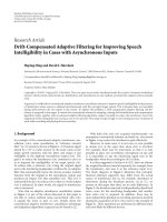
Figure 11.1 CT scan with bone windows demonstrates a right-sided opacification of the mastoid air
cells with bony expansion in the inferior aspect and bony sclerosis superiorly (white block arrows). The
left mastoid process is well aerated and normal in appearance (white line arrows).
Expert comment
Cortical thrombophlebitis is a major cause of cortical neurological deficits in patients with white
matter brain abscesses. This may be the result of occlusion of specific veins, such as the vein of Labbé
or a diffuse process involving cortical territory. Involvement of deep venous systems, although not as
common can occur.
The imaging studies demonstrated cerebellar and right mastoid abscesses in keeping with an otogenic origin. The MRV showed patent sinuses and large veins, with no
signs of lateral sinus thrombosis (Figure 11.6). Cultures obtained from the ear canal
swab grew Group A beta haemolytic streptococci and Pseudomonas and the patient
was started on intravenous ceftriaxone, 2g bd, and clindamycin, 600mg qds.
Case 11 Intracranial abscess
Figure 11.2 Non-enhanced CT scan with brain windows show an ill-defined right cerebellar
hypodensity (white block arrows).
Learning point Stages of brain abscess formation
Stages of brain abscess formation as defined by Britt et al. (pathological) and Osborn et al.
(radiological) (see Table 11.1) [1,2].
Table 11.1 Stages of brain abscess
Stage
Day
Early
cerebritis
0–3
Late
cerebritis
Early
capsule
Late
capsule
Microscopic features
Acute inflammatory reaction
with polymorphonuclear
leukocytes. Fibroblasts appear
and angiogenesis begins.
4–9
Necrosis. Macrophage
recruitment. Neovascularization
and associated vasogenic
oedema. Fibroblastic collagen
deposition.
10–13 Progressive central necrosis
and collagen deposition in
capsule. Peripheral gliosis.
14+
Reduction in inflammatory
cells. Multiple layers of
collegen capsule form
surrounded by increasing
numbers of reactive astrocytes.
MRI T1
MRI T1 + contrast
Poorly-defined hypo/ Patchy enhancement
isointense lesion
Hypointense centre Intense irregular rim
and iso/hyperintense enhancement
rim
Centre becomes
more hyperintense
than CSF and rim
more hyperintense
than white matter
Capsule thickens and
cavity may collapse
Samandouras G. The Neurosurgeon's Handbook. 2011. Oxford University Press.
Well-defined, thin-walled
capsule
Thick capsule with
possible cavity collapse.
Capsule is thicker on
cortical side and thinner
on ventricle side.
105
106
Challenging concepts in neurosurgery
Figure 11.3 MR T1, T2, and T2 FLAIR images, top to bottom, demonstrate a lesion near the right
cerebellopontine angle (CPA) involving the right cerebellar peduncle and abutting the brainstem (white
block arrow). The perifocal oedema affects the cerebellum, particularly the vermis, the pons, and
midbrain (white line arrows). The classical T2 hypointense rim caused by the susceptibility artefacts of a
maturing abscess is demonstrated (dotted arrow).
In this case the radiological features and clinical timeline are consistent with the
late capsule phase.
Clinical tip
Cases referred to neurosurgeons are often at the late abscess stage. When a cerebritis stage abscess
is suspected, microbiologists often request CSF analysis. This should be discouraged, as it is not
only dangerous in the presence of mass effect, but provides low diagnostic yield. CSF findings
when obtained, typically show normal glucose, raised protein, and raised WCC (1–1000/mm3) with
lymphocytes predominating.
Case 11 Intracranial abscess
Figure 11.4 MR T1 after gadolinium administration show a smooth, well-demarcated right cerebellar
ring-enhancing 3 × 3 × 2.5cm lesion with a thin wall (white block arrow). The hypointense surrounding
area is consistent with oedema. On the coronal views (middle) a second ring enhancing ‘daughter’ lesion is
seen in contact with the lesion superiorly (white line arrow). There is also ring-enhancement (1.8 × 1.5cm)
in the right mastoid. There is moderate enhancement of the right cerebellar tentorium.
The patient was taken to theatre urgently for aspiration of the abscess. In the
lateral position and without image-guided neuronavigation, a Dandy cannula was
inserted aiming just lateral to the right CP angle. Pus was aspirated at the first
attempt, but at the second attempt frank blood was aspirated. The resulting haemorrhage was difficult to control and, therefore, it was decided to convert the burr hole
to a small posterior fossa craniectomy. The haemorrhage was finally controlled and
it was felt by the operating surgeon that he could uneventfully remove the capsule
that was prominent in the operative field. After dissection, the abscess capsule was
excised. The post-operative CT is shown in Figure 11.7.
107
108
Challenging concepts in neurosurgery
Figure 11.5 Diffusion-weighted MRI shows a high signal lesion in the right CPA with restricted diffusion
(white block arrow). This is confirmed with the ADC map below that shows a central region of low signal
(white line arrow).
Figure 11.6 MRV. There is no evidence of thrombosis. The transverse sinuses bilaterally are gracile and
hypoplastic, but the sigmoid sinuses are of normal calibre. There is incidental anatomical variation as the
superior sagittal sinus divides caudally into two branches that drain to the internal jugular veins.
Post-operatively he was unable to abduct his right eye, but other movements were
unaffected. There was also a complete loss of right-sided facial cutaneous sensation
in V1–V3 distribution and an associated palsy of the muscles of mastication. He
was noted to have developed a right-sided House–Brackmann Grade 5 lower motor
neurone facial paresis. These findings were consistent with lesions of the abducens,
trigeminal, and facial nerves.
Case 11 Intracranial abscess
Figure 11.7 Enhanced axial CT shows complete capsule excision and a small amount of intra-operative air.
Expert comment
This case demonstrates that free-hand aspiration has a limited role in the management of cerebral
abscess, even in large or very superficial lesions.
Image guidance systems, either frameless or frame based are very useful in achieving target acquisition
and optimum aspiration of the centre of the volumetric space allowing maximum removal of the
infective material and planning of a minimally invasive trajectory.
An additional benefit of image guidance systems is the stabilization of a fixed trajectory. Hand-held
probes, even with minute hand movements, inadvertently and unnecessarily widen the tract disturbing
or damaging neural tissue at the walls of the tract.
The decision to excise the abscess capsule should be planned, especially when the capsule is adjacent
to eloquent areas, and is usually indicated when, despite repeated aspirations and targeted antibiotic
treatment, there is no radiological resolution and no clinical improvement of the patient.
The capsule of the abscess is adherent and tough and is not similar to the soft capsule of a metastatic
lesion or of a circumscribed meningioma.
Even removal of the capsule does not guarantee eradication of the abscess as recurrences have been
observed after complete abscess capsule removal.
Although not a common practice, the stereotactic insertion of an Ommoya reservoir to allow repeated aspiration and antibiotic infiltration has been described [3].
Learning point House–Brackmann classification
The original House–Brackmann classification of facial nerve weakness is shown in Table 11.2 [4].
Table 11.2 House–Brackmann classification
Grade
Description of facial weakness
Score
Percentage motor function
1
2
3
4
5
6
None
Slight
Moderate with full eye closure
Moderate with incomplete eye closure
Severe
Complete
8/8
7/8
5–6/8
3–4/8
1–2/8
0/8
100
76–99
51–75
26–50
1–25
0
(continued)
109
110
Challenging concepts in neurosurgery
The overall score is calculated by measuring the movement of the mid-portion of the superior aspect
of the eyebrow in a superior direction and the movement of the angle of the mouth laterally. The
eye is scored from 0 to 4 with one point given for each 0.25cm of cephalic movement. The mouth is
also scored out of 4 with 1 point given for each 0.25cm of lateral movement. The maximum score is
8. This does not consider the sensory or parasympathetic innervation of the facial nerve. A graphical
version was produced by Lazarini et al., this is simple to use and offers the advantage of speed over the
tabulated scale [5].
Two days later he underwent a mastoidectomy and tympanoplasty. A pneumatized mastoid cavity full of granulation tissue was drilled to healthy bone. The
facial nerve was decompressed by opening its bony canal. Within 2 weeks, his cranial neuropathies had improved with only mild weakness of mastication, normal
facial sensation, minimal diplopia, and a House–Brackman Grade 3 facial weakness
remaining. The intra-operative pus samples were sterile and a peripherally inserted
central catheter (PICC) line was inserted so the patient could receive 6 weeks of
intravenous antibiotics.
Discussion
The earliest successful series of posterior fossa intracranial operations were those of
the Sir William Macewen over 100 years ago. The original technique involved blind
drainage of a cerebellar abscess through a trephined opening in the temporal mastoid bone [6,7]. Macewen was also perhaps the first surgeon to champion the use
of the electric burr, a tool that in combination with the operating microscope and
suction irrigation would allow surgeons to unlock the complexities of the skull base.
Mastoidectomies were popularized by the German otologist Hermann Schwartze
and modified to achieve their current form under William F. House [8].
Chronic suppurative otitis media is a longstanding infective disease of the middle
ear. It is usually easily treated in the early stages with antibiotics, with or without
myringotomy. If treatment is delayed or ineffective the complications can be severe.
Most intracranial complications develop in patients with a chronically discharging
ear. The complications that are the primary concern to neurosurgeons are extraaxial (such as subdural empyema) or intra-axial (such as brain abscess). A brain
abscess is a focal suppurative process that involves the brain parenchyma. At least
50% of all adults brain abscesses are thought to be otogenic in origin [9]. The possible causes of brain abscesses are outlined in Table 11.3.
Clinical tip
Temporal lobe abscesses secondary
to middle ear infection are
best managed operatively in
conjunction with the ear, nose, and
throat (ENT) surgeon. Petrosectomy
or mastoidectomy are often
necessary and, ideally, when
indicated, should be performed at
the same operative session as the
abscess drainage.
Table 11.3 Aetiology of brain abscess from The Neurosurgeon’s Handbook by G. Samandouras
(reproduced with permission).
Primary infection
Micro–organisms
Frontal sinus
Aerobic and anaerobic streptococci
Strep milleri, Bacteroides species, Haemophilus species, Enterobacteriaceae,
Staph. aureus
Aerobic and anaerobic streptococci
Bacteroides fragilis, Enterobacteriaceae, Pseudomonas aeruginosa
Polymicrobial Bacteroides species, Streptococcus species
Staph. aureus, Clostridium species, Bacillus species, Enterobacteriaceae
Middle ear/mastoid bone
Haematogenous spread
Penetrating trauma
111
Case 11 Intracranial abscess
Spread of contiguous infection from an otogenic source to the brain is thought to
be a key cause of cerebellar abscesses and has been found in up to 93% of cases [10].
Intracranial entry can occur by a number of pathways.
The management of brain abscesses from an otogenic origin is controversial.
Otological infections that spread to the brain lie at the interface of neurosurgery,
ENT, and microbiology. Treatments include pure pharmacological management, single or repeat aspirations, capsule excision, and extensive ENT procedures.
Evidence base
Staged operative approach
The classical approach to intracranial complications of chronic supperative otitis media is first
to treat the intracranial disease (with either aspiration or capsule excision) and then remove the
offending source via mastoidectomy at a later date. This was described by Joe Pennybacker in 1948
who reported eighteen cases of otogenic cerebellar abscesses. Interestingly, he notes that only two
of the nine survived in the pre-antibiotic era, whereas after penicillin was introduced eight out of
nine survived, underlining the importance of antimicrobial therapy [13] (Class IV evidence). In 1981,
Shu-Yuan Yang reported 400 cases of brain abscess (115 cerebellar) treated over 20 years in China
without the aid of CT imaging. They found no difference in mortality between simple aspiration or
capsule excision (Class IV evidence). In 2011, a review of 973 brain abscesses (38.6% otorhinogenic)
over a 20-year period in Durban, South Africa recommended abscess drainage and separate
eradication of infection source. This study was limited by its lack of direct comparison with other
strategies [14]. A review of the literature pertaining to aspiration versus capsule excision over a 78-year
period by Ratnaike et al. favoured aspiration because of a 6.6% mortality rate versus 12.7% in the
capsule excision group [15]. However, the validity of this final conclusion is questionable because
abscesses location, aetiology or adjuvant therapy was not addressed. A modern consensus document
on treatment of bacterial brain abscesses states that the type of surgical approach does not appear to
be critical in determining outcome but that the speed of the therapeutic operation, including surgery,
appear to be the more decisive factors for the final outcome [16] (Class V evidence).
Combined approach (neurosurgery and ENT surgeons)
The location of cerebral abscesses that originate in the ear is remarkably constant. In twenty-six
cases of otogenic abscesses that were treated over 15 years all were found immediately adjacent to
the petrous temporal bone. In the pre-CT era, this consistency of location allowed blind drainage.
More recently, it has facilitated a concurrent approach to the abscess and mastoid infection through
a single incision [17]. Morwani & Jayashankar propose a single stage, transmastoid approach as a safe
treatment modality for otogenic intracranial abscesses [18] (Class IV evidence). They retrospectively
reviewed sixty-one patients who had undergone transmastoid abscess drainage and concurrent
tympanomastoidectomy (canal wall up or down depending on pathology). Follow-up was for a
minimum of 24 months. Their mortality was 3%, there was a 6% complication rate (CSF leak or
meningitis), and a 3% abscess recurrence rate. They conclude that this is a safe and effective treatment
strategy. This view is also supported by the work of Singh and Maharaj who found lower mortality
(13% versus 36%) when procedures were combined or performed within 12 hours of each other [19]
(Class IV evidence). Kurien et al. also adopted concurrent craniotomy and mastoidectomy, and in
their report of thirty-six patients found this to be a safe procedure [20] (Class IV evidence). It has been
suggested that early surgical intervention is important to achieve a good outcome and transtemporal
drainage of the abscess allows eradication of the primary mastoid disease at the same time as treating
the intracranial complications [11,21].
Non-surgical management
Wanna et al. have suggested that an initial non-surgical approach to otogenic intracranial abscess
with 6 weeks of broad-spectrum antibiotics (vancomycin, ceftriaxone, and metronidazole) and a
shorter intravenous steroid course is safe and effective (Class IV evidence) [12]. They reserve surgical
(continued)
Learning point Routes of
intracranial infection from the
middle ear
Direct spread through a bone
defect in the tegmen tympani
(the very thin layer of temporal
bone that separates the
tympanic cavity from the middle
cranial fossa) or via Trautmann’s
triangle (demarcated by the
angle between the sigmoid sinus,
the superior petrosal sinus and
the osseous labyrinth).
● A retrograde thrombophelbitis
of the emissary veins may allow
communication through the
skull into the venous sinuses and
then to the brain parenchyma
[11,12].
●
112
Expert comment
In the absence of definitive
diagnosis and clinical deterioration,
atypical causes of abscess should
be considered, including TB,
Nocardia, and fungal infections,
such as Aspergillus or Mucorales
order fungi.
Nocardia responds to
sulphonamides with or without
trimethoprim. Aspergillus responds
to voriconazole and mucormycosis
to amphotericin B.
Challenging concepts in neurosurgery
intervention for patients with an abscess that is expanding with mass effect, despite therapy, or
shows a poor response to treatment. They also state that neurosurgical intervention is indicated if
the abscess looks likely to rupture into the ventricles as this is a recognized poor prognostic marker,
with up to 80% mortality. Interval mastoidectomy is performed once the intracranial disease is stable
unless neurosurgical intervention is needed, in which case it is performed as a combined procedure
wherever possible. It has been suggested that perhaps mastoidectomy can also be avoided in these
patients. In separate studies, Kenna et al. and Dagan et al. treated mastoiditis with daily aural toilet
and intravenous antibiotics [22,23]. This approach has not been widely adopted and Wanna et al.
urge caution, given the severe potential complications of mastoiditis, including sinus thrombosis and
further abscess development. The results of Wanna et al. in ten consecutive patients with intracranial
complications from chronic supperative otitis media (four temporal abscesses, one cerebellar abscess,
four sagittal sinus thrombosis, and one subdural empyema) showed 0% mortality and no recurrence.
Mean hospital stay was 6.4 days. Although firm evidence is lacking, it has been suggested that medical
treatment alone may be more successful if it is begun during the cerebritis stage, if the lesion is less
than 2.5cm, if GCS is >12, and a specific organism is known (Class V evidence) [16,24]. Other authors
have found higher mortality rates with Hsiao et al. reporting an overall case fatality rate of 48% in
thirty-one cases of brain abscess that were managed non-operatively. They identified a low GCS as a
key poor prognostic marker in these patients (Class IV evidence) [25].
The optimum antibiotic regimen has not been firmly established, but an ‘Infection in Neurosurgery’
working party review in 2000 suggested ampicillin, metronidazole, and ceftazidime (or gentamicin), as
the first line empirical therapy (Class V evidence) [26].
A final word from the expert
To date, there has not been a blinded, randomized trial comparing the different treatment
approaches to intracranial abscesses. Neither has there been any meta-analysis of the
existing evidence. There remains clinical equipoise as to the most effective strategy for
treating otogenic posterior fossa brain abscesses. However, in the presence of a large
posterior fossa abscess at early- or late-stage capsule, stereotactic aspiration to obtain
diagnosis and reduce the microbial load appears to be a reasonable initial approach within
the context of multidisciplinary management.
References
1.Britt RH, Enzmann DR, Yeager AS. Neuropathological and computerized tomographic
findings in experimental brain abscess. Journal of Neurosurgery 1981; 55(4): 590–603.
2.Osborn AG, Salzman KL, Barkovich AJ. Diagnostic imaging: brain. Salt Lake City:
Amirsys, 2004.
3.Shen H, Huo Z, Liu L, et al. Stereotatic implantation of Ommaya reservoir in the
management of brain abscesses. British Journal of Neurosurgery. 2011; 25(5): 1–5.
4.House JW, Brackmann DE. Facial nerve grading system. Otolaryngology—head and neck
surgery 1985; 93(2): 146–7.
5.Lazarini P, Mitre E, Takatu E, et al. Graphic visual adaptation of House-Brackmann facial
nerve grading for peripheral facial palsy. Clinical Otolaryngology 2006; 31(3): 192–7.
6.Canale DJ. William Macewen and the treatment of brain abscesses: revisited after one
hundred years. Journal of Neurosurgery 1996; 84(1): 133–42.
7.Macewen SW. Pyogenic infective diseases of the brain and spinal cord. Basingstoke:
Macmillan, 1893.
Case 11 Intracranial abscess
8.Sunder S, Jackler RK, Blevins NH. Virtuosity with the Mallet and Gouge: the brilliant
triumph of the ‘modern’ mastoid operation. Otolaryngologic Clinics of North America.
2006; 39(6): 1191.
9.Syal R, Singh H, Duggal K. Otogenic brain abscess: management by otologist. Journal of
Laryngology & Otology 2006; 120(10): 837–41.
10.Hsu CW, Lu CH, Chuang MJ, et al. Cerebellar bacterial brain abscess: report of eight
cases. Acta Neurologica Taiwanica 2011; 20(1): 47–52.
11.Alaani A, Coulson C, McDermott AL, et al. Transtemporal approach to otogenic brain
abscesses. Acta Otolaryngologica 2010; 130(11): 1214–19.
12.Wanna GB, Dharamsi LM, Moss JR, et al. Contemporary management of intracranial
complications of otitis media. Otology & Neurotology 2010; 31(1): 111.
13.Pennybacker J. Cerebellar abscess: treatment by excision with the aid of antibiotics.
Journal of Neurology, Neurosurgery, and Psychiatry 1948; 11(1): 1.
14.Nathoo N, Nadvi S.S., Narotam PK, & van Dellen JR. Brain abscess: management and
outcome analysis of a computed tomography era experience with 973 patients. World
neurosurgery. 2011; 75(5): 716–726.
15.Ratnaike TE, Das S, Gregson BA, et al. A review of brain abscess surgical treatment,78
years: aspiration versus excision. World Neurosurgery 2011; 76(5): 431–6.
16.Arlotti M, Grossi P, Pea F, et al. Consensus document on controversial issues for
the treatment of infections of the central nervous system: bacterial brain abscesses.
International Journal of Infectious Diseases 2010; 14: S79–92.
17.Penido NDO, Borin A, Iha LCN, et al. Intracranial complications of otitis media: 15 years of
experience in 33 patients. Otolaryngology-Head and Neck Surgery. 2005; 132(1): 37–42.
18.Morwani K, Jayashankar N. Single stage, transmastoid approach for otogenic intracranial
abscess. Journal of Laryngology and Otology 2009; 123(11): 1216.
19.Singh B, Maharaj TJ. Radical mastoidectomy: its place in otitic intracranial complications. The Journal of Laryngology & Otology 1993; 107(12): 1113–18.
20.Kurien M, Job A, Mathew J, et al. Otogenic intracranial abscess: concurrent craniotomy and mastoidectomy—changing trends in a developing country. Archives of
Otolaryngology—Head and Neck Surgery 1998; 124(12): 1353.
21.Hippargekar P, Shinde A. Trans-mastoid needle aspiration for otogenic brain abscesses.
Journal of Laryngology & Otology 2003; 117(5): 422–3.
22.Kenna MA, Bluestone CD, Reilly JS, et al. Medical management of chronic suppurative
otitis media without cholesteatoma in children. Laryngoscope 1986; 96(2): 146–51.
23.Dagan R, Fliss DM, Einhorn M, et al. Outpatient management of chronic suppurative otitis media without cholesteatoma in children. Pediatric Infectious Disease Journal 1992;
11(7): 542–546.
24.Erdofüan E, Cansever T. Pyogenic brain abscess. Neurosurgery Focus 2008; 24(6): E2.
25.Hsiao SY, Chang WN, Lin WC, et al. The experiences of nonoperative treatment in
patients with bacterial brain abscess. Clinical Microbiology and Infection 2011; 17(4):
615–20.
26.De Louvois EB, Bayston R, Lees PD, et al. The rational use of antibiotics in the treatment
of brain abscess. British Journal of Neurosurgery 2000; 14(6): 525–30.
27.Kocherry XG, Hegde T, Sastry KVR, et al. Efficacy of stereotactic aspiration in deep-seated
and eloquent-region intracranial pyogenic abscesses. Neurosurgical Focus 2008; 24(6): 13.
28.Senft C, Seifert V, Hermann E, et al. Surgical treatment of cerebral abscess with the use
of a mobile ultralow-field MRI. Neurosurgical Review 2009; 32(1): 77–85.
113
CA SE
12
Deep brain stimulation
for debilitating
Parkinson’s disease
Jonathan A. Hyam
Expert commentary Alexander L. Green and Tipu Z. Aziz
Case history
A 70-year-old man was referred to the functional neurosurgical service with a diagnosis of idiopathic Parkinson’s disease (PD) for 16 years. His symptoms were rigidity, bradykinesia, and dyskinesias, causing disability and limitations in his activities
of daily living, such as washing himself, cutting up food, writing, and safely using
appliances unsupervised. He had no disturbance of awareness, sensory-perceptual
function, thought, or intellectual function.
Since the time of his diagnosis his oral medications included madopar (containing
L-dopa and a levodopa (L-dopa) decarboxylase inhibitor to reduce its breakdown outside the brain), selegiline (a monoamine oxidase inhibitor), pergolide (a dopaminergic
agonist), and he had an apomorphine (another dopaminergic agonist) pump in situ.
However, his medical therapy had resulted in dyskinesias, 6 years after the diagnosis
of PD. After adjustment to sinemet and apomorphine, he experienced a medication
‘off’ state for 75% of the day with motor symptoms breakthrough despite medication
with resulting bradykinesia and rigidity; and medication ‘on’ state for 25% of the day.
Learning point Pharmacological agents used in Parkinson’s disease
Levodopa therapy was a breakthrough in the management of PD during the 1960s [1]. Due to the
eventual motor complications of its use, a variety of other drugs have been developed for PD, which
act on dopaminergic and non-dopaminergic systems. In early PD, there is no single first choice
drug, and therapy with L-dopa, dopaminergic agonists, or monoamine oxidase-B inhibitors, is
recommended [2]. Table 12.1 describes the range of pharmacological agents currently used in PD [3].
Table 12.1 Pharmacological agents used in PD
Type
Dopamine
Example
L-dopa
Madopar, Sinemet (L-dopa
+ decarboxylase inhibitor)
Dopaminergic agonist
Bromocriptine,
apomorphine,
pramipexole
Anticholinergics
Benzhexol
Monoamine oxidase inhibitors Selegiline
Catechol-O-methyltransferase Entacapone
Inhibitors
Glutamate-antagonist; ?
Amantadine
dopamine reuptake blocker
Side effects
Motor fluctuations, dyskinesias
Hallucinations, sleepiness,
impulsive behaviour,
e.g. gambling, hypersexuality
Central/peripheral autonomic disturbance
Sleep disturbance, light-headedness
Augmented dopa-induced dyskinesias,
Hallucinations, depression
Expert comment
There are many treatments for
Parkinson’s disease and DBS should
be considered as one of many
options, although not always the
most suitable. As these are complex
patients, they should be assessed
by a multidisciplinary team
including neurologist, surgeon,
specialist nurse, neuropsychologist,
and other relevant healthcare
professionals
116
Challenging concepts in neurosurgery
On examination, he had no arm tremor. Movements were bradykinetic. Dyskinesias,
particularly on the right, and bilateral cog-wheel rigidity were present. Cranial nerve
and peripheral neurological examinations were otherwise normal. Micrographia was
demonstrated. Unified Parkinson’s disease Rating Scale (UPDRS) scores were Part 1:
5/16; Part 2: 3/56 on, 25/56 off; Part 3: 15/104 on, 43/104 off. Formal neuropsychological evaluation using interview and battery tests did not reveal significant psychological pathology. Brain MRI was normal.
Learning point Unified Parkinson’s disease Rating Scale
UPDRS was developed to monitor the severity and progression of PD, whereby several existing scales
were incorporated into one, allowing more efficient and flexible patient assessment. It is extensively
used by neurologists across the world with 87% reporting its use in trials and 70% using it in clinical
practice [4]. The Movement Disorders Society judged the UPDRS as providing a comprehensive
assessment of the motor aspects of PD especially, more than the non-motor aspect, although some
items had low or adequate inter- and intra-rater reliability [4]. It is divided into four categories
evaluating:
I: mentation, behaviour, and mood.
II: activities of daily living.
● III: motor examination.
● IV: complications of therapy.
●
●
The Modified Hoehn & Yahr Staging and Schwab & England ADL Scales were added later (Table 12.2).
Table 12.2 Unified PD Rating Scale
Part
Aspects of disease
Factors included
I
II
III
IV
V
VI
Mentation, behaviour and mood
Activities of daily living*
Motor examination
Complications of therapy
Modified Hoehn & Yahr staging
Schwab & England ADL Scale
Intellect, thought disorder, depression
Falls, dressing, swallowing, hygiene, utensil handling
Tremor, posture, rigidity, speech, gait, bradykinesia
Dyskinesias, on/off fluctuations, orthostasis
Disease severity, uni/bilateral, balance, independence
Independence/dependence, showering, swallowing,
bladder/bowel
*Denotes patients tested in both on and off PD states.
Modified from Fahn et al. [5].
When discussing the aims of surgery with the patient, amelioration of the bradykinesia, dyskinesias, and rigidity were agreed to be the most important. The patient
was offered and accepted bilateral subthalamic nucleus (STN) deep brain stimulation.
A stereotactic pre-operative MRI was performed to define the subcortical nuclei for
electrode targeting (see Figure 12.1). At the beginning of the procedure, a stereotactic
frame was applied to the patient’s head under local anaesthesia and a stereotactic CT
performed. CT is less susceptible to spatial artefacts and is registered with the MRI.
The subthalamic nucleus was identified using the patient’s imaging, and with the
assistance of registration with a brain atlas to help confirm and plot the target coordinates. The patient was taken to theatre and bilateral craniostomies were fashioned
under local anaesthetic. A 1.8-mm diameter radiofrequency electrode was passed
to target, monitoring impedance to detect transgression of the ventricle, which can
lead to electrode misplacement. The DBS electrode was then inserted in its place
(Figure 12.2a). A neurologist objectively assessed the contralateral limb rigidity
during test stimulation to ensure electrode position produced clinical benefit. The
Case 12 Deep brain stimulation for debilitating Parkinson’s disease
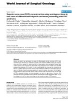
Third ventricle
Subthalamic
nucleus
traversed by
electrode
Red nucleus
Figure 12.1 Reconstructed three-dimensional brain MRI in axial section with deep brain electrode
trajectory to subthalamic nucleus (circled) planned on neuronavigation workstation.
procedure was repeated on the contralateral side. On placement of the contralateral
radiofrequency electrode to target, there was an improvement in rigidity, independent
of stimulation, a phenomenon known as ‘stun’, whereby the mechanical interruption or
microlesioning of the target nucleus produces a temporary therapeutic effect. Although
it confirms that the target produces clinical efficacy, it also prevents the fine-tuning of
electrode placement based on further clinical examination during the procedure.
(a)
(b)
Figure 12.2 (a) Deep brain electrode implanted using stereotactic frame attached to patient. (b)
Implanted pulse generator to be internalised within subclavicular pocket
117
118
Challenging concepts in neurosurgery
A post-operative stereotactic CT head was performed with head frame and localizer still attached, which verified the electrode contacts’ position in the STNs. On
returning to theatre, under general anaesthesia, extension leads were connected to
the electrodes and tunnelled behind the ear, into a subclavian pocket that had been
fashioned. The implanted pulse generator was connected to the extension leads and
placed in the pocket, which was then closed (Figure 12.2b). The patient was woken
in recovery and had suffered no neurological deterioration. The stimulator was not
initially activated. There was a unilateral improvement in rigidity and dyskinesia
as a result of the intra-operative stun effect. Stimulation was activated 2 weeks
post-operatively at 1.5V, 90 microseconds and 130Hz, allowing the acute changes of
surgery including stun to settle down so that stimulation titration was performed
without such a confounding factor.
Post-operative UPDRS scores at 6 months were Part 1: 1/16; Part 2: 8/56 on, 24/56
off; Part 3: 8/104 on, 34/104 off.
There was a marked improvement in Part III of the UPDRS with improvements in
rigidity, dyskinesias, and bradykinesia on examination. There was no deterioration
in mood or cognition detected.
Discussion
PD is a neurodegenerative disorder caused, in part, by the loss of dopaminergic
neurones in the substantia nigra (pars compacta). This results in disruption of the
normal oscillatory and synchronous neuronal activity between the cortex, globus
pallidus interna (GPi) and STN, The three cardinal clinical manifestations of PD are
bradykinesia, tremor, and rigidity. Gait and postural instability is often also seen
[6]. The place of surgery in the management of PD has been cyclical. It was once the
mainstay of treatment, in the form of ablative surgeries, such as pedunculotomy, and
was then made largely redundant by the advent of dopaminergic drugs. However, it
was found that dopaminergic drugs caused side effects including dyskinesias, which
could be severely incapacitating. Once again, surgery (commonly taking the form of
DBS) became an important modality in the management of PD, not only to treat the
cardinal symptoms of the disease itself, but also to treat the dyskinetic side effects
of medical therapy.
Patient selection
The commonest reasons for poor outcomes after DBS are: poor patient selection,
poor operative electrode placement, and inadequate stimulation programming [7]. In
DBS for PD, the ideal patient characteristics are a patient with idiopathic PD with an
excellent response to L-dopa, particularly the medication motor ‘on’ state. Broadly,
DBS surgery is offered to those who suffer from intractable tremor, debilitating side
effects of medical therapy, such as dyskinesia, are of a younger age, and have a
psychological and physical health sufficient to tolerate surgery and ongoing stimulation management as an outpatient. Patients with psychiatric diagnoses of major
depression, acute psychosis, and dementia are excluded [7]. Therefore, a movement
disorder neurologist and clinical psychologist/psychiatrist should form part of the
DBS team, as well as the surgeon. A multidisciplinary approach is key to the management of these patients. Furthermore, identification of the most debilitating symptoms for the individual patient are critical as this determines whether non-surgical
119
Case 12 Deep brain stimulation for debilitating Parkinson’s disease
therapies have been exhausted for a given symptom profile, the location of the deepbrain stimulator placement, and allows for an estimation of the chances that DBS
will be beneficial.
Location, location, location
Depending on the site targeted by DBS, a variety of symptoms can be ameliorated
(see ‘Learning point: Parkinson’s disease symptoms and relevant deep-brain stimulation target nuclei’). It is therefore critical to tailor the targeting to the individual
patient. DBS for PD is supported by the NICE Guidelines of 2006 with particular reference to STN, GPi and thalamic stimulation [2].
Subthalamic nucleus
The cardinal symptoms of PD, namely bradykinesia, rigidity, and tremor, as well
as dyskinesias resulting from medication, can all be ameliorated by STN DBS to
varying degrees. The STN was identified as a target in PD as a direct result of primate models [8,9]. As STN DBS diminishes these symptoms, the pharmacological therapy can be reduced together with their resulting side effects, particularly
the incidence and severity of dyskinesias. Krack et al. demonstrated that STN
DBS improved the motor symptoms of PD, improved activities of daily living, and
reduced medication requirements [10]. The multicentre PD SURG Trial randomly
assigned 366 patients with advanced PD to immediate surgery with best medical
therapy or best medical therapy alone [11]. This was effectively a study of STN
DBS surgery as only four patients received stimulation of a different nucleus, i.e.
the GPi. The PD SURG trial found that DBS produced clear advantages compared
with maximal medical therapy in clinical assessments and patient-assessed quality of life at 1 year follow-up. DBS conferred improvements in mobility and the
activities of daily living domains of the PDQ-39 questionnaire, the total UPDRS,
particularly part IV including time and severity of dyskinesias and ‘off’ periods,
and a fall in daily dopaminergic drug requirement by a third. PD SURG found an
adverse surgery-related event in 19% of patients. There was one procedure-related
death, but no suicides [11].
Adverse effects of STN DBS attributable to the subthalamic location itself include
psychiatric and cognitive disturbances, reflecting the STN’s role in association and
limbic circuits [12]. PD SURG found no decline in cognition as rated by the dementia
rating scale (DRS-II), although its sensitivity to cognitive decline has been questioned [13]. Speech decline after surgery was detected on detailed neuropsychological testing, with a reduced verbal fluency and vocabulary [11]. Decline in cognition
and mood has been inconsistently reported by other series, but appears to affect
1–2% of patients [14].
Globus pallidus interna
The GPi has been an important target in PD for the amelioration of dyskinesias.
Its impact on bradykinesia and rigidity is becoming increasingly recognized. In
a multicentre RCT, the cooperative studies programme (CSP) 468 Study Group
demonstrated that STN and GPi DBS were more efficacious than medical therapy
alone, in terms of increased time in the PD on state without dyskinesias, increased
motor function, and a variety of quality of life measures [15]. After randomization
between STN and GPi stimulation, they later demonstrated that both targets produced equivalent efficacy in motor function improvement measured by the UPDRS
Expert comment
Patient selection for DBS is one
of the most important aspects.
A good candidate is generally
one who has a good response to
L-dopa, but either the side effects
of the treatment are too severe
(dyskinesia) or motor on–off
fluctuations predominate. Tremor
can also be treated successfully.
Approximately 10% of patients with
PD are suitable.
120
Challenging concepts in neurosurgery
Part III at 24 months [16]. Although dopaminergic drug requirement was lowered
to a greater degree by STN stimulation, it also led to a decline in mood and visuomotor processing speed compared with GPi stimulation. Given that cognitive and
mood disturbance was also found less after GPi compared with STN stimulation
in other studies [17, 18, 19], pallidal stimulation is an important option for treating bradykinesia, rigidity, and dyskinesias in PD, and is a valid target in the case
presented here.
Thalamus
Tremor amelioration is one of the oldest indications for functional neurosurgery
after Irving Cooper’s serendipitous observations in the 1950s [20,21]. Tremor can be
treated by DBS of the motor thalamus or the STN. Thalamic DBS should be reserved
for cases in which tremor is the predominant debilitating symptom and where the
other cardinal symptoms of PD or drug side effects have not and are not expected to
manifest [7]. Within the motor thalamus, the ventralis intermedius nucleus (VIM) is
the commonest target, but the ventralis oralis nucleus (VOP), intimately related to it,
is an alternative [22]. As this patient was not troubled by tremor, thalamic stimulation would not be an appropriate choice for him.
Pedunculopontine nucleus
Postural instability and gait freezing have historically not responded well to
DBS nor L-dopa therapy. However, a novel target, the pedunculopontine nucleus
(PPN), was identified in primate studies [23, 24, 25], as a reticular nucleus located
at the junction of the mesencephalon and pons [Jenkinson et al. 2006; 26]. In
humans with advanced PD, PPN stimulation results in improvements in measurements of gait, posture, and balance [27, 28, 29]. As these were not prominent
symptoms in this gentleman’s PD, PPN stimulation would not be an appropriate
choice for him.
Clinical tip Indications and patient selection for DBS in PD
Patient selection is critical and only a minority of PD sufferers are appropriate for DBS. The factors
recommended to confer good outcome from DBS can be divided into three broad categories relating
to the PD itself and response to L-dopa, psychiatric and psychological factors, and general surgical
factors.
Parkinson’s disease features
Idiopathic.
Excellent response to L-dopa.
●Dyskinesias.
● Intractable tremor.
●
●
Psychiatric
No dementia.
No major depression.
● No acute psychosis.
● Cognition status good.
●
●
General
●
●
Younger age.
Fitness for neurosurgery.
Case 12 Deep brain stimulation for debilitating Parkinson’s disease
Learning point Parkinson’s disease symptoms and relevant deep-brain stimulation
target nuclei
Depending on the electrode target, DBS can confer benefit on a range of symptoms in PD.
Establishing the symptoms most deleterious to the individual patient is therefore crucial to planning
DBS in order to provide as much benefit as possible (Table 12.3). Some targets benefit a greater range
of symptoms than others [2,16,27].
Table 12.3 PD symptoms and relevant deep-brain
stimulation target nuclei
Symptom
Target
Tremor
Bradykinesia, rigidity
Dyskinesia
Postural instability, gait freezing
Thalamus, STN
STN, GPi
STN, GPi
PPN
Clinical tip Accurately implanting deep brain stimulation electrodes
Several measures to optimize the accuracy of deep brain electrode implantation are undertaken. Their
utilization varies depending on the case and the unit.
Neuroimaging
MRI provides definition of the subcortical structures for targeting. CT provides greater spatial accuracy
as it is less subject to artefacts than MRI. Fusion of the two modalities provides the advantages of both.
Intra-operative neurological assessment
Test stimulation and clinical assessment while the patient is awake provides rapid feedback on the
clinical effect of stimulation and adverse effects, and allows optimization of electrode depth. The
anaesthetist’s role is therefore crucial. This is not suitable for patients who would not tolerate surgery
while awake, such as those in whom their movement disorder is so severe.
Microelectrode recording
Localization of cell groups within the target nucleus by depth recordings from multiple fine
microelectrodes provides neurophysiological targeting feedback. Disadvantages include longer operative
time and a concern of increase risk of intracranial haemorrhage due to multiple electrode passes [30].
Lesional surgery
Creating a lesion, rather than chronically implanting an electrode is an important
alternative for clinicians and patients to consider. Historically, deep brain ablational
surgery preceded DBS, which is not suitable for all patients. Lesions of the GPi
(pallidotomy) or motor thalamus (thalamotomy) can confer similar efficacy to DBS
[31,32] and benefits from subthalamotomy have also been reported [17,33], and are
therefore useful to consider in PD patients. DBS is an expensive therapy on account
of the hardware costs of the electrode and pulse generator and also the subsequent
need for follow-up and battery replacement surgeries. The advantage of a lesion is
that it is a one-off therapy and does not require continued follow-up nor is there
any hardware to manage. Therefore, determining factors include the patient’s tolerance and compliance with intensive follow-up, and their agreement to undergo
further battery change procedures to maintain stimulation, their cognitive level,
expectations and level of neurological risk they deem acceptable, bilateral symptoms
(bilateral thalamotomy has an unacceptably high risk of speech and swallowing
121
122
Challenging concepts in neurosurgery
disturbance [34,35], hardware and infection fears, and local economic factors. The
lesion, however, is an irreversible and unmodifiable therapy. DBS electrodes have
the advantage that they can be switched-off or removed if causing adverse effects
and the stimulation parameters can be titrated to the patient’s needs in addition to
allowing adjustment over time as their tolerance or disease state changes.
A final word from the expert
There are likely to be two main future developments and these are equivalent to a ‘space
race’ between improving technology and other biological treatments. For example, electrode
design is advancing rapidly with improvements in electric field shaping and other modalities,
such as optogenetics. On the other hand, there have been huge recent developments
in stem cell research, viral vectors, and growth factor infusions with the aim of restoring
‘normal’ brain.
References
1.Cotzias GC, Papavasiliou PS, Gellene R. L-dopa in Parkinson’s syndrome. New England
Journal Medicine 1969; 281(5): 272.
2.National Institute for Health and Clinical Excellence (NICE). Parkinson’s diseases:
diagnosis and management in primary and secondary care, NICE Clinical Guideline
35. London: NICE, 2006. Available at: />live/10984/30088/30088.pdf.
3.Kalinderi K, Fidani L, Castor Z, et al. Pharmacological treatment and the prospect of
pharmacogenetics in Parkinson’s disease. International Journal of Clinical Practice 2011;
65(12): 1289–94.
4.Movement Disorder Society Task Force on Rating Scales for Parkinson’s Disease. The
Unified Parkinson’s Disease Rating Scale (UPDRS): status and recommendations.
Movement Disorders 2003; 18(7): 738–50.
5.Fahn S, Elton RL, Members of the UPDRS Development Committee. Unified Parkinson’s
Disease Rating Scale. In: S Fahn, CD Marsden, DB Calne, et al. (eds), Recent developments in Parkinson’s disease vol. 2 (pp. 153–64). Florham Park, NJ: Macmillan Health
Care Information 1987.
6.Williams D, Tijssen M, van Bruggen G, et al. Dopamine-dependent changes in the functional connectivity between basal ganglia and cerebral cortex in humans. Brain 2002;
125: 1558–69.
7.Volkmann J. Selecting appropriate Parkinson’s patients for deep brain stimulation. In:P
Bain, T Aziz, X Liu, et al. (eds), Deep brain stimulation (pp. 75–83). Oxford: Oxford
University Press, 2009.
8.Aziz TZ, Peggs D, Sambrook MA, et al. Lesion of the subthalamic nucleus for the alleviation of 1-methyl-4-phenyl-1,2,3,6-tetrahydropyridine (MPTP)-induced parkinsonism in
the primate. Movement Disorders 1991; 6: 288–92.
9.Bergman H, Wichmann T, Delong MR. Reversal of experimental parkinsonism by lesion
of the subthalamic nucleus. Science 1990; 249: 1436–8.
10.Krack P, Batir A, Van Blercom N, et al. Five year follow-up of bilateral stimulation of the
subthalamic nucleus in advanced Parkinson’s disease. New England Journal of Medicine
2003; 349: 1925–34.
11.Williams A, Gill S, Varma T, et al., on behalf of the Parkinson’s disease Surgical
Collaborative Group. Deep brain stimulation plus best medical therapy versus medical
Case 12 Deep brain stimulation for debilitating Parkinson’s disease
therapy alone for advanced Parkinson’s disease (PD SURG trial): a randomized, openlabel trial. Lancet Neurology 2010; 9 (6): 581–91.
12.Hamani C, Saint-Cyr SA, Fraser J, et al. The subthalamic nucleus in the context of movement disorders. Brain 2004; 127: 4–20.
13.Rodriguez-Oroz MC. Deep brain stimulation for advanced Parkinson’s disease. Lancet
Neurology 2010; 9(6): 558–9.
14.Woods SP, Fields JA, Troster AI. Neuropsychological sequelae of subthalamic nucleus
deep brain stimulation in Parkinson’s disease: a critical review. Neuropsychology
Reviews 2002; 12: 111–26.
15.Weaver FM, Follett K, Stern M, et al. Bilateral deep brain stimulation vs best medical
therapy for patients with advanced Parkinson disease: a randomized controlled trial.
Journal of the American Medical Association 2009; 301(1): 63–73.
16.Follett KA, Weaver FM, Stern M, et al. Pallidal versus subthalamic deep-brain stimulation for Parkinson’s disease. New England Journal of Medicine 2010; 362(22): 2077–91.
17.Walter BL, Vitek JL. Surgical treatment for Parkinson’s disease. Lancet Neurology 2004;
3: 719–28.
18.Volkmann J, Alert N, Voges J, et al. Safety and efficacy of pallidal or subthalamic
nucleus stimulation in advance PD. Neurology 2001; 56: 548–51.
19.Rodriguez-Oroz MC, Obeso JA, Lang AE, et al. Bilateral deep brain stimulation in
Parkinson’s disease: a multicentre study with 4 years follow-up. Brain 2005; 128:
2240–9.
20.Cooper IS. Effect of anterior choroidal artery ligation on involuntary movements and
rigidity. Transactions of the American Neurological Association 1953; 3(78th meeting):
6–7.
21.Das K, Benzil DL, Rovit RL, et al. Irving S. Cooper (1922–1985): a pioneer in functional
neurosurgery. Journal of Neurosurgery 1998; 89(5): 865–73.
22.Hyam J, Owen SLF, Kringelbach ML, et al. Contrasting connectivity of the ventralis
intermedius and ventralis oralis posterior nuclei of the motor thalamus demonstrated by
probabilistic tractography. Neurosurgery 2012; 70(1): 162–9.
23.Jenkinson N, Nandi D, Miall RC, et al. Pedunculopontine nucleus stimulation improves
akinesia in a Parkinsonian monkey. NeuroReport 2004; 15: 2621–4.
24.Jenkinson N, Nandi D, Oram R, et al. Pedunculopontine nucleus electric stimulation
alleviates akinesia independently of dopa-minergic mechanisms. NeuroReport 2006; 17:
639–41.
25.Nandi D, Aziz TZ, Giladi N, et al. Reversal of akinesia in experimental parkinsonism by
GABA antagonist microinjections in the pedunculopontine nucleus. Brain 2002; 125(11):
2418–30.
26.Zrinzo L, Zrinzo LV, Tisch S, et al. Stereotactic localization of the human pedunculopontine nucleus: atlas-based coordinates and validation of a magnetic resonance imaging
protocol for direct localization. Brain 2008; 131(6): 1588–98.
27.Plaha P, Gill SS. Bilateral deep brain stimulation of the pedunculopontine nucleus for
Parkinson’s disease. NeuroReport 2005; 16: 1883–7.
28.Moro E, Hamani C, Poon YY, et al. Unilateral pedunculopontine stimulation improves
falls in Parkinson’s disease. Brain 2010; 133(1): 215–24.
29.Thevathasan W, Coyne TJ, Hyam JA, et al. Pedunculopontine nucleus stimulation
improves gait freezing in Parkinson’s disease. Neurosurgery 2011; 69: 1248–54.
30.Zrinzo L, Foltynie T, Limousine P, et al. Reducing hemorrhagic complications in functional neurosurgery: a large case series and systematic literature review. Journal of
Neurosurgery 2012; 116(1): 84–94.
31.Bittar RG, Hyam J, Nandi D, et al. Thalamotomy versus thalamic stimulation for multiple
sclerosis tremor. Journal of Clinical Neuroscience 2005; 12(6): 638–42.
32.Gross RE. What happened to posteroventral pallidotomy for Parkinson’s disease and
dystonia? Neurotherapeutics 2008; 5: 281–93.
123
124
Challenging concepts in neurosurgery
33.Alvarez L, Macias R, Lopez G, et al. Bilateral dorsal subthalamotomy in Parkinson’s
disease (PD): initial response and evolution after 2 years. Movement Disorders 2002;
17(Suppl. 5): S95.
34.Alusi SH, Aziz TZ, Glickman S, et al. Stereotactic lesional surgery for the treatment of
tremor in multiple sclerosis: a prospective case-controlled study. Brain 2001; 124(8):
1576–89.
35.Samra K, Waltz JM, Riklan M, et al. Relief of intention tremor by thalamic surgery.
Journal of Neurology Neurosurgery and Psychiatry 1970; 33(1): 7–15.
CA SE
13
Endoscopic resection of a
growth hormone-secreting
pituitary macroadenoma
Alessandro Paluzzi
Expert commentary Paul Gardner
Case history
A 61-year-old male presented with a 2-year history of fatigue, erectile dysfunction, and increasing hand and shoe sizes (size 9 to size 11). He also complained
of visual problems affecting his driving. His wife had reported that he had
started snoring at night, and had noticed that his nose and jaw had grown to
change his facial features significantly compared with photographs of him several
years before.
His previous medical history was unremarkable except for hypertension.
Learning point The signs and symptoms of acromegaly
The acral changes (from Gr akron = extremity) are the most common clinical signs that lead to
the diagnosis. Hands and feet are broadened, and the fingers and toes are thickened and stubby.
The nose is widened, and the cheekbones and forehead become prominent, sometimes with
frontal bossing. Prognathism, maxillary widening, dental diastasis, and macroglossia are also
common.
In addition to the typical dysmorphic facial and body features, acromegaly is associated with a
number of systemic complications, including hypertension, caradiomyopathy, diabetes mellitus,
sleep apnoea syndrome, and colon cancer. These account for the associated mortality risk in
acromegalic patients compared with the normal population [1]. Treatment of each specific
co-morbidity greatly improves the general prognosis of the patients [2]. Furthermore, the systemic
comorbidities, together with the presence of macroglossia and jaw malocclusion, need to be
taken into account pre-operatively before removal of a pituitary adenoma, since they increase the
anaesthetic risk of these patients.
On examination, the features of acromegaly were noted. His blood pressure
was 170/102 on lisinopril/hydrocholorthyazide and random blood glucose was
8.3mmol/L (normal range 3.9–5.5mmol/L). Visual field assessment demonstrated
gross bitemporal hemianopia, and this was confirmed on Humphrey visual field
automated testing (Figure 13.2e).
Endocrine tests showed a random growth hormone (GH) level of 58ng/
mL (normal range 0–5 ng/mL) and IGF-1 level of 667ng/mL (reference range:
71–290ng/mL). Other endocrine tests revealed hypothyroidism with decreased
free T4 at 0.48ng/dL (normal range 0.8–1.8ng/dL) and normal TSH at 0.520μIU/mL
126
Challenging concepts in neurosurgery
(normal range 0.300–5000μIU/mL). He also displayed hypogonadotropic hypogonadism with decreased LH at 0.3mIU/mL (normal range 1–5.6mIU/mL) and
FSH at 1.5mIU/mL (normal range 1.5–14.3mIU/mL) and undetectable testosterone <1ng/dL (normal range 250–1100ng/dL). His AM cortisol was also low at
1μg/dL (normal AM range 7–25μg/dL) with an ACTH of 15pg/mL (normal range
9–46pg/mL).
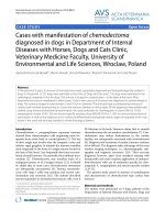
T1-weighted MRI with contrast revealed a large sellar lesion with suprasellar
extension consistent with pituitary macroadenoma measuring 3.3 × 2.6 × 3.7cm
(Figure 13.1). The tumour extended laterally beyond the lateral wall of the cavernous
internal carotid artery, suggesting a high probability of cavernous sinus invasion
(Knosp grade III) (Figure 13.1 a, c, e).
(a)
(b)
(c)
(d)
Naso-septal flap
(e)
(f)
Figure 13.1 Pre- and post-operative (12 months) gadolinium-enhanced T1 MR imaging of
the macroadenoma. (a, b) Axial views. The arrow points to the portion of the adenoma invading
the left cavernous sinus. (c, d) Coronal views. On the pre-operative scan (c) the most lateral border
of the adenoma on the left side extends beyond the lateral edge of the carotid artery indicating,
according to the Knosp classification, high probability of cavernous sinus invasion. (e, f ) sagittal
views. In the post-operative scan (f ) the enhancing tissue at the level of the planum sphenoidale
corresponds to the muco-perichondrial naso-septal flap used to repair the intra-operative
dural opening.
Case 13 Endoscopic resection of a macroadenoma
Learning point Knosp classification
In 1991, Engelbert Knosp proposed a radiological classification to predict the likelihood of cavernous
sinus invasion from a pituitary adenoma. He studied the pre-operative MRI scans of 25 pituitary
adenomas that were confirmed surgically to have invaded the cavernous sinus space. Five ‘Knosp
grades’ were defined by the relationship of the adenoma’s lateral edge with the internal carotid artery,
as shown on the most representative coronal post-contrast T1 slice. Grade 0 represents the normal
condition, and Grade 4 corresponds to the total encasement of the intracavernous carotid artery.
According to this classification, surgically proven invasion of the cavernous sinus space was present
in all Grade 4 and 3 cases and in all but one of the Grade 2 cases; no invasion was present in Grade 0
and Grade 1 cases.
In view of the recent history of visual deterioration and the diagnosis of acromegaly from a GH-secreting adenoma the patient was advised to undergo surgical
intervention. He consented to an expanded endonasal approach (EEA) for resection
of the pituitary macradenoma.
During the operation, marked expansion of the sella was noticed. After initial bony exposure of the sella and both cavernous sinuses (Figure 13.2a), the
tumour was debulked using a ‘2-sucker technique’ (Figure 13.2b). The adenoma
was found to have invaded the medial wall of the left cavernous sinus and to
extend into the medial compartment of the cavernous sinus. Complete resection of this component of the tumour was achieved with the help of a 45-degree
angled endoscope. The inferior hypophyseal artery was identified and coagulated
(Figure 13.2c). To avoid herniation of arachnoid through the enlarged diafragma
sellae during the initial steps of the tumour debulking, the suprasellar portion
of the tumour was addressed only at the end, using again a 45-degree angled
endoscope (Figure 13.2d). Both superior hypophyseal arteries were visualized
and preserved. Gross total resection of the tumour was achieved. The repair of
the dural defect was carried out using a pedicled muco-perichondrial naso-septal
flap (Figure 13.1f).
The patient made a satisfactory post-operative recovery. His vision subjectively
improved immediately post-operatively and formal visual field assessment 2 weeks
and 6 months later demonstrated an objective substantial decrease in the visual field
defects bilaterally (Figure 13.2f). Post-operative MRI scans at 3, 6, and 12 months
(Figure 13.1b, d, f) demonstrated gross total resection without any evidence of residual or recurrent tumour.
His GH on the first post-operative day was down to 0.74ng/mL (normal range
0–5 ng/mL), while the IGF-1 was still abnormal at 412ng/mL (reference range:
71–290ng/mL). Two weeks later, both levels were normal, with a random GH of
0.40ng/mL and IGF-1 of 113ng/mL and the MRI scan at 1 month showed no evidence of residual adenoma. Both endocrinological and radiographic results were
taken with caution at this stage, since it is well known that during the first 3 months
post-operatively they can be misleading. During subsequent follow-up, the clinical
features of acromegaly gradually improved and biochemical cure was maintained at
7 months and at his last follow-up 1 year post-operatively.
The patient was also medically treated with oral hydrocortisone 10mg bd,
transdermal testosterone 5g/day, and levothyroxine 100μg/day for panhypopituitarism that was present preoperatively.
127