Ebook High-Yield neuroanatomy (4th edition): Part 1
Bạn đang xem bản rút gọn của tài liệu. Xem và tải ngay bản đầy đủ của tài liệu tại đây (3.31 MB, 89 trang )
LWBK110-3895G-FM[i-xviii].qxd 8/14/08 5:57 AM Page i Aptara Inc.
High-Yield
TM
Neuroanatomy
FOURTH EDITION
LWBK110-3895G-FM[i-xviii].qxd 8/14/08 5:57 AM Page ii Aptara Inc.
LWBK110-3895G-FM[i-xviii].qxd 8/14/08 5:57 AM Page iii Aptara Inc.
High-Yield
TM
Neuroanatomy
FOURTH EDITION
James D. Fix, PhD
Professor Emeritus of Anatomy
Marshall University School of Medicine
Huntington, West Virginia
With Contributions by
Jennifer K. Brueckner, PhD
Associate Professor
Assistant Dean for Student Affairs
Department of Anatomy and Neurobiology
University of Kentucky College of Medicine
Lexington, Kentucky
LWBK110-3895G-FM[i-xviii].qxd 8/14/08 5:57 AM Page iv Aptara Inc.
Acquisitions Editor: Crystal Taylor
Managing Editor: Kelley Squazzo
Marketing Manager: Emilie Moyer
Designer: Terry Mallon
Compositor: Aptara
Fourth Edition
Copyright © 2009, 2005, 2000, 1995 Lippincott Williams & Wilkins, a Wolters Kluwer business.
351 West Camden Street
Baltimore, MD 21201
530 Walnut Street
Philadelphia, PA 19106
Printed in the United States of America.
All rights reserved. This book is protected by copyright. No part of this book may be reproduced or transmitted in
any form or by any means, including as photocopies or scanned-in or other electronic copies, or utilized by any
information storage and retrieval system without written permission from the copyright owner, except for brief
quotations embodied in critical articles and reviews. Materials appearing in this book prepared by individuals as
part of their official duties as U.S. government employees are not covered by the above-mentioned copyright. To
request permission, please contact Lippincott Williams & Wilkins at 530 Walnut Street, Philadelphia, PA 19106,
via email at , or via website at (products and services).
987654321
Library of Congress Cataloging-in-Publication Data
Fix, James D.
High yield neuroanatomy / James D. Fix, Jennifer K. Brueckner. — 4th ed.
p. ; cm.
Includes bibliographical references and index.
ISBN 978-0-7817-7946-3
1. Neuroanatomy—Outlines, syllabi, etc. 2. Neuroanatomy—Examinations, questions, etc. I. Brueckner,
Jennifer K., 1970- II. Title.
[DNLM: 1. Nervous System—anatomy & histology—Examination Questions. 2. Nervous System—
anatomy & histology—Outlines. 3. Nervous System Diseases—Examination Questions. 4. Nervous System
Diseases—Outlines. WL 18.2 F566h 2009]
QM451.F588 2009
611'.8076—dc22
2008024078
DISCLAIMER
Care has been taken to confirm the accuracy of the information present and to describe generally accepted
practices. However, the authors, editors, and publisher are not responsible for errors or omissions or for any
consequences from application of the information in this book and make no warranty, expressed or implied,
with respect to the currency, completeness, or accuracy of the contents of the publication. Application of this
information in a particular situation remains the professional responsibility of the practitioner; the clinical
treatments described and recommended may not be considered absolute and universal recommendations.
The authors, editors, and publisher have exerted every effort to ensure that drug selection and dosage set
forth in this text are in accordance with the current recommendations and practice at the time of publication.
However, in view of ongoing research, changes in government regulations, and the constant flow of information relating to drug therapy and drug reactions, the reader is urged to check the package insert for each drug
for any change in indications and dosage and for added warnings and precautions. This is particularly important when the recommended agent is a new or infrequently employed drug.
Some drugs and medical devices presented in this publication have Food and Drug Administration (FDA)
clearance for limited use in restricted research settings. It is the responsibility of the health care provider to
ascertain the FDA status of each drug or device planned for use in their clinical practice.
To purchase additional copies of this book, call our customer service department at (800) 638-3030 or fax
orders to (301) 223-2320. International customers should call (301) 223-2300.
Visit Lippincott Williams & Wilkins on the Internet: . Lippincott Williams & Wilkins customer service representatives are available from 8:30 am to 6:00 pm EST.
LWBK110-3895G-FM[i-xviii].qxd 8/14/08 5:57 AM Page v Aptara Inc.
Preface
Based on your feedback on previous editions of this text, the fourth edition has been reorganized
and updated significantly. New features include chapter reorganization, terminology updates consistent with Terminologica Anatomica, addition of a table of common neurologic disease states, and
an online ancillary of board-style review questions. To make the most effective use of this book,
study the computed tomography scans and magnetic resonance images carefully and read the legends. Test your knowledge of each topic area with board-style questions provided online. Finally,
remember these tips as you scan the chapters:
Chapter 1: What is the difference between Lewy and Hirano bodies? Nerve cells contain Nissl substance in their perikarya and dendrites but not in their axons. Remember that Nissl substance
(rough endoplasmic reticulum) plays a role in protein synthesis. Study Figure 1-3 on the localization and prevalence of common brain and spinal cord tumors. Remember that in adults, glioblastoma multiforme is the most common brain tumor, followed by astrocytoma and meningioma. In
children, astrocytoma is the most common brain tumor, followed by medulloblastoma and ependymoma. In the spinal cord, ependymoma is the most common tumor.
Chapter 2: The neural crest and its derivatives, the dual origin of the pituitary gland, and the difference between spina bifida and the Arnold-Chiari malformation are presented. Study the figures
that illustrate the Arnold-Chiari and Dandy-Walker malformations.
Chapter 3: The mini-atlas provides you with the essential examination structures labeled on computed tomography scans and magnetic resonance images.
Chapter 4: Cerebrospinal fluid pathways are well demonstrated in Figure 4-1. Cerebrospinal fluid is
produced by the choroid plexus and absorbed by the arachnoid villi that jut into the venous sinuses.
Chapter 5: The essential arteries and the functional areas that they irrigate are shown. Study the
carotid and vertebral angiograms and the epidural and subdural hematomas in computed tomography scans and magnetic resonance images.
Chapter 6: The adult spinal cord terminates (conus terminalis) at the lower border of the first lumbar vertebra. The newborn’s spinal cord extends to the third lumbar vertebra. In adults, the cauda
equina extends from vertebral levels L-2 to Co.
Chapter 7: The important anatomy of the autonomic nervous system is clearly seen in Figures 7-1
and 7-2.
Chapter 8: The tracts of the spinal cord are reduced to four: corticospinal (pyramidal), dorsal
columns, pain and temperature, and Horner’s. Know them cold.
Chapter 9: Study the eight classic national board lesions of the spinal cord. Four heavy hitters are
the Brown-Sequard syndrome, B12 avitaminosis (subacute combined degeneration), syringomyelia,
and amyotrophic lateral sclerosis (Lou Gehrig’s disease).
Chapter 10: Study the transverse sections of the brain stem and localize the cranial nerve nuclei.
Study the ventral surface of the brain stem and identify the exiting and entering cranial nerves.
On the dorsal surface of the brain stem, identify the only exiting cranial nerve, the trochlear
nerve.
v
LWBK110-3895G-FM[i-xviii].qxd 8/14/08 5:57 AM Page vi Aptara Inc.
vi
PREFACE
Chapter 11: This chapter on the cranial nerves is pivotal. It spawns more neuroanatomy examination questions than any other chapter. Carefully study all of the figures and legends. The seventh
cranial nerve deserves special consideration (see Figures 11-5 and 11-6). Understand the difference between an upper motor neuron and a lower motor neuron (Bell’s palsy).
Chapter 12: Cranial nerve (CN) V-1 is the afferent limb of the corneal reflex. CN V-1, V-2, III, IV,
and VI and the postganglionic sympathetic fibers are all found in the cavernous sinus.
Chapter 13: Figure 13-1 shows the auditory pathway. What are the causes of conduction and sensorineural deafness? Describe the Weber and Rinne tuning fork tests. Remember that the auditory
nerve and the organ of Corti are derived from the otic placode.
Chapter 14: This chapter describes the two types of vestibular nystagmus: postrotational and caloric
(COWS acronym). Vestibuloocular reflexes in the unconscious patient are also discussed (see Figure 14-3).
Chapter 15: Know the lesions of the visual system. How are quadrantanopias created? There are
two major lesions of the optic chiasm. Know them! What is Meyer’s loop?
Chapter 16: The three most important lesions of the brain stem are occlusion of the anterior spinal
artery (Figure 16-1), occlusion of the posterior inferior cerebellar artery (Figure 16-1), and medial
longitudinal fasciculus syndrome (Figure 16-2). Weber’s syndrome is the most common midbrain
lesion (Figure 16-3).
Chapter 17: Figure 17-1 shows everything you need to know about what goes in and what comes
out of the thalamus. Know the anatomy of the internal capsule; it will be on the examination. What
is the blood supply of the internal capsule (stroke)?
Chapter 18: Figures 18-1 and 18-2 show that the paraventricular and supraoptic nuclei synthesize
and release antidiuretic hormone and oxytocin. The suprachiasmatic nucleus receives direct input
from the retina and plays a role in the regulation of circadian rhythms.
Chapter 19: Bilateral lesions of the amygdala result in Klüver-Bucy syndrome. Recall the triad hyperphagia, hypersexuality, and psychic blindness. Memory loss is associated with bilateral lesions of
the hippocampus. Wernicke’s encephalopathy results from a deficiency of thiamine (vitamin B1).
Lesions are found in the mamillary bodies, thalamus, and midbrain tegmentum (Figure 19-3).
Know the Papez circuit, a common board question.
Chapter 20: Figure 20-1 shows the most important cerebellar circuit. The inhibitory ␥-aminobutyric acid (GABA)-ergic Purkinje cells give rise to the cerebello-dentatothalamic tract. What are
mossy and climbing fibers?
Chapter 21: Figure 21-6 shows the circuitry of the basal ganglia and their associated neurotransmitters. Parkinson’s disease is associated with a depopulation of neurons in the substantia nigra.
Huntington’s disease results in a loss of nerve cells in the caudate nucleus and putamen. Hemiballism results from infarction of the contralateral subthalamic nucleus.
Chapter 22: This chapter describes the cortical localization of functional areas of the brain. How
does the dominant hemisphere differ from the nondominant hemisphere? Figure 22-5 shows the
effects of various major hemispheric lesions. What symptoms result from a lesion of the right inferior parietal lobe? What is Gerstmann’s syndrome?
Chapter 23: In this chapter, the pathways of the major neurotransmitters are shown in separate
brain maps. Glutamate is the major excitatory transmitter of the brain; GABA is the major
inhibitory transmitter. Purkinje cells of the cerebellum are GABA-ergic. In Alzheimer’s disease,
there is a loss of acetylcholinergic neurons in the basal nucleus of Meynert. In Parkinson’s disease,
there is a loss of dopaminergic neurons in the substantia nigra.
LWBK110-3895G-FM[i-xviii].qxd 8/14/08 5:57 AM Page vii Aptara Inc.
PREFACE
vii
Chapter 24: This chapter describes apraxia, aphasia, and dysprosody. Be able to differentiate Broca’s
aphasia from Wernicke’s aphasia. What is conduction aphasia? This is board-relevant material.
While we have worked hard to ensure accuracy, we appreciate that some errors and omissions may
have escaped our attention. We would welcome your comments and suggestions to improve this
book in subsequent editions.
We wish you good luck.
James D. Fix
Jennifer K. Brueckner
LWBK110-3895G-FM[i-xviii].qxd 8/14/08 5:57 AM Page viii Aptara Inc.
LWBK110-3895G-FM[i-xviii].qxd 8/14/08 5:57 AM Page ix Aptara Inc.
Acknowledgments
The authors applaud all of the individuals at Lippincott Williams & Wilkins involved in this revision,
including Crystal Taylor, Kelley Squazzo, Jennifer Verbiar, and Wendy Druck, Aptara Project Manager. Without their hard work, dedication, cooperation, and understanding, our vision for this new
edition would not have been realized.
ix
LWBK110-3895G-FM[i-xviii].qxd 8/14/08 5:57 AM Page x Aptara Inc.
LWBK110-3895G-FM[i-xviii].qxd 8/14/08 5:57 AM Page xi Aptara Inc.
Contents
Preface . . . . . . . . . . . . . . . . . . . . . . . . . . . . . . . . . . . . . . . . . . . . . . . . . . . . . . . . . . . . . . . v
Acknowledgements . . . . . . . . . . . . . . . . . . . . . . . . . . . . . . . . . . . . . . . . . . . . . . . . . . . . . ix
1 Neurohistology
I.
II.
III.
IV.
V.
VI.
VII.
VIII.
IX.
X.
XI.
XII.
XIII.
........................................................1
Neurons . . . . . . . . . . . . . . . . . . . . . . . . . . . . . . . . . . . . . . . . . . . . . . . . . . . . . . . . . . 1
Nissl substance . . . . . . . . . . . . . . . . . . . . . . . . . . . . . . . . . . . . . . . . . . . . . . . . . . . . 1
Axonal transport . . . . . . . . . . . . . . . . . . . . . . . . . . . . . . . . . . . . . . . . . . . . . . . . . . . 1
Wallerian degeneration . . . . . . . . . . . . . . . . . . . . . . . . . . . . . . . . . . . . . . . . . . . . . . 2
Chromatolysis . . . . . . . . . . . . . . . . . . . . . . . . . . . . . . . . . . . . . . . . . . . . . . . . . . . . . 3
Regeneration of nerve cells . . . . . . . . . . . . . . . . . . . . . . . . . . . . . . . . . . . . . . . . . . . . 3
Glial cells . . . . . . . . . . . . . . . . . . . . . . . . . . . . . . . . . . . . . . . . . . . . . . . . . . . . . . . . . 3
The blood–brain barrier . . . . . . . . . . . . . . . . . . . . . . . . . . . . . . . . . . . . . . . . . . . . . . 5
The blood–CSF barrier . . . . . . . . . . . . . . . . . . . . . . . . . . . . . . . . . . . . . . . . . . . . . . . 5
Pigments and inclusions . . . . . . . . . . . . . . . . . . . . . . . . . . . . . . . . . . . . . . . . . . . . . . 5
The classification of nerve fibers . . . . . . . . . . . . . . . . . . . . . . . . . . . . . . . . . . . . . . . 5
Tumors of the CNS and PNS . . . . . . . . . . . . . . . . . . . . . . . . . . . . . . . . . . . . . . . . . . 5
Cutaneous receptors . . . . . . . . . . . . . . . . . . . . . . . . . . . . . . . . . . . . . . . . . . . . . . . . . 7
2 Development of the Nervous System
I.
II.
III.
IV.
V.
VI.
VII.
VIII.
IX.
X.
.......................................9
The neural tube . . . . . . . . . . . . . . . . . . . . . . . . . . . . . . . . . . . . . . . . . . . . . . . . . . . . 9
The neural crest . . . . . . . . . . . . . . . . . . . . . . . . . . . . . . . . . . . . . . . . . . . . . . . . . . . . 9
The anterior neuropore . . . . . . . . . . . . . . . . . . . . . . . . . . . . . . . . . . . . . . . . . . . . . 11
The posterior neuropore . . . . . . . . . . . . . . . . . . . . . . . . . . . . . . . . . . . . . . . . . . . . . 11
Microglia . . . . . . . . . . . . . . . . . . . . . . . . . . . . . . . . . . . . . . . . . . . . . . . . . . . . . . . . 11
Myelination . . . . . . . . . . . . . . . . . . . . . . . . . . . . . . . . . . . . . . . . . . . . . . . . . . . . . . 11
Positional changes of the spinal cord . . . . . . . . . . . . . . . . . . . . . . . . . . . . . . . . . . . 13
The optic nerve and chiasm . . . . . . . . . . . . . . . . . . . . . . . . . . . . . . . . . . . . . . . . . . 13
The hypophysis . . . . . . . . . . . . . . . . . . . . . . . . . . . . . . . . . . . . . . . . . . . . . . . . . . . 13
Congenital malformations of the CNS . . . . . . . . . . . . . . . . . . . . . . . . . . . . . . . . . . 13
3 Cross-Sectional Anatomy of the Brain . . . . . . . . . . . . . . . . . . . . . . . . . . . . . . . . . . . . . 17
I.
II.
III.
IV.
V.
VI.
VII.
Introduction . . . . . . . . . . . . . . . . . . . . . . . . . . . . . . . . . . . . . . . . . . . . . . . . . . . . . . 17
Midsagittal section . . . . . . . . . . . . . . . . . . . . . . . . . . . . . . . . . . . . . . . . . . . . . . . . . 17
Coronal section through the optic chiasm . . . . . . . . . . . . . . . . . . . . . . . . . . . . . . . 17
Coronal section through the mamillary bodies . . . . . . . . . . . . . . . . . . . . . . . . . . . . 17
Axial image through the thalamus and internal capsule . . . . . . . . . . . . . . . . . . . . . 17
Axial image through the midbrain, mamillary bodies, and optic tract . . . . . . . . . . . 17
Atlas of the brain and brain stem . . . . . . . . . . . . . . . . . . . . . . . . . . . . . . . . . . . . . . 17
xi
LWBK110-3895G-FM[i-xviii].qxd 8/14/08 5:57 AM Page xii Aptara Inc.
xii
CONTENTS
4 Meninges, Ventricles, and Cerebrospinal Fluid . . . . . . . . . . . . . . . . . . . . . . . . . . . . . . 30
I.
II.
III.
IV.
Meninges . . . . . . . . . . . . . . . . . . . . . . . . . . . . . . . . . . . . . . . . . . . . . . . . . . . . . . . . 30
Ventricular system . . . . . . . . . . . . . . . . . . . . . . . . . . . . . . . . . . . . . . . . . . . . . . . . . 32
Cerebrospinal fluid . . . . . . . . . . . . . . . . . . . . . . . . . . . . . . . . . . . . . . . . . . . . . . . . . 33
Herniation . . . . . . . . . . . . . . . . . . . . . . . . . . . . . . . . . . . . . . . . . . . . . . . . . . . . . . . 34
5 Blood Supply
I.
II.
III.
IV.
V.
VI.
VII.
VIII.
The spinal cord and lower brain stem . . . . . . . . . . . . . . . . . . . . . . . . . . . . . . . . . . . 38
The internal carotid system . . . . . . . . . . . . . . . . . . . . . . . . . . . . . . . . . . . . . . . . . . 38
The vertebrobasilar system . . . . . . . . . . . . . . . . . . . . . . . . . . . . . . . . . . . . . . . . . . . 40
The blood supply of the internal capsule . . . . . . . . . . . . . . . . . . . . . . . . . . . . . . . . 41
Veins of the brain . . . . . . . . . . . . . . . . . . . . . . . . . . . . . . . . . . . . . . . . . . . . . . . . . . 41
Venous dural sinuses . . . . . . . . . . . . . . . . . . . . . . . . . . . . . . . . . . . . . . . . . . . . . . . 41
Angiography . . . . . . . . . . . . . . . . . . . . . . . . . . . . . . . . . . . . . . . . . . . . . . . . . . . . . 41
The middle meningeal artery . . . . . . . . . . . . . . . . . . . . . . . . . . . . . . . . . . . . . . . . . 43
6 Spinal Cord
I.
II.
III.
IV.
V.
. . . . . . . . . . . . . . . . . . . . . . . . . . . . . . . . . . . . . . . . . . . . . . . . . . . . . . . . . 38
. . . . . . . . . . . . . . . . . . . . . . . . . . . . . . . . . . . . . . . . . . . . . . . . . . . . . . . . . . 49
Gray and white rami communicans . . . . . . . . . . . . . . . . . . . . . . . . . . . . . . . . . . . . 49
Termination of the conus medullaris . . . . . . . . . . . . . . . . . . . . . . . . . . . . . . . . . . . 49
Location of the major motor and sensory nuclei of the spinal cord . . . . . . . . . . . . . 49
The cauda equina . . . . . . . . . . . . . . . . . . . . . . . . . . . . . . . . . . . . . . . . . . . . . . . . . . 50
The myotatic reflex . . . . . . . . . . . . . . . . . . . . . . . . . . . . . . . . . . . . . . . . . . . . . . . . 50
7 Autonomic Nervous System . . . . . . . . . . . . . . . . . . . . . . . . . . . . . . . . . . . . . . . . . . . . . . 53
I.
II.
III.
IV.
V.
Introduction . . . . . . . . . . . . . . . . . . . . . . . . . . . . . . . . . . . . . . . . . . . . . . . . . . . . . . 53
Cranial nerves (CN) with parasympathetic components . . . . . . . . . . . . . . . . . . . . . 53
Communicating rami . . . . . . . . . . . . . . . . . . . . . . . . . . . . . . . . . . . . . . . . . . . . . . . 53
Neurotransmitters . . . . . . . . . . . . . . . . . . . . . . . . . . . . . . . . . . . . . . . . . . . . . . . . . 56
Clinical correlation . . . . . . . . . . . . . . . . . . . . . . . . . . . . . . . . . . . . . . . . . . . . . . . . . 57
8 Tracts of the Spinal Cord
I.
II.
III.
IV.
V.
Introduction . . . . . . . . . . . . . . . . . . . . . . . . . . . . . . . . . . . . . . . . . . . . . . . . . . . . . . 58
Posterior (dorsal) column–medial lemniscus pathway . . . . . . . . . . . . . . . . . . . . . . 58
Lateral spinothalamic tract . . . . . . . . . . . . . . . . . . . . . . . . . . . . . . . . . . . . . . . . . . . 59
Lateral corticospinal tract . . . . . . . . . . . . . . . . . . . . . . . . . . . . . . . . . . . . . . . . . . . . 63
Hypothalamospinal tract . . . . . . . . . . . . . . . . . . . . . . . . . . . . . . . . . . . . . . . . . . . . 63
9 Lesions of the Spinal Cord
I.
II.
III.
IV.
V.
VI.
VII.
. . . . . . . . . . . . . . . . . . . . . . . . . . . . . . . . . . . . . . . . . . . . . . . 58
. . . . . . . . . . . . . . . . . . . . . . . . . . . . . . . . . . . . . . . . . . . . . . 65
Diseases of the motor neurons and corticospinal tracts . . . . . . . . . . . . . . . . . . . . . . 65
Sensory pathway lesions . . . . . . . . . . . . . . . . . . . . . . . . . . . . . . . . . . . . . . . . . . . . . 65
Combined motor and sensory lesions . . . . . . . . . . . . . . . . . . . . . . . . . . . . . . . . . . . 65
Peripheral nervous system (PNS) lesions . . . . . . . . . . . . . . . . . . . . . . . . . . . . . . . . 68
Intervertebral disk herniation . . . . . . . . . . . . . . . . . . . . . . . . . . . . . . . . . . . . . . . . . 68
Cauda equina syndrome (spinal roots L3 to C0) . . . . . . . . . . . . . . . . . . . . . . . . . . 68
Conus medullaris syndrome (cord segments S3 to C0) . . . . . . . . . . . . . . . . . . . . . . 69
LWBK110-3895G-FM[i-xviii].qxd 8/14/08 5:57 AM Page xiii Aptara Inc.
CONTENTS
xiii
10 Brain Stem . . . . . . . . . . . . . . . . . . . . . . . . . . . . . . . . . . . . . . . . . . . . . . . . . . . . . . . . . . . 70
I.
II.
III.
IV.
V.
Overview . . . . . . . . . . . . . . . . . . . . . . . . . . . . . . . . . . . . . . . . . . . . . . . . . . . . . . . . 70
Cross section through the medulla . . . . . . . . . . . . . . . . . . . . . . . . . . . . . . . . . . . . . 70
Cross section through the pons . . . . . . . . . . . . . . . . . . . . . . . . . . . . . . . . . . . . . . . 70
Cross section through the rostral midbrain . . . . . . . . . . . . . . . . . . . . . . . . . . . . . . . 72
Corticonuclear fibers . . . . . . . . . . . . . . . . . . . . . . . . . . . . . . . . . . . . . . . . . . . . . . . 73
11 Cranial Nerves . . . . . . . . . . . . . . . . . . . . . . . . . . . . . . . . . . . . . . . . . . . . . . . . . . . . . . . . 74
I.
II.
III.
IV.
V.
VI.
VII.
VIII.
IX.
X.
XI.
XII.
The olfactory nerve (CN I) . . . . . . . . . . . . . . . . . . . . . . . . . . . . . . . . . . . . . . . . . . . 74
The optic nerve (CN II) . . . . . . . . . . . . . . . . . . . . . . . . . . . . . . . . . . . . . . . . . . . . . 74
The oculomotor nerve (CN III) . . . . . . . . . . . . . . . . . . . . . . . . . . . . . . . . . . . . . . . 75
The trochlear nerve (CN IV) . . . . . . . . . . . . . . . . . . . . . . . . . . . . . . . . . . . . . . . . . 76
The trigeminal nerve (CN V) . . . . . . . . . . . . . . . . . . . . . . . . . . . . . . . . . . . . . . . . . 76
The abducent nerve (CN VI) . . . . . . . . . . . . . . . . . . . . . . . . . . . . . . . . . . . . . . . . . 79
The facial nerve (CN VII) . . . . . . . . . . . . . . . . . . . . . . . . . . . . . . . . . . . . . . . . . . . . 79
The vestibulocochlear nerve (CN VIII) . . . . . . . . . . . . . . . . . . . . . . . . . . . . . . . . . . 82
The glossopharyngeal nerve (CN IX) . . . . . . . . . . . . . . . . . . . . . . . . . . . . . . . . . . . 82
The vagal nerve (CN X) . . . . . . . . . . . . . . . . . . . . . . . . . . . . . . . . . . . . . . . . . . . . . 84
The accessory nerve (CN XI) . . . . . . . . . . . . . . . . . . . . . . . . . . . . . . . . . . . . . . . . . 85
The hypoglossal nerve (CN XII) . . . . . . . . . . . . . . . . . . . . . . . . . . . . . . . . . . . . . . . 87
12 Trigeminal System
I.
II.
III.
IV.
V.
Overview . . . . . . . . . . . . . . . . . . . . . . . . . . . . . . . . . . . . . . . . . . . . . . . . . . . . . . . . 88
The trigeminal ganglion . . . . . . . . . . . . . . . . . . . . . . . . . . . . . . . . . . . . . . . . . . . . . 88
Trigeminothalamic pathways . . . . . . . . . . . . . . . . . . . . . . . . . . . . . . . . . . . . . . . . . 88
Trigeminal reflexes . . . . . . . . . . . . . . . . . . . . . . . . . . . . . . . . . . . . . . . . . . . . . . . . . 90
The cavernous sinus . . . . . . . . . . . . . . . . . . . . . . . . . . . . . . . . . . . . . . . . . . . . . . . . 92
13 Auditory System
I.
II.
III.
IV.
. . . . . . . . . . . . . . . . . . . . . . . . . . . . . . . . . . . . . . . . . . . . . . . . . . . . . . 93
Overview . . . . . . . . . . . . . . . . . . . . . . . . . . . . . . . . . . . . . . . . . . . . . . . . . . . . . . . . 93
The auditory pathway . . . . . . . . . . . . . . . . . . . . . . . . . . . . . . . . . . . . . . . . . . . . . . 93
Hearing defects . . . . . . . . . . . . . . . . . . . . . . . . . . . . . . . . . . . . . . . . . . . . . . . . . . . 95
Auditory tests . . . . . . . . . . . . . . . . . . . . . . . . . . . . . . . . . . . . . . . . . . . . . . . . . . . . . 95
14 Vestibular System
I.
II.
III.
IV.
. . . . . . . . . . . . . . . . . . . . . . . . . . . . . . . . . . . . . . . . . . . . . . . . . . . . 88
. . . . . . . . . . . . . . . . . . . . . . . . . . . . . . . . . . . . . . . . . . . . . . . . . . . . . 97
Overview . . . . . . . . . . . . . . . . . . . . . . . . . . . . . . . . . . . . . . . . . . . . . . . . . . . . . . . . 97
The labyrinth . . . . . . . . . . . . . . . . . . . . . . . . . . . . . . . . . . . . . . . . . . . . . . . . . . . . . 97
The vestibular pathways . . . . . . . . . . . . . . . . . . . . . . . . . . . . . . . . . . . . . . . . . . . . . 97
Vestibuloocular reflexes . . . . . . . . . . . . . . . . . . . . . . . . . . . . . . . . . . . . . . . . . . . . . 99
15 Visual System . . . . . . . . . . . . . . . . . . . . . . . . . . . . . . . . . . . . . . . . . . . . . . . . . . . . . . . . 101
I.
II.
III.
IV.
Introduction . . . . . . . . . . . . . . . . . . . . . . . . . . . . . . . . . . . . . . . . . . . . . . . . . . . . . 101
The visual pathway . . . . . . . . . . . . . . . . . . . . . . . . . . . . . . . . . . . . . . . . . . . . . . . 101
The pupillary light reflex pathway . . . . . . . . . . . . . . . . . . . . . . . . . . . . . . . . . . . . 105
The pupillary dilation pathway . . . . . . . . . . . . . . . . . . . . . . . . . . . . . . . . . . . . . . . 105
LWBK110-3895G-FM[i-xviii].qxd 8/14/08 5:57 AM Page xiv Aptara Inc.
xiv
CONTENTS
V. The near reflex and accommodation pathway . . . . . . . . . . . . . . . . . . . . . . . . . . . . 106
VI. Cortical and subcortical centers for ocular motility . . . . . . . . . . . . . . . . . . . . . . . 106
VII. Clinical correlation . . . . . . . . . . . . . . . . . . . . . . . . . . . . . . . . . . . . . . . . . . . . . . . . 107
16 Lesions of the Brain Stem
. . . . . . . . . . . . . . . . . . . . . . . . . . . . . . . . . . . . . . . . . . . . . 109
I. Lesions of the medulla . . . . . . . . . . . . . . . . . . . . . . . . . . . . . . . . . . . . . . . . . . . . . 109
II. Lesions of the pons . . . . . . . . . . . . . . . . . . . . . . . . . . . . . . . . . . . . . . . . . . . . . . . 110
III. Lesions of the midbrain . . . . . . . . . . . . . . . . . . . . . . . . . . . . . . . . . . . . . . . . . . . . 111
IV. Acoustic neuroma (schwannoma) . . . . . . . . . . . . . . . . . . . . . . . . . . . . . . . . . . . . 112
V. Jugular foramen syndrome . . . . . . . . . . . . . . . . . . . . . . . . . . . . . . . . . . . . . . . . . . 113
VI. “Locked-in” syndrome . . . . . . . . . . . . . . . . . . . . . . . . . . . . . . . . . . . . . . . . . . . . . 114
VII. Central pontine myelinolysis . . . . . . . . . . . . . . . . . . . . . . . . . . . . . . . . . . . . . . . . 114
VIII. “Top of the basilar” syndrome . . . . . . . . . . . . . . . . . . . . . . . . . . . . . . . . . . . . . . . 115
IX. Subclavian steal syndrome . . . . . . . . . . . . . . . . . . . . . . . . . . . . . . . . . . . . . . . . . . 115
X. The cerebellopontine angle . . . . . . . . . . . . . . . . . . . . . . . . . . . . . . . . . . . . . . . . . . 115
17 Thalamus
I.
II.
III.
IV.
. . . . . . . . . . . . . . . . . . . . . . . . . . . . . . . . . . . . . . . . . . . . . . . . . . . . . . . . . . . 116
Introduction . . . . . . . . . . . . . . . . . . . . . . . . . . . . . . . . . . . . . . . . . . . . . . . . . . . . . 116
Major thalamic nuclei and their connections . . . . . . . . . . . . . . . . . . . . . . . . . . . . 116
Blood supply . . . . . . . . . . . . . . . . . . . . . . . . . . . . . . . . . . . . . . . . . . . . . . . . . . . . 118
The internal capsule . . . . . . . . . . . . . . . . . . . . . . . . . . . . . . . . . . . . . . . . . . . . . . . 118
18 Hypothalamus
. . . . . . . . . . . . . . . . . . . . . . . . . . . . . . . . . . . . . . . . . . . . . . . . . . . . . . . 120
I. Introduction . . . . . . . . . . . . . . . . . . . . . . . . . . . . . . . . . . . . . . . . . . . . . . . . . . . . . 120
II. Functions . . . . . . . . . . . . . . . . . . . . . . . . . . . . . . . . . . . . . . . . . . . . . . . . . . . . . . . 122
III. Clinical correlation . . . . . . . . . . . . . . . . . . . . . . . . . . . . . . . . . . . . . . . . . . . . . . . . 124
19 Limbic System
I.
II.
III.
IV.
Introduction . . . . . . . . . . . . . . . . . . . . . . . . . . . . . . . . . . . . . . . . . . . . . . . . . . . . . 125
Major components and connections . . . . . . . . . . . . . . . . . . . . . . . . . . . . . . . . . . . 125
The Papez circuit . . . . . . . . . . . . . . . . . . . . . . . . . . . . . . . . . . . . . . . . . . . . . . . . . 127
Clinical correlation . . . . . . . . . . . . . . . . . . . . . . . . . . . . . . . . . . . . . . . . . . . . . . . . 127
20 Cerebellum
I.
II.
III.
IV.
V.
. . . . . . . . . . . . . . . . . . . . . . . . . . . . . . . . . . . . . . . . . . . . . . . . . . . . . . . 125
. . . . . . . . . . . . . . . . . . . . . . . . . . . . . . . . . . . . . . . . . . . . . . . . . . . . . . . . . 130
Function . . . . . . . . . . . . . . . . . . . . . . . . . . . . . . . . . . . . . . . . . . . . . . . . . . . . . . . 130
Anatomy . . . . . . . . . . . . . . . . . . . . . . . . . . . . . . . . . . . . . . . . . . . . . . . . . . . . . . . 130
The major cerebellar pathway . . . . . . . . . . . . . . . . . . . . . . . . . . . . . . . . . . . . . . . . 131
Cerebellar dysfunction . . . . . . . . . . . . . . . . . . . . . . . . . . . . . . . . . . . . . . . . . . . . . 132
Cerebellar syndromes and tumors . . . . . . . . . . . . . . . . . . . . . . . . . . . . . . . . . . . . 132
21 Basal Nuclei (Ganglia) and Striatal Motor System
. . . . . . . . . . . . . . . . . . . . . . . . . . 135
I. Basal nuclei (ganglia) . . . . . . . . . . . . . . . . . . . . . . . . . . . . . . . . . . . . . . . . . . . . . . 135
II. The striatal (extrapyramidal) motor system . . . . . . . . . . . . . . . . . . . . . . . . . . . . . 135
III. Clinical correlation . . . . . . . . . . . . . . . . . . . . . . . . . . . . . . . . . . . . . . . . . . . . . . . 136
LWBK110-3895G-FM[i-xviii].qxd
8/14/08
8:07 PM
Page xv KSC
CONTENTS
22 Cerebral Cortex
I.
II.
III.
IV.
V.
VI.
VII.
VIII.
xv
. . . . . . . . . . . . . . . . . . . . . . . . . . . . . . . . . . . . . . . . . . . . . . . . . . . . . . 142
Introduction . . . . . . . . . . . . . . . . . . . . . . . . . . . . . . . . . . . . . . . . . . . . . . . . . . . . . 142
The six-layered neocortex . . . . . . . . . . . . . . . . . . . . . . . . . . . . . . . . . . . . . . . . . . 142
Functional areas . . . . . . . . . . . . . . . . . . . . . . . . . . . . . . . . . . . . . . . . . . . . . . . . . . 142
Focal destructive hemispheric lesions and symptoms . . . . . . . . . . . . . . . . . . . . . . 148
Cerebral dominance . . . . . . . . . . . . . . . . . . . . . . . . . . . . . . . . . . . . . . . . . . . . . . . 148
Split-brain syndrome . . . . . . . . . . . . . . . . . . . . . . . . . . . . . . . . . . . . . . . . . . . . . . 148
Other lesions of the corpus callosum . . . . . . . . . . . . . . . . . . . . . . . . . . . . . . . . . . 149
Brain and spinal cord tumors . . . . . . . . . . . . . . . . . . . . . . . . . . . . . . . . . . . . . . . . 149
23 Neurotransmitters . . . . . . . . . . . . . . . . . . . . . . . . . . . . . . . . . . . . . . . . . . . . . . . . . . . . 151
I. Important transmitters and their pathways . . . . . . . . . . . . . . . . . . . . . . . . . . . . . . 151
II. Functional and clinical considerations . . . . . . . . . . . . . . . . . . . . . . . . . . . . . . . . . 156
24 Apraxia, Aphasia, and Dysprosody
. . . . . . . . . . . . . . . . . . . . . . . . . . . . . . . . . . . . . . . 158
I. Apraxia . . . . . . . . . . . . . . . . . . . . . . . . . . . . . . . . . . . . . . . . . . . . . . . . . . . . . . . . 158
II. Aphasia . . . . . . . . . . . . . . . . . . . . . . . . . . . . . . . . . . . . . . . . . . . . . . . . . . . . . . . . 158
III. Dysprosody . . . . . . . . . . . . . . . . . . . . . . . . . . . . . . . . . . . . . . . . . . . . . . . . . . . . . 160
Appendix I. Table of Cranial Nerves . . . . . . . . . . . . . . . . . . . . . . . . . . . . . . . . . . . . . . . 162
Appendix II. Table of Common Neurologic Disease States . . . . . . . . . . . . . . . . . . . . . . 165
Glossary . . . . . . . . . . . . . . . . . . . . . . . . . . . . . . . . . . . . . . . . . . . . . . . . . . . . . . . . . . . . 169
Index . . . . . . . . . . . . . . . . . . . . . . . . . . . . . . . . . . . . . . . . . . . . . . . . . . . . . . . . . . . . . . 181
LWBK110-3895G-FM[i-xviii].qxd 8/14/08 5:57 AM Page xvi Aptara Inc.
LWBK110-3895G-FM[i-xviii].qxd 8/14/08 5:57 AM Page xvii Aptara Inc.
High-Yield
TM
Neuroanatomy
FOURTH EDITION
LWBK110-3895G-FM[i-xviii].qxd 8/14/08 5:57 AM Page xviii Aptara Inc.
LWBK110-3895G-C01[1-8].qxd 7/10/08 7:15 AM Page 1 Aptara Inc.
Chapter
1
Neurohistology
✔ Key Concepts
1) What is the difference between Lewy and Hirano bodies?
2) Nerve cells contain Nissl substance in their perikarya and dendrites but not in their
axons. Remember that Nissl substance (rough endoplasmic reticulum) plays a role in
protein synthesis.
3) Study Figures 1-3 and 1-4 on the localization and prevalence of common brain and
spinal cord tumors. Remember that, in adults, glioblastoma multiforme is the most
common brain tumor, followed by astrocytoma and meningioma. In children, astrocytoma is the most common brain tumor, followed by medulloblastoma and ependymoma. In the spinal cord, ependymoma is the most common tumor.
I
NEURONS are classified by the number of processes (Figure 1-1).
A. PSEUDOUNIPOLAR NEURONS are located in the spinal (dorsal root) ganglia and sensory ganglia of cranial nerves (CN) V, VII, IX, and X.
B. BIPOLAR NEURONS are found in the cochlear and vestibular ganglia of CN VIII, in
the olfactory nerve (CN I), and in the retina.
C. MULTIPOLAR NEURONS are the largest population of nerve cells in the nervous system. This group includes motor neurons, neurons of the autonomic nervous system,
interneurons, pyramidal cells of the cerebral cortex, and Purkinje cells of the cerebellar cortex.
D. There are approximately 1011 neurons in the brain and approximately 1010 neurons in
the neocortex.
NISSL SUBSTANCE
II
is characteristic of neurons. It consists of rosettes of polysomes and
rough endoplasmic reticulum; therefore, it has a role in protein synthesis. Nissl substance
is found in the nerve cell body (perikaryon) and dendrites, not in the axon hillock or axon.
III
mediates the intracellular distribution of secretory proteins,
organelles, and cytoskeletal elements. It is inhibited by colchicine, which depolymerizes
microtubules.
AXONAL TRANSPORT
1
LWBK110-3895G-C01[1-8].qxd 7/10/08 7:15 AM Page 2 Aptara Inc.
2
CHAPTER 1
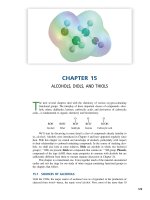
● Figure 1-1 Types of nerve cells. Olfactory neurons are bipolar and unmyelinated. Auditory neurons are bipolar and
myelinated. Spinal (dorsal root) ganglion cells (cutaneous) are pseudounipolar and myelinated. Motor neurons are multipolar and myelinated. Arrows indicate input through the axons of other neurons. Nerve cells are characterized by the
presence of Nissl substance and rough endoplasmic reticulum. (Modified from Carpenter MB, Sutin J, Human Neuroanatomy. Baltimore: Williams & Wilkins, 1983:92, with permission.)
A. FAST ANTEROGRADE AXONAL TRANSPORT is responsible for transporting all
newly synthesized membranous organelles (vesicles) and precursors of neurotransmitters. This process occurs at the rate of 200 to 400 mm/day. It is mediated by neurotubules and kinesin. (Fast transport is neurotubule-dependent.)
B. SLOW ANTEROGRADE TRANSPORT is responsible for transporting fibrillar
cytoskeletal and protoplasmic elements. This process occurs at the rate of 1 to
5 mm/day.
C. FAST RETROGRADE TRANSPORT returns used materials from the axon terminal
to the cell body for degradation and recycling at a rate of 100 to 200 mm/day. It
transports nerve growth factor, neurotropic viruses, and toxins, such as herpes
simplex, rabies, poliovirus, and tetanus toxin. It is mediated by neurotubules and
dynein.
IV
WALLERIAN DEGENERATION
is anterograde degeneration characterized by the disappearance of axons and myelin sheaths and the secondary proliferation of Schwann cells. It
occurs in the central nervous system (CNS) and the peripheral nervous system (PNS).
LWBK110-3895G-C01[1-8].qxd 7/10/08 7:15 AM Page 3 Aptara Inc.
NEUROHISTOLOGY
3
CHROMATOLYSIS
V
is the result of retrograde degeneration in the neurons of the CNS
and PNS. There is a loss of Nissl substance after axotomy.
VI
REGENERATION OF NERVE CELLS
A. CNS. Effective regeneration does not occur in the CNS. For example, there is no
regeneration of the optic nerve, which is a tract of the diencephalon. There are no
basement membranes or endoneural investments surrounding the axons of the CNS.
B. PNS. Regeneration does occur in the PNS. The proximal tip of a severed axon grows
into the endoneural tube, which consists of Schwann cell basement membrane and
endoneurium. The axon sprout grows at the rate of 3 mm/day (Figure 1-2).
VII
GLIAL CELLS are the nonneural cells of the nervous system.
A. MACROGLIA consist of astrocytes and oligodendrocytes.
1. Astrocytes perform the following functions:
a. They project foot processes that envelop the basement membrane of capillaries, neurons, and synapses.
b. They form the external and internal glial-limiting membranes of the CNS.
c. They play a role in the metabolism of certain neurotransmitters [e.g.,
␥-aminobutyric acid (GABA), serotonin, glutamate].
d. They buffer the potassium concentration of the extracellular space.
e. They form glial scars in damaged areas of the brain (i.e., astrogliosis).
f. They contain glial fibrillary acidic protein (GFAP), which is a marker for astrocytes.
g. They contain glutamine synthetase, another biochemical marker for astrocytes.
h. They may be identified with monoclonal antibodies (e.g., A2B5).
2. Oligodendrocytes are the myelin-forming cells of the CNS. One oligodendrocyte
can myelinate as many as 30 axons.
B. MICROGLIA arise from monocytes and function as the scavenger cells (phagocytes) of
the CNS.
C. EPENDYMAL CELLS are ciliated cells that line the central canal and ventricles of the
brain. They also line the luminal surface of the choroid plexus. These cells produce
cerebrospinal fluid (CSF).
D. TANYCYTES are modified ependymal cells that contact capillaries and neurons. They
mediate cellular transport between the ventricles and the neuropil. They project to
hypothalamic nuclei that regulate the release of gonadotropic hormone from the adenohypophysis.
E. SCHWANN CELLS are derived from the neural crest. They are the myelin-forming cells
of the PNS. One Schwann cell can myelinate only one internode. Schwann cells invest
all myelinated and unmyelinated axons of the PNS and are separated from each other
by the nodes of Ranvier.
LWBK110-3895G-C01[1-8].qxd 7/10/08 7:15 AM Page 4 Aptara Inc.
4
CHAPTER 1
● Figure 1-2 Schematic diagram of peripheral nerve regeneration.
LWBK110-3895G-C01[1-8].qxd 7/10/08 7:15 AM Page 5 Aptara Inc.
NEUROHISTOLOGY
5
THE BLOOD–BRAIN BARRIER
VIII
consists of the tight junctions of nonfenestrated
endothelial cells; some authorities include the astrocytic foot processes. Infarction of brain
tissue destroys the tight junctions of endothelial cells and results in vasogenic edema,
which is an infiltrate of plasma into the extracellular space.
IX
consists of the tight junctions between the cuboidal
epithelial cells of the choroid plexus. The barrier is permeable to some circulating peptides
(e.g., insulin) and plasma proteins (e.g., prealbumin).
X
PIGMENTS AND INCLUSIONS
THE BLOOD–CSF BARRIER
A. LIPOFUSCIN GRANULES are pigmented cytoplasmic inclusions that commonly
accumulate with aging. They are considered residual bodies that are derived from
lysosomes.
B. MELANIN (NEUROMELANIN) is blackish intracytoplasmic pigment found in the substantia nigra and locus coeruleus. It disappears from nigral neurons in patients who
have Parkinson’s disease.
C. LEWY BODIES are neuronal inclusions that are characteristic of Parkinson’s disease.
D. NEGRI BODIES are intracytoplasmic inclusions that are pathognomonic of rabies. They
are found in the pyramidal cells of the hippocampus and the Purkinje cells of the cerebellum.
E. HIRANO BODIES are intraneuronal, eosinophilic, rodlike inclusions that are found in
the hippocampus of patients with Alzheimer’s disease.
F.
NEUROFIBRILLARY TANGLES consist of intracytoplasmic degenerated neurofilaments. They are seen in patients with Alzheimer’s disease.
G. COWDRY TYPE A INCLUSION BODIES are intranuclear inclusions that are found in
neurons and glia in herpes simplex encephalitis.
XI
THE CLASSIFICATION OF NERVE FIBERS is shown in Table 1-1.
XII
TUMORS OF THE CNS AND PNS are shown in Figures 1-3 and 1-4.
A. One-third of brain tumors are metastatic, and two-thirds are primary. In metastatic
tumors, the primary site of malignancy is the lung in 35% of cases, the breast in 17%,
the gastrointestinal tract in 6%, melanoma in 6%, and the kidney in 5%.
B. Brain tumors are classified as glial (50%) or nonglial (50%).