Ebook Operative thoracic surgery (6/E): Part 2
Bạn đang xem bản rút gọn của tài liệu. Xem và tải ngay bản đầy đủ của tài liệu tại đây (14.3 MB, 258 trang )
17
Uniportal video-assisted thoracoscopic surgery
(VATS)
GAETANO ROCCO
INTRODUCTION
Single-port (uniportal) video-assisted thoracoscopic surgery (VATS) represents an evolution of traditional VATS
principles and, at the same time, a formidable return to the
geometric configuration of classic open thoracotomies.1–3
In a way, the uniportal concept is the center of a star system
whose satellites exchange technical aspects with the other
known thoracic surgical approaches (see Figure 17.1). The
main feature of the uniportal VATS approach consists of
targeting, through a caudocranial (sagittal) plane, any area
of surgical interest inside the chest (see Figure 17.2). Two
advantages result from such a perspective: (1) the procedure
allows for a similar approach as is used for open surgery and
(2) the reacquisition of the depth of visualization lost with
conventional three-port VATS.3 The latter is based on the
17.1 Uniportal VATS seen as the fulcrum of the
armamentarium of the modern thoracic surgeon.
17.2 Caudocranial approach (i.e., sagittal plane) for
uniportal VATS.
206 Uniportal video-assisted thoracoscopic surgery (VATS)
17.3 Schematic of the simultaneous insertion of the
development of a transversal latero-lateral (or anteroposterior) plane, along which the operative instruments are
deployed to address the target area.3 With the current 2-D
technology, the surgical maneuvers impede in-depth visualization through a centrally located videothoracoscope because
of the torsion angle created by the operative instruments
(see Figure 17.3).3,4 As a result, traditional three-port VATS
demands an extent of hand–eye coordination to overcome
the geometrical obstacle originating from this torsion angle
(see Figure 17.4a).4 This hand–eye coordination represents
an added difficulty, especially during hilar dissection during
VATS lobectomy, and this has possibly undermined the more
universal acceptance of the procedure, which is otherwise
appealing. Conversely, in the uniportal approach, the eye
“accompanies” in depth the stems of the instruments, which
are deployed parallel to each other along the sagittal plane,
and effectively represents an extension of the surgeon’s hands
(see Figure 17.4b).4 At present, the similarity between open
and uniportal VATS is as close as it can get. In addition, the
articulated jaws or graspers can be positioned so as to avoid
bite closure on the target area, which could, in turn, obstruct
the in-depth view. Furthermore, the fulcrum of the operative
instruments is inside the chest—at a short distance from the
actual lesion. This characteristic assimilates uniportal VATS
to robotic surgery; indeed, robotic surgery is considered to
be the minimally invasive surgical approach that most closely
duplicates the technical features of open thoracotomy (see
Figure 17.1).
videothoracoscope and instrument ensemble during uniportal VATS.
x
0
2
4
z
0
2
A
4
0
B
17.4a–b
2
4
y
(a) The torsion angle resulting from instrument interaction along a transversal plane obstructing in-depth visualization
through 2-D imaged conventional three-port VATS; (b) 2-D imaged uniportal VATS enabling improved in-depth visualization of the
surgical field.
Uniportal VATS for diagnostic purposes 207
The concept of using a thoracoscope and instrumentation
through the same small incision dates back to a report by
Singer in 1924.5 Uniportal VATS has since been described for
sympathectomy and the diagnosis of pleural conditions.6,7
The general consensus is that the main advantage of uniportal VATS is to provide a minimally invasive approach that can
be used in conjunction with loco-regional anesthesia to fast
track surgical candidates to diagnostic or therapeutic procedures.1 In this setting, the triad one port–one intercostal–less
pain seems justified, albeit that definitive evidence (i.e., a
prospective, randomized trial) has yet to be published.8,9
from incisions located anterior to the scapular angle line.
The intercostal space selected depends on the caudocranial
level where the lesion is found in the lung. As an example, if
the lesion is in the apex of the right upper lobe, an incision
should be placed at the fourth or fifth intercostal space. Once
the incision is made (see Figure 17.5a), the distribution of
the surgical personnel varies so that the first surgeon and his/
her assistant work from the same side, looking at the same
monitor (see Figure 17.5b).
PREOPERATIVE PLANNING
Recurrent pleural or pericardial effusions, early empyemas,
interstitial lung disease, peripheral pulmonary nodules,
or ground glass opacities, as well as pleural or mediastinal
masses and lymph node biopsy, are all amenable to uniportal
VATS, yielding precise histological diagnosis and short hospitalizations.2,6,10,11 Interestingly, selected awake patients can be
operated on under a combination of loco-regional anesthesia
and sedation.12 Typically, an epidural catheter is positioned
at the T5-6 level and a single shot of 1% Ropivacain solution
(10 mg/mL diluted to 5 mg/mL, for a total dose of 15 mL =
75 mg) is administered.12,13 In addition, the patient is given
intravenous (IV) midazolam (4 mg), fentanyl (100 mcg)
and propofol (0.5 mg/kg/h up to a total of 30 mg in 1 hour),
along with supplemental oxygen by nasal prongs in order to
maintain arterial oxygen saturation above 90%.12,13
The technical feasibility of uniportal VATS is heavily dependent on preoperative planning of the surgical coordinates
necessary to identify the location of the single incision. In this
setting, the scapular angle line—that is, longitude—defines
the distinction between anteriorly and posteriorly located
incisions. The latitude is defined by the intercostal space at a
level that must warrant sufficient distance between the single
port and target lesion to avoid videothoracoscope-instrument interference.2 Longitudinal and latitudinal coordinates
usually allow for placing the incision so as to “face” the target area inside the chest. Accordingly, lesions located in the
middle lobe are best approached through incisions located
posterior to the scapular angle line; conversely, lesions located
in the apical segment of the lower lobe are best addressed
UNIPORTAL VATS FOR DIAGNOSTIC PURPOSES
(a)
17.5a–b Distribution of the theater personnel before the incision (a) and after the incision (b) for a uniportal VATS procedure.
(b)
208 Uniportal video-assisted thoracoscopic surgery (VATS)
SURGICAL TECHNIQUE FOR UNIPORTAL VATS
FOR PLEURAL CONDITIONS
As a rule, diagnostic uniportal VATS is performed through
a single 1.0–1.5 cm incision located along a virtual thoracotomy line in the fifth intercostal space, usually anterior to
the scapular line if the pleural effusion occupies two-thirds
or more of the chest cavity.14 When the pleural effusion is
less significant, needle probing is used to identify the most
recumbent site compatible with safe performance of the procedure and convenient chest drain placement. A 24 Fr chest
drain is passed through a 10 mm trocar inserted through the
single incision and the pleural fluid aspirated and routinely
sent for cytology. As a rule, a 5 mm trocar is then used to
introduce a 5 mm 0-degree videothoracoscope to explore
the posterior chest wall and the diaphragm. The trocar is
removed along the stem of the videothoracoscope to gain
more operative space at the incision level. Later, the videothoracoscope is tilted toward the assistant’s side, and the
17.6 Length of the incision for uniportal VATS
wedge resection.
anterior chest wall, pericardium, and diaphragm are visualized. At this point, biopsy forceps are introduced parallel
to the videothoracoscope. If talc pleurodesis is needed, the
insufflator is inserted parallel to the thoracoscope, which
is slightly retracted to visualize the tip of the insufflators in
order to better direct talc aspersion. Talc poudrage is completed by rotating the thoracoscope and insufflator ensemble
to cover all areas of the chest cavity.
SURGICAL TECHNIQUE FOR UNIPORTAL VATS
WEDGE RESECTION
The perfect size for single-port VATS—in line with the
extreme minimally invasive philosophy behind this technique—is one fingerbreadth measured at the knuckle—that
is, 2.5 cm (see Figures 17.6 and 17.7).3 The intercostal space
is opened flush to the superior border of the underlying rib
17.7 The standard length of incision has to accommodate one
surgeon’s fingerbreadth.
Surgical technique for uniportal VATS wedge resection 209
17.8 The endostapler is articulated outside the chest and
17.9 Intraoperative view of the simultaneous insertion of the
so as to allow for 1 cm lateral movements on each side. The
following step is the introduction of a 0- or 30-degree 5 mm
videothoracoscope without trocar, which is retracted along
the thoracoscope stem.3 Next, articulating endograspers
and an endostapler are inserted to suspend and resect the
pulmonary target area along a craniocaudal (sagittal) plane
(see Figures 17.8 and 17.9). The reciprocal position of the
instruments and the thoracoscope can vary during the procedure to facilitate surgical maneuvers.3 The placement of soft
tissue retractors is discouraged, to avoid subtracting room
for the instruments and thoracoscope. Once the nodule is
visualized or identified with an ultrasound probe,15 the area
of parenchyma containing the nodule is marked and resected
(see Figure 17.10).
Uniportal VATS wedge resection of the lung; the
endograsper is suspending the parenchyma to be resected while
the endostapler is positioned at the base of the parenchyma to
complete the resection.
inserted in the same fashion as one would insert a mediastinoscope
under the pre-cervical fascia.
videothoracoscope and instrument ensemble.
17.10
210 Uniportal video-assisted thoracoscopic surgery (VATS)
RESULTS OF UNIPORTAL VATS FOR
DIAGNOSIS AND TREATMENT OF
INTRATHORACIC CONDITIONS
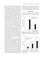
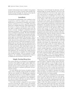
A 10-year study reported that uniportal VATS for the diagnosis and treatment of intrathoracic conditions was performed
in up to 28% of thoracic surgical candidates.2 Of the 644
uniportal VATS procedures, over 50% were used to diagnose pleuropericardial conditions, while 29% were needed
for wedge resections. The remaining 21% of surgeries were
performed for pre-thoracotomy exploration of the chest
cavity, diagnosis of mediastinal masses, sympathectomy,
and debridement of early stage empyemas or hemothoraces. The median operative time was 18 and 22 minutes for
diagnostic uniportal VATS and wedge resection, respectively.
In addition, median postprocedure chest tube duration was
4 days (range, 2–20) and 2 days (range, 0–6) for pleural
effusions and wedge resections, respectively, inclusive of
the day of chest drain insertion. Furthermore, the median
postoperative hospitalizations were 5 and 4 days, respectively,
for pleural effusions and wedge resections; these figures
included the operative day. Overall, 146 pulmonary nodules
were resected by uniportal VATS; the median size was 1.6 cm
(range, 0.4–3.2) and the median margin from the nodule was
1.2 cm (range, 0.5–2.1). Of the 146 nodules, 69 were proven
to be primary lung cancers, 77 secondary deposits from an
extrathoracic cancer, and 33 benign lesions.2
UNIPORTAL VATS FOR PNEUMOTHORAX
One of the most appropriate indications for uniportal VATS
seems to be represented by the management of pneumothorax.3,16 The presence of a chest drain, often placed in an
emergency setting, and of a usually visible target lesion (i.e.,
a bleb or bulla) make the single-port approach immediately
feasible both under general or loco-regional anesthesia.13
Wedge resection of the apex and apical pleurectomy or talc
pleurodesis are easily accomplished through uniportal VATS
using articulating instruments.3 In particular, a scratch pad
appropriately folded and cut to size can be mounted on the
articulating arm of an endograsper.16 The scratch pad can
be applied to the entire circumference of the inner chest
wall by rotating the endograsper arm.3,16 The initial tear
induced in the parietal pleura can be used as starting point
for an apical pleurectomy using endo Kitners to elevate the
parietal pleura from the endothoracic fascia.16 Alternatively,
a thorough abrasion can be easily obtained by extending the
procedure, under visual control, onto the remaining chest
wall and diaphragm. Likewise, any blebs or bullae can be
resected concomitantly in any peripheral area of the lung
by changing the orientation of the videothoracoscope and
operative instrument ensemble. Talc pleurodesis is also a
viable choice in selected patients with bilateral symptomatic
recurrent pneumothoraces.
UNIPORTAL VATS SYMPATHECTOMY
My colleagues’ and my initial experience with bilateral single
access sympathectomy was reported in 2004 and updated
in 2007.17 The main indications were palmar hyperhidrosis
and facial blushing. The technique consists of sequentially
entering the chest cavities during the same operative session
through a single 0.5–1.0 cm incision located in the axilla.17
Through this incision, a 5 mm 0-degree videothoracoscope
is inserted along with an endograsper. In our experience, the
use of an articulating endograsper is preferred to be able to
mobilize the lung apex as necessary. As a rule, the sympathetic chain, with its T2 and T3 ganglia, was identified and
divided by means of a diathermy hook.17 The diathermy
hook is pressed against the rib; by applying low voltage electricity, the surgeon makes sure to separate the nerve endings
and to laterally extend the sympathectomy for 3–5 cm to
include the so-called Kuntz fibers.17
References 211
UNIPORTAL VATS MAJOR LUNG RESECTIONS
CONCLUSIONS
Gonzalez-Rivas and his colleagues from Coruña University
Hospital deserve the credit for having recently expanded
the indications of uniportal VATS to include major lung
resections.18,19 The authors have described the evolution
of the single-port technique from multiple-port down to
only two-port lobectomy.18 Of the original uniportal VATS
technique,3 Gonzalez-Rivas and colleagues have maintained
the caudocranial approach to the target structure in the lung
hilum and the introduction of multiple instruments through
the same incision along with the videothoracoscope, which
is usually located at one edge of the incision; the full use of
laterality for the surgical maneuvers; and, the insertion of
the chest drain through the same incision at the end of the
procedure.18 However, the typical approach to uniportal
VATS major pulmonary resection is an anterior one for
all possible lobar resections and pneumonectomy,20 with a
length for the utility and operative incision, which is larger
(up to 5 cm) than the one used for the classic uniportal
VATS wedge resection to accommodate the extracted specimen (see Figure 17.11).18 The anterior single-port incision
was sufficient to ensure safe lobar resection and adequate
nodal dissection, as later demonstrated in the work of other
groups.21,22 Standard open instrumentation can be used,
although articulated or specifically devised instruments have
also been recommended to facilitate hilar dissection. After
uniportal VATS lobectomy, while the mean operative time
was 154 minutes, the median duration of chest drain insertion was 2 days (range, 1–16) whereas the median length of
stay in the hospital was 3 days (range, 1–14) with neither
operative nor 30-day mortality.18
By 2014, virtually all routine thoracic surgical procedures
could be done by uniportal VATS.9 While the issues of feasibility and safety seem to have been solved, the jury is still
out as to the results of the uniportal technique compared
with those of conventional three-port VATS. It appears intuitive that conditions like pleural effusions, pneumothoraces,
and hyperidrosis need to be managed through a singleport incision to fast track patients by reducing morbidity.
When it comes to major resections, postoperative pain,
and long-term oncologic outcomes will provide the crucial
benchmark for comparison between uniportal and other
surgical approaches.
17.11
REFERENCES
1. Rocco G. One-port (uniportal) video-assisted thoracic
surgical resections: a clear advance. Journal of Thoracic and
Cardiovascular Surgery. 2012; 144(3): S27–31.
2. Rocco G, Martucci N, La Manna C, Jones DR, De Luca G, La
Rocca A et al. Ten-year experience on 644 patients undergoing
single-port (uniportal) video-assisted thoracoscopic surgery.
Annals of Thoracic Surgery. 2013; 96(2): 434–8.
3. Rocco G, Martin-Ucar A, Passera E. Uniportal VATS wedge
pulmonary resections. Annals of Thoracic Surgery. 2004; 77(2):
726–8.
4. Bertolaccini L, Rocco G, Viti A, Terzi A. Geometrical
characteristics of uniportal VATS. Journal of Thoracic Disease.
2013; 5(Suppl. 3): S214–16.
5. Moisiuc FV, Colt HG. Thoracoscopy: origins revisited.
Respiration. 2007; 74(3): 344–55.
6. Rocco G. History and indications of uniportal pulmonary wedge
resections. Journal of Thoracic Disease. 2013; 5(Suppl. 3):
S212–13.
Instrument disposition for uniportal VATS lobectomy; the videothoracoscope is routinely kept at one side of the incision to
facilitate surgical maneuvers.
212 Uniportal video-assisted thoracoscopic surgery (VATS)
7. Rocco G. VATS and uniportal VATS: a glimpse into the future.
Journal of Thoracic Disease. 2013; 5(Suppl. 3): S174.
8. Atkinson JL, Fode-Thomas NC, Fealey RD, Eisenach JH, Goerss
SJ. Endoscopic transthoracic limited sympathotomy for
palmar-plantar hyperhidrosis: outcomes and complications
during a 10-year period. Mayo Clinic Proceedings. 2011; 86(8):
721–9.
9. Roubelakis A, Modi A, Holman M, Casali G, Khan AZ. Uniportal
video-assisted thoracic surgery: the lesser invasive thoracic
surgery. Asian Cardiovascular and Thoracic Annals. 2014; 22(1):
72–6.
10. Rocco G, Brunelli A, Jutley R, Salati M, Scognamiglio F, La
Manna C et al. Uniportal VATS for mediastinal nodal diagnosis
and staging. Interactive Cardiovascular and Thoracic Surgery.
2006; 5(4): 430–2.
11. Rocco G, La Rocca A, La Manna C, Scognamiglio F, D’Aiuto M,
Jutley R et al. Uniportal video-assisted thoracoscopic surgery
pericardial window. Journal of Thoracic and Cardiovascular
Surgery. 2006; 131(4): 921–2.
12. Rocco G, Romano V, Accardo R, Tempesta A, La Manna C, La
Rocca A et al. Awake single-access (uniportal) video-assisted
thoracoscopic surgery for peripheral pulmonary nodules in a
complete ambulatory setting. Annals of Thoracic Surgery. 2010;
89(5): 1625–7.
13. Rocco G, La Rocca A, Martucci N, Accardo R. Awake singleaccess (uniportal) video-assisted thoracoscopic surgery
for spontaneous pneumothorax. Journal of Thoracic and
Cardiovascular Surgery. 2011; 142(4): 944–5.
14. Salati M, Brunelli A, Rocco G. Uniportal video-assisted
thoracic surgery for diagnosis and treatment of intrathoracic
conditions. Thoracic Surgery Clinics. 2008; 18(3): 305–10, vii.
15. Rocco G, Cicalese M, La Manna C, La Rocca A, Martucci
N, Salvi R. Ultrasonographic identification of peripheral
pulmonary nodules through uniportal video-assisted thoracic
surgery. Annals of Thoracic Surgery. 2011; 92(3): 1099–101.
16. Jutley RS, Khalil MW, Rocco G. Uniportal vs standard
three-port VATS technique for spontaneous pneumothorax:
comparison of post-operative pain and residual paraesthesia.
European Journal of Cardio-thoracic Surgery. 2005; 28(1):
43–6.
17. Rocco G. Endoscopic VATS sympathectomy: the uniportal
technique. Multimedia Manual of Cardiothoracic Surgery. 2007;
2007(507): MMCTS.2004.000323.
18. Gonzalez-Rivas D, Paradela M, Fernandez R, Delgado M, Fieira
E, Mendez L et al. Uniportal video-assisted thoracoscopic
lobectomy: two years of experience. Annals of Thoracic Surgery.
2013; 95(2): 426–32.
19. Gonzalez-Rivas D, Fieira E, Mendez L, Garcia J. Single-port
video-assisted thoracoscopic anatomic segmentectomy and
right upper lobectomy. European Journal of Cardio-thoracic
Surgery. 2012; 42(6): e169–71.
20. Gonzalez-Rivas D, Delgado M, Fieira E, Mendez L, Fernandez
R, de la Torre M. Uniportal video-assisted thoracoscopic
pneumonectomy. Journal of Thoracic Disease. 2013; 5(Suppl. 3):
S246–52.
21. Tam JK, Lim KS. Total muscle-sparing uniportal video-assisted
thoracoscopic surgery lobectomy. Annals of Thoracic Surgery.
2013; 96(6): 1982–6.
22. Wang BY, Tu CC, Liu CY, Shih CS, Liu CC. Single-incision
thoracoscopic lobectomy and segmentectomy with radical
lymph node dissection. Annals of Thoracic Surgery. 2013; 96(3):
977–82.
18
Segmentectomy
WENTAO FANG, CHENXI ZHONG, AND ZHIGANG LI
RATIONALE FOR SEGMENTECTOMY
Segmentectomy was first performed in 1939 for the treatment of benign pulmonary diseases such as bronchiectasis
and tuberculosis. Shortly thereafter, anatomic pulmonary
segmentectomy was also employed for primary lung cancers.
The study by Jensik et al. in 1979 showed that segmentectomy
was safe and feasible for selected patients with non-small-cell
lung cancer (NSCLC).1 Since then, whether segmentectomy
is comparable to lobectomy has been an area of controversy.
In 1995, the Lung Cancer Study Group reported a randomized trial in stage IA (T1N0M0) NSCLC, comparing
limited resection in 122 patients (82 segmentectomies and
40 wedge resections) with lobectomy in 125 patients.2 The
results showed that, compared with lobectomy, limited resection was associated with 75% increase in recurrence (p = .02),
tripling of local recurrence (p = .008), 30% increase in
overall death (p = .08), and 50% increase in cancer death
(p = .09). The inclusion of nonanatomic wedge resections in
the limited resection group tends to bias the results in favor
of lobectomy and subsequent studies have not confirmed
the results found in the Lung Cancer Study Group report.
Thereafter, lobectomy has been considered the standard
procedure for early stage NSCLC, while sublobar resection is
reserved only for those who could not tolerate lobectomy due
to marginal lung function and/or significant comorbidities.
However, the size of the lesion to be resected should be taken
into consideration, given that, in the seventh edition of the
Union for International Cancer Control staging system for
NSCLC, T1 disease is now subdivided into T1A (≤2 cm) and
T1B (>2 cm).3 The Lung Cancer Study Group trial included
all T1N0M0 tumors of size up to 3 cm, and it did not stratify
the results between T1A and T1B.2 In a more detailed retrospective study involving 1272 stage I NSCLC patients, the
5-year cancer-specific survivals were similar after lobectomy
(92.4%) or segmentectomy (96.7%) when the tumor size
was ≤20 mm.4
It should also be noted that the Lung Cancer Study Group
trial came from the time when only TNM (tumor node
metastasis) staging was considered for surgical strategy. With
the increased use of computed tomography (CT) screening, small peripheral ground glass opacity (GGO) lesions,
which would have been difficult or even impossible to detect
on routine chest X-ray, have been encountered more frequently in daily practice. These lesions often correspond to
rather indolent early stage adenocarcinomas. Emerging data
have shown that these GGO lesions seldom have lymphatic
involvement. Compared with standard lobectomy, sublobar
resection may offer equivalent local control and disease-free
survival for these patients. The International Association
for the Study of Lung Cancer, together with the American
Thoracic Society and European Respiratory Society, recently
proposed a new histologic classification system for lung
adenocarcinomas, highlighted by the introduction of adenocarcinoma in situ (AIS; small adenocarcinomas <3 cm in
diameter with pure lepidic growth) and minimally invasive
adenocarcinoma (MIA; small solitary adenocarcinomas
showing predominant lepidic growth with ≤5 mm invasion).5
It is appropriate at this time to reevaluate the indication and
selection of surgical approach and specifically, the extent of
resection, incorporating both anatomical (TNM) and biological behavior (histologic subtyping) of the tumor.
Meanwhile, segmentectomy should be distinguished from
nonanatomic wedge resection, as the latter was applied to
up to one-third of the patients in the limited resection arm
of the Lung Cancer Study Group trial.2 The advantages of
segmentectomy over nonanatomic wedge resection are at
least twofold: first, by dissecting the segmental vessels and
bronchus, hilar and segmental lymph nodes can be harvested
systematically; second, anatomic segmentectomy also enables
a deeper parenchymal resection and a safer margin for relatively centrally located lesions.
Moreover, surgical management of early stage lung cancer has changed greatly with the introduction of minimally
214 Segmentectomy
invasive video-assisted thoracoscopic surgery (VATS).6 In
the case of lobectomy, there is a large body of evidence
demonstrating that VATS is associated with decreased morbidity and mortality, shorter hospital stay, less postoperative
pain, earlier return to normal life, better quality of life, and
superior compliance with adjuvant therapy. VATS even has
potentially better oncologic results, making it now the preferred approach over open lobectomy. When segmentectomy
is performed via VATS, it is not simply to revive a procedure
that previously was used infrequently but to add new meaning to “minimally invasive” lung cancer surgery to include
parenchymal sparing, in addition to the other advantages
of VATS noted above. For small early stage lung cancers,
VATS segmentectomy may be expected to achieve excellent
oncologic results with very low morbidity and mortality.
A retrospective study conducted at our hospital compared
clinical outcomes between VATS segmentectomy and lobectomy in patients with small-sized (≤2 cm) stage IA tumors.7
There were no in-hospital deaths in either group. Local recurrence rates were similar after VATS segmentectomy (5.1%)
and lobectomy (4.9%), and no significant difference was
observed in 5-year overall or disease-free survivals following
both procedures.
INDICATIONS FOR SEGMENTECTOMY
Pulmonary segmentectomy is often indicated for benign
lesions such as those caused by infectious diseases, and may
also be used selectively in patients with NSCLC. For small
GGO lesions, segmentectomy is sometimes used to establish
a histologic diagnosis, as fine needle biopsy has been shown
to be quite unsatisfactory in such situations. The overall
diagnostic yield from fine needle aspiration is merely 51%
for GGO dominant lesions (GGO ratio >50%) and only 35%
for GGO dominant lesions smaller than 10 mm. In addition,
these lesions are sometimes extremely difficult to locate
when using a VATS approach, making a wedge resection very
challenging.
As mentioned earlier, segmentectomy has been accepted
and used as an alternative for those high-risk lung cancer patients who are deemed unable to tolerate lobectomy.
The potential benefits of segmentectomy compared with
lobectomy are less surgical risk and better preservation
of pulmonary function, while its advantage over nonanatomic wedge resection is superior oncologic outcome. Until
recently, the indication for segmentectomy in good-risk
patients who have no contraindication to lobectomy was not
only unclear but questionable on oncologic grounds. Both
tumor size and biology should be considered in determining
the feasibility and efficacy of segmentectomy. Retrospective
data from single or multiple institutions demonstrate that
segmentectomy provides acceptable local control for tumors
sized 2 cm or smaller, provided that at least a 2 cm resection margin can be achieved.8 GGO-type tumors represent
an excellent indication for segmentectomy. For pure GGO
lesions corresponding to AIS or MIA, even tumors up to 3 cm
can be considered for segmentectomy. A near 100% diseasefree survival rate can be expected after complete resection.9
Several studies have shown that width of resection margin
is an important factor in maintaining local control following
segmentectomy.7 A safe margin of greater than 2 cm might
be reasonable, as resection margins less than 2 cm have been
shown to be associated with an increased incidence of local
recurrence. Based on this concern, if a tumor is located on the
edge of diseased segment or a safe resection margin cannot
be guaranteed intraoperatively, multiple segmental resections
or lobectomy should be performed.
For lung cancer patients, preoperative staging should be
completed to confirm the absence of nodal (mediastinal or
hilar) disease. Small tumors, especially those appearing on
CT to be air-containing lesions, are associated with a lower
likelihood for lymphatic spread, which is another reason why
they are excellent candidates for segmental resection. Still,
careful intraoperative exploration of hilar and mediastinal
lymph nodes should be performed to exclude occult metastases and ensure the appropriateness of segmentectomy.
Conversion to standard lobectomy is indicated when a frozen
section of a mediastinal or hilar lymph node demonstrates
the presence of metastatic disease. Segmentectomy should be
oncologically more effective than nonanatomic wedge resection, since it includes dissection of intersegmental, intralobar,
and interlobar lymph nodes.
While anatomically less lung parenchyma is resected by
segmentectomy than lobectomy, it does not necessarily result
in a similar amount of pulmonary function preserved. This
is affected by multiple factors, including the number, location, and quality of the segment resected. Resecting more
than three segments has been shown to leave only 0.1 L of
forced expiratory volume in 1 second in the remaining lobe.
Recognizing this, basal segmentectomy of the lower lobes
with preservation only of the superior segment, though
technically feasible, is seldom indicated.
GENERAL STRATEGY FOR SEGMENTECTOMY
Technically, all segments can be approached surgically. The
superior segments of the lower lobes, the lingular segment
and the upper division of the left upper lobe, and posterior
segment of the right upper lobe, in decreasing order of
frequency, are the most common segmentectomies performed. Other individual segmental resections, such as upper
lobe superior or anterior segmentectomy, are feasible but
less commonly performed. Basal segmentectomy is seldom
indicated, as it saves very little pulmonary function of the
remaining lower lobe.
Segmentectomy can be performed thorough standard
lateral thoracotomy or via a VATS approach. Compared
with thoracoscopic lobectomy, VATS has been applied to
anatomic segmentectomy only recently. Technically, thoracoscopic segmentectomy is considered to be more difficult
than thoracoscopic lobectomy. Thoracic surgeons should be
familiar with the three-dimensional anatomical relationship
General strategy for segmentectomy 215
of pulmonary segments to accomplish a segmentectomy successfully. Still, it has been proven to be safe and oncologically
effective. No matter whether via an open or minimally invasive approach, it is imperative to make certain that standard
dissection and oncologic principles are not compromised.
Open segmentectomies are often approached through a
lateral thoracotomy via the fifth intercostal space. In performing a minimally invasive thoracoscopic segmentectomy,
a standard three- or four-hole approach, with the major utility port in the fourth or fifth intercostal space, is the usual
technique. The entire chest cavity should first be inspected
to rule out signs of unexpected advanced disease, such as
pleural dissemination or concomitant additional pulmonary
nodules. Except for high-risk patients who cannot tolerate
lobectomy, mediastinal or hilar nodal involvement should
always lead to conversion to standard lobectomy, so as to
ensure lymphatic clearance. Usually, the tumor should be
palpated to confirm that segmentectomy is the correct procedure to ensure an adequate resection margin; otherwise, a
bi-segmentectomy or lobectomy would be a better choice.
During segmentectomy, the segmental pulmonary veins,
arteries, and bronchus are dissected and stapled separately.
Thoracoscopic segmentectomy usually begins with identification and dissection of the segmental vein. Subsequently,
the bronchus or the artery is divided, depending on the segment to be resected. Alternatively, the arterial branches can
be identified and mobilized before the segmental veins are
divided, but the more logical approach takes the segmental
vein first. Some authors stated that this might minimize
engorgement of the segment and facilitate further maneuvering, but in our experience, this has not been the case.
Mobilizing arterial branches to the posterior segment of the
upper lobes or the apical segment of the lower lobes often
requires dissection of the major fissure. In the major fissure,
the main pulmonary artery can be exposed, demonstrating
its continuation into the lower lobe. On the right side, the
lower lobe superior segmental branch can be identified at the
posterior part of the major fissure. The posterior ascending
and the middle lobe branches originate opposite each other,
and go, respectively, to the posterior segment of the upper
lobe and the middle lobe.
On the left side, the pulmonary artery crosses superiorly
above the left main bronchus to become the most posterior
structure in the hilum. The apicoposterior and anterior
segmental branches are located anteriorly and superiorly. A
separate posterior segmental branch is often found posteriorly on the main pulmonary artery, just at or above the major
fissure. In the major fissure, the lingular branches, directed
anteriorly, and the superior segment branch, posteriorly, are
located across from each other on the continuation of the
pulmonary artery. The surgeon must be mindful of the high
variability in pulmonary artery branching, and carefully
identify and confirm each branch before ligation.
In performing VATS segmentectomy, the pulmonary vessels are usually divided using endostaplers or endo-clips,
with or without the help of energetic devices such as a
Harmonic scalpel. After vascular division, the segmental
bronchus is then identified and divided with an endostapler,
or divided sharply and closed with interrupted absorbable
sutures. The segmental bronchus is first clamped and the
lung inflated before stapling for further confirmation of the
correct anatomic location. Alternatively, a bronchoscopy can
be helpful to confirm the correct segmental bronchus has
been identified.
Division of the intersegmental plane is sometimes the
most challenging part of a segmentectomy. Selected jet
ventilation in the diseased segmental bronchus may help
delineate the correct plane.10 In our experience, identification of the intersegmental plane can be achieved by repeated
ventilation of the ipsilateral lung after the segmental bronchus is clamped. The first several puffs will probably serve to
delineate the parenchyma aerated by that bronchus. Due to
the large degree of intersegmental cross-ventilation through
collateral pores of Kohn, it may be helpful to inflate the entire
lung, clamp the segmental bronchus, and then collapse the
lung while observing the delineation between residually
inflated and actively deflating lung. In addition, the divided
vascular and bronchial structures can be used as landmarks
to guide this process. There are two ways to divide the segmental parenchyma: via the so-called open division or with
the use of a stapling device. The advantage of open division
with electrocautery or simply by “stripping” the intersegmental plane using the venous supply as a guide, is greater
preservation of lung volume. However, this technique is
associated with increased risk of air leak and oozing from
the raw surface of the lung, which could be problematic after
operation, though both the air leak and bleeding usually stop
spontaneously in a short period if the correct plane has been
entered. Staple division results in a pneumostatic separation
of the intersegmental plane, minimizing the troublesome
issue of air leak, but this comes at the expense of more volume loss, as the visceral pleural layers are drawn together
during the act of stapling. The intersegmental plane is stapled according to the inflation–deflation line and at least a
2 cm parenchymal resection margin should be guaranteed in
segmentectomy for malignant diseases. When using staplers
to divide the intersegmental plane, care should be taken to
ensure they are placed exactly in the right position so as to
avoid inadvertently stapling the adjacent segmental vein or
bronchus. This may result in engorgement or atelectasis and
repeated infection of the remaining lobe. Inflation of the
remaining lung after the stapler is approximated but not yet
fired is often helpful in avoiding inadvertent injury of the
adjacent segmental bronchus.
216 Segmentectomy
SPECIFIC SEGMENTAL RESECTIONS
Upper division segmentectomy of the
left upper lobe
This segmentectomy begins with the dissection of the anterior hilum. After the upper division branches of the left
superior pulmonary vein are divided (see Figure 18.1), the
upper division bronchus, located directly behind the pulmonary vein, is readily exposed (see Figure 18.2). Under
thoracoscopy, this can easily be visualized. It is then divided
18.1 The upper division branches of the left superior
pulmonary vein are divided.
with an endostapler, after the location of the lingular segment bronchus is confirmed. Alternatively, the anterior and
apical pulmonary artery branches can be exposed and dissected first. This may also facilitate passing of endostapler
through the bronchus during VATS segmentectomy (see
Figure 18.3). As described earlier, there is usually a posterior
arterial branch located just at or above the major fissure. This
can be dissected either anteriorly after the segmental bronchus is divided, or posteriorly from the major fissure (see
Figure 18.4). Segmentectomy is then completed with division
of the intersegmental plane, as previously described. In case
of a fully developed major fissure, fixation of the remaining
lingular segment to the left lower lobe is advisable to prevent
torsion of this segment.
18.2 The upper division bronchus of the left upper lobe is
exposed and stapled, sparing the lingular bronchus.
Notes: LSPV, left superior pulmonary vein.
18.3 The anterior and apical pulmonary artery branches are
divided and stapled.
18.4 The posterior segmental artery is exposed and stapled.
Notes: LSPV, left superior pulmonary vein.
Specific segmental resections 217
18.5 The lingular branch of the left superior pulmonary vein
is exposed.
18.6 The lingular branches of the left pulmonary artery are
exposed after the major fissure is opened.
Notes: LSPV, left superior pulmonary vein.
Left upper lobe lingular segmentectomy
Resection of the left lingular segment is somewhat similar to
right middle lobectomy. The lung is retracted posteriorly, and
the hilar pleura is incised to expose the lingular branch of the
superior pulmonary vein (see Figure 18.5). After the lingular
vein is divided, dissection of the lingular bronchus can be
undertaken at its bifurcation from the left upper lobe bronchus. The major fissure is then opened, beginning anteriorly,
to expose the lingular branches of the pulmonary artery (see
Figure 18.6). There are usually two branches that supply this
segment that originate either separately side by side or from
one single stem at the anterior end of the pulmonary artery
before it continues on to the left lower lobe. Division of the
intersegmental plane starts from the hilum anteriorly to the
midline of the major fissure posteriorly, with stapling devices,
after this plane is identified and confirmed.
Superior segmentectomy of the lower lobes
Removal of the lower lobe superior segment is often initiated
with dissection of the pulmonary artery in the major fissure.
The superior segment branch can be approached directly if
the major fissure is well developed. Otherwise, the posterior
portion of the major fissure can be opened and divided with a
stapler. Once this is done, the segmental artery can be isolated
and divided (see Figure 18.7). This will provide excellent
exposure to the superior segment bronchus, which runs deep
to the artery. The superior segment vein can be identified as
the uppermost separate tributary running into the inferior
pulmonary vein, and can be approached after opening the
hilar pleura posteriorly (see Figure 18.8).
18.7 The apical segment artery of the right lower lobe is
divided and stapled after the posterior portion of the major fissure
is developed.
218 Segmentectomy
along the hilum. The anterior segmental vein is then exposed,
ligated, and divided. Care must be taken not to compromise
other branches of the superior pulmonary vein. The anterior
segmental pulmonary artery, likewise, can be identified as
it branches from the anterior trunk. The horizontal fissure
is then opened to expose the anterior segmental bronchus
posterior to the pulmonary vein. When the bronchus is
divided and its distal stump retracted up and forward, the
intersegmental plane can be stapled without injury to the
remaining hilar structures.
REFERENCES
18.8 The apical segmental vein of the right lower lobe is
identified as the upper most separate tributary running into the
inferior pulmonary vein.
Right upper lobe segmentectomies
Individual segmental resections of the right upper lobe are
technically more demanding. Apical segment resection begins
by opening the hilar pleura adjacent to the azygous vein. The
apical branch of the anterior pulmonary artery trunk is
identified and divided. The apical segmental bronchus is then
approached posteriorly and dissected after dividing the right
posterior bronchial artery branch. The apical segment branch
of the pulmonary vein is usually encompassed in the staple
line when dividing the intersegmental plane using staplers.
In performing posterior segmentectomy, related branches
of pulmonary artery and vein can be exposed and divided in
the major fissure when opened. Alternatively, the bronchus
to this segment can be tracked along the right upper lobe
bronchus posteriorly and distally, dissected first, and divided.
The anterior segment is often approached from the medial
aspect, beginning with incision of the mediastinal pleura
1. Jensik RJ, Faber LP, Kittle CF. Segmental resection for
bronchogenic carcinoma. Ann Thorac Surg. 1979; 28: 475–83.
2. Lung Cancer Study Group, Ginsberg RJ, Rubinstein LV.
Randomized trial of lobectomy versus limited resection for
T1N0 non-small cell lung cancer. Ann Thorac Surg. 1995; 60:
615–22.
3. Sobin LH, Gospadarowicz MK, Wittekind C (eds). (2009) TNM
Classification of Malignant Tumours, 7th edition. Oxford, UK.
Wiley-Blackwell. pp. 138–47.
4. Okada M, Nishio W, Sakamoto T et al. Effect of tumor size
on prognosis in patients with non-small cell lung cancer: the
role of segmentectomy as a type of lesser resection. J Thorac
Cardiovasc Surg. 2011; 129: 87–93.
5. Travis WD, Brambilla E, Noguchi M et al. International
Association for the Study of Lung Cancer/American
Thoracic Society/European Respiratory Society international
multidisciplinary classification of lung adenocarcinoma.
J Thorac Oncol. 2011; 6: 244–85.
6. Onaitis MW, Petersen RP, Balderson SS et al. Thoracoscopic
lobectomy is a safe and versatile procedure: experience with
500 consecutive patients. Ann Surg. 2006; 244: 420–5.
7. Zhong C, Fang W, Mao T et al. Comparison of thoracoscopic
segmentectomy and thoracoscopic lobectomy for small-sized
stage IA lung cancer. Ann Thorac Surg. 2012; 94: 362–7.
8. Swanson SJ. Video-assisted thoracic surgery segmentectomy:
the future of surgery for lung cancer? Ann Thorac Surg. 2010;
89: S2096–7.
9. Smith CB, Swanson SJ, Mhango G et al. Survival after
segmentectomy and wedge resection in stage I non-small-cell
lung cancer. J Thorac Oncol. 2013; 8: 73–8.
10. Okada M, Mimura T, Ikegaki J et al. A novel video-assisted
anatomic segmentectomy technique: selective segmental
inflation via bronchofiberoptic jet followed by cautery cutting.
J Thorac Cardiovasc Surg. 2007; 133: 753–8.
19
Combined bronchial and pulmonary artery sleeve
resections
ABEL GÓMEZ-CARO AND LAUREANO MOLINS
INTRODUCTION
In centrally located lung cancer, resection is frequently associated with massive parenchyma extirpation and high rates of
morbidity and mortality. Pneumonectomy (PN) has a significantly greater incidence of mortality compared with lesser
pulmonary resections and results in substantial declines in
lung function and quality of life, precluding adjuvant treatments or further lung resection. In the search for alternative
strategies, sleeve lobectomy (SL) has become the gold standard for centrally located lung tumors that otherwise would
not be resectable by simple lobectomy. Sparing lung function
may allow patients with very limited lung function and those
treated with chemoradiotherapy to overcome prohibitive
surgical risk and be candidates for intervention. About 10%–
14% of all lung tumors and nearly 60% of central tumors
may be amenable to sleeve resection with combined pulmonary artery (PA) and bronchial reconstruction techniques.
Several thoracic surgery teams have developed an aggressive
parenchyma-sparing policy, with a reported PN:SL ratio of
at least 1:3, decreasing the PN rate to 5%.
Management of centrally located non-small-cell lung
cancer may combine various surgical techniques to avoid
PN without compromising the long-term oncological results.
Surgical options include PA reconstruction or replacement,
alleviation of bronchial mismatch, and in some cases, resection of more than one lobe and airway anastomoses in
segmental bronchi.
PREOPERATIVE EVALUATION
Preoperative assessment of potential surgical candidates
includes taking the clinical history; performing a physical
examination; standard blood tests; chest radiographic analysis; bronchoscopy; and thoracic, abdominal, and cerebral
computed tomography scan, as well as 18F-fluoro-D-glucose
positron emission tomography. Suggestion of ipsilateral
mediastinal lymph node metastases (N2 disease) requires
histologic confirmation using the most appropriate invasive
methods; if confirmed, neoadjuvant treatment is needed
before the candidate can be considered for resection, based
on response to therapy. Functional tolerance of PN must
be established before SL can be attempted. In very carefully
selected cases with high probability of complete resection
without neoadjuvant therapy, the SL strategy could be considered even with poor lung function that precludes PN.
The predicted postoperative forced expiratory volume in
1 second is estimated either with the 19-segment method,
which multiplies baseline function by the percentage of lung
segments that remain after resection, or with isotopic scanning where needed.
ANESTHESIA
Systematic bronchoscopy is done before surgery and repeated
in theater by the operating surgeon to assess intraluminal
tumor extension from segmental or main bronchi in order to
macroscopically anticipate the potential site of anastomoses.
If laser or mechanical resection is needed, rigid bronchoscopy should be performed. Double-lumen tube intubation
is preferred over a bronchial blocker in these operations. If
extended SL (lobe plus one or two segments) is carried out,
jet ventilation may be employed if desaturation occurs during
the procedure and is useful to identify the segmental plane if
extended SL is needed. Epidural catheterization is routinely
used, if not contraindicated, to improve postoperative care
and physiotherapy. Antibiotics may be started if there is
evidence of ongoing infection; if not, regular prophylactic
protocol is followed.
220 Combined bronchial and pulmonary artery sleeve resections
SURGICAL TECHNIQUE
Posterolateral thoracotomy with or without serratus dorsi
muscle sparing is the preferred approach. Comfortable and
excellent exposure is essential for technically demanding
procedures such as bronchial and PA reconstruction. If vascular reconstruction is required positioning the clamps also
requires adequate exposure and precise surgical technique,
following accepted vascular principles in order to avoid
postoperative anastomotic complications.
1. During thoracotomy, if bronchovascular reconstruction
is planned, an intercostal flap including the parietal
pleural is harvested and preserved before any rib
spreading, to be used to cover the anastomosis and to
separate the PA and bronchial sutures. An exploration
19.1a–d
of the thoracic cavity is completed before performing
any irreversible steps in the procedure. Technical and
oncological feasibility of the parenchymal-sparing
technique is evaluated preoperatively in the outpatient
clinic, with the final decision made by the surgeon
during the procedure.
Left-side double-sleeve resection
PA reconstruction—lateral resection, end-to-end anastomoses, patch reconstruction, or replacement—is most frequent
on the left side (60%–70% of cases), mainly because of the
short left main PA and its relation to the mainstem bronchus.
Lateral PA resection, patches, or end-to-end anastomoses
may be performed on the right side, but replacement by
(a)
(b)
(c)
(d)
(a) Tumor involving the PA branch at take-off; (b) Tangential suture (with clamps); (c) tangential inverted suture (with
clamps); (d) patch for PA reconstruction (with clamps).
Surgical technique 221
conduit is rarely required. In general, lateral resection is performed when the branch take-off or less than 25% of the PA
caliber is tumor involved. Although lateral clamping is the
simplest procedure, systemic heparin and central clamping
are safer and more easily achieve an adequate artery caliber
and healthy anastomosis. When more than about a third of
the artery is involved, reconstruction should be performed
using either a patch (autologous or bovine pericardium,
autologous vein, etc.) or end-to-end anastomosis, depending
on the surgeon’s experience or preferences. In our experience,
end-to-end anastomosis tends to be preferred because it is
simple, quick, and easily performed along with the bronchial
sleeve resection. A long artery segment invaded by the tumor
may require PA replacement with biological conduit (see
Figure 19.1).
2. On either side, when the PA is involved, intrapericardial
control of the main PA should be achieved. Lymph
nodes of the aortopulmonary window may complicate
the main artery and bronchus dissection. The superior
pulmonary vein is encircled intra- or extrapericardially
and divided, allowing full exposure of the proximal PA
and better exposing the artery to permit optimal clamp
placement for proximal control. The left main PA is
clamped as far proximally as possible, with distal control
achieved by clamping the artery within the fissure.
Fused fissures and inflamed tissues are frequent in
these cases, and may result in persistent postoperative
air leak that can cause concern regarding anastomotic
failure. Careful surgical technique is required to avoid
this problem, allowing the surgeon to sleep better at
night. These central tumors usually extend throughout
the fissure and may involve the superior segment of the
lower lobe. When an extended SL (lobe plus one or two
segments) is required, the intersegmental plane must
be identified, with or without the use of jet ventilation
in order to complete the anatomic segmentectomy.
The segments involved are removed en bloc with the
lobe by developing the intersegmental plane, usually
with electrocautery and scissors. We avoid the use of
mechanical staplers in order to optimize reexpansion
of remnant lung in an attempt to fill the entire thoracic
cavity. Once the specimen is removed, the raw surface
of the lung parenchyma is checked for bleeding and air
leaks and may be reinforced with pulmonary sealant
(see Figure 19.2).
3. When the PA segment is involved by the tumor (<25%
of all sleeve reconstructions), it must be resected en
bloc with the specimen. Systemic heparin sodium (5000
units/h) is intravenously administered before any PA
clamping and not reversed during operation. Soft
atraumatic vascular clamps are used on the proximal PA
(Satinsky curve clamp) and distal (bulldog or femoral
clamp) disease-free segments of the PA. Proximal clamp
placement must provide sufficient space to allow for
construction of the anastomosis. If an extensive PA
reconstruction is planned, division of the ligamentum
arteriosum prior to placing the proximal clamp greatly
facilitates mobilization of the proximal portion of the
PA, leaving enough space for the anastomosis. The
phrenic and vagus nerves and, specifically, the left
recurrent laryngeal nerve should be identified and
preserved, if possible, but there should be no hesitation
in sacrificing these structures if doing so will permit
a complete resection. Ideally, one should avoid taking
both the phrenic and the vagus nerves. If resection of
the vagus nerve is necessary, one should try to take it
distal to the take-off of the left recurrent laryngeal nerve.
To avoid injuries after both anastomoses are complete,
systematic mediastinal dissection with en bloc lymph
node resection of station 7 is performed before the
reconstruction and clamping (see Figure 19.3).
19.2 Before clamping.
19.3 Clamping of left PA.
222 Combined bronchial and pulmonary artery sleeve resections
4. Once the fissure is opened, the PA and bronchus are
circumferentially divided using a scalpel. The distal
bronchial opening is always close to the origin of
segmental bronchi (if not oncologically precluded);
a trapezium-like section, involving less of the distal
bronchus wall, is recommended to minimize the
tension of the anastomoses (see Figure 19.4a).
Bronchial and arterial margins are assessed routinely
by frozen section to ensure R0 resection. The bronchial
anastomosis should be performed prior to the vascular
reconstruction. En bloc resection of the tumor, lung
parenchyma, and PA is performed. The PA section
should be placed at least 5 mm distal to the proximal
clamp to allow for construction of the anastomosis (see
Figure 19.4b).
5. Avoidance of excessive tension on both the bronchial
and vascular anastomoses is essential and should not
be a problem. Bronchial tension can be decreased
with several maneuvers, including the routine use
of division of the inferior pulmonary ligament. A
U-shaped pericardial release incision around the
inferior pulmonary vein allows for an extra 1–2 cm and
causes no additional morbidity. If necessary for better
exposure, rolled packing can be placed at the bottom
of the thoracic cavity to lift the lower lobe and facilitate
anastomosis. If tension tears the tissues (damaged by
inflammation, previous chemoradiotherapy, fissure
(a)
19.4a–b
dissection, etc.) during PA anastomoses, a PN or
PA replacement should be considered at this stage.
Completion PN in a reoperation has a high incidence
of complications and mortality. Bronchus manipulation
must be very gentle to protect the tissues and bronchial
blood supply. Use of the electrocautery of surrounding
tissues should be avoided and bronchial arteries must be
spared during dissection and lymphadenectomy.
The bronchial anastomosis is begun on the
membranous aspect using an absorbable monofilament
4-0 suture with a double needle. The initial stitch is
placed in the middle of the membranous portion of
the distal bronchial segment and main bronchus to
avoid torsion of the bronchial axis, with running suture
leading away from the surgeon until the cartilaginous
junction. The other needle is used at this point and
membranous portion is completed. Corner stitches are
placed and tension of the running suture is checked
and tied with the knots outside. The first stitch (again,
double needle and absorbable monofilament 4-0) is
placed at the middle of the cartilaginous portion and
the anastomosis is completed by interrupted stitches
every 2–3 mm, alternating sides to avoid telescoping.
The cartilage sutures should encompass the entire
bronchial wall and involve approximately a 3–4 mm
length of bronchus to ensure a solid anastomosis (see
Figure 19.5). Extremely large caliber discrepancies
(b)
Trapezoid bronchial cut. (a) Lines show the cuts to be made in the mainstem bronchus. The proximal cut is made first to
assure complete resection. The cut is made between cartilage rings to assure a clean edge to facilitate the anastomosis. The distal cut is
made beyond the lesion but as close as possible so as to preserve distal length. (b) The sleeve of bronchus has been resected. Note the clean
bronchial edges that allow for an accurate anastomosis with the best chance of healing.
Surgical technique 223
(a)
(b)
19.5a–b Detail of the suture technique used for the anastomosis. (a) membranous face; (b) cartilaginous face.
between the proximal and distal bronchial segments are
uncommon in routine SL, but are a frequent finding
in extended SL. These can be reconciled by narrowing
the proximal stump by passing 4-0 absorbable
monofilament sutures through the membranous
portion and adjacent ends of the stump’s cartilaginous
ring to achieve plication and substantial narrowing.
We prefer this small variation over telescopic suture,
which could result in healing problems during the
postoperative course. In general, we consider this hybrid
anastomosis (running and interrupted suture) quicker,
safe, and equivalent to using all interrupted sutures to
adjust the caliber discrepancies. After filling the thoracic
cavity with saline, we routinely check the suture line for
air leaks using a peak airway pressure of 30 mmHg, and
we perform bronchoscopy prior to leaving theater. Any
air leak on the bronchial suture should be reinforced
using interrupted sutures, ignoring the needle hole leaks.
If the bronchial anastomosis is not perfect, this is the
moment to redo or correct. A few hours or days later,
correction will be more difficult for both the surgeon
and the patient (see Figure 19.5a and b).
6. PA anastomoses are performed using systemic and local
heparin to avoid in situ thrombosis. If distal clamping
is very tight after bronchial anastomosis, the clamp can
be removed and the inferior pulmonary vein can be
clamped discontinuously to avoid intralobar venous
thrombosis. After 20 minutes, when the bulldog clamp is
removed, there is very little backflow due to the surgical
atelectasis, and distal anastomosis can be carried out
without further maneuvers.
End-to-end anastomosis is started using a
nonabsorbable monofilament 5-0 to 6-0 running
suture, beginning in front of the principal surgeon
and at the bottom of the anastomosis. The PA is then
refilled by local heparin-saline and the proximal clamp
is partially opened to allow 25%–50% flow reperfusion,
while a gentle ventilation of the spared lobe quickly
enhances lung perfusion. The anastomosic suture is
tied after air purge during the low-flow reperfusion,
and the clamp is totally removed after 10–15 minutes.
A pedicled intercostal flap is used to wrap the bronchial
anastomoses and split vascular anastomoses, especially
in the case of a double sleeve, large caliber discrepancies,
and neoadjuvant chemoradiotherapy. Close surveillance
of the spared lobe is needed during closing to detect
thrombosis or any other technical complications. As the
PA is a low-pressure system, a small arterial leak may
go unnoticed in the operating room. In our experience,
postoperative anticoagulation or antiplatelet therapy is
not needed for PA reconstruction when using biological
materials; we start it only when indicated because of
associated diseases (see Figure 19.6).
19.6 Arterial anastomosis.
224 Combined bronchial and pulmonary artery sleeve resections
Right-side double-sleeve resection
The basic and most frequent location for this resection is
tumor at the origin of the right upper lobe bronchus. If an
associated PA reconstruction is required, usually a lateral
resection suffices and only very rarely is an end-to-end
anastomosis necessary. On the right side, dissection of the
mainstem should be performed from the posterior aspect,
and subcarinal lymphadenectomy is performed prior to
division of the bronchus. The bronchus intermedius dissection is performed from behind and the bronchus encircles
just proximal to the take-off of the bronchus to the superior
segment of the lower lobe. Once the artery to the superior
segment is identified, the posterior fissure is dissected and the
adequacy of resection is confirmed. In general, the superior
pulmonary vein control is less challenging because the central
tumors are more distant.
Dissection and division of the superior pulmonary vein,
preserving the middle lobe vein, allows excellent exposure of
the artery for clamping. Azygos vein division can facilitate
access to paratracheal and hilar lymph nodes and facilitate
exposure of the right main PA and main bronchus. On the
right side, intrapericardial control of the PA is recommended
to allow adequate room for clamping if the central tumor is
close to the right PA origin. Essentially, PA reconstruction is
performed as described for the left side, using systemic and
local heparin (see Figures 19.7 through 19.9).
19.7 Right-side arterial control.
19.8 Bronchial anastomosis right side.
19.9 PA anastomosis right side.
Surgical technique 225
Lower sleeve resection
This type of resection is performed when the upper lobe is
spared of a central tumor involving the bronchial division.
On the right side, the tumor usually involves the bronchus
intermedius and extends proximal to upper lobe bronchus
take-off. If the upper lobe bronchus or membranous portion close to the main bronchus is involved, the upper lobe
bronchus can be anastomosed in the right main bronchus
after middle and lower lobe resection. On the left side, lower
lobe tumors involving the mainstem bronchus proximal to
the upper lobe take-off but sparing the upper lobe are candidates for sparing the upper lobe and anastomosing it to the
left mainstem bronchus. These procedures, at times, may be
more complex than regular SL, due to caliber discrepancies
and frequently associated vascular resection.
Caliber discrepancies between proximal and distal bronchial stumps can be corrected by reducing the proximal
stump, inserting 5-0 absorbable monofilament stitches
through the membranous portion and adjacent ends of the
stump’s cartilaginous ring to achieve plication and substantial narrowing. Correcting the size discrepancy allows the
anastomosis to be carried out as previously described. A
continuous running 4-0 or 5-0 absorbable monofilament
suture is placed from the cartilaginous membranous juncture
to the middle of the cartilaginous wall. The rest of the anastomosis is performed using interrupted sutures. Each suture
is inserted through the full thickness of the bronchial wall,
and all knots are tied outside. During these anastomoses,
torsion should be carefully prevented due to the weakness of
the lobar bronchus and the direction change of the bronchial
axis (see Figures 19.10 through 19.12).
(b)
(a)
19.10a–b Lower SL in right side.
Note: Right lower lobe
226 Combined bronchial and pulmonary artery sleeve resections
(b)
(a)
19.11a–b Lower resection left side.
(a)
19.12a–b
(b)
(a) The anastomosis is begun by approximating the membranous portion of the bronchus using a continuous suture.
(b) Following completion of the membranous portion interrupted sutures are used to complete the cartilaginous portion of the anastomosis.
Surgical technique 227
Pulmonary artery reconstruction by patch
Pulmonary artery reconstruction by conduit
Patches are a widely accepted option for PA reconstruction
when tumor involvement is lateral and exceeds 50% of the
caliber, or when a direct suture is either impossible or may
result in a very narrow artery. All cases amenable to patch
reconstruction can be easily performed using an end-to-end
anastomosis with an excellent result. Following resection of
the bronchus and the PA, the arterial reconstruction, whether
patch or end to end, should be done prior to bronchial reconstruction to avoid prolonged clamp time. Biological patch
(autologous or heterologous pericardium or pulmonary
vein) can be used and results in a very low rate of thrombosis
and excellent performance. Autologous patch material from
the pericardium should be harvested anterior to phrenic
nerve. Bovine pericardial tissue is another available option
that requires no extra preparation time. The patch should
be oval shaped and as small as possible to maintain artery
tension, and suturing should be done with double-armed,
monofilament 6-0 running suture. Double landmarks at the
superior and inferior edges are sutured first to maintain the
tension during suturing. A small needle minimizes tissue
injury and needle hole bleeding, and is essential if a pericardial patch is used. The suture starts from the top of the
artery and is tied using the landmark stitches. This technique
is not always easy, for several reasons; poor malleability of
the pericardial patch is probably the most important of
these, because oozing may result during the first hours after
surgery and a small leak may go unnoticed, with serious
consequences (see Figure 19.13).
The last option to avoid PN is PA replacement using biological material. In our opinion, the use of other foreign materials
should be avoided due to the high incidence of thrombosis
and infection and the need for lifelong anticoagulation.
Options such as autologous or cryopreserved allograft arteries or bovine pericardium have been suggested to replace the
artery and avoid PN.
In general, anastomoses are performed using systemic
and local heparinization to avoid in situ thrombosis, as
described earlier. The selected conduit should be constructed
to the right caliber and size. Similar caliber to the proximal
stump artery should be achieved, with a smooth decrease in
caliber to match the distal stump. The conduit length must
be as short as possible to prevent kinking but avoid tension.
Traction sutures inserted in stumps should be gently pulled
to reduce tension when tying anastomotic sutures, and can
also be used as landmarks to prevent artery twist. Running
nonabsorbable 6-0 monofilament suture is used for the endto-end distal anastomosis. Usually, this reconstruction is the
more technically demanding due to the proximity to the
origin of the superior segment arterial branch and should
be performed first. The distal clamp should be removed for
this anastomosis, and the corresponding pulmonary vein can
be intermittently clamped if there is a major backflow. Most
often the backflow after 15–25 minutes is very low because
of the surgical atelectasis of the lobe, and the vascular anastomosis can be carried out without any distal clamp. After
completion, the anastomosis is checked for any leak with
heparin-saline before starting the proximal anastomosis.
Sometimes the superior segment branch is very close to the
anastomosis and should be divided to avoid unexpected
thrombosis starting at that point. After division, the proximal
anastomosis is performed checking for correct size, length,
and absence of twisting in the conduit.
Once the anastomoses are completed, backflow is allowed
before the distal anastomosis is tied, allowing air drainage
from the circuit. The proximal clamp is then removed and
a close surveillance of the spared lobe is carried out during
the preclosure protocol to detect thrombosis, color change, or
any other technical complications such as small arterial leaks.
Once the lobe is reinflated, the arterial conduit should
be carefully assessed. If the conduit is too large, unexpected
kinking can occur at the anastomosis of the implanted conduit and lead to thrombotic or ischemic complications.
After both anastomoses are complete, we routinely cover
the bronchial anastomosis, especially when the patient has
undergone induction chemoradiotherapy, or if there was a
large caliber discrepancy between the proximal and distal
bronchial segments.
The use of biological conduits and autologous or bovine
pericardium is an intriguing option, primarily because of
some presumed resistance to infection and avoidance of the
need for anticoagulation or antiplatelet therapy beyond the
first month—and it may be the only option when an unexpected PA replacement is needed. Cryopreserved allografts
19.13
Following resection of a portion of the circumference
of the PA a pericardial patch is placed to close the defect so as to
prevent any narrowing of the artery.
228 Combined bronchial and pulmonary artery sleeve resections
19.14 Right-side conduit.
have the added advantage of better malleability and adaptability to any kind of intrathoracic vessel, particularly in
intrapulmonary PA replacement. The thrombosis risk with
a nonbiological prosthesis mandates lifelong anticoagulation,
and the grafts are not completely resistant to infection. In
addition, these conduits lack malleability and adaptability
compared with biological grafts, especially cryopreserved
allografts, and are therefore less appropriate for most
intrapulmonary PA replacement.
Overall, PA reconstruction has proven to be a reliable
and useful operation for parenchymal sparing in central
tumors and, compared with PN, offers better immediate
and long-term results in terms of complications, survival,
quality of life, and substantially better respiratory function
(see Figure 19.14).
POSTOPERATIVE CARE
All patients should spend at least the first 24 hours in the
intensive care unit. Postoperative care starts in theater, with
a bronchoscopy to check the anastomoses, clean the airway,
and take samples. In some cases, surgical revision will be
mandatory to avoid short- and long-term healing problems
that will be impossible to resolve later. Bronchoscopy should
be repeated in the event of sputum retention during the first
postoperative days. Some patients may require a mini- or
regular tracheotomy for airway cleaning. Pain relief and
physiotherapy to avoid lung infection and cleaning of airways
to prevent anastomosis failure are essential. A routine bronchoscopy is recommended on the seventh postoperative day,
or before discharge, whichever comes first. In general, clear
dehiscence should be surgically treated by completion PN,
especially if a vascular reconstruction also has been done.
Early dehiscence within 5 days is frequently related to technical issues and reanastomosis can be attempted, although the
reported success rate is low.
PA reconstructions (lateral resection, end-to-end anastomoses, and patch or conduit replacement) usually do not
require anticoagulation or antiplatelet agents if biological
patch material or conduits are used. Postoperative lowmolecular weight heparin is routinely used, as in other
pulmonary resections. Low steroid doses are recommended
to reduce secretion retention and atelectasis, facilitate parenchymal reexpansion, and minimize the risk of dehiscence and
granuloma formation.
Daily chest X-ray is performed, even in absence of clinical symptoms. Any clinical or radiological change should be
taken seriously, and angio-CT scan may reveal any patency
problems. Partial artery thrombosis can be treated with
heparinization if there is no associated pulmonary infarction
and the pulmonary vein is unobstructed. In our experience
artery thrombosis after reconstruction typically leads to
completion PN.
Finally, any residual pleural space following extended
SL can be managed by adjusting the duration of drainage
(depending on clinical and radiological follow-up) and level
of suction (gentle during mechanical ventilation and interrupted as soon as possible).
OUTCOMES
Sleeve lobectomies can be performed safely and should be
considered in all central tumors in lieu of PN. Induction
chemoradiotherapy does not preclude these parenchymal-sparing techniques; indeed, it may even minimize
postoperative complications. There is valuable information
in the literature concerning the safety of SL after chemoradiotherapy showing no increased incidence of anastomosic
complications, morbidity, and mortality.
Sleeve resection to spare well-functioning pulmonary
parenchyma is an excellent strategy to reduce postoperative
complications and respiratory impairment and improve
quality of life and length of survival. In addition, there is
reliable information about higher rates of adjuvant therapy
completion with sleeve resection patients compared with
PN patients.
PA reconstruction for lung-sparing surgery is an infrequent
procedure. Among the experienced centers, SL represents less
than 14% of all pulmonary resections for lung cancer and
only 25% of these require pulmonary reconstruction. Most
vascular reconstructions are tangential patches, followed by
end-to-end anastomoses, and, finally, very few PA replacements by various types of conduits. Prosthetic and biological
substitutes have been used for this purpose. Prosthetic materials, including polytetrafluoroethylene and Gore-Tex, are
readily available, easy to use, and can be adjusted perfectly
to the PA diameter. The main issues related to their use are
the high frequency of early thrombosis, potential infectious
Further reading 229
complications (especially in the case of double-sleeve resection), and the need for long-term anticoagulation therapy.
Biological substitutes—arterial or venous allografts or
autologous pericardium conduits—have been used with satisfactory results, and homologous saphenous or pulmonary
veins are possible alternatives. However, the latter require
time-consuming intraoperative procedures, produce variable outcomes related to graft shrinkage or twisting, and are
not always available. Cryopreserved arterial allografts offer
substantial advantages: availability in tissue banks, bacteriologic safety, and no need for anticoagulation therapy. Their
ability to resist infection has been demonstrated by vascular
surgeons in the routine use of cryopreserved allografts to
address aortic prosthesis infection.
The most-feared complications after SL are bronchial
fistulae (<3%). Most often, if the anastomosis is still viable,
these should be conservatively managed with antibiotics,
thoracic drain, etc. Flap cover may offer an excellent solution
without extra morbidity during the first surgery, if performed
well. Any air leakage should be monitored for cessation or
increase, and bronchoscopy will reveal whether the healing
process is satisfactory or PN completion is needed. Mortality
after failed spared lobe resection is very high, with technical
issues that can be impossible to resolve.
When PA reconstruction is associated with SL, any bleeding
(drain or hemoptysis) should be taken seriously to rule out
PA fistulae. Reoperation to assess the anastomosis and flap
health is recommended over waiting for massive hemoptysis.
Early PA thrombosis is rare and usually linked to technical pitfalls. An angio-CT scan allows for the identification of
PA flow and will inform the surgical decision. Conservative
treatment with heparin should not be attempted and PN
completion is mandatory in these cases.
Our experience suggests that PA reconstruction after
extended resection of centrally located lung tumors is feasible
with acceptable morbidity. These procedures could avoid
PN in selected patients. Long-term follow-up seems to make
clear the beneficial effects of avoiding PNs with similar local
recurrence rates and extended long-term survival. Therefore,
increased use of these techniques may be desirable and,
despite their complexity, promote better surgical results.
FURTHER READING
Berthet JP, Boada M, Paradela M, Molins L, Matecki S, MartyAné CH, Gómez-Caro A. Pulmonary sleeve resection in
locally advanced lung cancer using cryopreserved allograft
for pulmonary artery replacement. Journal of Thoracic and
Cardiovascular Surgery. 2013; 146(5): 1191–7.
Berthet JP, Paradela M, Jimenez MJ, Molins L, Gómez-Caro A.
Extended sleeve lobectomy: one more step toward avoiding
pneumonectomy in centrally located lung cancer. Annals of
Thoracic Surgery. 2013; 96(6): 1988–97.
Fadel E, Yildizeli B, Chapelier AR, Dicenta I, Mussot S, Dartevelle
PG. Sleeve lobectomy for bronchogenic cancers: factors
affecting survival. Annals of Thoracic Surgery. 2002; 74(3):
851–8; discussion 858–9.
Gómez-Caro A, Boada M, Reguart N, Viñolas N, Casas F, Molins L.
Sleeve lobectomy after induction chemoradiotherapy. European
Journal of Cardio-thoracic Surgery. 2012; 41(5): 1052–8.
Gómez-Caro A, Garcia S, Reguart N, Cladellas E, Arguis P,
Sanchez M, Gimferrer JM. Determining the appropriate sleeve
lobectomy versus pneumonectomy ratio in central non-small
cell lung cancer patients: an audit of an aggressive policy of
pneumonectomy avoidance. European Journal of Cardio-thoracic
Surgery. 2011; 39(3): 352–9.
Gómez-Caro A, Martinez E, Rodríguez A, Sanchez D, Martorell J,
Gimferrer JM, Haverich A, Harringer W, Pomar JL, Macchiarini P.
Cryopreserved arterial allograft reconstruction after excision of
thoracic malignancies. Annals of Thoracic Surgery. 2008; 86(6):
1753–61; discussion 61.
Venuta F, Ciccone AM, Anile M, Ibrahim M, De Giacomo T, Coloni
GF et al. Reconstruction of the pulmonary artery for lung
cancer: long-term results. Journal of Thoracic and Cardiovascular
Surgery. 2009; 138(5): 1185–91.