Surgery result assessment of C1 lateral mass and C2 pedicle screw fixation in treating unstable C2 odontoid/dens fracture in Phu Tho general hospital
Bạn đang xem bản rút gọn của tài liệu. Xem và tải ngay bản đầy đủ của tài liệu tại đây (849.82 KB, 7 trang )
JOURNAL OF MEDICAL RESEARCH
SURGERY RESULT ASSESSMENT OF C1 LATERAL MASS AND
C2 PEDICLE SCREW FIXATION IN TREATING UNSTABLE C2
ODONTOID/ DENS FRACTURE IN PHU THO GENERAL HOSPITAL
Son Nguyen Van1, Toan Do Thi Thanh2, Ngoc Nguyen Huy1, Hoat Luu Ngoc2
1
Phu Tho General Hospital, Vietnam; 2Hanoi Medical University, Vietnam
Superior cervical spinal lesions account for 25% of cervical spinal lesion. Due to the special structure of
superior cervical spine and the diversity of anatomical lesions, various non-surgical treatment methods such
as Mini Verve powder, Halo frame, continuous traction, as well as surgical methods such as occipital splints,
screwing through joints, C1 - C2 posterior arch binding, screwing through the odontoid process...have been
applied to treat these lesions.Comparing these techniques, the fixation of the C1 lateral mass and C2
pedicle with polyaxial screws has many advantages and has been widely applied in recent years. The aim of
this study was to assess the result of Atlantal lateral mass and axis pedicle screw fixation for the treatment of
unstable C2 odontoid fracture. We investigated the clinia records of 20 patients suffered from unstable C2
odontoid fracture whom received an Atlantal lateral mass and axis pedicle screw fixation at Phu Tho General
hospital from 1/2012 to 12/2015. Of all the patients, no intraoperative complications were observed. The average recovery time was 15 days, as being judged by clinical systems after surgery without major neurological complications and wound infection. We concluded that the C1 lateral mass and C2 pedicle screw fixation
for treatment of unstable odontoid fracture is a suitable option for these conditions with a high success rate
and few complications.
Keywords: C1 lateral mass, C2 pedicle screw fixation, unstable C2 odontoid/dens fracture
I. INTRODUCTION
line, solidifying the vertebrae and maintaining
C2 Odontoid fractures account for 10 -
C1 - C2 rotational movement. The bone weld-
20% of cervical spine fractures. However, only
ing rate with this technique is approximately
type II unstable odontoid fractures, or included
90%. However, this technique is difficult to
with C1 - C2 dislocation, are candidates for
perform when the fractured odontoid is com-
surgery [1 - 3]. These surgeries would solidify
bined with C1 - C2 dislocation [3].
the vertebrae and decompress it if needed.
There are multiple treatment methods for C2
odontoid fracture and two main types of treatments are anterior fusion and posterior fusion:
Posterior fusion to achieve stable fixation
of C1 - C2 junction consists of multiple different methods such as Magerl’s screw interarticulation C1 - C2 surgery with a relatively
Anterior fusion by screw inter-articulation
high bone welding rate of 78 - 99%. However,
odontoid was first performed by Bohler. This
the risk of damage to the vertebral artery is
technique involves directly fixing the fractured
high and this method is difficult to perform to
severe C1 - C2 dislocation in patients with a
Corresponding author: Do Thi Thanh Toan, Ha Noi
Medical University
Email:
Received: 15/7/2018
Accepted: 18/11/2018
JMR 116 E3 (7) - 2018
thoracic kyphosis (Hunchbacked) [4; 5]. In
2001, Harms and Melcher disseminated the
technique of C1 lateral mass and C2 pedicle
screw fixation. Harms’ C1 - C2 fixation method
53
JOURNAL OF MEDICAL RESEARCH
is a suitable option with low risk, easily mend-
vertebrae, classification of the spinal damage,
able C1 - C2 and high bone welding rate [6].
determination of the anatomical structure of
We studied and applied the posterior C1
the vertebrae and C1 lateral mass and meas-
lateral mass and C2 pedicle screw fixation for
urement the size of C2 stem and to have a
treating unstable C2 odontoid fracture and
post-op precision of the position and location
dislocated C1 - C2 in order to evaluate the
of screw insertion by 64 rows of computed
result as well as the advantages and disad-
tomography with perioperative vertebral artery
vantages of this method.
rendering.
II. METHODS
1. Subjects
Patients who suffered from unstable C2
odontoid fracture and received a atlantal lateral mass and axis pedicle screw fixation at
Phu Tho General Hospital in 3 years, from
01/2012 to 12/2015.
+ Assessment of the spinal cord, soft tissue
and to determine patient prognosis through
MRI.
Operative technique
The patients received tracheal intubation in
the prone position and their heads were fixed
on May-Few frame. The incision was made
along to the ligamentum flavum, in the bottom
Inclusion criteria
of the occipital C5, C6. The lower bone mem-
+ Suffered from unstable C2 odontoid frac-
brane of muscle mass next to spondylosis was
ture.
+ Were operated in atlantal lateral mass
and axis pedicle screw fixation.
then incised to the outer edge of C2 - C3 joint.
The lower bone membrane above the C1 posterior arch was then exposed to enable the
visualization of the C1 lateral mass and to al-
2. Methods
low for the inner side of the C1 lateral mass.
- Study design: A case series study .
The vertebral artery runs along artery channel
- Time: from 1/2012 to 12/2015.
above C1 posterior arch, and the C2 nerve
- Place: Phu Tho General hospital.
- Sample size and Sampling: Total sampling during the study period is 20 patients met
criteria. They were all included in the study.
root usually lies from lateral mass to the C1
posterior arch. After protecting vertebral artery
and C2 root by high-speed grinding with 3mm
drill bit into the center of C1 lateral mass, aim
the screw a 10 - 15 degrees to the center ac-
- Variables
cording to horizontal plane. The screw fixa-
+ Perioperative clinical symptoms.
tions into the C1 lateral mass are 26 – 34 mm
+ Assessment of nerve damage according
to ASIA scale.
long and 3.5 mm in diameter was then performed. The guiding mark to place the screw
on the C2 stem was one third the length in the
+ Assessment and classification of C1 - C2
center of C2 joint block. The screw was then
damage through conventional X-ray imaging
directed to the inner, upper edge of C2 arch,
(straight, lateral, open- mouth).
and thereafter was aimed 15 -20 degrees to
+ Assessment of the condition of the
54
center and upwards 20 degrees. The fixation
JMR 116 E3 (7) - 2018
JOURNAL OF MEDICAL RESEARCH
rod for the C1 - C2 was then placed. The outer
to refuse answering questions that they did not
section of the C1 and C2 posterior arches
want to answer and stop at anytime they
were then grinded and grafted by part of the
wanted.
C3 spondylosis. Muscles are then grafted into
III. RESULTS
the spondylosis behind the C2. Closure of the
skin by sutures.
The average age of patients was 35.2
years old; the youngest patient was 15 years
3. Research ethics
old while the oldest was 75 years old. There
The research protocol was conformed to
were 18 male and 2 female patients who par-
the Helsinki Declaration and all the interviews
ticipated in the study.
have been conducted with the consent form
The primary causes for fractured odontoid
sent to the study subjects or to their parents if
were traffic accidents and falls. Among 20
they were under 17 years old at the point of
study patients, traffic accidents accounted for
being interviewed. Respondents had the right
8 and falls accounted for 12.
Table 1. Clinical signs
Clinical signs
Cases number
Ratio %
High neck pain
18
90%
Sensory disorder
10
50%
Circular muscle disorder
2
10%
Quadriplegia
1
5%
According to the Anderson and D’Alonzo classification, all 20 study participants had type 2
odontoid fracture. Result from Table 1 shows that 18 patients (90%) had neck pain, 10 patients
(50%) reported numbness on both hands and 2 (10%) patients had post-traumatic circular muscle
disorder. There was only 1 patient who has quadriplegia, MRI showed spinal stenosis corresponding to the injury but no obtrusion to the artery.
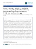
The C1 lateral mass and the C2 pedicle screw fixation had been, as indicated by comparing
pre- and postoperative images, successfully performed in all patients. There were no cases of
vertebral artery injury or other complications during operation. Mending was performed relatively
easily. Two patients with sensory disorder and circular muscle disorder were required to decompress C1 posterior arc, MRI revealed compressed muscles caused by dislocated C1 - C2. After
the surgery, 18 patients had reported fewer bouts of neck pain as well as hand numbness. Ninety
percent of them report no circular muscle disorder post-operation. One patient who had quadriplegia due to compressed C2 - C3 spinal cord are able to walk again after 1 year undergoing surgery
but have not completely recovered with numbness in both hands still occurred.
JMR 116 E3 (7) - 2018
55
JOURNAL OF MEDICAL RESEARCH
Pre-surgery images
a. Odotoid/
dens
Post surgery images
fracture pre-surgery
images
b. Post surgery images of C1 lateral mass
screw fixation
c. Images of vertebral arteries
e. Images of posterior C2 spinous process
d. Image of post C1 and C2 screw fixation
f. Post screw fixation image of C1 and C2
fracture
Figure 1. Pre and post surgery images
56
JMR 116 E3 (7) - 2018
JOURNAL OF MEDICAL RESEARCH
Case illustration
Placing the screw on the inner upper edge
A 33-year-old male patient was the victim
of the C2 pedicle, direct toward the center at
of a road accident with his head and neck
an angle of 20 degrees and upward of 20 de-
bump against the road. After falling, he
grees. Set fixed rod to join C1 - C2. The corti-
remained conscious but with severe neck
cal bone in the outermost of arcs C1 and C2
pain, exacerbated by movement. With neck
was crusted and transplanted by bone of C3
movement restrained, CT scan of the neck
posterior spinosity. The tendon was stitched
spine showed C2 odontoid process fracture
into C2 posterior spinosity. Close the skin ac-
type II.
cording to the anatomical layers.
The patient was scanned by 64-slide CT
Postoperative patient was given antibiotics,
scanner to determine the anatomical structure
, then could sit up and exercise early, thread
of the C1 - C2 vertebral body, the C1 lateral
cut and discharged from the hospital 15 days
mass, examine the size of the pedicle of C2,
after the surgery.
and the vertebral artery location when passing
C2 and C1.
IV. DISCUSSION
Surgerical Technique: Endotracheal anes-
Surgical treatment for C2 odontoid process
thesia with the posture of lying in the prone
fracture has many different methods. In gen-
position. The head was fixed on May-Few
eral, there are two main types of surgery: fron-
frame. Incision of skin was along the posterior
tal way surgery and posterior way surgery
interspinous line from the point under the outer
Frontal way surgery by screwing through the
occiput to the C5, C6 spinous process. Dis-
odontoid apophysis was first performed by
section of the muscle mass beside the bilat-
Bohler [7; 8]. This technique directly fixes the
eral spinal spinosity under the periosteum to
fracture, strengthens the spine of the neck,
the outer edge of joint C2 - C3. Dissection be-
preserves the rotation of C1C2. Bone weld
neath the periosteum, above C1 posterior arch
rate is about 90%. But this technique is difficult
to both sides to see the C1 lateral mass. The
to perform when odontoid process fractures
inner edge of C1 lateral mass was palpable.
involve severe C1C2 dislocations.
The vertebral artery was in the artery groove
Posterior way surgery to harden C1C2 also
above the C1 posterior arch. Normally, C2
has many different methods such as lateral
radicle is located from lateral mass to the C1
mass screwing surgery C1 and the C2 pedicle
posterior arch. After protecting the vertebral
that has many advantages. In 2002, Author
artery and C2 radicle, high-speed grinding
Goel reported 160 cases that were operated
drills with a 3-mm bit were used in the middle
with splint screw at C1 lateral mass and C2
of C1 lateral mass to screw toward the center
pedicle; no patient then had neurological and
crossing horizontal plane made the angle of
vascular complications [9]. In 2001, Harms
10 degrees. After Taro set screw 34mm long,
and Melcher reported 37 cases of screwing C1
diameter 3.5 mm into the C1 lateral mass. The
lateral mass and C2 pedicle that also resulted
point to place the screw through the C2 pedi-
in 100% bone and no vascular and neurologic
cle was in the middle of the upper one third of
complication [6]. In 2010, Mummaneni studied
the C2 pedicle.
42 cases of C1 lateral mass screwing showing
JMR 116 E3 (7) - 2018
57
JOURNAL OF MEDICAL RESEARCH
high bone weld rate, reduced neck pain and
pedicle, because if the C2 pedicle were too
improved neurological function [4].
small, this technique could not be applied.
Regarding to determining the bolt point on
During surgery, continuously use C-Arm 3D to
C1 lateral mass, there are many different
well control the path of the screw to reduce the
views: The authors Harms and Goel screwed
risk of injury to vertebral artery and spinal
directly to the mass under posterior arch C1
cord.
after rolling up the C2 radicle downward [1].
Tan, Wang and associates screwed on C1 to
the lateral mass for good results. In this case,
we screwed directly on C1 posterior arch and
found many advantages: there is no need to
roll up C2 radicle, the screw was quite firm
because the part of screw in the bone was
long,
which
was
favorable
for
C1C2
dislocation treatment. However, the bolt point
was just on the posterior arch, near the
vertebral artery groove, thus, vertebral artery
is vulnerable [10]. Therefore, according to us,
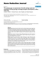
Figure 2. Vertebral arterial abnormality
understanding of the path of the vertebral
image
artery before the surgery by using 64-slide
construction scan would help to avoid this
complication.
Through
V. CONLUSION
Screwing surgery through C1 lateral mass
the
study,
we
found
that
and C2 pedicle should be applied to patient
preoperative screening was of paramount
with sprained C1C2, odontoid process fracture
importance. Patients are required to have a 64
-slide construction scan to determine the
anatomy of the path of lateral vertebral artery
because, according to the literature, 15 to 20%
of patients have abnormalities of anatomical
vertebral artery. We met a case of odontoid
process fracture type II, 64-slide construction
scan showed abnormality of the path of left
vertebral artery that went over the posterior
anterior facet of C1 lateral mass. In that case,
we had to use the surgical method of screwing
through the C1C2 joint and the patient also
had surgery successfully.
fix but keeping ability to exercise cervical
spine after surgery and it’s safe. However, it is
needed to carry out preoperative examination
thoroughly together with modern medical
equipment, and it needs high accuracy and
surgeon’s experiences.
ACKNOWLEDGEMENTS
We highly appreciate the Phu Tho provincial general hospital for providing the data,
allowing and supporting us to perform our research. We would like to thank Department of
Vertebral arterial abnormality image
Biostatistic and Health Informatic, Hanoi Medi-
It is important to take 64-slide scan to have
cal University for technical assistance and ed-
anatomical determination of the size of the C2
58
type II as it results in high bone weld rate, firm
iting the manuscript.
JMR 116 E3 (7) - 2018
JOURNAL OF MEDICAL RESEARCH
REFERENCES
1. Aryan HE., Newman CB., Acosta FL
(2008). Stabilization of the atlantoaxial complex via C1 lateral mass and C2 pedicle screw
fixation in multicenter clinical experience in
102 patients: modification of the Harms and
Goel technique. J Neurosurg Spine, 8, 222 229.
2. Fessler RG., Sekhar L (2006). Posterior
6. Harms J., Melcher RP (2011). Posterior
C1-C2 fusion with polyaxial screw and rod
fixation. Spine, 26, 246 - 247.
7. Nguyen Van Thach., Nguyen Le Bao
Tien., Dinh Ngoc Son et al (2013). Evaluation of initial results of C1 lateral mass and C2
pecicle screw fixation in treating post trauma
unstable high cervical spine. Vietnamese
Journal of Trauma and Orthopedic, 12 - 19.
atlantoaxial fusion: Surgical Anatomy and
8. Borne GM., Bedou GL., Pinaudeau M
Technique option. Atlas of neurosurgical tech-
(1984). Treatment of pedicular fractures of the
niques, 128 - 139.
axis. J Neurosurg, 60, 88 - 93.
3. Ryken TC., Hadley MN., Walter BC
9. Goel A., Desai KI., Muzumdar DP
(2013). Management of isolated fractures of
(2002). Atlantoaxial fixation using plate and
the axis in adults. Neurosurgery, 72, 132 - 150.
screw method: A report of 160 treated
4. Mummaneni PV., Lu DC (2010). C1
lateral mass fixation: A comparison of constructs. Neurosurgery, 66, A68 - A82.
5. Pryputniewicz DM., Hadley MN (2010).
Axis fractures. Neurosurgegy, 66, A68 - A82.
JMR 116 E3 (7) - 2018
patients. Neurosurgery, 51, 1351 - 1357.
10. Tan M., Wang H., Wang Y (2003).
Morphometric evaluation of screw fixation in
atlas via posterior arch and lateral mass.
Spine, 28, 888 - 895.
59