Ebook Clinical manual of alzheimer disease and other dementias: Part 2
Bạn đang xem bản rút gọn của tài liệu. Xem và tải ngay bản đầy đủ của tài liệu tại đây (4.41 MB, 218 trang )
9
Frontotemporal Dementia
and Other Tauopathies
Anne M. Lipton, M.D., Ph.D.
Adam Boxer, M.D., Ph.D.
I
n its broadest sense, the term frontotemporal dementia (FTD) refers to a number of neurodegenerative diseases that vary in clinical presentation and pathological findings. FTD is also known as frontotemporal lobar degeneration (FTLD)
(Neary et al. 1998). The clinical and research nosology for this disease continue
to evolve and sometimes create controversy or confusion. Frontal-variant FTD
(fvFTD) refers to the specific FTD clinical subtype characterized by executive
dysfunction and apathy. Although the clinical syndromes vary, they characteristically involve problems with language, behavior, and/or motor findings, such
Preparation of portions of this chapter was supported by National Institutes of Health
Grant K23NS048855 and the John Douglas French Foundation.
219
220 Clinical Manual of Alzheimer Disease and Other Dementias
as parkinsonism. Research in FTD, including genetic discoveries and the application of modern neuroimaging techniques, has led to remarkable advances.
History
The archetypal FTD is Pick disease, first clinically delineated by Arnold Pick
(1892), who described language impairments and behavioral disturbances in
the setting of focal brain atrophy. Alois Alzheimer (1911) provided the first histopathological description of Pick disease with argyrophilic inclusions (later
called Pick bodies) and swollen, achromatic cells (later called Pick cells). The
Lund-Manchester criteria (Lund and Manchester Groups 1994) delineated the
clinical features of FTD; these criteria were later refined by a consensus panel
that used the term frontotemporal lobar degeneration (Neary et al. 1998). Additional clinical consensus criteria for FTD have been published (McKhann et al.
2001).
FTD occurs, on average, in individuals in their 50s and may be the most
common cause of dementia in this age group (Knopman et al. 2004). Onset before age 65 years is one of the clinical diagnostic criteria for FTD (Neary et al.
1998).
Clinical Subtypes of FTD
Patients with FTD present with the insidious onset of a behavioral syndrome
or a language variant. FTD progresses gradually, but survival is generally shorter
than for Alzheimer disease. Hodges et al. (2003) reported that median survival from symptom onset and from diagnosis was about 6 years for fvFTD and
about 3 years for FTD associated with motor neuron disease.
Frontal Variant FTD
The frontal or behavioral variant of FTD is an FTD subtype characterized by
executive dysfunction and problems with social conduct and interpersonal skills
associated with abnormalities of the right frontotemporal lobe on neuroimaging (Mychack et al. 2001). Lack of insight is a hallmark of the fvFTD subtype.
Patients are often impulsive and oblivious to societal or other limitations on
their actions. Compulsions, hoarding, and decline in hygiene frequently occur.
Frontotemporal Dementia and Other Tauopathies 221
An individual with fvFTD may display disinhibition, apathy, or both. Patients
with orbitofrontal dysfunction are more “disagreeable” and less modest and altruistic (Rankin et al. 2004). Damage in the ventromedial frontal lobes is associated with disinhibited, impulsive, antisocial, and compulsive behaviors
(Rosen et al. 2002a). Patients with fvFTD may have some aspects of a KlüverBucy syndrome, including eating (or drinking) to excess, with an emphasis on
carbohydrate-laden junk food.
Primary Progressive Aphasia
Patients with a language variant of FTD—either progressive nonfluent aphasia
or semantic dementia—frequently have one or more extensive evaluations for
stroke due to their aphasia. The aphasia worsens, and they may become mute.
Some also develop behaviors similar to those seen in fvFTD or in motor dysfunctions such as amyotrophic lateral sclerosis (ALS) or parkinsonism.
Artistic abilities often manifest in patients with a language variant of FTD,
but they may emerge in patients with nonlanguage presentations of FTD as
well (Miller et al. 1998). These talents may manifest de novo or as a modification of a skill previously evident in an individual.
Progressive Nonfluent Aphasia
Progressive nonfluent aphasia involves expressive aphasia with word finding
difficulty, agrammatism, and phonemic paraphasias. Unlike patients with the
other forms of FTD, patients with progressive nonfluent aphasia usually have
little functional or behavioral impairment until late in their disease.
Semantic Dementia
Semantic dementia, also called the temporal lobe variant of FTD, is caused by a
progressive loss of information about the world and is associated with degeneration of the anterior temporal lobes. It usually manifests as a fluent dysphasia
with impairment of semantic verbal memory (severe difficulty in naming and
in understanding the meaning of words) and an associative agnosia (e.g., difficulty in stating or demonstrating the function of an object, such as a tool or
utensil) in individuals with more left temporal lobe involvement. Prosopagnosia
(inability to recognize faces) may rarely occur and is associated with right temporal lobe damage. More commonly, behavioral problems similar to those in
fvFTD occur in individuals with more right lobar dysfunction.
222 Clinical Manual of Alzheimer Disease and Other Dementias
Overlap of FTD Clinical Syndromes
Because the three FTD clinical syndromes often overlap (as can be seen in
some of the above examples), and because they may also overlap with motor
syndromes such as motor neuron disease/ALS and parkinsonism (including
corticobasal syndrome and progressive supranuclear palsy [PSP]), some authors suggest the term Pick complex to encompass all of these syndromes.
The current consensus clinical criteria for FTD are useful but still lack precision. New guidelines are in development. The current clinical criteria fail to
account for many neurogenetic and neuroimaging aspects of the diagnosis of
FTD. Rosen et al. (2002b) found that the Neary et al. (1998) clinical consensus
criteria efficiently separated 30 autopsy-proven cases of Alzheimer disease and
30 autopsy-proven cases of FTLD. They found that the following five clinical
features best distinguished FTLD from Alzheimer disease: presence of social
conduct disorders, hyperorality, akinesia, and absence of amnesia and perceptual disorder.
Clinical Syndromes Associated With FTD
A number of diseases overlap clinically and pathologically with FTD, including motor neuron disease/ALS, corticobasal syndrome, and PSP.
Motor Neuron Disease/Amyotrophic Lateral Sclerosis
Of 100 ALS patients studied prospectively with extensive neuropsychological
assessment, about one-third met criteria for FTLD (Lomen-Hoerth et al.
2003). Many patients clinically diagnosed with FTLD have motor neuron–
type inclusions on histopathology, either with or without clinical motor neuron
disease (Bigio et al. 2003). Moreover, both chronic traumatic encephalopathy
and FTLD may include TAR-DNA binding protein 43 (TDP-43)–positive inclusions in the brain. These inclusions have been shown in the spinal cord in
a few cases of chronic traumatic encephalopathy associated with motor neuron
disease (McKee et al. 2010).
Corticobasal Syndrome
Corticobasal syndrome is the current nomenclature used to describe the unifying
clinical and pathological characteristics of FTD and corticobasal degeneration,
Frontotemporal Dementia and Other Tauopathies 223
also known as corticobasal ganglionic degeneration (CBGD). CBGD is a Parkinson-plus syndrome (classically manifested as unilateral rigidity, apraxia, the
alien hand syndrome, reflex myoclonus, and/or cortical sensory loss) that tends
to progress more rapidly than Parkinson disease and is usually less amenable to
treatment.
Progressive Supranuclear Palsy
PSP is another Parkinson-plus syndrome possessing clinical and pathological
overlap with FTD. Both FTD and PSP are tauopathies (pathologically classified as abnormalities of the cytoskeletal protein tau) with clinical onset in late
life. PSP is characterized by balance difficulty, falls, visual disturbances, slurred
speech, dysphagia, and personality change (Richardson et al. 1963). The dementia of PSP is consistent with FTD. A characteristic triad of ophthalmoplegia, pseudobulbar palsy, and axial dystonia develops. First, downward gaze is
impaired, then upward gaze, then voluntary gaze in all directions. If the eyes are
fixed on a target and the head is turned, full eye movement occurs (doll’s eye
phenomenon), indicating that the motor nerves are intact.
The etiology of PSP is unknown. Pathological findings include loss of
neurons; gliosis; and the presence of neurofibrillary tangles in the surviving
neurons in the midbrain, cerebellar peduncles, and subthalamic nucleus.
Functional impairment proceeds to anarthria and total immobility, usually
within a few years.
Neuropathology
FTD is pathologically distinct from Alzheimer disease. Historically, the FTD
disorders have been divided into Pick disease and non-Pick lobar atrophy
(Dickson 1998). Both have grossly appreciable frontal and temporal atrophy.
Pick bodies are seen only in Pick disease.
Tau and ubiquitin immunohistochemistries are important in classifying
pathological FTD subtypes. Motor neuron–type, ubiquitin-positive inclusions
are the most common histopathological type of FTLD (Lipton et al. 2004). The
chief protein associated with ubiquitinated inclusions is now recognized to be
TDP-43 (Neumann et al. 2006). Frontotemporal degeneration with neuronal
loss and spongiosis has no tau or ubiquitin inclusions, but some of these cases
are classifiable as FTLD–motor neuron disease (Lipton et al. 2004). Cortico-
224 Clinical Manual of Alzheimer Disease and Other Dementias
basal degeneration has tau-positive neuronal inclusions and glial plaques, along
with ballooned neurons, in cortex, basal ganglia, brain stem, and cerebellum.
Despite the shared pathology in patients with FTD, there may be a variety of pathological findings within the same clinical FTD subtype. Familial
multiple system tauopathy is one of the many cases of familial FTD and parkinsonism linked to chromosome 17 (FTDP-17). These families have a variety of clinical presentations, including disinhibition-dementia-parkinsonismamyotrophy complex, and neuropathological findings always associated with
tau deposition. In contrast, individuals with progranulin mutations, an even
more common form of autosomal dominant FTD, are found to have ubiquitin pathology at autopsy. Validity of the FTLD diagnostic consensus criteria
has been verified histopathologically (Knopman et al. 2005).
Diagnostic Evaluation
Clinical evaluation, including history from a reliable collateral source, such as a
close family member, is crucial in the diagnosis of FTD. Family history of neurological disease and psychiatric illness is important, because FTD is hereditary
in some cases and is often not diagnosed as FTD per se, but rather may manifest
as motor neuron disease or parkinsonism, go undiagnosed, or be misdiagnosed
(as depression, bipolar disorder, another form of dementia, etc.). Neurological
evaluation may elicit abnormalities, such as motor weakness, parkinsonism, or
frontal reflexes, that may provide additional diagnostic certainty. Patients with
FTD, particularly the FTD clinical profile, will often display echopraxia (imitating the examiner), perseveration, and motor impersistence. Patients can also
be tested for frontal release signs, such as suck, snout, rooting, palmomental,
and Babinski reflexes.
Neuropsychological Testing
A comprehensive neuropsychological evaluation is often helpful in diagnostic
differentiation (see Chapter 3, “Neuropsychological Assessment”), if the patient
can comprehend and cooperate with such testing. Usual clinical tests, such as
the Mini-Mental State Examination (MMSE; Folstein et al. 1975), do not directly assess executive functioning and may be relatively normal in patients
with FTD (due to relative sparing of memory) or may show profound impairment in patients with the language variants of FTD. However, MMSE scores
Frontotemporal Dementia and Other Tauopathies 225
do decline at a greater rate in FTD than in Alzheimer disease (Chow et al.
2006). Neuropsychological evaluation may reveal executive dysfunction on
commonly performed assessments, including the Stroop Test, the Trail Making Test, tests of verbal and design fluency, and the Wisconsin Card Sorting
Test (Hodges and Graham 2001).
Tests reported to be sensitive to FTD include the Frontal Behavioral Inventory (Kertesz et al. 1997) and the Frontal Assessment Battery (FAB; Dubois et
al. 2000). The FAB has been shown to distinguish healthy control subjects from
patients with mild Parkinson disease, multiple system atrophy, corticobasal degeneration, and PSP. Total FAB scores did not differentiate FTLD from Alzheimer disease, but some subscores (of mental flexibility and environmental
autonomy) did (Lipton et al. 2005), and patients with Alzheimer disease and
FTLD patients actually performed comparably on the Luria maneuver (Weiner
et al. 2011).
Speech-Language Cognitive Evaluation
A speech-language cognitive evaluation is often helpful, especially in diagnosing
specific language variants (see also Chapter 1, “Neuropsychiatric Assessment
and Diagnosis”). Some patients may also benefit from further therapy to assist
in maintaining communication.
Neuroimaging
Prominent frontal lobe atrophy on structural magnetic resonance imaging is
a common feature of FTD, particularly in individuals without motor neuron
disease (Figure 9–1). Neuroimaging with 18F-labeled fluorodeoxyglucose
positron emission tomography (FDG-PET) is sometimes helpful in the differential diagnosis of FTD (Foster et al. 2007) and has been approved by
Medicare for this purpose in the context of a comprehensive clinical evaluation (see also Chapter 4, “Neuroimaging”). The amyloid imaging agent
Pittsburgh compound B, or PIB, may be even more valuable for ruling out
atypical forms of Alzheimer disease that mimic FTD (Rabinovici et al. 2007).
Electroencephalography
Electroencephalography (EEG) is not generally helpful for diagnosis. EEG has
been shown to be normal in many cases. One study showed that electroenceph-
226 Clinical Manual of Alzheimer Disease and Other Dementias
alographic abnormalities correlated with severity of FTD but that this correlation was not helpful in differentiating FTD from Alzheimer disease (Chan et al.
2004).
Genetics
Genetic tests are not available commercially but are a major area of research interest. Multiple genetic loci (on chromosomes 3p, 9p, 9q, 17q21, and 17q24)
and five genes (those for microtubule-associated protein tau, progranulin,
valosin-containing protein, and charged multivesicular body protein 2B
[CHMP2B]) have been associated with inherited FTD (Mackenzie and Rademakers 2007; Rademakers and Hutton 2007). FTD with parkinsonism (FTDP17) has been linked to mutations in the gene coding for the microtubule-associated protein tau (Hutton et al. 1998). FTD with ubiquitin-positive inclusions
(FTDU-17) is caused by loss-of-function mutations in the TAR-DNA binding
protein gene coding for progranulin (PGRN), a growth factor involved in neuronal survival (Baker et al. 2006).
Treatment
No treatment for FTD has been approved by the U.S. Food and Drug Administration, but antidepressants, including selective serotonin reuptake inhibitors, are useful in treating many of the behavioral symptoms (Huey et al.
2006). Trazodone is the only medication for FTD behavioral symptoms studied in a double-blind, randomized controlled trial (Lebert et al. 2004). Trazodone is beneficial for a number of behavioral problems in FTD, including
irritability, agitation, depressive symptoms, and eating disorders.
FTD does not entail a cholinergic deficit, and the use of cholinesterase inhibitors is controversial. In an open-label study, rivastigmine ameliorated behavioral problems in FTD (Moretti et al. 2004), but donepezil worsened
behavioral symptoms (Mendez et al. 2007). Other symptomatic treatments
that have been tried are dopaminergic therapies for parkinsonism and language
problems. A prospective 26-week open-label trial of memantine 20 mg/day in
FTD showed that patients with progressive nonfluent aphasia maintained relative cognitive stability over the 26 weeks, whereas the subjects with semantic
aphasia had a decline in cognitive ability (Boxer et al. 2009). In a double-blind
study of memantine 20 mg/day in 18 human subjects with primary progres-
Frontotemporal Dementia and Other Tauopathies 227
Figure 9–1. Magnetic resonance imaging (MRI) findings in frontotemporal dementia (FTD).
FTD: parasagittal and coronal images from T1-weighted MRI. Note asymmetric right
frontal atrophy on coronal image (*), and lack of significant atrophy posterior to frontal lobe on sagittal image.
Semantic dementia (SD): axial and coronal images; atrophy is most severe anteriorly
and involves both medial and lateral temporal lobe structures (*).
Progressive nonfluent aphasia (PNFA): axial and coronal images show asymmetric left
frontal atrophy with minimal temporal lobe involvement (*).
Source. Reprinted from Lipton AM, Boxer A: “Frontotemporal Dementia,” in The
American Psychiatric Publishing Textbook of Alzheimer Disease and Other Dementias. Edited
by Weiner MF, Lipton AM. Washington, DC, American Psychiatric Publishing, 2009,
pp. 219–227. Copyright 2009, American Psychiatric Publishing. Used with permission.
228 Clinical Manual of Alzheimer Disease and Other Dementias
sive aphasia, the treated group showed less decline on the Western Aphasia Battery than did the placebo group (Johnson et al. 2010).
Key Clinical Points
• Frontotemporal dementia (FTD) may be the most common cause
of dementia for adults under age 65.
• Gradual personality change with impaired judgment in the fifth
or sixth decade of life should elicit suspicion for the frontal/
behavioral variant of FTD.
• FTD may manifest as a disorder of language expression or comprehension.
• FTD overlaps clinically and pathologically with a number of neurological syndromes, including amyotrophic lateral sclerosis, corticobasal syndrome, and progressive supranuclear palsy.
References
Alzheimer A: Über eigenartige Krankheitsfälle des späteren Alters. Zeitscrift für die
gesamte Neurologie und Psychiatrie 4:356–385, 1911
Baker M, Mackenzie IR, Pickering-Brown SM, et al: Mutations in progranulin cause
tau-negative frontotemporal dementia linked to chromosome 17. Nature 24:916–
919, 2006
Bigio EH, Lipton AM, White CL III, et al: Frontotemporal and motor neuron degeneration with neurofilament inclusion bodies: additional evidence for overlap between FTD and ALS. Neuropathol Appl Neurobiol 29:239–253, 2003
Boxer AL, Lipton AM, Womack K, et al: An open-label study of memantine treatment
in 3 types of frontotemporal lobar degeneration. Alzheimer Dis Assoc Disord
23:211–217, 2009
Chan D, Walters RJ, Sampson EL, et al: EEG abnormalities in frontotemporal lobar
degeneration. Neurology 62:1628–1630, 2004
Chow TW, Hynan LS, Lipton AM: MMSE scores decline at a greater rate in frontotemporal degeneration than in AD. Dement Geriatr Cogn Disord 22:194–199, 2006
Dickson DW: Pick’s disease: a modern approach. Brain Pathol 8:339–354, 1998
Dubois B, Slachevsky A, Litvan I, et al: The FAB: a frontal assessment battery at bedside.
Neurology 55:1622–1625, 2000
Frontotemporal Dementia and Other Tauopathies 229
Folstein MF, Folstein SE, McHugh PR: “Mini-mental state”: a practical method for
grading the cognitive state of patients for the clinician. J Psychiatr Res 12:189–
198, 1975
Foster NL, Heidebrink JL, Clark CM, et al: FDG-PET improves accuracy in distinguishing frontotemporal dementia and Alzheimer’s disease. Brain 130:2616–
2635, 2007
Hodges JR, Graham KS: Episodic memory: insights from semantic dementia. Philos
Trans R Soc Lond B Biol Sci 356:1423–1434, 2001
Hodges JR, Davies R, Xuereb J, et al: Survival in frontotemporal dementia. Neurology
61:349–354, 2003
Huey ED, Putnam KT, Grafman J: A systematic review of neurotransmitter deficits
and treatments in frontotemporal dementia. Neurology 66:17–22, 2006
Hutton M, Lendon CL, Rizzu P: Association of missense and 5′-splice-site mutations
in tau with the inherited dementia FTDP-17. Nature 393:702–705, 1998
Johnson NA, Rademaker A, Weintraub S, et al: Pilot trial of memantine in primary
progressive aphasia (letter). Alzheimer Dis Assoc Disord 24:308, 2010
Kertesz A, Davidson W, Fox H: Frontal Behavioral Inventory: diagnostic criteria for
frontal lobe dementia. Can J Neurol Sci 24:9–36, 1997
Knopman DS, Petersen RC, Edland SD, et al: The incidence of frontotemporal lobar
degeneration in Rochester, Minnesota, 1990 through 1994. Neurology 62:506–
508, 2004
Knopman DS, Boeve BF, Parisi JE, et al: Antemortem diagnosis of frontotemporal
lobar degeneration. Ann Neurol 57:480–488, 2005
Lebert F, Stekke W, Hasenbroekx C: Frontotemporal dementia: a randomised, controlled trial with trazodone. Dement Geriatr Cogn Disord 17:355–359, 2004
Lipton AM, White CL III, Bigio EH: Frontotemporal lobar degeneration with motor
neuron disease–type inclusions predominates in 76 cases of frontotemporal degeneration. Acta Neuropathol 108:379–385, 2004
Lipton AM, Ohman KA, Womack KB, et al: Subscores of the FAB differentiate frontotemporal lobar degeneration from AD. Neurology 65:726–731, 2005
Lomen-Hoerth C, Murphy J, Langmore S, et al: Are amyotrophic lateral sclerosis patients cognitively normal? Neurology 60:1094–1097, 2003
Lund and Manchester Groups: Clinical and neuropathological criteria for frontotemporal dementia. J Neurol Neurosurg Psychiatry 57:416–418, 1994
Mackenzie IR, Rademakers R: The molecular genetics and neuropathology of frontotemporal lobar degeneration: recent developments. Neurogenetics 8:237–248, 2007
McKee AC, Gavett BE, Stern RA, et al: TDP-43 proteinopathy and motor neuron
disease in chronic traumatic encephalopathy. J Neuropathol Exp Neurol 69:918–
929, 2010
230 Clinical Manual of Alzheimer Disease and Other Dementias
McKhann GM, Albert MS, Grossman M, et al: Clinical and pathological diagnosis of
frontotemporal dementia. Arch Neurol 58:1803–1809, 2001
Mendez MF, Shapira JS, McMurtray A, et al: Preliminary findings: behavioral worsening on donepezil in patients with frontotemporal dementia. Am J Geriatr Psychiatry 15:84–87, 2007
Miller BL, Cummings J, Mishkin F, et al: Emergence of artistic talent in frontotemporal
dementia. Neurology 51:978–982, 1998
Moretti R, Torre P, Antonello RM, et al: Rivastigmine in frontotemporal dementia: an
open-label study. Drugs Aging 21:931–937, 2004
Mychack P, Kramer JH, Boone KB, et al: The influence of right frontotemporal dysfunction on social behavior in frontotemporal dementia. Neurology 56 (suppl 4):S11–
S15, 2001
Neary D, Snowden JS, Gustafson L, et al: Frontotemporal lobar degeneration: a consensus on clinical diagnostic criteria. Neurology 51:1546–1554, 1998
Neumann M, Sampathu DM, Kwong LK, et al: Ubiquitinated TDP-43 in frontotemporal
lobar degeneration and amyotrophic lateral sclerosis. Science 314:130–133, 2006
Pick A: Über die Beziehungen der senilen Hirnatrophie zur Aphasie. Prager medizinische Wochenschrift 17:165–167, 1892
Rabinovici GD, Furst AJ, O’Neil JP, et al: 11C-PIB PET imaging in Alzheimer disease
and frontotemporal lobar degeneration. Neurology 68:1205–1212, 2007
Rademakers R, Hutton M: The genetics of frontotemporal lobar degeneration. Curr
Neurol Neurosci Rep 7:434–442, 2007
Rankin KP, Rosen HJ, Kramer JH, et al: Right and left medial orbitofrontal volumes
show an opposite relationship to agreeableness in FTD. Dement Geriatr Cogn
Disord 17:328–332, 2004
Richardson JC, Steele J, Olszewski J: Supranuclear ophthalmoplegia, pseudobulbar
palsy, nuchal dystonia and dementia. Trans Am Neurol Assoc 88:25–29, 1963
Rosen HJ, Hartikainen KM, Jagust W, et al: Utility of clinical criteria in differentiating
frontotemporal lobar degeneration from Alzheimer’s disease. Neurology 58:1608–
1615, 2002a
Rosen HJ, Perry RJ, Murphy J, et al. Emotion comprehension in the temporal variant
of frontotemporal dementia. Brain 125:2286–2295, 2002b
Weiner MF, Hynan LS, Rossetti H, et al: Luria’s three-step test: what is it and what
does it tell us? Int Psychogeriatr May 4, 2011 (Epub ahead of print)
Further Reading
Brun A: Identification and characterization of frontal lobe degeneration: historical perspective on the development of FTD. Alzheimer Dis Assoc Disord 21:3–4, 2007
Frontotemporal Dementia and Other Tauopathies 231
Caselli R, Yaari R: Medical management of frontotemporal dementia. Am J Alzheimers
Dis Other Demen 22:489–498, 2007
Hallam BJ, Silverberg ND, Lamarre AK, et al: Clinical presentation of prodromal
frontotemporal dementia. Am J Alzheimers Dis Other Demen 22:456–457, 2007
Levy JA, Chelune GJ: Cognitive-behavioral profiles of neurodegenerative dementias:
beyond Alzheimer’s disease. J Geriatr Psychiatry Neurol 20:227–238, 2007
This page intentionally left blank
10
Traumatic Brain Injury
Erin D. Bigler, Ph.D.
More than 80,000 individuals become disabled from traumatic brain in-
juries (TBIs) each year (Thurman 2007). Thus, TBI represents a substantial
source of neuropsychiatric morbidity and disability. From a cognitive perspective, the three likely proximal outcomes of a TBI are complete recovery after
a period of recuperation, mild to moderate residual cognitive impairment, and
dementia. However, even in those who appear to fully recover from the proximal effects of TBI, the brain injury may become a vulnerability factor that
during aging interacts with other environmental, constitutional, and genetic
factors to produce later cognitive decline and earlier onset of frank dementia
The technical expertise and manuscript assistance of Tracy Abildskov, Craig Vickers, and
Jo Ann Petrie are gratefully acknowledged. Much of the research reported on in this
chapter was supported by a grant from the Ira Fulton Foundation.
233
234 Clinical Manual of Alzheimer Disease and Other Dementias
in late life (Gavett et al. 2010; van den Heuvel et al. 2007). Acute TBI induces
several histopathological changes that also occur in age-related degenerative
diseases such as Alzheimer disease (AD) (DeKosky et al. 2010). Indeed, much
has been written about TBI as a substantial risk factor for dementia and other
neuropsychiatric problems later in life (Rao and Lyketsos 2002; Starkstein and
Jorge 2005), although much needs to be discovered and scientifically established about the distant effects of TBI (Blennow et al. 2006).
By the standards of DSM-IV-TR (American Psychiatric Association 2000),
dementia due to head trauma is diagnosed when the dementia is judged to be a
direct pathophysiological consequence of head trauma (see Table 10–1). Head
trauma is a common cause of acquired dementia (Kim et al. 2011). By definition, when head injury is the proximal cause of a dementia syndrome, the person
never recovers sufficiently to overcome or compensate for the substantial cognitive and behavioral residuals of the brain injury. However, the majority of TBIs
are in the mild to moderate range, and although cognitive impairments are
commonplace, most TBIs at this level of severity do not cause dementia. A more
common diagnosis attributable to TBI is cognitive disorder not otherwise specified (NOS).
Head injury can be a remote contributor to the later development of dementia even if the individual experienced an apparent complete recovery from the
original brain injury (Bigler 2007). These remote effects of TBI are discussed
later in this chapter after a discussion of proximal effects.
Proximal Effects of TBI:
Dementia Due to Head Trauma
Dementia due to head trauma usually results from a moderate to severe brain
injury. As indicated in Table 1–2 of Chapter 1, DSM-IV-TR criteria for dementia require multiple cognitive deficits, including memory impairment and at
least one of the following cognitive disturbances: aphasia, apraxia, agnosia, or a
disturbance in executive functioning. The deficits that make up dementia are
diagnosed clinically. The deficits must be sufficient to cause functional impairment in home or work life and must represent a decline from previous functioning. Because a distinct antecedent event is known in TBI-associated cognitive
disorders, little doubt exists about the causal relationship of the head injury in
dementia due to head trauma.
Traumatic Brain Injury 235
Table 10–1. Dementia due to head trauma
The essential feature of dementia due to head trauma is the presence of a dementia
that is judged to be the direct pathophysiological consequence of head trauma. The
degree and type of cognitive impairments or behavioral disturbances depend on the
location and extent of the brain injury. Posttraumatic amnesia is frequently present,
along with persisting memory impairment. A variety of other behavioral symptoms
may be evident, with or without the presence of motor or sensory deficits. These
symptoms include aphasia, attentional problems, irritability, anxiety, depression or
affective lability, apathy, increased aggression, or other changes in personality. Alcohol
or other substance intoxication is often present in individuals with acute head injuries,
and concurrent substance abuse or dependence may be present. Head injury occurs
most often in young males and has been associated with risk-taking behaviors. When
it occurs in the context of a single injury, dementia due to head trauma is usually
nonprogressive, but repeated head injury (e.g., from boxing) may lead to a progressive
dementia (so-called dementia pugilistica). A single head trauma that is followed by
a progressive decline in cognitive function should raise the possibility of another
superimposed process such as hydrocephalus or a major depressive episode.
Source. Reprinted from American Psychiatric Association: Diagnostic and Statistical Manual of
Mental Disorders, 4th Edition, Text Revision. Washington, DC, American Psychiatric Association, 2000, p. 164. Copyright 2000, American Psychiatric Association. Used with permission.
In the most severe cases, TBI-related cognitive impairment can be detected
during standard mental status examination, using screening psychometric tests
such as the Mini-Mental State Examination (MMSE; Lorentz et al. 2002). In
some cases, more detailed neuropsychological testing may be necessary. The
drawing presented in Figure 10–1 is by a patient with dementia due to head injury. He had sustained a severe TBI as an adolescent, and despite extensive inpatient and outpatient treatment and although physically intact, he never
recovered enough cognitive and praxic functions to live independently. When
the patient was tested postinjury as a young adult, his Full Scale IQ score was
78 and his MMSE score was 17. Preinjury school records reflected average academic performance with no history of learning or developmental disorder, and
he had never been diagnosed prior to injury with a neuropsychiatric condition.
He had striking impairment in short-term memory and severe constructional
apraxia, as evidenced in the drawing shown in Figure 10–1. Over a 5-year span
of monitoring, he never changed significantly. Unlike dementias associated
with progressive degenerative diseases such as AD, dementia associated with
head trauma is static.
236 Clinical Manual of Alzheimer Disease and Other Dementias
Neuroimaging for Estimating Severity of
Brain Injury in TBI
Significant advancements in detecting TBI abnormalities have come from
contemporary high-field magnetic resonance imaging (MRI) and functional
neuroimaging techniques (Metting et al. 2007; Taber and Hurley 2007). Using neuroimaging findings to visualize the degree and extent of structural and
functional damage greatly assists clinicians in understanding the effects of TBI
(Bigler 2011). Computed tomography (CT) studies have demonstrated that
the extent of TBI-induced structural brain damage is linearly related to the severity of brain injury and that both are coarsely related to the degree of cognitive impairment (Cullum and Bigler 1986). Wilde et al. (2006) examined the
association between posttraumatic amnesia (PTA) and the development of
MRI-identified cerebral atrophy in patients with TBI. PTA is often used as a
marker of initial injury severity: PTA<1 hour is consistent with mild TBI,
PTA=1–24 hours indicates moderate TBI, and PTA>24 hours indicates severe injury (Lezak et al. 2004). Wilde et al. (2006) calculated that the odds of
developing generalized cerebral atrophy on quantitative MRI increases by 6%
with each day of PTA. In addition to greater amounts of cerebral atrophy,
longer PTA is associated with worse functional outcome. The combination of
longer PTA and greater amounts of cerebral atrophy is associated in turn with
the poorest TBI outcome (Bigler et al. 2006). Thus, for the clinician making
predictions about clinical outcome, markers of brain injury severity, including
PTA, or severity of coma, as indicated by the Glasgow Coma Scale (GCS), and
their duration directly relate to the likelihood of developing dementia following TBI.
MRI studies of the brain readily demonstrate clinically relevant TBI-related
atrophy, when present. The MRI findings of the patient whose apraxia was evident from the drawing in Figure 10–1 are shown in Figure 10–2; they demonstrate extensive structural damage to the entire brain, particularly in frontotemporal regions, readily appreciated by viewing the reconstructed brain in
three-dimensional views. The scan, in conjunction with the patient’s neuropsychological test performance, initial GCS score of 3 (GCS scores range from 3
[deep coma] to 15 [alert, oriented, and following commands]), and history of
weeks of coma and months of PTA, points to the greater likelihood of a residual
dementia due to head trauma.
Traumatic Brain Injury 237
Figure 10–1. Drawing by patient with dementia due to head injury (lower
portion) in response to model (upper portion).
At the time of neuropsychological assessment and neuroimaging, this patient was 22 years
old and 2 years post–traumatic brain injury. He had severe constructional apraxia, as demonstrated by his inability to copy the Rey-Osterrieth Complex Figure (Lezak et al. 2004).
He performed below the first percentile on all measures of short-term memory and was
unable to perform any standardized executive function tasks.
238 Clinical Manual of Alzheimer Disease and Other Dementias
As shown in Figure 10–2, because of the particular vulnerability for focal
damage in TBI to occur in the frontal and temporal lobe regions of the brain,
frontal and temporal lobe atrophy is often observed to develop after injury (Bigler 2011). Injury significant enough to produce focal damage typically occurs
amid a backdrop of diffuse injury. TBI-induced damage to frontotemporal systems increases the likelihood of cognitive impairment and disability (Wilde et
al. 2005), in part because of the disruption of cholinergic systems subserved by
these regions and the critical role that cholinergic neurons play in cognition
(Salmond et al. 2005). Damage to these regions represents another common
connection between TBI and the development of a dementing illness such as
AD later in life.
Progression of Atrophy From Day of
Injury Until Stabilization
Viewing the progression of cerebral damage from acute to chronic stage helps
to demonstrate how TBI alters brain structure that is pertinent to developing
dementia. This progression can be straightforwardly observed in sequential
neuroimaging, as shown in Figure 10–3. The day-of-injury (DOI) scan demonstrates multiple hemorrhagic lesions, intraventricular hemorrhage, and generalized edema in a brain with no identifiable preinjury abnormalities. Although
the acute scan demonstrates prominent neuropathological changes, the otherwise intact features help the clinician establish baseline information for future
comparison. Subsequent neuroimaging shows over time how hydrocephalus ex
vacuo emerges as a reflection of brain parenchyma volume loss. In a postmortem study of patients who would likely have met criteria for dementia due to
head trauma, Adams et al. (2011) found that the majority of individuals with
severe to moderate disability from TBI who subsequently died had cortical contusions, diffuse traumatic axonal injury (TAI), and ventricular dilation as a reflection of cerebral atrophy; specific to level of disability, the extensiveness of
TAI, presence of thalamic lesions, and increased ventricular dilation were particularly prognostic for worse outcome. Viewing the scans in Figure 10–3 serially, one can see that the brain injury has resulted in extensive cerebral atrophy
that stabilizes a few months posttrauma. Given the severity of his TBI (GCS=
3), the extensive nature of the cerebral damage documented by the emergence
of cerebral atrophy, his impaired cognition on examination and MMSE score
of <10, and his unchanging status for several years postinjury, this patient also
Traumatic Brain Injury 239
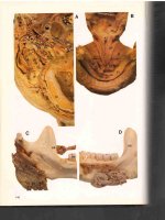
Figure 10–2. Neuroimaging studies for the patient with dementia due to
head injury whose drawing is shown in Figure 10–1 (see color plate 10).
B is an axial T1-weighted magnetic resonance image showing extensive frontal damage
(white arrow) as a result of the severe traumatic brain injury. D is a sagittal T1-weighted
image showing the extensive frontal pathology present in this patient (white arrow). A,
C, and F are three-dimensional magnetic resonance image reconstructions visualizing
the ventricles (shown in blue in Plate 10; shown here in gray) in the dorsal view in A, the
extensive frontotemporal wasting (black arrows) in C, and the bifrontal atrophy, particularly in the inferior frontal region in F. E—a view of single-photon emission computed tomography findings at the same axial level depicted in B—shows extensive loss
of frontal perfusion. There is generalized ventricular dilation (see Figure 10–4H for a
normal dorsal view). These imaging findings demonstrate diffuse brain damage and
generalized loss of total brain volume.
Source. Reprinted from Bigler ED: “Traumatic Brain Injury,” in The American Psychiatric Publishing Textbook of Alzheimer Disease and Other Dementias. Edited by Weiner
MF, Lipton AM. Washington, DC, American Psychiatric Publishing, 2009, pp. 229–
246. Copyright 2009, American Psychiatric Publishing. Used with permission.
240 Clinical Manual of Alzheimer Disease and Other Dementias
meets the criteria for dementia due to head trauma. Therefore, starting with the
DOI scan, the degree of resultant cerebral atrophy can be documented over
time and typically stabilized within 6 months postinjury, with level of atrophy
coarsely associated with degree of cognitive impairment. Such neuroimaging
findings in association with the mental status findings reflective of cognitive impairment are the type most likely to be associated with dementia due to head
trauma.
Additional Factors That Contribute to
Severity of Functional Injury
Damage to Critical Limbic System Structures
Both animal models and human studies have demonstrated the vulnerability of
the hippocampus to TBI (Bigler et al. 2010). In humans, this vulnerability of the
medial temporal lobe and hippocampus is due in part to their location in the
middle cranial fossa and also to excitotoxic reactions that occur in traumatically
injured hippocampal neurons (Geddes et al. 2003); diaschisis plays a role as well
because hippocampal neurons have diverse afferent and efferent cortical connections throughout the brain (Wilde et al. 2007). Because the medial temporal cortex (and in particular the hippocampus) is so critical to all cognitive functions,
damage to this region has a high likelihood for disrupting cognition; however,
even with extensive damage, the patient may not meet criteria for dementia.
Figure 10–4 shows scans from an adolescent patient who sustained a severe
TBI in a motor vehicle accident (GCS score=3). The scan demonstrates medial
temporal lobe atrophy with prominent hippocampal atrophy. Positron emission tomography imaging confirmed reduced radiotracer uptake throughout
the medial temporal lobes bilaterally, yet neuropsychological studies demonstrated only mild memory impairment and related cognitive impairments. The
patient’s MMSE score was 26. Thus, despite these rather dramatic imaging
findings and the presence of some cognitive sequelae from the TBI that certainly met criteria for cognitive disorder NOS, the level of cognitive impairment did not warrant a diagnosis of dementia.
Wilde et al. (2007) have also shown that in comparison to all other brain
structures, the hippocampus exhibits the greatest atrophic changes in response
to TBI. From this and other research, one can conclude that hippocampal injury is found in most cases of moderate to severe TBI. Additionally, it should
Traumatic Brain Injury
DOI
8 days
4 months
9 months
241
17 months
Figure 10–3. Progression of cerebral damage from acute to chronic stage
of traumatic brain injury (TBI).
This patient sustained a severe TBI (Glasgow Coma Scale score=3) with months of
coma and persistent posttraumatic amnesia. Since the patient regained consciousness,
his MMSE score has been consistently below 10. The sequential imaging shows brain
changes over time. The day-of-injury (DOI) computed tomography scan shows multiple intraparenchymal and intraventricular hemorrhagic lesions scattered throughout
the brain, some of which “‘blossom” 8 days postinjury. However, by 4 and 9 months
postinjury, ventricular dilation, as a sign of generalized brain volume loss, has peaked
and shows little change thereafter.
Source. Reprinted from Bigler ED: “Traumatic Brain Injury,” in The American Psychiatric Publishing Textbook of Alzheimer Disease and Other Dementias. Edited by Weiner
MF, Lipton AM. Washington, DC, American Psychiatric Publishing, 2009, pp. 229–
246. Copyright 2009, American Psychiatric Publishing. Used with permission.
be noted that the hippocampus, which also plays a role in emotional control, is
potentially injured by stress-related hormones that are part of both the physical
and emotional reaction to injury (Wolkowitz et al. 2007). The high incidence
of neuropsychiatric sequelae, including depression, in individuals with TBI
(Holsinger et al. 2002), as well as the potential part that damage to limbic structures such as the hippocampus may play in mood disorders following TBI
(Jorge et al. 2007), underscores the role that hippocampal damage may play in
the emotional and cognitive aftermath of TBI. There is even evidence that injury may disrupt hippocampal neurogenesis and that the presence of amyloid
may reduce the rate of neurogenesis (Morgan 2007). Because neuropsychiatric
disorders may also be a vulnerability factor for later expression of dementia
(Starkstein and Jorge 2005), anything that increases the likelihood of neuropsychiatric disorder over the life span may have adverse consequences on the aging process.
242 Clinical Manual of Alzheimer Disease and Other Dementias
Speed-of-Processing Deficits
A nearly universal consequence of TBI, directly related to the severity of injury
and persistence of neurobehavioral symptoms, is reduced speed of cognitive
processing (Ben-David et al. 2011). Two main neuropathological consequences of TBI impair processing speed. Because recovery from focal pathology is probably due to alternate, redundant, or adaptive pathways taking over
function, this less direct way of processing increases response time. The other
main factor is the selective vulnerability of white matter to TBI (Vannorsdall
et al. 2010). Diminished white matter integrity results in less efficient neural
transmission. In normal aging, the extent of white matter pathology directly
relates to speed of processing, and both are related to the clinical presentation
of age-related mild cognitive impairment (MCI) and dementia (Burns et al.
2005). Because diminished speed of processing is a natural consequence of aging that impacts executive function, and alterations in processing speed mirror
normal changes in white matter integrity with aging, the older the individual
is at the time of sustaining a TBI, the less resilient the brain is to injury.
Genetics
Many studies have demonstrated that presence of the apolipoprotein E4 allele
(APOE4) may adversely affect the outcome of any type of acquired brain injury
(Mayeux et al. 1995; Verghese et al. 2011). The role of APOE4 or any other genetic factor in recovery from TBI is beyond the scope of this review, and there
are negative reports or findings of minimal association (Han et al. 2007). Nonetheless, genetic factors likely affect recovery from TBI.
Associated Vascular Effects
TAI is associated with microvascular damage in addition to direct damage to
the axon, and damage to the underlying cerebral microvasculature can, by itself,
cause dementia (Holsinger et al. 2007). The combination of TAI and microvascular damage can lead to widespread changes in cerebral integrity (Petrov and
Rafols 2001). Ueda et al. (2006) reported changes in the vascular reactivity and
local autoregulation of cerebrovasculature following a TBI, suggesting that localized cerebral perfusion may be disrupted, affecting the energy needs of neurons. In this scenario, neurons may not be specifically damaged but are nonetheless rendered functionally impaired because of diminished autoregulatory
factors resulting from vascular rather than neuronal injury.
Traumatic Brain Injury
243
Figure 10–4. Neuroimaging studies for an adolescent patient who sustained a severe traumatic brain injury (TBI) in a motor vehicle accident, contrasted with those of an age-matched control (see color plate 11).
The coronal T1-weighted magnetic resonance image shown in A shows pronounced
hippocampal atrophy (arrow), along with dilated anterior horns of the lateral ventricular system (arrowhead) and prominence of cortical sulci, all indicating generalized
cerebral volume loss due to TBI as compared with an age-matched control (B). The
three-dimensional (3D) reconstructions of the TBI patient (E) and an age-matched
control subject (F) show the frontotemporal atrophy present in the TBI case patient
(E), defined by more prominent sulci than in the age-matched control. The TBI patient has profound hippocampal volume loss that can be readily appreciated in the 3D
reconstruction (C, ventral view) of the hippocampal formation and fornix as shown in
yellow (see color plate 11), compared with the normal appearance of the hippocampus
in the control subject (D). A 3D dorsal view reconstruction of the surface anatomy
shows generalized atrophy with prominent sulcal widening in the TBI patient (G)
compared with the control subject (H), along with a dilated ventricular system.
Source. Reprinted from Bigler ED: “Traumatic Brain Injury,” in The American Psychiatric Publishing Textbook of Alzheimer Disease and Other Dementias. Edited by Weiner
MF, Lipton AM. Washington, DC, American Psychiatric Publishing, 2009, pp. 229–
246. Copyright 2009, American Psychiatric Publishing. Used with permission.