Some clinical features and image diagnosis features in patients with multi-level cervical stenosis
Bạn đang xem bản rút gọn của tài liệu. Xem và tải ngay bản đầy đủ của tài liệu tại đây (272.3 KB, 5 trang )
Journal of military pharmaco-medicine n08-2017
SOME CLINICAL FEATURES AND IMAGE DIAGNOSIS
FEATURES IN PATIENTS WITH MULTI-LEVEL
CERVICAL STENOSIS
Nguyen Khac Hieu*; Pham Hoa Binh*; Vu Van Hoe**
SUMMARY
Objectives: To describe some clinical and imaging diagnosis features of patients with multilevels cervical stenosis. Subjects and methods: Prospective study, clinical and imaging
diagnosis description of 31 cases that had multi-levels cervical stenosis at 108 Central Military
Hospital from February, 2011 to October, 2015. Results and conclusion: The average age of
patients was 56.8 years. The male/female ratio was 2.1/1. The average illness duration was
16.19 months. The patient's clinical condition was evaluated by JOA scale before surgery with
0
an average JOA score of 7.65 ± 4.28. The median lordosis angle was 22.35 and average ROM
0
angle was 45.26 . Torg- avlov’s ratio of C5 was 0.64. The average diameter of anteroposterior
(AP) of the cervical spinal canal on CT-Scanner at C3 was 10.52 mm, C4: 9.78 mm; C5: 9.57 mm;
C6: 9.95 mm; C7: 11.63 mm. Spinal cord hyperintensity on T2-weighted magnetic resonance
imaging (MRI) was 96.8%.
* Keywords: Cervical stenosis; Clinical features; Imaging diagnosis.
INTRODUCTION
Cervical stenosis resulting from
degeneration is a common spine disease
in middle-aged people. It has various
clinical symptoms at varying degrees
such as neck pain, shoulder pain,
radiculopathy or myelopathy. Treatment
of cervical stenosis restores neurological
functions, relieves pain, helps patients
recuperate and bring them back to normal
life. There are many treatment procedures
that depend on the stage of the disease
such as conservative treatment to operation.
The diagnosis of cervical stenosis resulting
from degeneration is based on clinical
examination and imaging diagnostic tests.
The right diagnosis of this disease helps
to make appropriate treatment. Based on
these reasons, the aim of this study is:
To describe some clinical and image
diagnosis features of patients with multilevel cervical stenosis.
SUBJECTS AND METHODS
1. Subjects.
31 patients, who were diagnosed as
multi-levels cervical stenosis, were operated
by laminoplasty at 108 Military Central
Hospital from February, 2011 to October,
2015.
- Selective standards: Patients were
diagnosed as cervical stenosis with over
2 levels, determined by cervical myelopathy
and MRI, and operated by laminoplasty
using titanium mini plate.
* 108 Military Central Hospital
** 103 Military Hospital
Corresponding author: Nguyen Khac Hieu ()
Date received: 23/03/2017
Date accepted: 26/09/2017
232
Journal of military pharmaco-medicine n08-2017
- Exclusive criteria: Patients were diagnosed as cervical stenosis under 3 levels and
cervical stenosis after traumatic cervical injury.
2. Methods.
- Prospective and descriptive study.
- Clinical stage was evaluated by JOA score (min is 0 and max is 17 points).
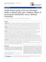
- On the standard plain X-ray film, we measured lordosis angle and range of motion
(ROM) angle based on flexion and extension angle and Cobb method.
Figure 1: Lordosis angle (A) and ROM = (Ɵ ± Ɵ1) + (Ɵ2 – Ɵ) Ɵ: Lordosis angle.
- Measuring the AP diameter of cervical
canal on the computerized tomography at
the pedicle position.
- Taking MRI to determine the level of
stenosis the patients got. We found the
reasons including bulging disc, disc
herniation, yellow ligament hypertrophy,
hyperintensity on T2-weighted or hyporintensity
on T1-weighted of spinal cord.
- Data storage, analysis and processing
by SPSS 16.0 software.
RESULTS AND DISCUSSION
1. Sex and age.
In 31 patients, there were 21 males
(67.7%) and 10 females (32.3%). The
male per female ratio was 2.1/1.
According to the researches of cervical
stenosis disease, the number of male
patients was higher than female ones.
In our study, the male/female ratio was
2.1/1. Compared with Nguyen Van Thach's
study [2], the proportion was similar.
The average of patients was 56.84 ± 8.23
years old (from 38 to 73). Most patients
were in 2 groups of age, from 51 to 60
and from 61 to 70 years old. The number
of 51 to 70 years old patients accounted
for 77.4%. The average age in our study
matched with Phan Quang Son’s one
[1].
Studies indicated that age related
cervical degeneration was more common
in middle age and less common in age
groups under 40 [3]. The average age of
56.8 in the study was consistent with local
and national studies.
233
Journal of military pharmaco-medicine n08-2017
2. Illness duration.
The duration (unit:month) was from
symptoms onset to admission. The shortest
time was 1 month and the longest time
was 96 months. The average illness duration
was 16.19 months. In our study, most of
patients admitted to the hospital within
12 months of illness, accounted for 71%.
The duration of illness in our study was
similar to Phan Quang Son’s one [1]
(p > 0.05) but was shorter than that
of Nguyen Van Thach [2] (p < 0.05).
Long duration of illness affected the results
of surgery.
3. Clinical conditions of hospitalized
patients based on JOA score.
The patient's clinical condition was
evaluated on JOA before surgery with an
average JOA score of 7.65 ± 4.28. The
lowest score was 3 and the highest one
was 13. Most of the patients in the study
had JOA score ≤ 12 (96.8%). The
preoperative JOA score was 7.65. A JOA
score ≤ 7 indicated severe myelopathy
while 8 to 12 points showed medium
myelopathy and 13 was mild myelopathy.
A mild myelopathy was usually treated by
conservative procedure. In the case,
when the JOA score was less than or
equal to 12 [6], surgical treatment was
indicated. In Cheng's study, the JOA
score before surgery was 7.9 ± 2.8.
Duetzmann et al, who conducted a series
of studies on cervical laminoplasty
(n = 4.949) had an average JOA score of
9.91 ± 1.65. JOA score in our study was
not significantly different from Cheng
(p > 0.05), but different from Duetzmann
(p < 0.01).
234
4. Imaging diagnosis.
* Standard X-ray:
In this study, we used Cobb method to
measure and classify lordosis angle as
well as evaluate range of motion (ROM)
of the cervical spine.
With 31 patients, the average lordosis
angle was 22.35 ± 9.03 0 (1 - 35) and
median ROM: 45.26 ± 10.250 (24 - 63).
* Computerized tomography scanner:
23 cases had been taken with
computerized tomography scanner before
surgery. CT-scanner images clearly
showed vertebral body, ossification of
posterior longitudinal ligaments (OPLL),
bone spur, etc. We measured the diameter
of AP of the cervical spinal canal by
computerized tomography.
Table 1: The average diameter of AP
of the cervical spinal canal.
Vertebrae
Diameter (mm)
C3
10.52 ± 1.13
C4
9.78 ± 1.40
C5
9.57 ± 2.05
C6
9.95 ± 1.56
C7
11.63 ± 1.48
n
23
The proportion of patients with AP
cervical spinal canal diameter less than or
equal to 12 mm at C3: 95.7%, C4: 100%,
C5: 95.7%, C6: 100%, C7: 73.9%.
Preoperative CT-scanner not only
accurately measured the AP cervical
canal diameter but also accurately
diagnosed cases of OPLL. According to
Kokubun [4], the AP cervical spinal canal
diameter ≤ 12 mm was called spinal
stenosis. In our study, most of patients
had diameter of AP of the cervical spinal
canal ≤ 12 mm.
Journal of military pharmaco-medicine n08-2017
* Magnetic resonance imaging:
31 patients who took MRI without
gadolinium enhanced on T1-weighted and
T2-weighted on axial and sagittal, had the
characteristics of cervical stenosis such as
yellow ligament hypertrophy, bulging disc,
disc herniation and signal change in the
spinal cord.
Table 2: Number level of stenosis.
Number level of
stenosis
Number of
patients
Ratio
(%)
Three levels
9
29
Four levels
10
32.3
Five levels
12
38.7
Sum stenosis level
127
100%
Compared with Phan Quang Son [2],
we found that 2 studies had the same
results in the percentage of lesions
between four and five levels. When the
lesion was 3 levels, some surgeons
could choose anterior approach such as
vertebra ecorpectomy, discectomy fixation
and bone grafts. However, when spinal
stenosis had 4 or more levels, most surgeons
chose posterior approach.
* Morphology lesions on MRI:
Researching on 31 patients who took
MRI (in which 5 patients took dynamic MRI),
we found that:
Table 3: Morphology lesions on MRI.
Morphology lesions
Number of Ratio
patients
(%)
Bulging disc
31
100
Disc herniation
11
35.5
Yellow ligament hypertrophy
29
93.5
Hypertensive signal on T2W
30
96.8
Hyportensive signal on T1W
4
12.9
According to results of studies,
hyperintensity signal on T2-weighted
image was a recovery prognostic factor.
Groups with hyperintensity on T2-weighted
image showed higher recovery rates than
non - hyperintensity one. In the Secer’s
study (2017), the recovery rate of the
T2-weighted hyperintensity group was
73.5 ± 25.2%. This figure was significantly
higher than that of the control group
without T2-weighted hyperintensity
(37.1 ± 1.68) [7].
For those patients who had marked
clinical symptoms of cervical myelopathy
but the basis of MRI did not clearly show
the cause as well as the location of
compression, the dynamic MRI was a
good choice for clarification diagnosis.
There had been a lot of studies in the
world [5] that showed the diagnostic
efficiency of the method. However, this
issue was rarely mentioned in Vietnam.
CONCLUSION
Studying 31 patients with multi-levels
cervical myelopathy who underwent
cervical laminoplasty by using titanium
mini plate at 108 Central Military Hospital
from February, 2011 to October, 2015,
we draw some conclusions about clinical
features and imaging diagnosis as follows:
- Clinical features: The average age
was 56.8 and the most common age
group was from 51 to 70, accounted for
77.4%. The number of male patients was
higher than females and the ratio of
male/female was 2.1/1. The duration of
illness from onset to admission was 16.1
months. The average JOA score before
operation was 7.65 ± 2.48.
235
Journal of military pharmaco-medicine n08-2017
- Diagnosis imaging: The average
lordosis angle was 22.35 ± 9.030 and the
median ROM angle was 45.26 ± 10.250.
The average diameter of AP of the cervical
spinal canal on CT-Scanner at C3: 10.52 mm;
C4: 9.78 mm; C5: 9.57 mm; C6: 9.95 mm;
C7: 11.63 mm. There were total 127 cervical
levels with stenosis in which 12 patients
had 3 levels stenosis, 10 patients had 4
levels stenosis and 9 patients had 5 levels
stenosis. The rate of spinal cord hyperintensity
on T2-weighted MRI was 96.8%.
REFERENCES
1. han uang Sơn. Nghiên cứu điều trị
bệnh lý hẹp ống sống cổ bằng phương pháp
tạo hình bản sống kết hợp ghép san hô. Luận
án Tiến sỹ Y học. Trường ại học Y Dược
TP. H Chí Minh. 2015.
2. Nguyễn Văn Th ch. ánh giá kết quả
điều trị bệnh lý hẹp ống sống cổ đa tầng bằng
236
phương pháp tạo hình cung sau đường gi a.
Tạp chí Y học Th c hành. 2011, 779 + 780,
tr.577-581.
3. Kelly J.C et al. The natural history and
clinical syndromes of degenerative cervical
spondylosis. Advances in Orthopedics. 2011,
2012.
4. Kokubun S, Sato T. Cervical myelopathy
and its management. Current Orthopaedics.
1981, 12, pp.7-12.
5. Muhle C et al. Dynamic changes of the
spinal canal in patients with cervical spondylosis
at flexion and extension using MRI. Investigative
Radiology. 1998, 33 (8), pp.444-449.
6. Mark S.G. Cervical spinal stenosis.
Handbook of Neurosurgery. Thiem. New York.
2010.
7. Secer H.I et al. Open-door laminoplasty
with preservation of muscle attachments of
C2 and C7 for cervical spondylotic myelopathy:
Retrospective study. Turk Neurosurg. 2015,
p.1.