Evaluation of the critical care period results after isolated mitral valve replacement or simultaneous mitral and aortic valve surgery in patients with pulmonary hypertension
Bạn đang xem bản rút gọn của tài liệu. Xem và tải ngay bản đầy đủ của tài liệu tại đây (110.64 KB, 7 trang )
Journal of military pharmaco-medicine no2-2018
EVALUATION THE CRITICAL CARE PERIOD RESULTS AFTER
ISOLATED MITRAL VALVE REPLACEMENT OR
SIMULTANEOUS MITRAL AND AORTIC VALVE SURGERY IN
PATIENTS WITH PULMONARY HYPERTENSION
Kieu Van Khuong*; Pham Thi Hong Thi**; Nguyen Quoc Kinh***
SUMMARY
Objectives: To assess the early outcome after elective isolated or concomitant mitral valve
replacement (MVR) in patients with pulmonary hypertension (PH). Subjects and methods: The
study included patients with baseline systolic pulmonary artery pressure (PAPs) of at least
35 mmHg measured by echo who underwent elective MVR and/or aortic valve replacement
(AVR). The systemic and pulmonary hemodynamic changes, arterial and mixed venous blood
gas parameters were reported at various time points before and after operation. Preoperative
and postoperative transthoracic echocardiography was performed. Results: Sixty seven patients
(15 males and 52 females), arithmetic mean age was 45.51 ± 10.74 years (min - max: 20 - 68)
were included in the study. The operative mortality rate was 4.5%. The receiver operating
characteristic curves identified PAPs as a good predictor of operative mortality. Postoperatively,
there was a significant reduction in left atrial diameter (LAd). The arithmetic mean PAPs and
pulmonary artery occlusion pressure (PAOP) decreased significantly after cardiopulmonary
bypass (CPB) and persisted throughout the study period. Central venous pressure (CVP)
decreased after CPB time and remained so to PAC removing time point, postoperatively. A
decrease in SvO2 was significant after operation. Conclusion: Proper perioperative care and
anesthetic techniques resulted in the improvement of LAd, PAPs, PAOP, with acceptable
operative mortality in patients with PH who was performed elective isolated MVR or
simultaneous mitral and AVR.
* Keywords: Pulmonary hypertension; Aortic valve replacement; Mitral valve replacement.
INTRODUCTION
All around the world, rheumatic heart
disease remains a major health problem,
although its prevalence in the developed
countries is much reduced. Involvement
of the mitral valve and aortic valve results
in stenosis and/or regurgitation heart
valve diseases. Where surgery is indicated,
MVR is usually necessary [1]. The
development of pulmonary arterial
hypertension (PAH) has been considered
a risk factor for poor outcomes in patients
undergoing MVR and/or AVR [2]. However,
* 103 Military Hospital
** Vietnam National Heart Institute
*** Vietduc Hospital
Corresponding author: Kieu Van Khuong ()
Date received: 10/12/2017
Date accepted: 22/01/2018
154
Journal of military pharmaco-medicine no2-2018
there is no consensus on the outcome of
patients with PAH after MVR in the
literature; some studies have shown that
severe PAH is associated with poorer
outcome and higher mortality rate [3],
whereas others do not agree that severe
PAH implies a higher risk during corrective
surgery [4, 5, 6, 7].
This study was designed to: Assess
the early clinical, hemodynamic, and
echocardiographic changes after elective
isolated MVR or concomitant MVR and
AVR in patients with PH.
SUBJECTS AND METHODS
1. Subjects.
Between April, 2017 and November,
2017, 67 consecutive adult patients with a
baseline PAPs of at least 35 mmHg (as
measured by preinduction transthoracic
echocardiography) who had performed
mitral and/or simultaneous AVR at Heart
Center of Hue Center Hospital. Patients
with coronary artery disease or idiopathic
PAP were excluded from the study.
2. Methods.
All preoperative assessments were
carried out by two-dimensional transthoracic
echocardiography. A pulmonary artery
catheter (PAC) was placed in the pulmonary
artery to measure PAPs and PAOP.
General anesthesia was induced with
fentanyl, 3 - 5 µg/kg. All patients were
operated on through a arithmetic mean
sternotomy on CPB with moderate general
hypothermia (28 - 30°C). We used two
kinds of mechanical prosthesis: ATS valve
and St Jude medical bileaflet mechanical
prosthesis. The hemodynamic and arterial
blood gas parameters were reported at
time points: T0: baseline or pre-induction;
T1: post-intubation; T2: immediate postCPB; T3: at ICU; T4: 6 hour post-ICU; T5:
24 hour post-ICU and Toff: before PAC
removing and the hemodynamics had been
stabilized postoperatively. Hemodynamic
parameters that were recorded included
mean arterial pressure (MAP), PAPs,
PAOP, and central venous pressure
(CVP). All data were expressed as mean
± standard deviation, min - max or
number and percent as appropriate.
The preoperative and postoperative
echocardiographic parameters, and the
hemodynamic and arterial blood gas
parameters obtained at various time
intervals were compared with the baseline
values. The receiver operating characteristic
(ROC) curves were used to estimate the
relationship between sensitivity (proportion
of true positive cases) and 1-specificity
(proportion of false-positive cases) of
PAPs in the prediction of operative
mortality. A p-value of 0.05 or less was
considered significant.
RESULTS
Table 1: Patient’s characteristics.
Variables
Result
Min - max
Age (year)
45.5 ± 10.7
20 - 68
Gender [m/f, (%)]
15/52
(22.4/77.6)
2
Body surface area (m )
1.44 ± 0.11
Body mass index
2
(kg/m )
19.8 ± 2.4
15.4 - 25.2
155
Journal of military pharmaco-medicine no2-2018
Weight (kg)
48.0 ± 6.5
33 - 67
Height (cm)
155.9 ± 7.0
142 - 170
NYHA II (n, %)
21 (31.3)
NYHA III (n, %)
44 (65.7)
NYHA IV (n, %)
2 (3)
Atrial fibrillation (n, %)
31 (46.3)
Cardiothoracic ratio
> 50% (n, %)
40 (59.7)
The study group was mainly female
(77.6%) at arithmetic mean age of 45.51 ±
10.74 years. The patients were classified
as follows: 21 patients (31.3%) in NYHA II
class and 44 patients (65.7%) in NYHA III
class and 2 cases in NYHA class IV.
Table 2: Intraoperative and postoperative
clinical outcome variables.
Variables
Result
Min Max
CPB time (min),
arithmetic mean
114.2 ±
57.7
54 - 466
ACC time (min),
arithmetic mean
79.8 ±
36.8
31 - 185
Tricuspid repair [n (%)]
15
(22.4%)
LAA exclusion [n (%)]
18 (26.9)
Thromboembolic
removing [n (%)]
11 (16.4)
Time of ventilator (h)
Mechanical assist by
IABP [n (%)]
Operative mortality
[n (%)]
156
20.9 ±
31.7
4 (6%)
3 (4.5%)
(ACC: Aortic cross-clamp time; CPB:
Cardiopulmonary bypass time; IABP:
Intra-aortic balloon pump; ICU: Intensive
care unit. LAA: Left atrial appendage)
The arithmetic mean CPB time was
114.18 ± 57.71 mins (range: 54 - 466)
and the arithmetic mean aortic crossclamp time was 79.76 ± 36.78 mins
(range: 31 - 185). Tricuspid repair was
performed in 15 patients (22.39%).
Table 3: Comparison of preoperative
and postoperative echocardiographic
variables.
Variables
T0
T3
Toff
Lad (mm)
51.03 ±
b
8.19
42.13 ±
b
2.30
43.67 ±
b
2.88
LVEDD
(mm)
48.28 ±
b
8.31
45.92 ±
b
5.59
45.45 ±
b
5.19
LVESD
(mm)
34.66 ±
b
7.15
32.61 ±
b
5.40
31.87 ±
a
5.31
EF (%)
53.49 ±
8.02
53.28 ±
7.38
55.21 ±
8.22
(a: Significant difference < 0.0001;
b: Significant difference < 0.05. Abbreviation:
EF: Ejection fraction; Lad: Left atrium
diameter; LVEDD: Left ventricular enddiastolic diameter; LVESD: Left ventricular
end-systolic diameter; CTR: Cardiothoracic
ratio)
There was a significant difference
between LAd, LVEDD, LVESD at points
time of postoperation and when to remove
PAC in comparision with baseline time
results but no significant difference in EF.
Journal of military pharmaco-medicine no2-2018
Table 4: Early hemodynamic parameter changes.
2
CVP (mmHg)
2.43 ± 0.77
a
7.55 ± 4.11
b
94.48 ± 12.41
a
49.15 ± 17.52
a
23.43 ± 9.84
T1
1.66 ± 0.42
a
7.08 ± 3.21
67.66 ± 10.22
a
29.91 ± 11.08
a
16.48 ± 7.54
T2
2.58 ± 0.61
a
8.49 ± 2.80
74.54 ± 14.22
a
32.45 ± 10.31
a
11.40 ± 4.16
T3
3.02 ± 0.80
a
6.21 ± 2.99
b
93.01 ± 13.34
33.13 ± 11.94
a
9.63 ± 5.91
a
T4
2.65 ± 0.55
a
6.10 ± 2.79
b
74.00 ± 11.63
a
32.18 ± 11.89
a
9.91 ± 5.43
a
Toff
3.00 ± 0.69
a
6.21 ± 3.20
b
79.16 ± 10.70
a
31.45 ±1 1.99
a
9.63 ± 5.06
a
Time points
CI (L/min/m )
T0
MAP (mmHg)
PAPs (mmHg)
PAOP (mmHg)
a
a
a
(a: Significant difference < 0.0001; b: Significant difference < 0.05. Abbreviation: CI:
Cardiac output index; CVP: Central venous pressure; MAP: Mean arterial pressure;
POAP: Pulmonary artery occlusion pressure; PAPs: Pulmonary systolic arterial
pressure)
There was a significant decrease in PAPs, PAOP after induction, CPB stop, and this
change persisted throughout at removing PAC time point postoperatively. CVP and
MAP decreased, but it kept in normal range. CI decreased after induction anesthesia
(T1) and increased significantly at T2, T3, Toff time point.
Table 5: Arterial and mixed venous blood gas parameter changes.
Time points
pH
PaCO2 (mmHg)
PaO2 (mmHg)
SaO2 (%)
SvO2 (%)
T0
7.42 ± 0.03
38.24 ± 4.22
210.64 ± 96.08
99.19 ± 1.99
72.57 ± 9.34
T1
7.48 ± 0.06
a
32.48 ± 5.54
a
319.56 ± 80.85
a
99.94 ± 0.37
b
72.37 ± 8.10
T2
7.45 ± 0.07
b
31.53 ± 6.40
a
301.00 ± 82.54
a
99.85 ± 0.76
74.53 ± 8.78
T3
7.40 ± 0.07
b
34.84 ± 8.35
b
175.43 ± 62.35
b
99.05 ± 1.20
69.19 ± 10.55
T4
7.39 ± 0.07
b
36.29 ± 5.88
b
163.98 ± 36.34
b
99.14 ± 0.66
63.34 ± 12.43
a
Toff
7.45 ± 0.07
b
39.46 ± 6.19
104.50 ± 36.74
a
96.86 ± 2.67
a
59.24 ± 11.17
a
a
(a: Significant difference < 0.0001, b: Significant difference < 0.05)
There was a significant difference of blood gas parameters between baseline time
point (T0 time point) with other time points, excepted PaCO2 (Toff time point), PaO2 (T2
time point), SaO2 (T2, T3, T4 time point) and SvO2 (T1, T2, T3 time point).
157
Journal of military pharmaco-medicine no2-2018
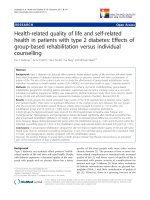
Sensitivity
PAPs
100
80
60
40
20
0
Sensitivity: 66.7
Specificity: 85.9
Criterion : >65
0
40
80
100-Specificity
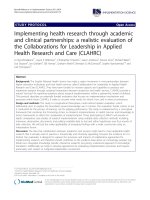
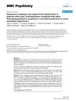
Figure 1: The receiver operating characteristic curve of systolic pulmonary arterial
hypertension as a predictor of operative mortality.
DISCUSSION
There were 67 patients involved in the
study, the lowest age was 20, the highest
was 68, the mean was 45.51 ± 10.74 years.
This finding is consistent with recent studies
in cardiac valve surgery in India and
Vietnam. The study by Nirmal Kumar et al
[7] in severe PH patients soon after MVR
had an arithmetic mean age of 32.1 years,
lower than our result. This difference was
due to the fact that study only included
patients with mitral valve disease with
severe PH (PAPs > 50 mmHg) and this
feature was more common in acute
rheumatoid arthritis in India [10]. Age in
our study was similar to result of Gökhan
Lafçı [4] and two other local authors: Vu
Quynh Nga (44.2 ± 11.5 years), Doan
Duc Hoang (46.69 ± 12.57 years) [1, 2].
The patients were mainly in NYHA III
with 46.3% rhythm dysfunction as atrial
fibrillation (AF) and 59.7% of them
increased cardiothoracic ratio over 50%.
Some intraoperative and postoperative
results showed in table 2. The arithmetic
mean CPB time (114.2 ± 57.7 mins) and
the arithmetic mean ACC time (79.7 ±
158
36.8 mins) was similar to Xiaochun Song’s
study (CPB: 119.9 ± 37.4 mins and ACC:
82.5 ± 31.8 mins) [7]. Our result was
higher than Vu Thuc Phuong’s study
(CPB time of group D, using dobutamin:
95.2 ± 35.1 mins; CPB time of group E,
using epinephrine: 86.5 ± 24.1 mins. ACC
time of group D: 73.5 ± 32.4 mins; ACC
time of group E 67.2 ± 20.8 mins). It may
be due to their patient groups were mainly
replaced one valve surgery (> 60% in both
groups) and no preoperative heart failure.
A comparison of preoperative and
postoperative (T3 time point), removing
PAC time point (Toff) echocardiographic
variables is presented in table 3.
Postoperatively, there was a significant
reduction in LAd, LVEDD, LVESD
(p < 0.05). The increased left atrial (LA)
pressure in mitral valve disease is passively
transmitted to the pulmonary vasculature
and can lead to an increase in pulmonary
vascular resistance (PVR). Some other
factors such as reactive pulmonary
vasoconstriction and organic changes in
pulmonary vasculature are also responsible
for this increase in PVR [11]. Following
mitral valve surgery, LA loading can
Journal of military pharmaco-medicine no2-2018
be adequately decompressed. This
decompression is very influential in the
regression of PH [4]. LA enlargement is a
pathophysiological response to volume
overload resulting from valvular diseases
which is known as LA remodeling, and
has been shown previously to be associated
with cardioembolic events. Following MVR,
the LA may undergo reverse remodeling
characterized by LA volume reduction. LA
size reduces with the return of normal
sinus rhythm and a decrease in the
gradient across the mitral valve.
Our results showed a significant decrease
in PAPs and PAOP after CPB (T2 time
point), and this change persisted to time
point of PAC removing, postoperatively
(table 4). These findings were in agreement
with some other researchers who have
reported hemodynamic changes in patients
with rheumatic mitral valve disease at
different intervals after MVR, with an
immediate reduction in PAPs. In the study
by Kumar [9], the mean PAPs, PAOP, and
pulmonary vascular resistance decreased
significantly soon after CPB in patients
with severe PH. The mean PAPs
approached near-normal values (26 ±
5 mmHg) 6 hours and 24 hours,
postoperatively. The study by Mubeen et
al [12] showed that the mean PAPs
decreased by 38% from a mean
preoperative level of 59.8 to 37.1 mmHg
immediately following MVR. Although it
continued to decrease over the next
24 hour, this further decrease was not
statistically significant. In a recent study
by Bayat et al, PAP in patients with severe
PAH showed no significant reduction
immediately after MVR, but it decreased
significantly below the range of severe
PAP over the first 24 hours after operation.
The present study showed that MVR
could be performed in patients with
rheumatic valvular disease and severe
PH with an acceptable operative mortality
of 10%. The study by Mubeen et al [12]
showed that the operative mortality was
5.5% in patients with subsystemic PAP,
with a mean of 58.1 mmHg and 28.5% in
patients with a suprasystemic PAP of
83.2 mmHg. The operative mortality rate
in our study was 4.5% (table 2). The ROC
curves (figure 1) identified PAPs as a
good predictor of operative mortality (area
under the ROC curve: 0.794; p < 0.05),
and the value greater than 65 mmHg has
the highest specificity (85.9%) and
sensitivity (66.7%) for the risk of operative
mortality in those patients. Similarly, the
recent study by Corciova et al identified
PAPs value greater than 65 mmHg to
have the highest specificity and sensitivity
for the risk of perioperative death in mitral
regurgitation patients (area under the
ROC curve: 0.782; p < 0.001).
SvO2 did not change at T1, T2 time
point (table 5) but it decreased significantly
after operation even when hemodynamic
in stable (at Toff time point). SvO2 can be
used to assess the adequacy of tissue
perfusion and oxygenation. When analyzing
in conjunction with other hemodynamic
parameters, following trends in the SvO2
does offer insight into cardiac performance
and tissue oxygen delivery. In the
postoperative cardiac surgical patient, a
fall in SvO2 generally reflects decreased
oxygen delivery or increased oxygen
extraction by tissues and is suggestive of
a reduction in cardiac output. However,
other constantly changing factors that
affect oxygen supply and demand may
159
Journal of military pharmaco-medicine no2-2018
also influence SvO2 and must be taken
into consideration. These include shivering,
temperature, anemia, alteration in FiO2,
and the efficiency of alveolar gas exchange.
A decresae in SvO2 at T3, T4 time point
(patients in ventilation support) (table 5)
can result from a decrease CO, low
hemoglobin level or an increase in oxygen
consumption. SvO2 decreased at Toff
may be relative with a decrease in FiO2
(in room air), fever or anemia. It is very
important to take care the patients in this
stage because SvO2 reduction under
threshold leads to the danger of organ
dysfunction that is reason why they come
back to intensive care unit ward.
CONCLUSION
Isolated MVR or concomitant MVR and
AVR was safe and effective even in
patients with PH, with acceptable operative
mortality and a significant improvement in
left atrial diameter, pulmonary hemodynamics
(PAPs, PAOP), but a decrease in mixed
venous saturation early after operation.
The anesthetic technique and perioperative
care can be useful in improving the
outcome in such patients.
4. Lafci G, A.I Diken, H.S Gedik et al.
Alterations in pulmonary artery pressure
following mitral valve replacement. Turk
Kardiyol Dern Ars. 2012, 40 (3), pp.235-241.
5. Svein Simonsen K.F. Anders Andersen
and Leif Efskind Hospital mortality after mitral
valve replacement. 1974.
6. Parvathy U.T, R. Rajan, A.G. Faybushevich.
Reversal of abnormal cardiac parameters
following mitral valve replacement for severe
mitral stenosis in relation to pulmonary artery
pressure: A retrospective study of noninvasive
parameters - early and late pattern. Interv
Med Appl Sci. 2016, 8 (2), pp.49-59.
7. Song X.C, Zhang X. Chen et al. An
excellent result of surgical treatment in
patients with severe pulmonary arterial
hypertension following mitral valve disease. J
Cardiothorac Surg. 2015, 10, p.70.
8. Thunberg C.A, B.D Gaitan, A. Grewal
et al. Pulmonary hypertension in patients
undergoing cardiac surgery: pathophysiology,
perioperative management and outcomes. J.
Cardiothorac Vasc Anesth. 2013, 27 (3),
pp.551-572.
REFERENCE
8. Kumar N.P. Sevta S, Satyarthy et al.
Early results of mitral valve replacement in
severe pulmonary artery hypertension - An
institutional prospective study. World Journal
of Cardiovascular Surgery. 2013, 3 (2), pp.63-69.
1. Hoằng Đ.Đ. Nghiên cứu vai trò của chỉ
số SvO2 trong hồi sức huyết động ở bệnh
nhân phẫu thuật tim có nguy cơ cao. 2017.
10. Padmavati S. Present status of rheumatic
fever and rheumatic heart disease in India.
Indian Heart J. 1995, 47 (4), pp.395-398.
2. Nga V.Q. Nghiên cứu một số thông số
huyết động và chức năng tim bằng siêu âm
Doppler ở bệnh nhân phẫu thuật thay van hai
lá Sorin Bicarbon. 2010.
11. Ott B. Valvular heart disease a companion
to Braunwald's heart disease expert consult
third edition. 2009.
3. Zakkar M, E. Amirak, K.M. Chan et al.
Rheumatic mitral valve disease: current surgical
status. Prog Cardiovasc Dis. 2009, 51 (6),
pp.478-481.
160
12. Mohammad Mubeen, Amrendra K
Singh, Surendra K Agarwal, Jeewan Pillai,
Shalini Kapoor, Ashok K Srivastava. Mitral
valve replacement in severe pulmonary
arterial hypertension. 2008.