Ebook Oxford handbook of neurology (2th edition): Part 2
Bạn đang xem bản rút gọn của tài liệu. Xem và tải ngay bản đầy đủ của tài liệu tại đây (29.71 MB, 234 trang )
Chapter 6
Neurology in medicine
Neurological symptoms: cardiac disease 396
Neurological disease: cardiac pathology 400
Neurological features of respiratory disease 402
Respiratory failure in neurology 404
Neurological disorders: gastroenterological symptoms 42
Gastroenterological disorders: neurological presentations 46
Neurology and renal medicine 420
Hereditary disorders of the nervous system and the kidneys 422
Neurology and haematological disorders 424
Neurology and connective tissue disorders and vasculitides 428
Endocrine neuroanatomy 432
Neurology in endocrine disorders 436
Primary pituitary disorders 440
The neuroendocrine syndromes 442
Dermatology in neurology 444
Inherited neurocutaneous syndromes 452
Neurological and neurosurgical issues in pregnancy 456
395
396
CHAPTER 6
Neurology in medicine
Neurological symptoms:
cardiac disease
Cardiac arrhythmias
• Risk factor for strokes (e.g. AF).
• Paroxysmal arrhythmias should be considered as cause for syncope,
collapse, seizures—review ECG, consider 24-hour ECG.
• See Table 6. for ECG changes in neurological disease.
Long QT syndrome:
• Potential cause of avoidable sudden cardiac death from ventricular
arrhythmias.
• Autosomal dominant with variable expression.
•Prevalence: /2000–3000.
• In one series, 39% with long QT were most misdiagnosed as epilepsy.
• Clinical features:
• cerebral hypoperfusion can manifest as myoclonic jerks or epileptic
type movements;
• vasovagal attacks;
• exertional syncope (long QT);
• syncope during emotional stress (long QT2);
• sudden syncope at rest (long QT3);
• rapid recovery;
• otherwise normal examination;
• may have family history of unexplained sudden death.
• Numerous drug triggers:
• antiarrhythmics, e.g. amiodarone, sotalol;
• antibiotics, e.g. erythromycin, clarithromycin;
• antihistamines, e.g. ondansetron;
• antidepressants, e.g. amitriptyline, fluoxetine;
• antipsychotics, e.g. quetiapine, haloperidol.
• Syncope with long QT on an ECG (> 450 ms in males, > 460 ms
in females) in the absence of causative medications or disorders, is
suggestive of the diagnosis.
• Automated measurement is frequently inaccurate.
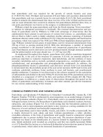
• Manual measurement of QTc using the tangent technique (see Fig. 6.).
Endocarditis
• Infectious endocarditis, non-bacterial thrombotic endocarditis, Libman–
Sacks endocarditis in SLE.
• All are associated with embolic phenomena causing stroke.
Infectious endocarditis
• Neurological complications relatively common.
• Focal:
• embolism (from valvular vegetations), haemorrhage secondary to
rupture of infective arteritis, mycotic aneurysm, cerebral abscess.
•Diffuse:
• multiple microemboli, DIC, multi-organ failure.
Neurological symptoms: cardiac disease
RR INTERVAL
Tangent
T
P
Lead II or V5
QRS
Baseline
QT
INTERVAL
QTC = QT/√RRseconds
Fig. 6. The tangent technique for calculating corrected QT interval. Adapted
from Postema PG, de Jong JSSG, van der Bilt IAC, Wilde AAM (2008). Accurate
electrocardiographic assessment of the QT interval: teach the tangent. Heart Rhythm,
5, 05–8, with permission.
Non-bacterial thrombotic endocarditis
•Occurs in hypercoagulable states, e.g. advanced malignancies and AIDs.
• Similar presentation to infective endocarditis; haemorrhage less likely.
Cardiac surgery
• Neurological complications either focal or global.
• In order of frequency, the following neurological events are found after
cardiac surgery (incidence):
• persistent cognitive deficits (20–40%);
• reversible encephalopathy (3–2%);
• peripheral neuropathies—brachial plexus (.5–24%), phrenic
(0–60%), recurrent laryngeal, sciatic, femoral, saphenous,
sympathetic chain;
• stroke (3–6%);
• seizures (especially in children) (3%).
• Several proposed mechanisms including embolic (fat, air), cerebral
hypoperfusion, metabolic abnormalities, impaired cerebral
autoregulation, or systemic inflammatory response.
• Identified risk factors: cross-clamping time, type of surgery (lowest
risk in aortic valve replacement), open heart surgery, comorbidities,
atherosclerotic ascending aorta disease.
397
398
Rate & rhythm
Axis
PR duration
QTc
QRS
duration
ST segment
T wave
Notes
Subarachnoid
haemorrhage
Ventricular/atrial
arrhythmias
Varied
Varied
i
Varied
i/d
Inverted
Common within 48 h of onset,
lasts up to 6 weeks
Prominent U waves
Most arrhythmias have been
described in SAH
Friedreich’s
ataxia
N
R or L
(R > L)
N
N
May be i
N
Inversion
Hypokinetic dilated
cardiomyopathy
Often inferolateral T-wave
inversion
Muscular
dystrophies
Onset of
arrhythmias with
heart block (AF/
junctional)
N/L
N/i
N
May be i
i/d
Flattened and Dominant R wave V, V2
inverted
Pseudoinfarction
Anterolateral Q waves
Heart block
Cardiomyopathy
Myotonic
dystrophy type I
N*
N
N/i
N
N
N
N
*Develops heart block severity
linked to CTG repeat length
Channelopathies: periodic paralysis
Hypokalaemic
N/d
N
i
N
N
N/d
Flattened
U waves
Hyperkalaemic
N/ectopic
N
N/d
N/d
N/i
N
Tall/tented
Potassium rarely in cardiotoxic range
Neurology in medicine
Condition
CHAPTER 6
Table 6. ECG changes in neurological disease
Anderson’s
syndrome
Often bigeminy
See note
N/absent
i
N/i
N
N*
*High incidence of ventricular
arrythmias (torsade/VT/VF)
Bidirectional VT (axis switches each
beat) unique to subset of patients
Metabolic disorders
N
N
N/d
N
N
N
N
Pre-excitation LVH
Danon disease
(LAMP-2
mutation)
N
N
N/d
N
N
N
N
Pre-excitation WPW due to
myocardial hypertrophy
Lafora disease
N*
N
N/i
N
N
N
N
May develop heart block
Mitochondrial cytopathies
Kearns–Sayre
syndrome
N*
N/L
i
N
N/i
N
Normal/
inverted
*Complete heart block may
develop after ophthalmoplegia
Ocular
myopathy
N
N/R
N
N
N
N/d
Normal/
inverted
Cardiac involvement rare
MERFF
N
N/L
N
N
N
N/d
N
LVH
MELAS
N
N
N/d
N
N
N/d
Normal/
inverted
WPW LVH
LHON
N
N
N/d
N/i
N
N/d
Normal/
inverted
Pre-excitation syndromes (WPW/
LGL)
LVH (hypertrophic)
N, normal; L, left; R, right; LVH, left ventricular hypertrophy; WPW, Wolff–Parkinson–White syndrome; LGL, Lown–Ganong–Levine syndrome; VT, ventricular tachycardia.
Neurological symptoms: cardiac disease
Fabry’s disease
399
400
CHAPTER 6
Neurology in medicine
Neurological disease: cardiac
pathology
Cushing’s response
Bradycardia and hypertension in response to i
i intracranial pressure.
See Table 6.2 for neurological diseases associated with cardiac pathology.
Subarachnoid haemorrhage
• ECG abnormalities seen in 80–90% (see Table 6.); more frequently in
severe neurological impairment.
•Onset within 48 hours; may last up to 6 weeks.
• Should not delay surgery unless malignant arrhythmia present or
probability of infarction is very high.
Muscular dystrophy
• The muscular dystrophies often have cardiac muscle involvement and
can be divided into two groups:
Muscular dystrophy with prominent cardiomyopathy
Becker’s muscular dystrophy
• Dilated cardiomyopathy in males aged 20–40 years; later in female
carriers.
• May be the first sign in subclinical cases.
Duchenne’s muscular dystrophy
•Occurs late in disease; may be under-recognized as patients are less
active.
•Mortality from dilated cardiomyopathy 0–5%.
• Serum atrial natriuretic peptide useful for detecting cardiomyopathy and
instituting early treatment with beta-blockers plus an ACE inhibitor.
Muscular dystrophy with prominent cardiac conduction disturbance
• Early detection of conduction defects: heart block, AV standstill, severe
bradycardia, AF.
•May not be symptomatic and is associated with sudden death.
Pacemakers may be life-saving.
• Late-onset dilated cardiomyopathy.
Emery–Dreifuss muscular dystrophy
• Cardiac involvement at any age; may be present at onset.
• Nomal myocardium is replaced by fibro-adipose tissue.
• Inability to pace the atrial paralysis is pathognomonic.
Limb girdle muscular dystrophy
• Neuromuscular symptoms precede cardiovascular symptoms.
Neurological disease: cardiac pathology
Table 6.2 Neurological diseases associated with cardiac pathology
Cardiac pathology
Neurological condition
Long QT syndrome
Periodic paralysis
Cardiac arrhythmia
Epilepsy
MELAS (WPW)
GBS
Conduction block
Kearns–Sayre syndrome
Lafora disease
Ventricular hypertrophy
MELAS (symmetrical)
MERRF (asymmetrical)
Cardiomyopathy
Muscular dystrophies (dilated)
Friedreich’s ataxia (dilated)
Glycogen storage disease (dilated/hypertrophic/
restrictive)
MERRF, MELAS (dilated/hypertrophic)
Epilepsy
• Ictal tachycardia: the most common finding (82%).
• Ictal bradycardia: occurs in 3–4% and can cause syncope. Male > female.
Most common in temporal lobe seizures.
• Ictal asystole: rare but important finding (temporal and frontal seizures).
Possible increased risk of SUDEP; pacemaker should be considered if
duration of asystole > 4 seconds.
Guillain–Barré syndrome
• Autonomic dysfunction manifesting as arrhythmias.
•Often tachycardia, though life-threatening brady- and tachyarrhythmias
can occur, usually in ventilated patients.
Mitochondrial cytopathies
• Patients with Kearns–Sayre syndrome should be evaluated and
monitored for AV conduction disturbances.
• Heart block develops after the ophthalmoplegia. Permanent pacemaker
improves survival.
• Patients with MERRF and MELAS should be followed for cardiac
hypertrophy and dilated cardiomyopathy.
401
402
CHAPTER 6
Neurology in medicine
Neurological features of respiratory
disease
Often arise due to changes in PaO2 and PaCO2, or as a side effect of
medication.
Hypoxia (PaO2 < 0 kPa)
•Symptoms:
• cognitive dysfunction;
• confusion;
• amnesia;
• behavioural change/aggression;
• hallucinations;
• gait disturbance.
Physical signs may include petechial retinal haemorrhages and saccade
disruption (seen in altitude sickness).
Hypercapnoea (PaCO2 > 6 kPa)
•Symptoms:
• drowsiness/fatigue;
• headaches (especially in morning);
• episodic confusion;
• excessive daytime somnolence;
• impotence;
• poor concentration.
• Symptoms depend on the rate of CO2 rise, and can differ between
acute and chronic.
• Can arise insidiously in patients with neuromuscular weakness, and so
symptoms should be actively sought.
• Neurological signs include tremor (on outstretched hands),
papilloedema (which can cause blindness), d GCS, and seizures.
• Can also precipitate d GCS and seizures by over-hyperventilating
hypercapnoeic patients.
Hypocapnoea (PaCO2 < 4.5 kPa)
•Symptoms:
• light-headedness;
• breathlessness;
• ‘tingling’ sensations;
• headache;
• palpitations;
• tetany;
• tinnitis;
• chest pain;
• tremor;
• visual blurring;
• transient LOC;
• unsteadiness.
Neurological features of respiratory disease
403
404
CHAPTER 6
Neurology in medicine
Respiratory failure in neurology
See Table 6.3 for differential diagnosis and Table 6.4 for central disorders of
ventilatory control. See Fig. 6.2 for examples of central respiratory patterns.
Acute respiratory failure
•May be acute presentation or decompensation of a chronic condition.
• In neurological patients consider common causes first, e.g. chest
infection, PE, or exacerbation of pre-existing respiratory illness.
• Clues in the background history, tempo of onset, clinical signs, and
arterial blood gas (ABG).
• Review medications, especially for opiates, benzodiazepines, and
anticholinergics (look for signs of overdose).
• In an acute setting, resuscitate using the ABC approach (Box 6.).
Chronic respiratory failure
• Often identified by symptomatic hypercapnoea.
• Additional symptoms may include dyspnoea on immersion in water or
on lying flat (indicating diaphragm paralysis).
• Paradoxical (inward) inspiratory movement of abdominal muscles
indicates a 70% reduction in normal respiratory muscle strength.
• Lying and standing FVC should also be documented as it is often
diagnostic.
• Initial non-invasive specialist tests to investigate suspected respiratory
muscle weakness include maximal sniff nasal pressure, magnetic/electric
phrenic nerve stimulation, and mouth pressure during phrenic nerve
stimulation.
• Formal specialist tests required when respiratory muscle weakness
cannot be confirmed or refuted on non-invasive tests or if precise
sequential measurements are required. These require the placement of
oesophageal and gastric balloons.
• Further tests are possible in specific circumstances, e.g. suspected
hemi-diaphragm disease.
• Polysomnography is indicated if the patient has:
• proven severe weakness but denies hypercapnoea symptoms;
• sleep symptoms without demonstrable severe respiratory muscle
weakness;
• sleep symptoms but fails to meet the British Thoracic Society
desaturation criteria for obstructive sleep apnoea.
Respiratory failure: differential diagnosis
The differential diagnosis is shown in Table 6.3.
Central disorders of ventilatory control
Central disorders of ventilatory control are listed in Table 6.4.
Respiratory failure in neurology
Box 6. ABC approach
• Secure airway.
• Give high flow oxygen in first instance regardless of cause; adjust
according to ABG and clinical response.
• Secure IV access and send bloods for U&Es, bone profile, Mg, CRP,
FBC, clotting ± blood cultures.
• ABG (document FiO2).
•Obtain CXR and ECG.
• Document FVC (if possible).
• Get early anaesthetic support and transfer to ITU if:
• falling GCS or GCS < 8 (unable to support airway);
• not responding to initial therapy;
• becoming progressively exhausted or increasingly shocked despite
treatment;
• FVC < 5 mL/kg (< L).
405
406
CHAPTER 6
Neurology in medicine
Table 6.3 Differential diagnosis for respiratory failure in the neurological
patient
Neuromuscular
junction pathology
Muscular
disorder
Peripheral
neuropathy
Central causes
Anticholinesterase
overdoseA
HypokalaemiaA
GBS
(demyelinating &
axonal)A
Toxic: alcohol,
opiates,
barbiturates,
benzodiazepinesA
Acute
organophosphate
poisoningA
Periodic paralysisA
GBS mimics
e.g. HIV,
Lyme disease,
sarcoidosis,
CMVA
Pontomedullary
SOLA:
haemorrhage,
AVM, tumour,
syrinx, etc.
BotulismA
HypophosphataemiaA
CIDPC
Brachial
neuritisA/C
Transtentorial
herniationA
Myasthenia
gravisA
Acute
rhabdomyolysisA
Critical illness
polyneuropathyC
Bilateral tegmental
medullary infarctsA
HypermagnesaemiaA
Polymyositis
Toxins: organophosphates,
thallium, arsenic,
lead, gold,
lithiumA/C
EncephalitisA
Snake, spider,
scorpion biteA
Thyroid diseaseA/C
Drugs:
vincristineC
Cord lesions C3–5
or higher. Either
intrinsic (e.g.
MS, transverse
myelitis) or
compressive (e.g.
trauma, disc)A
Fish, shellfish, crab
poisoningA
Combined
neuromuscular
blockade and
steroidsA/C
LymphomaA
Infections,
e.g. polio,
tetanus, rabies,
leptospirosisA
Tick paralysisA
Barium
intoxicationA
Vasculitis–SLEA
Post-polio
syndromeC
Antibiotic-induced
paralysisA
Myotonic
dystrophyA/C
Metabolic–acute
intermittent
porphyriaA
Respiratory failure in neurology
Table 6.3 (Contd.)
Neuromuscular
junction pathology
Muscular
disorder
Peripheral
neuropathy
Lambert–Eaton
syndromeC
Limb–girdle
syndromesC
Hereditary
tyrosinaemiaC
Mitochondrial
myopathyC
DiphtheriaA
Central causes
Inflammatory
myopathyC
Hereditary
myopathyC
Acid maltase
deficiency
Carnitine palmityl
transferase
deficiency
Superscript indicates usual mode of initial presentation: A, acute (history usually minutes–hours);
C, chronic (history longer than several days/detected incidentally). Note that all chronic causes
can present as acute on chronic.
407
408
CHAPTER 6
Neurology in medicine
Table 6.4 Central disorders of ventilatory control
Disorder
Mechanism
Notes
Cheyne–Stokes
respiration
Due to an instability
in normal feedback
control for respiration
Slow oscillation crescendo–
decrescendo hyperventilation
followed by apnoea
May occur due to several
factors that cause widespread
cortical dysfunction (i ICP
& metabolic disturbances)
which results in i oscillations
in blood gas levels
Causes include heart failure,
sleep apnoea, stroke, uraemia
Short-cycle
periodic breathing
ICP, lower pontine
lesions, and expanding
posterior fossa lesions
Similar to Cheyne–Stokes but
much faster oscillations (~ 2:4
ratio of hypo- to hyperventilation)
Central
neurogenic
hyper-ventilation
Infiltrative central
tegmental pontine
lesion causing
stimulation of pontine
respiratory group
Definition: hyperventilation
that persists during sleep with
respiratory alkalosis and absence
of another organic cause
RR: 40–70 breaths/min
Common lesions: lymphoma,
astrocytoma
If lesion is treatable
(very rare), recovery is
possible (e.g. lymphatoid
granulomatosis)
Morphine used for palliative
symptomatic treatment
Cluster breathing
Lower pontine
tegmental lesion
Rapidly alternating
hyperventilation followed by
apnoeic episodes of variable
length
Apneustic
breathing
Lesion at dorsolateral
lower half of pons
Prolonged inspiratory gasp with
pause at full inspiration
Ataxic breathing
(‘Biot’s breathing’)
Medullary insult
(e.g. poliomyelitis)
Irregular pattern and amplitude
Combined with bilateral
CN VI is warning sign
of impending brainstem
compression (posterior fossa
lesion)
Respiratory failure in neurology
Table 6.4 (Contd.)
Disorder
Mechanism
Notes
Ondine’s curse
Removal of chemical
control of breathing
with preservation
of voluntary control
Lower medullary lesion
Classic syndrome: in children
associated with Hirschsprung’s
disease and GORD; in adults often
secondary to trauma
High risk of nocturnal sudden
death; therefore nocturnal
respiratory support required
Selected patients benefit from
diaphragmatic pacing
Transtentorial
herniation
Progressive
compromise of
respiratory nuclei with
breakdown of normal
regulatory mechanisms
Cheyne–Stokes
d
Central neurogenic hyperventilation
d
Eupnoea (quiet breathing)
d
Irregular gasping (pre-terminal)
Brainstem/high
cervical spine
injury
C3–5 and above
Loss of voluntary and
involuntary control of
respiratory muscles
Ventilatory support via
tracheostomy
Bilateral diaphragmatic
pacing may be of benefit if
available; only indicated if
phrenic nerves are functioning
but corticodiaphragmatic
is interrupted on cervical
magnetic stimulation
Kussmaul
breathing
Metabolic acidosis
Deep regular respiration
GORD, gastro-oesophageal reflux disease.
409
410
CHAPTER 6
Neurology in medicine
Cheyne-Stokes
Central
Neurogenic
Hyperventilation
Cluster
Apneustic
Ataxic
Fig. 6.2 Examples of central respiratory patterns. Graphs represent respiratory
pattern (y-axis, tidal volume; x-axis, time). Diagrams indicate the location of the
characteristic lesion.
Respiratory failure in neurology
411
412
CHAPTER 6
Neurology in medicine
Neurological disorders:
gastroenterological symptoms
See Table 6.5 for differential diagnosis.
Dysphagia
• Neurogenic dysphagia suggested by:
• drooling of saliva;
• coughing/choking during swallowing;
• nasal regurgitation.
•May arise from dysfunction at any point in the swallowing pathway.
Clues in the history of onset and associated symptoms.
• Assessment should be made with speech and language therapy
assessment and videofluoroscopy.
Gastrointestinal motility disorders
Defects in innervation
Achalasia
• Absence of peristalsis with failure of relaxation of the lower
oesophageal sphincter (LOS).
• Caused by degeneration of oesophageal myenteric nerves and loss of
inhibitory ganglion cells leading to unopposed contraction of the LOS.
• Presents with progressive dysphagia to fluids and solids with
regurgitation of undigested food.
• Primary: e.g. triple A syndrome (achalasia, Addison’s disease, and alacrima).
• Secondary: uncommon—Chagas’ disease, oesophageal cancer, diabetes.
• Diagnosis with oesophageal manometry.
Hirschprung’s disease
• Caused by loss of parasympathetic ganglion cells (aganglionosis) from the
myenteric and submucosal plexus due to failure of neural migration.
• Always involves the anus and continues proximally for a variable
distance.
• Presents soon after birth with constipation and abdominal distension.
• Affected segment is contracted, with colonic dilatation proximally.
• Diagnosis: full-thickness rectal biopsy taken .5 cm above dentate line
showing aganglionosis with associated increase in acetylcholinesterase
staining.
Gastroparesis
• Caused by dysfunction of both the parasympathetic and sympathetic
supply of the stomach leading to delayed gastric emptying.
• Presents with nausea, vomiting, reflux, early satiety, abdominal pain, and
distension.
•Most common cause: diabetes mellitus.
•Other causes: post-gastric surgery, Parkinson’s disease, intestinal
pseudo-obstruction, collagen vascular disorders (SLE, scleroderma), stiff
man syndrome, CMT, paraneoplastic syndrome.
• Diagnosis: gastric emptying study.
• Treatment: majority respond to prokinetics, e.g. metoclopramide.
• Laparoscopic gastric pacing available for highly treatment-resistant cases.
Neurological disorders: gastroenterological symptoms
Table 6.5 Differential diagnosis for lesion of the swallowing pathway
Pathway
component
Differential diagnosis
Afferent
Recurrent laryngeal nerve palsy: tumour (neck, lung,
mediastinum), surgical trauma (thyroidectomy), aortic
aneurysm
Palatal hypo-aesthesia in Arnold–Chiari type I
Brainstem nuclei
MS, lateral medullary syndrome, Arnold–Chiari type I
Cortical
Stroke (common), MS, PD, PSP, HD, Wilson’s disease
Efferent
(including NMJ)
GBS, ALS, myasthenia gravis, recurrent laryngeal nerve palsy
Muscle
Polymyositis, dermatomyositis, myotonic dystrophy, muscular
dystrophy
Pseudo-obstruction
• Presentation of the signs, symptoms, and radiological appearance of
bowel obstruction but with no evidence of an obstructive cause.
• Acute or chronic (rarer, but likely to have neurological aetiology).
• Caused by an imbalance in the autonomic innervation leading to
increased sympathetic tone causing inhibition of colonic motility.
• Colon may become massively distended with risk of perforation.
• Broad neurological differential diagnosis, including:
• Myopathic:
– NMJ disorder: myasthenia gravis;
– myopathy: myositis, myotonic dystrophy, muscular dystrophy.
• Neuropathic:
–Metabolic: diabetes, porphyria;
– Infiltrative: systemic sclerosis, amyloidosis;
– Infection: Chagas’ disease, CMV infection;
– Drugs: anticholinergics, opiates, tricyclic antidepressants,
vincristine;
– Spinal cord injury (trauma, intrinsic/extrinsic lesion, recent
surgery);
– Paraneoplastic (anti-Hu in small cell lung cancer);
– Primary autonomic failure.
• Mitochondrial:
–MNGIE syndrome (mitochondrial neurogastrointestinal
encephalomyopathy syndrome).
• Rule out common causes (electrolyte or thyroid abnormalities, sepsis).
• Conservative management initially (NBM and NG decompression).
• Neostigmine can be used if no response to conservative treatment
• Endoscopic/surgical input if failing conservative/i risk of perforation.
413
414
CHAPTER 6
Neurology in medicine
Examples of neurological associations with common
symptoms
Constipation
Parkinson’s disease (20 year prodrome before clinical presentation), MS,
spinal cord injury, autonomic neuropathy, pseudo-obstruction.
Diarrhoea
Autonomic neuropathy, coeliac disease, Whipple’s disease.
Faecal incontinence
Spinal cord lesions, Parkinson’s disease, MSA, MS, spinal cord injury, autonomic neuropathy, pudendal nerve injury.
Neurological disorders: gastroenterological symptoms
415
416
CHAPTER 6
Neurology in medicine
Gastroenterological disorders:
neurological presentations
Nutritional deficiency syndromes
Water-soluble vitamins
B (thiamine)
•Often associated with alcohol abuse, but may also emerge in the context
of peptic ulcer disease, acute pancreatitis, gastric/oesophageal cancer,
anorexia nervosa, bariatric surgery, hyperemesis gravidarum, or starvation.
• Treatment initially with IV thiamine (50–00 mg IV/IM) before any
IV glucose solutions as this can worsen symptoms. Maintain PO.
• Wernicke’s encephalopathy:
• triad of ocular abnormalities, ataxia (usually gait), and encephalopathy.
• Ocular findings: ophthalmoplegia (horizontal gaze palsy or gaze
paresis), nystagmus (horizontal > vertical); less frequently anisocoria,
sluggish pupils, ptosis.
• Other symptoms: vestibular dysfunction, peripheral neuropathy
(d proprioception, foot drop), hypothermia, hypotension, coma.
• Korsakoff syndrome:
• disproportionate impairment in memory relative to other cognitive
deficits in an otherwise alert and responsive patient;
• often follows Wernicke’s encephalopathy (Wernicke–Korsakoff );
• extensive retrograde amnesia, antegrade amnesia (less),
confabulation.
•Beri-beri:
• ‘wet’ beri-beri—cardiac involvement (high output failure);
• ‘dry’ beri-beri—CNS involvement (often Wernicke’s ± Korsakoff ’s);
additionally brisk reflexes, polyneuritis (lower limbs > upper limbs),
weakness, pain, paralysis, and seizures.
• cardiac and CNS involvement may present together.
B3 (nicotinamide):
Pellagra (see b ‘Dermatology in neurology/Pellagra’, pp. 444–50): triad of
dementia, diarrhoea, and dermatitis.
B6 (pyridoxine):
• Very rare; usually a consequence of a genetic enzymatic defect
preventing pyridoxine conversion in the liver.
• Seizures, muscle cramps, paraesthesiae.
B2 (cobalamin):
• Requires intrinsic factor (IF) for absorption. Due to inadequate intake
(e.g. vegetarians), IF deficiency, nitrous oxide abuse, malnutrition,
resection of stomach or terminal ileum, terminal ileum disease.
•Often presents with symmetric paraesthesiae (feet > hands) and gait
ataxia.
• Sensory: dorsal column involvement (d vibration, d proprioception).
•Motor weakness usually lower limb (axonal neuropathy); often
hyporeflexic (though may be hyper-reflexic), Lhermitte’s sign.
GASTROENTEROLOGICAL DISORDERS
• Autonomic features may also be present.
• Neuropsychiatric features include impaired memory, personality change,
hypomania, psychosis, hallucinations, and emotional lability.
• Visual changes: cecocentric scotoma, optic atrophy.
•MRI may show T2 and FLAIR white matter hyperintensities.
•Measure serum B2: normal value does not exclude deficiency. Measure
serum MMA (methylmalonic acid) and homocysteine which are both
elevated in B2 deficiency, if normal effectively excludes deficiency.
•B2: replacement. Replace before starting any folate supplements (to
avoid precipitating subacute combined degeneration of the cord). Treat
underlying aetiology.
• Without neurological involvement: hydroxocobalamin mg
3x week, for 2 weeks, then every 3 months.
• With neurological involvement: hydroxocobalamin mg alternate
days until no improvement then 2-monthly.
Fat-soluble vitamins
Can arise from fat malabsorption states such as liver disease, biliary
obstruction, pancreatitis, cystic fibrosis, abetalipoproteinaemia (vitamin E),
small bowel resection.
Vitamin D:
• Proximal myopathy.
Vitamin E (tocopherol):
• Ataxia, dysarthria, d vibration and proprioception, d deep
tendon reflexes, Babinski sign positive, pes cavus, kyphoscoliosis,
cardiomyopathy.
• Presentation similar to Friedreich’s ataxia.
Other intestinal neurological disorders
Coeliac disease
Neurological features may develop independent of nutritional deficiency
(e.g. B2—Ramsay Hunt syndrome).
• Cerebellar ataxia.
• Peripheral neuropathy.
•Dementia.
•Myoclonus.
•Seizures.
Imaging may show cerebellar atrophy or bilateral cortical calcification.
Inflammatory bowel disease (IBD)
• i Thromboembolic disease in both Crohn’s disease and ulcerative
colitis.
•Myositis seen more commonly in Crohn’s disease.
• Neuropathy (more common in ulcerative colitis):
• Crohn’s disease—sensory axonal polyneuropathy;
• ulcerative colitis—AIDP or CIDP.
417
418
CHAPTER 6
Neurology in medicine
Whipple’s disease
• Caused by Gram-positive actinomycete Tropheryma whippelii.
• Begins as polyarthralgia, chronic diarrhoea, abdominal pain, and
weight loss.
• CNS: rhythmic myoclonus, dementia, ophthalmoplegia, neuropsychiatric
changes, hypothalamic disturbance (e.g. insomnia, hyperphagia), ataxia.
•Oculomasticatory myorhythmia (pendular convergence nystagmus with
palatal, tongue, and mandibular movements) and/or oculofacialskeletal
myorhythmia (plus limb involvement) and vertical supranuclear
ophthalmoplegia are pathognomic. (NB: Movements persist during
sleep.)
• CSF: inflammatory (± PAS-containing macrophage). PCR for
T. whippelii.
• May require gut biopsy to demonstrate PAS-positive macrophages.
• Treatment: chloramphenicol and co-trimoxazole for –2 years.
GASTROENTEROLOGICAL DISORDERS
419