Ebook Challenging concepts in cardiovascular medicine - A case based approach with expert commentary: Part 2
Bạn đang xem bản rút gọn của tài liệu. Xem và tải ngay bản đầy đủ của tài liệu tại đây (4.31 MB, 96 trang )
16
Paroxysmal atrial fibrillation
Shouvik Haldar
Expert commentary Professor John Camm
Case history
A 55-year-old man was referred to cardiology outpatients by his general practitioner
(GP) with a 2-month history of intermittent palpitations. He was taking ramipril for
hypertension and had no other relevant medical history. He drank 30 units of alcohol
per week and was a lifelong non-smoker. There was no significant family history.
He described four recent episodes of self-terminating palpitations. They were of
sudden onset, occurring both at rest and during mild exertion, and had each lasted
between 15 and 60 minutes. The first episode had occurred after he had returned from
a party, having consumed a significant amount of alcohol. The others had occurred
whilst at work. On each occasion, he had felt his heart pounding fast and chaotically
and during the more prolonged attacks, he had felt dizzy and breathless. Clinical
examination revealed a regular pulse of 75 beats per minute (bpm) with a blood
pressure (BP) of 145/80 mmHg. He had normal heart sounds with no signs of cardiac
failure.
His 12-lead electrocardiogram (ECG) confirmed a normal sinus rhythm with a normal electrical axis. Transthoracic echocardiography (TTE) confirmed a normal cardiac
structure and function with a mildly dilated left atrial size of 40 mm (normal 27–38
mm). Exercise stress testing did not induce any arrhythmias and was negative for
ischaemia. Routine blood tests, including thyroid function, were normal.
At this stage, there was a high clinical suspicion of paroxysmal atrial fibrillation
(PAF). However, in the absence of ECG evidence to confirm this diagnosis, treatment
was not commenced. He was advised to reduce his alcohol and caffeine intake
and an outpatient 7-day event recorder was requested with subsequent follow-up
arranged.
By the time of his 6-week follow-up, he had had a further two symptomatic episodes. Neither of these had occurred during his 7-day event recorder which had not
documented any arrhythmias. Fortunately, the patient had attended Accident &
Emergency (A&E) with a symptomatic episode. Despite spontaneously reverting to
sinus rhythm, an initial ECG had captured fast AF. In view of his history, the A&E
specialist had given him a copy of the ECG, which he had been instructed to bring
along to his follow-up appointment (Figure 16.1).
Now with firm evidence of PAF, treatment options were discussed at his outpatient
review. Although his paroxysms were fairly infrequent, the patient was highly symptomatic from them. With no evidence of structural heart abnormalities or ischaemic
heart disease, a class I AAD in the form of flecainide 300 mg was initiated as a pillin-the-pocket strategy. The CHADS2 criteria (Table 16.1) were used to stratify the
patient’s thromboembolic risk which duly scored the patient at ‘1’. This gave the
patient a ‘moderate risk’ of thromboembolism and the patient was commenced on
aspirin 75 mg once daily.
Expert comment
Treatment with an anti-arrhythmic
drug (AAD) should not be readily
considered without a definitive
ECG diagnosis. However, if you
have tried and failed to get a
recording, it may be reasonable to
try a beta-blocker.
Expert comment
It is a good idea to give the patient
a letter requesting A&E to do an
ECG as soon as the patient turns
up complaining of an arrhythmia.
A&E can then be asked to give a
copy to the patient and fax a copy
to the physician. If involving the GP,
you should first check that they
have an ECG machine and again
give the patient a letter for the GP
practice.
Expert comment
With a CHADS2 score of 1, current
guidelines allow the doctor/patient
to choose aspirin or warfarin.
However, the evidence base for
aspirin is relatively poor.
Expert comment
A ‘pill-in-the-pocket’ strategy
should really be tested in A&E or
Coronary Care Unit (CCU) before
patients can be discharged to take
the medication themselves in the
community.
166
Challenging concepts in cardiovascular medicine
Clinical tip Diagnosis of
arrhythmias with regard to
temporal relation
A thorough history and clinical
evaluation is essential in
diagnosing arrhythmias, but tools
such as ambulatory ECG monitors
can be invaluable. In those with
frequent (daily) symptoms, a
Holter monitor can continuously
record and save data for up to
48 hours. Patients are encouraged
to keep an event diary, allowing
the correlation of symptoms with
ECG recordings. Patients with
infrequent symptoms require loop
event recorders that can
continuously record data, with
information stored only upon
activation by the patient. Once
activated, they are programmed to
capture the preceding and
subsequent two minutes of data.
Compared to Holter monitors,
event recorders can be used for
longer periods, have a higher yield
in diagnosing arrhythmias, and
have been proven to be more
cost-effective and efficacious for
the evaluation of palpitations [1].
If prolonged external ambulatory
event monitors fail to document
an arrhythmia, an implantable
loop recorder (e.g. Reveal™ device)
can be used. This device is
implanted subcutaneously and has
a battery life of up to two years. It
continuously scans for arrhythmias
and automatically stores
tachycardia or bradycardia events
for future analysis, in addition to
information when activated by the
patient.
Figure 16.1 ECG on arrival to Accident & Emergency (courtesy of Jonas de Jong).
Learning point Aetiology of atrial fibrillation
AF is a complex re-entrant arrhythmia based on the coexistence of multiple wavelets of electrical
activity within the atria. The exact aetiology remains unclear, but multiple mechanisms have been
implicated in the genesis of AF. These include ectopic activity in the form of pulmonary and nonpulmonary vein triggers, susceptible atrial substrates (e.g. atrial tissue that perpetuates AF secondary to
structural or electrical remodelling, fibrosis or gap junction mutations), and areas with excessive
autonomic activity. Of these, the pulmonary vein foci, which represent muscular ‘sleeves’ of atrial
myocardium that extend into the pulmonary veins, have been shown to exhibit the majority of ectopic
activity, leading to the triggering of AF [2].
Table 16.1 Adapted CHADS2 scheme for the assessment of stroke risk in patients with
(non-valvular) AF [3]
CHADS2 risk factor
Points
Congestive heart failure
Hypertension (systolic >160 mmHg)
Age > 75 years
Diabetes
Prior stroke or TIA
1
1
1
1
2
Total CHADS2 score
Risk of stroke
Annual stroke rate (%) Antithrombotic therapy indicated
0
1
2–6
Low
Moderate
High
1.9
2.8
4.0–18.2
Aspirin
Warfarin or aspirin
Warfarin
Learning point How to reduce the risk of stroke in atrial fibrillation
The most feared complication of AF is stroke secondary to thromboembolism. As the prevalence of AF
increases with an ageing population, prophylaxis against thromboembolism remains the fundamental
issue in the therapeutic management of AF. In practice, the risk of stroke is increased four- to five-fold
in non-valvular AF. This is regardless of whether patients have paroxysmal or more prolonged bouts of
AF, i.e. persistent or permanent.
Prophylaxis with antiplatelet agents or oral anticoagulants is determined by a patient’s risk of
thromboembolism. Well-validated and simple risk stratification models, such as the CHADS2 (Table 16.1)
and the NICE thromboprophylaxis guideline schemes, are commonly used to aid decision-making [4].
Both of these schemes classify patients into low-risk, moderate-risk, and high-risk categories.
continued
167
Case 16 Paroxysmal atrial fibrillation
Low-risk patients are managed with aspirin (75–300 mg daily). Those at moderate risk can be treated
with either aspirin or warfarin. The majority of patients fall into this intermediate category with the
decision to anticoagulate based on risk-benefit assessments and a patient’s preference rather than
robust data. Patients at high risk should be anticoagulated with warfarin, aiming for a target INR of 2–3.
Combination therapy with aspirin and clopidogrel should only be used in patients whose risk warrants
warfarin for thromboprophylaxis, but who are unable to tolerate it.
The CHADS2 score does have its limitations as it does not take into account all risk factors for stroke.
Many patients fall into the moderate-risk category where data exist to show that these patients may
well benefit more from warfarin than aspirin. A more risk factor-orientated approach in stroke risk
stratification is the CHA2DS2-VASc score which refines the score by considering additional factors (refer
to Case 3, Table 3.1), thus providing a more accurate assessment of thromboembolism risk.
The left atrial appendage (LAA) is the origin for a large proportion of thromboemboli. Early surgical
efforts to obliterate this structure proved favourable in reducing the risk of thromboembolism and
current guidelines recommend routine surgical excision of the LAA, in addition to mitral valve surgery
to reduce the risk of stroke [5-8]. In those who warrant oral anticoagulation, but have
contraindications, a new approach, based on this principle, has evolved. Closing the LAA with a
percutaneous device (Figure 16.2) appears to be a promising alternative, with encouraging results in a
recent study using the Watchman® device [9].
Clinical tip National
Institute of Health and Clinical
Excellence (NICE) guidelines for
the ‘pill-in-the-pocket’ strategy [4]
In patients with PAF, relatively
infrequent (<1/month) symptomatic
episodes of AF which do not cause
significant haemodynamic
compromise (e.g. hypotension) may
be treated with a single loading
dose of an AAD. This is known as
the ‘pill-in-the-pocket’ strategy and
should be considered in those who
fulfill all of the following:
●
●
●
●
Learning point Pharmacological cardioversion of acute onset atrial fibrillation
Pharmacological cardioversion should be considered in haemodynamically stable patients with acute
onset (new or paroxysmal) AF. Class I and III AADs are the most effective in cardioversion and
maintenance of sinus rhythm. Ideally, they should be used as soon as possible after arrhythmia onset
for optimal efficacy. Randomized trial data comparing flecainide, propafenone, and amiodarone in
cardioversion of recent onset AF found conversion to sinus rhythm occurred in 90%, 72%, and 64% of
patients, respectively [10].
Class I AADs are negatively inotropic, may block conduction, and can be pro-arrhythmic. Therefore,
they are contraindicated in those with left ventricular impairment, significant conduction tissue disease,
or a history of myocardial infarction. In this population, the drug of choice is amiodarone although
cardioversion may take longer (days to weeks) [11].
Occasionally, class IC drugs may
cause ventricular proarrhythmia,
atrial flutter with 1:1 conduction,
or profound bradycardia
immediately after pharmacological
cardioversion. Hence the
‘pill-in-the-pocket’ approach
should ideally be first tested in
hospital under close monitoring.
Clinical tip Rate or rhythm
control in paroxysmal atrial
fibrillation
●
●
Figure 16.2 The WATCHMAN® LAA Closure Technology. The device is inserted via a catheter into the
left atrial appendage. Once in the correct position, the device is expanded and remains lodged here
(courtesy of Atritech, Inc. Plymouth, MN, USA).
No history of left ventricular
dysfunction or valvular or
ischaemic heart disease;
History of infrequent
symptomatic episodes of PAF;
Systolic BP >100 mmHg and a
resting heart rate above 70 bpm;
Able to understand how and
when to take the medication.
In those with PAF, either rhythm
control or rate control may be
used as the initial strategy.
However, there are minimal
clinical data on which is the
better approach. This is due to
the fact that only 25% of patients
involved in the major clinical
trials comparing rhythm control
vs rate control had PAF.
Interestingly, those who were
highly symptomatic were also
excluded from the trials.
Generally speaking, in PAF,
rhythm control to reduce the
number and length of
paroxysms should be the initial
approach. If this fails having tried
the different AADs available,
then a rate control strategy to
control the ventricular response
is acceptable. Of course, the
option of a more interventional
approach remains and is dealt
with later in the text.
168
Challenging concepts in cardiovascular medicine
Learning point How to maintain sinus rhythm post-cardioversion
Expert comment
Class I agents can only be used in
patients without significant
underlying heart disease. Oral
sotalol may cause cardioversion.
Intravenous sotalol may also be
effective although it is not widely
used. Intravenous amiodarone is
highly effective, but it may take
24 hours or more to achieve
cardioversion.
Expert comment
The administration of adenosine in
these circumstances highlights the
fact that adenosine does not slow
atrial flutter frequency; if anything,
it tends to accelerate the
arrhythmia. Despite the fact that
the direct effects of adenosine are
transient, adenosine may also
cause atrial flutter to degenerate
into atrial fibrillation which may
then persist.
The majority of drugs used for pharmacological cardioversion are also used to maintain sinus rhythm.
Amiodarone has consistently been shown to be the most effective, but chronic use is limited due to its
extensive side effect profile [12-14]. Standard beta-blockers offer an attractive combination of modest
efficacy and limited adverse effects. Therefore, they are recommended as first-line in the prevention of
PAF, followed by class I agents [4,15].
Sotalol is equally as effective as amiodarone in converting AF into sinus rhythm [12]. It is also effective
in the maintenance of sinus rhythm [16]. Its class III action requires doses above 80 mg twice daily and
the ECG should be checked after dose adjustments to look for possible QT interval prolongation.
Although this is part of its therapeutic effect, when the QTc is >500 ms, the dose should be cut back.
Sotalol should be avoided in those with significant conduction disease (second- or third-degree
atrioventricular block [AVB]), significant left ventricular dysfunction, and renal impairment due to the
risk of QT prolongation and pro-arrhythmia.
Two months later, the patient re-presented to the A&E department. Having taken a
dose of flecainide for an episode of his palpitations, he experienced a sudden acceleration in his heart rate, rendering him very symptomatic. He was found to be haemodynamically stable with a narrow complex tachycardia (NCT) and a ventricular rate of
240 bpm. He was given 6 mg of intravenous adenosine which transiently revealed
atrial flutter waves at a rate of 240 bpm before reverting back to the tachycardia.
At this point, a cardiology consult was requested. The specialist diagnosed atrial flutter
with 1:1 AV conduction. He administered 5 mg of intravenous verapamil to the patient
which achieved 2:1 AV block and slowed the flutter rate down to 150 bpm. One hour
later, the arrhythmia terminated and the patient was back in sinus rhythm. Prior to
discharge, the cardiology team reviewed his medical therapy. As his symptoms were
becoming increasingly distressing, it was felt that the ‘pill-in-the-pocket’ approach was
no longer appropriate. He was switched to regular flecainide, with the addition of betablockers to prevent accelerated AV conduction in the event of further atrial flutter.
Three months later, the patient was reviewed in outpatients. As a result of increasing lethargy and daytime somnolence, he had stopped taking beta-blockers and his GP
had prescribed diltiazem as an alternative with continued flecainide. Unfortunately,
Clinical tip Class IC anti-arrhythmic drugs and co-prescribing an atrioventricular nodal
blocking agent
●
●
●
Class IC AADs (flecainide, propafenone, and quinidine) are sodium channel blockers and when used
in atrial flutter, can slow the rate of the arrhythmia. Therefore, having administered these agents, a
narrow complex atrial flutter at 300 bpm with 2:1 AV conduction at 150 bpm may paradoxically
convert to a faster NCT at 200–250 bpm. This is because the atrial flutter rate may slow enough to
allow the AV node to conduct in a 1:1 fashion [17].
It is also important to note that the faster ventricular response may occasionally result in a wider
QRS morphology because of enhanced sodium channel blockade at these rates. The resulting broad
complex tachycardia created may be mistaken for ventricular tachycardia [18]. In a
haemodynamically stable patient where this is suspected, intravenous adenosine is a safe way to
establish the diagnosis; if flutter is confirmed, acute rate control with an intravenous calcium
channel blocker or beta-blocker should be commenced immediately.
In either circumstance, the resultant accelerated ventricular response may lead to haemodynamic
instability and needs to be treated accordingly. Importantly, this effect can also occur in AF as these drugs
can ‘organize’ AF into atrial flutter, as in this case. Therefore, experts advocate co-prescribing an AV nodal
blocking agent with class I AADs in atrial arrhythmias to prevent accelerated ventricular responses.
169
Case 16 Paroxysmal atrial fibrillation
despite combination therapy, his symptoms remained refractory. Although not keen on
invasive procedures, the patient was keen for symptomatic relief and agreed to discuss
the option of catheter ablation with an electrophysiologist.
Learning point Catheter ablation for atrial fibrillation explained
Early catheter-based attempts to cure AF focussed on replicating the surgical Cox–Maze procedure.
Linear lesions via radiofrequency catheter ablation were made to isolate parts of the atria, thus
preventing the propagation of AF. This technique gave credence to the concept of susceptible atrial
substrates [19]. It was during these procedures in 1998 when Haissaguerre et al. discovered that
ectopic pulmonary vein foci played a significant role in the initiation of AF [2]. Subsequent ablation
procedures were aimed at pulmonary vein isolation (PVI) to eliminate the triggering of AF. These
procedures produced encouraging results, so much so that PVI has gone on to become the
cornerstone of all current AF ablation techniques (Figure 16.3).
Expert comment
Young patients often find
beta-blocker therapy very
debilitating, especially when trying
to prevent an occasional AF
recurrence. The alternative agents
to protect the ventricles from a
rapid rate in PAF are nondihydropyridine calcium
antagonists (verapamil or
diltiazem), but digoxin should not
be used in this setting since it may
encourage recurrence of the
arrhythmia.
The procedure is generally done under general anaesthetic preceded by on-table transoesophageal
echocardiography to exclude LAA thrombus. After transvenous femoral access, the left atrium is
entered by means of trans-septal puncture. Mapping and ablation is performed in the region of the
pulmonary vein antrum to isolate the veins electrically from the atrium. In more refractory cases,
additional procedures may be required to check and ensure successful PVI and/or map and ablate
additional susceptible atrial substrates [20].
PAF is predominantly a trigger-dependent phenomenon (particularly in recent onset cases), unlike
persistent or permanent AF where electrical and structural remodelling has had time to alter the atrial
substrate. Ablation techniques reflect these differences with PVI enough to ‘cure’ most patients with
PAF whereas persistent or permanent AF requires PVI plus additional substrate modification
(as mentioned above). This may entail additional linear lesions in the left atrium and/or targeting areas
of abnormal electrical activity in either atrium (complex fractionated electrograms) to eliminate
arrhythmogenic areas that maintain AF propagation [21].
Success rates in PAF patients are as high as 80 to 90% (1-year follow-up data) whereas in persistent/
permanent AF, it is in the region of 50 to 70% (mean follow-up data of <15 months) with many
patients requiring multiple procedures to achieve this. In terms of complication rates, a worldwide
survey has shown a 6% risk of major complications with a 0.05% chance of peri-procedural death [22].
A more recent meta-analysis shows the following breakdown of morbidity and mortality rates:
●
●
●
Cardiac tamponade (0.7%);
Stroke or transient ischaemic attack (0.3%);
Pulmonary vein stenosis (1.6%).
Rarer complications include phrenic nerve injury and atrio-oesophageal fistula formation [23]. Patient
selection, therefore, is of paramount importance and it should be noted that ablation is generally
contraindicated in those with severe heart failure, untreated coronary artery disease, valvular
abnormalities, and left atrial thrombi.
Upon consultation with the electrophysiologist, he was informed that catheter ablation was an effective treatment for PAF in those whose symptoms remained refractory
to drug therapy. He was quoted success rates in the order of 70% with a significant
chance of requiring a second procedure. The major complication rate was quoted as
<1% for stroke, death, and cardiac tamponade with a 1.6% risk of asymptomatic or
symptomatic pulmonary vein stenosis [23]. On the basis of these figures, the patient
chose to proceed.
Six weeks later, he had undergone a successful PVI procedure. He was able to discontinue his AADs after three months and at his 6-month review, he remained completely free of symptoms with a Holter monitor confirming sinus rhythm throughout.
He was advised to remain on aspirin indefinitely and was given a further follow-up
appointment at twelve months.
Expert comment
Although AF ablation is generally
contraindicated in severe heart
failure, in situations where the
heart failure is thought to be
caused by or aggravated by AF,
ablation may be a very useful
technique to improve left
ventricular dysfunction and heart
failure, even in patients with
already well controlled ventricular
rates.
170
Challenging concepts in cardiovascular medicine
Figure 16.3 Three-dimensional electro-anatomical map of the left atrium viewed from the posterior
aspect, showing ablated areas (in yellow) encircling the pulmonary veins (this figure was published in
British Medical Bulletin, 88(1), Bajpai A, Savelieva I, Camm AJ, Treatment of atrial fibrillation, p. 89,
Copyright Oxford University Press 2008).
Learning point Future directions in the management of atrial fibrillation
The need for better AADs has led to significant research into agents with novel modes of action.
Dronedarone, a non-iodinated amiodarone derivative with multiple electrophysiological properties,
marks an important step forward in AF management. It has lower extracardiac toxicity and has a
significant impact on both maintaining sinus rhythm and controlling rate in AF when compared to
placebo. In a head-to-head comparison study with amiodarone (DIONYSOS trial), dronedarone was less
efficacious, but also less toxic than amiodarone in persistent AF patients [24]. The ATHENA trial put
dronedarone on the map by showing a significant reduction in the risk of all-cause mortality or
cardiovascular hospitalization when dronedarone was compared with placebo (54.5% vs 71.7%,
respectively, hazard ratio 0.76, p value <0.001) in over 4,600 patients with AF (mean follow-up of 21
months) [25]. However, the ANDROMEDA trial demonstrated an increase in mortality when dronedarone
was used in patients with recent severe heart failure, limiting its use [26]. Dronedarone appears to have
advantages in those patients with stable or no significant structural heart disease and this has been
reflected in the recently published European AF guidelines [27]. Longer-term safety and efficacy data are
needed, but it already seems to have carved out its niche in the management of AF. In the UK, the drug
has yet to be recommended in the NICE AF guidelines and so prescribing experience remains limited.
Several other agents are in the later stages of development. Vernakalant is relatively specific for atrial
ion channels and delays atrial repolarization, thus prolonging the effective refractory period. It has
minimal effects on ventricular tissue, a favourable side effect profile, and is currently in phase 3 trials.
Other innovative modes of action under investigation are those that attempt to modulate the
electrophysiological consequences of structural remodelling. This includes agents that target the
regulation of intracellular homeostasis such as sodium-calcium exchanger blockade and late sodium
channel blockade as well as gap junction modulators and stretch receptor antagonists [19,28].
Another interesting development has been the use of upstream therapy which aims to modify the
substrate for AF pharmacologically. Inhibitors of the renin-angiotensin system (ACE inhibitors and
angiotensin II receptor antagonists) as well as anti-inflammatory agents (statins and omega-3 fatty
acids) may confer protection against the structural and electrical remodelling process that occurs in AF.
Several studies have now shown that these agents may have a role in preventing recurrent AF and
maintaining sinus rhythm post-cardioversion. Hence in the future, these drugs may be used ‘upstream’
in a preventative role in those deemed at high risk of developing AF [19,28].
In terms of anticoagulation, the search continues for an alternative to warfarin. Agents with similar or
better efficacy without the need for monitoring and fewer bleeding complications are highly sought
continued
171
Case 16 Paroxysmal atrial fibrillation
after. Dabigatran, a direct thrombin inhibitor, has emerged as a worthy contender in a recent trial, the
Randomized Evaluation of Long-Term Anticoagulant Therapy (RE-LY) (see Landmark Trial) [29].
Advantages of the drug include its rapid onset of action, minimal drug-drug interactions, and the fact
that monitoring is not required. It is already in use for primary prevention of venous thromboembolic
events in adult orthopaedic surgery and has recently been approved by the US Food and Drug
Administration for the prevention of stroke and systemic embolism in patients with AF. Other ongoing
trials are assessing the suitability of factor Xa inhibitors in AF in both parenteral (fondaparinux,
idraparinux) and oral (rivaroxaban, apixaban, edoxaban) forms.
Finally, advances to refine catheter ablation for AF continue unabated. Recurrence of AF after
radiofrequency ablation often represents conduction recovery in the ablated myocardium [30]. This
has led to the use of alternative, and hopefully more efficacious, energy sources such as cryoenergy,
laser, and ultrasound. Alongside these energy forms are enhanced, balloon-based, radiofrequency
energy delivery techniques that are designed to give greater coverage and thus reduce procedure
times. Magnetic navigation systems are another exciting prospect, offering combined 3-dimensional
steering and imaging capabilities in a single system. They allow remote-controlled mapping and
ablation and have the potential to improve safety, reduce learning curves, and procedure times as well
as limit radiation exposure. All of these technological advancements have yet to prove their
effectiveness as compared to the ‘traditional’ RF ablation [31].
Landmark trial Randomized Evaluation of Long-term Anticoagulant Therapy (RE-LY) [29]
●
●
●
A landmark study comparing the efficacy and safety of a novel oral anticoagulant called dabigatran
etixilate with warfarin in the prevention of stroke in those with non-valvular AF.
The trial was one of the largest AF outcomes trials ever conducted enrolling over 18,000 patients in
44 countries worldwide.
Dabigatran given at 150 mg twice daily reduced the relative risk of stroke by 34% (p < 0.001) and
reduced the relative risk of haemorrhagic stroke by 74% (p < 0.001) compared to warfarin.
Landmark trial The Atrial Fibrillation Follow-up Investigation of Rhythm Management (AFFIRM) [32]
●
●
●
●
●
This was one of the largest randomized, multicentre studies comparing rhythm control vs rate
control strategies for AF.
The results found no statistical difference in the primary endpoint of total mortality between the two
groups at five years (23.8% in the rhythm control group vs 21.3% in the rate control group, p=0.08).
It is important to note that there was significant patient crossover from the rhythm control group to
the rate control group. This was due to either failure to maintain sinus rhythm or drug intolerance.
During the study, more patients were on warfarin in the rate control group as compared to the
rhythm control group (85% vs 70%) with no difference between the two groups in the stroke rate.
Hospitalizations occurred more frequently in the rhythm control group than in the rate control
group (80.1% vs 73.0%, respectively, p<0.001). This was probably due to the need to control rhythm
and perhaps reflects the poor efficacy/safety profile of current AADs.
Discussion
AF is the commonest arrhythmia worldwide and is a rising epidemic. Its sequelae can
lead to significant morbidity and mortality as a result of stroke and heart failure.
Physicians treating patients with this arrhythmia face a daunting array of management
options. Choosing the correct one requires a careful and logical approach whilst taking
into account individual circumstances and preferences. In PAF, the aim is to reduce the
frequency and duration of paroxysms, and in the longer term to maintain sinus rhythm,
initially by pharmacological means. This case highlights the limited efficacy and potential pro-arrhythmogenic nature of the AADs currently available, whilst re-emphasizing,
that treatment of AF should be guided by symptoms.
Expert comment
In this case, only flecainide was
tried as an AAD. Most physicians
would try several agents and in
most cases, both physicians and
patients would try amiodarone.
Amiodarone is the most effective
drug for the prevention of AF
recurrence, but it is associated with
many potentially serious side
effects.
172
Challenging concepts in cardiovascular medicine
A final word from the expert
The management of PAF is always challenging. Some cases are asymptomatic and for those, the major
clinical question is whether anticoagulation is needed. There is no firm evidence base from which to
make this decision, but most would agree that an asymptomatic paroxysm of six hours or more
warrants a risk assessment for anticoagulation. For symptomatic cases, in addition to anticoagulation
treatment according to current guidelines, it is usually recommended that patients should try at least
one AAD before considering an interventional approach. In my practice, I usually try several
anti-arrhythmic agents before electing for PVI because there is no solid basis on which to select any
particular anti-arrhythmic agent and patients may respond to one drug whilst being completely
refractory to others. However, I would not hesitate to refer active and fit people with refractory PAF
for PVI, particularly if they had minimal or no heart disease. The value of left atrial ablation procedures
for those with significant underlying heart disease is less certain and in these patients, rate control
would be appropriate unless the patient remained highly symptomatic.
References
1 Zimetbaum P, Josephson ME. Evaluation of patients with palpitations. N Engl J Med 1998;
338: 1369–1373.
2 Haissaguerre M, Jais P, Shah DC, et al. Spontaneous initiation of atrial fibrillation by ectopic
beats originating in the pulmonary veins. N Engl J Med 1998; 339: 659–666.
3 Gage BF, Waterman AD, Shannon W, et al. Validation of clinical classification schemes for
predicting stroke: results from the National Registry of Atrial Fibrillation, JAMA 2001; 285:
2864–2870.
4 National Institute for Health and Clinical Excellence. Atrial Fibrillation. NICE Guidance
CG36 [issued June 2006]. Available from: />5 Johnson WD, Ganjoo AK, Stone CD, et al. The left atrial appendage: our most lethal human
attachment! Surgical implications. Eur J Cardiothorac Surg 2000; 17: 718–722.
6 Garcia–Fernandez MA, Perez–David E, Quiles J, et al. Role of left atrial appendage
obliteration in stroke reduction in patients with mitral valve prosthesis: a transoesophageal
echocardiographic study. J Am Coll Cardiol 2003; 42: 1253–1258.
7 Blackshear JL, Odell JA. Appendage obliteration to reduce stroke in cardiac surgical patients
with atrial fibrillation. Ann Thorac Surg 1996; 61: 755–759.
8 Bonow RO, Carabello BA, Chatterjee K, et al. ACC/AHA 2006 guidelines for the management
of patients with valvular heart disease: a report of the American College of Cardiology/
American Heart Association Task Force on Practice Guidelines developed in collaboration
with the Society of Cardiovascular Anesthesiologists endorsed by the Society for
Cardiovascular Angiography and Interventions and the Society of Thoracic Surgeons. J Am
Coll Cardiol 2006; 48: e1–148.
9 Holmes DR, Reddy VY, Turi Z, et al. Percutaneous closure of the left atrial appendage versus
warfarin therapy for prevention of stroke in patients with atrial fibrillation: a randomized
non-inferiority trial. Lancet 2009; 374: 534–542.
10 Martinez–Marcos FJ, Garcia–Garmendia JL, Ortega–Carpio A, et al. Comparison of
intravenous flecainide, propafenone, and amiodarone for conversion of acute atrial
fibrillation to sinus rhythm. Am J Cardiol 2000; 86: 950–953.
11 Galve E, Rius T, Ballester R, et al. Intravenous amiodarone in treatment of recent onset atrial
fibrillation: results of a randomized, controlled study. J Am Coll Cardiol 1996; 27: 1079–1082.
12 Singh BN, Singh SN, Reda DJ, et al. Amiodarone versus sotalol for atrial fibrillation. N Engl
J Med 2005; 352: 861–872.
13 Roy D, Talajic M, Dorian P, et al. Amiodarone to prevent recurrence of atrial fibrillation.
N Engl J Med 2000; 342: 913–920.
14 Kochiadakis GE, Marketou ME, Igomenidis NE, et al. Amiodarone, sotalol or propafenone in
atrial fibrillation: which is preferred to maintain normal sinus rhythm? Pacing Clin
Electrophysiol 2000; 23: 1883–1887.
Case 16 Paroxysmal atrial fibrillation
15 Gronefeld GC, Hohnloser SH. Beta-blocker therapy in atrial fibrillation. Pacing Clin
Electrophysiol 2003; 26: 1607–1612.
16 Benditt DG, Williams JH, Jin J, et al. Maintenance of sinus rhythm with oral d,l-sotalol
therapy in patients with symptomatic atrial fibrillation and/or atrial flutter. Am J Cardiol
1999; 84: 270–277.
17 Roden DM. Anti-arrhythmic drugs: from mechanisms to clinical practice. Heart 2000; 84:
339–346.
18 Crijns HJ, van Gelder IC, Lie KI. Supraventricular tachycardia mimicking ventricular
tachycardia during flecainide treatment. Am J Cardiol 1988; 62: 1303–1306.
19 Lubitz SA, Fischer A, Fuster V. Catheter ablation of atrial fibrillation. BMJ 2008; 336:
819–826.
20 Bajpai A, Savelieva I, Camm AJ. Treatment of atrial fibrillation. Br Med Bull 2008; 88: 75–94.
21 O’Neill MD, Jais P, Hocini M, et al. Catheter ablation for atrial fibrillation. Circulation 2007;
116: 1515–1523.
22 Cappato R, Calkins H, Chen SA, et al. Worldwide survey on the methods, efficacy, and
safety of catheter ablation for human atrial fibrillation. Circulation 2005; 111: 1100–1105.
23 Calkins H, Reynolds MR, Spector P, et al. Treatment of atrial fibrillation with anti-arrhythmic
drugs or radiofrequency ablation: two systematic literature reviews and meta-analyses. Circ
Arrhythm Electrophysiol 2009; 2: 349–336.
24 Le Heuzey J, De Ferrari GM, Radzik D, et al. A short-term, randomized, double-blind,
parallel group study to evaluate the efficacy and safety of dronedarone versus amiodarone in
patients with persistent atrial fibrillation: the DIONYYSOS study. J Cardiovasc Electrophysiol
2010; 21: 597–605.
25 Hohnloser SH, Crijns HJ, van Eickels M, et al. Effect of dronedarone on cardiovascular
events in atrial fibrillation. N Engl J Med 2009; 360: 668–678.
26 Sanofi–Synthelabo Italy. Discontinuation of one of the studies (ANDROMEDA) with
dronedarone [issued 17 Jan 2003]. Available from: />en/layout.jsp?cnt=F4BA0D93-F93C-408C-B594-0EF1A67DA40F.
27 The Task Force for the management of atrial fibrillation of the European Society of
Cardiology. Guidelines for the management of atrial fibrillation. Eur Heart J 2010; 31:
2369–2429.
28 Savelieva I, Camm AJ. Anti-arrhythmic drug therapy for atrial fibrillation: current antiarrhythmic drugs, investigational agents and innovative approaches. Europace 2008; 10:
647–665.
29 Connolly SJ, Ezekowitz MD, Yusuf S, et al; the RE-LY Steering Committee and Investigators.
Dabigatran versus warfarin in patients with atrial fibrillation. N Engl J Med 2009; 361:
1139–1151.
30 Katritsis DG, Camm AJ. Catheter ablation of atrial fibrillation. Do we know what we are
doing? Europace 2007; 9: 1002–1005.
31 Ernst S. The future of atrial fibrillation ablation: new technologies and indications: atrial
fibrillation. Heart 2009; 95: 158–163.
32 AFFIRM First Anti-arrhythmic Drug Substudy Investigators. Maintenance of sinus rhythm in
patients with atrial fibrillation: an AFFIRM substudy of the first anti-arrhythmic drug. J Am
Coll Cardiol 2003; 42: 20–29.
173
This page intentionally left blank
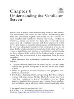
17
Ventricular tachycardia
in a ‘normal’ heart
Shouvik Haldar
Expert commentary Dr Anthony Chow
Case history
A 25-year-old woman attended the Accident & Emergency (A&E) department, complaining of fast palpitations. She led a very active lifestyle and had no medical history
of note. She was a non-smoker and drank 15 units of alcohol per week. She also drank
a significant amount of caffeine. There was a family history of ischaemic heart
disease.
She had first noticed her symptoms whilst at the gym two months earlier. Whilst
training on the exercise bike, she had felt her heart suddenly start pounding quickly.
She denied any associated chest pain, breathlessness, or syncope, but had felt dizzy.
Having stopped exercising, the palpitations continued for ten minutes before suddenly
stopping whilst drinking cold water. Similar episodes had occurred on three other
occasions, all during exercise. She had also experienced numerous short bursts of palpitations, lasting for two to three minutes each time. Notably, they had occurred whilst
under stressful situations at work.
By the time she was assessed, her symptoms had improved, but she was still aware
of an irregular heartbeat. Examination of her cardiovascular system was unremarkable
with an irregular pulse of approximately 75 beats per minute (bpm) with a blood
pressure (BP) of 130/80 mmHg. Her heart sounds were normal with no murmurs on
auscultation.
Her 12-lead electrocardiogram (ECG) (Figure 17.1) showed frequent ventricular
ectopics and routine blood tests, including full blood count (FBC), urea and electrolytes (U&E), thyroid function, and inflammatory markers were all within normal limits.
As she was a young, healthy adult, she was reassured that her symptoms were related
to benign ‘extra beats’, possibly precipitated by stress. She was advised to reduce her
caffeine intake and was discharged home.
Six weeks later, she re-presented to A&E, again with palpitations. The medical
registrar who reviewed her documented the ECG as sinus rhythm interspersed with
short bursts of non-sustained broad complex tachycardia, with left bundle branch
block (LBBB) morphology (Figure 17.2). As the patient was young, he felt that this
was most likely to represent short paroxysms of supraventricular tachycardia (SVT)
with aberrant conduction. Again, her laboratory tests including FBC, U&E, and magnesium levels were normal. She was given 2.5 mg of intravenous (IV) metoprolol
which settled her symptoms and was admitted overnight for observation.
Three hours later, the medical team was called to review the patient urgently. She
was now in a sustained broad complex tachycardia (BCT) at 190 bpm (Figure 17.3),
176
Challenging concepts in cardiovascular medicine
Figure 17.1 ECG on first admission.
Expert comment
The more variable QRS morphology
and subtle change in R-R intervals
between the QRS complexes
during non-sustained salvos of
ventricular activity (Figure 17.2)
would be more in keeping with a
focal tachycardia or micro
re-entrant tachycardia or even
paroxysmal atrial fibrillation (PAF)
with aberrant conduction rather
than a ‘common SVT’.
Figure 17.2 Second admission ECG showing non-sustained, repetitive bursts of monomorphic VT with
LBBB morphology and an inferior axis.
with the same LBBB morphology as seen in the earlier ECG (Figure 17.2). She was
symptomatic although haemodynamically stable with a BP of 115/75 mmHg. Despite
being doubtful about the origin of the arrhythmia, the presumed diagnosis was one
of ventricular tachycardia (VT) and the patient was started on an IV infusion of
amiodarone via a central venous line.
Learning point Bundle branch block patterns: features suggestive of ventricular
tachycardia [1,2]
BCT with bundle branch block patterns can either be SVT with aberrancy or VT. If the QRS complexes
have a LBBB type pattern, features suggestive of ventricular tachycardia are (Figure 17.4):
● QRS complexes with duration >160 ms;
● Presence of an R wave in V1 or V2 of >30 ms in width;
● Time from the start of the QRS wave to the nadir of the S is >70 ms in V1 or V2;
● A slurred or notched S in V1 or V2;
continued
177
Case 17 Ventricular tachycardia in a ‘normal’ heart
●
●
A qR complex in V6;
Inferior axis (QRS complexes are positive in inferior leads) or right axis deviation.
If there is a right bundle branch block (RBBB) type pattern, features suggestive of VT are:
QRS complex with duration >140 ms;
● Superior axis (negative in inferior leads);
● A QS wave or predominantly negative complex in lead V6;
● Concordance throughout the chest leads with all deflections positive;
● A single (R) or biphasic (QR or RS) R wave in lead V1;
● A triphasic R wave in lead V1, with the initial R wave taller than the secondary R wave and an S wave
that passes through the isoelectric line.
●
Clinical tip Features that
favour supraventricular
tachycardia with aberrant
conduction in broad complex
tachycardia
●
●
QRS morphology looks like
‘typical’ right or left bundle
branch block morphology;
QRS morphology in sinus
rhythm shows bundle branch
block or pre-excitation with a
pattern similar to the QRS
morphology during tachycardia.
Clinical tip Features that
favour ventricular tachycardia in
broad complex tachycardia
●
●
●
●
Evidence of independent atrial
activity (dissociated P waves);
Different broad QRS
morphologies during
tachycardia and sinus rhythm;
QRS concordance in leads V1 to
V6 (i.e. all leads show deflections
in the same direction);
Patient has history of structural
or ischaemic heart disease.
Expert comment
Figure 17.3 ECG showing sustained RVOT tachycardia with LBBB morphology and an inferior axis with
arrows denoting dissociated P waves.
V1 and V2
A
B
C
Figure 17.4 Features suggestive of VT in
QRS complexes with LBBB morphology
(reproduced with permission from BMJ
Publishing Group Ltd) [1].
A: > 30 ms favours VT
B: Notching, slurring favours VT
C: > 70 ms favours VT
The 12-lead ECG during sustained
tachycardia clearly confirms that
this is a VT. The QRS morphology is
exactly the same as the nonsustained VT appearance. There is
clear dissociation of P waves with
the QRS morphology, as can be
seen on the lead II rhythm strip
(black arrows on Figure 17.3); this
is diagnostic of VT. The inferior axis
with LBBB suggests that this is an
outflow tract tachycardia, but more
subtle changes can be used to
distinguish a right from left
ventricular outflow tract origin.
178
Expert comment
Beta-blockers can produce a
significant reduction in ectopic
activity and VT episodes, but in the
majority of cases, results are often
disappointing with little impact on
quality of life [3]. Beta-blockers can
be tried as first-line before other
more powerful anti-arrhythmic
drugs are used. Distinction of
RVOT VT from arrhythmogenic
right ventricular cardiomyopathy
(ARVC) with further imaging is of
paramount importance.
Scrutinizing a resting ECG without
ectopics or tachycardia may well
confirm T wave abnormalities and
epsilon waves characteristic
of ARVC.
Challenging concepts in cardiovascular medicine
Two hours after the amiodarone infusion had commenced, the patient remained
symptomatic although the rate had slowed marginally to 180 bpm. Electrical cardioversion was, therefore, attempted under sedation and was successful with a single 100 J
biphasic shock. The following day, a transthoracic echocardiogram (TTE) confirmed a
structurally normal heart. Upon review by a consultant cardiologist, her arrhythmia
was diagnosed as originating from the right ventricular outflow tract (RVOT). She was
prescribed 2.5 mg of bisoprolol daily and discharged with arrangements for an urgent
outpatient cardiac magnetic resonance imaging (MRI) and follow-up in clinic.
Learning point Key electrocardiogram features of ventricular tachycardia
In VT, atrial activity is predominantly independent of ventricular activity. P waves are, therefore,
dissociated from QRS complexes; this is known as atrioventricular (AV) dissociation. This independent
atrial activity may be difficult to discern due to the broad and bizarre morphology of the QRS
complexes as well as its fast rate.
Evidence of independent atrial activity:
●
●
●
●
Independent P waves which are dissociated from QRS complexes;
More QRS complexes than P waves as atrial rates are generally slower;
Capture beats (They represent occasional atrial impulses capturing the ventricles via the normal
conduction system. These QRS complexes occur earlier and are narrow.);
Fusion beats (They represent the simultaneous activation of the ventricles via the normal conduction
system and from the ventricles themselves. The QRS complex, therefore, looks like a cross between a
normal and a tachycardia complex and occurs slightly earlier than expected.).
It should be noted that some VTs conduct regularly to the atria, producing retrograde P waves seen
after the QRS complex. Therefore, typical AV dissociation is not seen, but instead there is
ventriculoatrial (VA) conduction which can signify VT.
Learning point What is right ventricular outflow tract tachycardia?
VT occurs predominantly in the setting of structural heart disease. However, up to 10% of patients with
VT have no obvious structural abnormalities [4]. They generally have a normal baseline ECG,
echocardiogram, and coronary angiogram, although subtle abnormalities may be found on MRI.
These ventricular arrhythmias can be caused by RVOT VT, LVOT VT, and idiopathic left ventricular
tachycardia (ILVT). These are monomorphic, not familial, and collectively termed idiopathic VT. Others
types are due to inherited channelopathies such as Brugada syndrome, long QT syndrome, and
catecholaminergic VT, giving rise to polymorphic VT.
RVOT VT constitutes 90% of the outflow tract tachycardias and the majority of patients have a good
prognosis [5]. It is a distinctive wide QRS complex tachycardia with LBBB morphology and inferior axis,
and is sensitive to adenosine. The ECG in sinus rhythm is predominantly normal although a small
proportion will have RBBB. There is a female preponderance and patients present in the third to fifth
decade of life [6]. Common symptoms include palpitations, dizziness, and pre-syncope. Frank syncope
is unusual. Precipitants include exercise and emotional stress.
There are two distinct forms of RVOT VT: firstly a non-sustained, repetitive, monomorphic VT which is
often suppressed by exercise; secondly a paroxysmal, exercise-induced sustained VT [7]. Patients may
exhibit overlapping features of both forms. Symptomatic episodes may occur as rare or frequent
isolated premature ventricular complexes (PVCs), bursts of non-sustained VT, or as discrete episodes of
sustained tachycardia. If symptoms are frequent and left untreated, then a tachycardia-induced
cardiomyopathy may result. The mechanism is thought to be due to the activation of cyclic AMP
(cAMP), mediated by catecholamines, which leads to high intracellular calcium concentrations. This in
turn causes delayed after-depolarizations in the action potential repolarization phase, triggering the
onset of a tachycardia [4].
179
Case 17 Ventricular tachycardia in a ‘normal’ heart
At her review appointment, the patient reported that her symptoms had improved,
but were not completely resolved. She was able to manage gentle exercise, but more
strenuous exertion brought on symptomatic palpitations. Her cardiac MRI was reported
as normal, showing no evidence of an underlying cardiac muscle disease that could
predispose to a rhythm disturbance. She was referred to a cardiac electrophysiologist
in view of her ongoing symptoms.
After consultation with the electrophysiologist, the patient agreed to a catheter
ablation procedure. Under conscious sedation, multipolar catheters were introduced
percutaneously via the right femoral vein and positioned in the right side of the heart.
The pace-mapping technique was used to localize the site of origin of the tachycardia.
This area was then successfully ablated without complication using radiofrequency
energy. The patient remained symptom-free one year post-procedure, off all medication
and with an unrestricted exercise capacity.
Learning point Management of right ventricular outflow tract tachycardia
Terminating RVOT VT in the acute setting can be achieved with vagal manoeuvres, IV adenosine
(suppresses cAMP-mediated triggered activity), beta-blockers, verapamil, and lidocaine.
Long-term management options include medical therapy or catheter ablation. Beta-blockers and
calcium channel blockers are generally used as first-line drugs and are effective in 25–50% of patients.
Alternative anti-arrhythmic drugs (AADs) include those in class IA and class IC whilst amiodarone and
sotalol are also useful [8].
Catheter ablation requires intra-cardiac mapping, using either activation or pace-mapping techniques
to identify the exact origin of the tachycardia. Pace-mapping involves pacing at different sites in the
RVOT tract until identifying the site that reproduced the exact QRS morphology to that of the clinical
tachycardia. In contrast, activation mapping aims to identify the earliest site of ventricular activation
during the clinical tachycardia. Once identified, radiofrequency energy is applied to disrupt the circuit.
It is curative in 90% of cases and these high success rates can be attributed to the focal origin of the
tachycardia [9]. Complications such as cardiac perforation and tamponade occur in less than 1%. In the
current joint European and American guidelines, catheter ablation has a class 1C recommendation for
those who are ‘drug-refractory, drug–intolerant, or do not want long-term drug therapy’ [10].
Lastly, it is worth noting that VT of LBBB morphology is also seen in a more serious condition called
ARVC (see Case 14). ARVC can cause sudden cardiac death (SCD) in young adults and is commonly
associated with structural abnormalities in the right ventricle although these may be subtle and easily
missed. It is important, therefore, to maintain a high index of suspicion when assessing patients with
LBBB morphology VT (Table 17.1).
Clinical tip Acute
management of a broad complex
tachycardia
Management of a BCT (even when
VT is suspected, but the diagnosis
is unconfirmed) is dependent on
the patient’s haemodynamic status.
If signs of haemodynamic
instability (chest pain, systolic
blood pressure < 90 mmHg, heart
failure, decreased level of
consciousness) are present, then
emergent electrical cardioversion is
warranted.
If the patient is stable, then
intravenous amiodarone, lidocaine
± beta-blockers may be used.
Don’t forget to treat ischaemia if
VT is in the context of an acute
coronary syndrome. If
pharmacological therapy fails to
cardiovert VT in a
haemodynamically stable patient,
then electrical cardioversion
should be used. It is also
mandatory to ensure that
electrolytes such as potassium and
magnesium are adequately
replaced, aiming for the upper
range of normal.
The use of IV adenosine as a
diagnostic or therapeutic
manoeuvre should be limited to
those with a haemodynamically
stable BCT where there is an
unconfirmed diagnosis of VT
vs SVT.
If there is doubt about the origin of
a BCT, it is best to treat as VT. For
example, giving IV verapamil to a
BCT mistakenly identified as SVT
with aberrancy could be fatal.
Discussion
Expert comment
VT in structurally normal hearts represents a small, but important, proportion of
patients within the wide clinical spectrum of VT. Generally, these patients have a good
prognosis with a benign clinical course and their risk of SCD is reassuringly very low.
AADs have a modest efficacy and may be sufficient to suppress the arrhythmia in
some patients. For those in whom AADs fail, catheter ablation is recommended and
can provide freedom from both the troublesome arrhythmia and the side effects of
long-term medical therapy. Advances in radiofrequency ablation have improved the
success rates in outflow tract VT to approximately 90% with a minimal risk of complications. It is easy to understand why catheter ablation, with its favourable safety and
efficacy profile, has revolutionized the management of this condition.
Catheter ablation is indicated in
those with recurrent symptomatic
RVOT arrhythmias, adverse effects
from drug therapy, or tachycardiainduced cardiomyopathy, which
can be reversible by ablation
therapy [11]. Procedural risks are
low at approximately 1–2% and are
generally preferred by most
patients as a definitive cure.
180
Challenging concepts in cardiovascular medicine
Table 17.1 Differentiating RVOT tachycardia from ARVC
Family history of
arrhythmia or SCD
Arrhythmias
SCD
Frontal plane QRS
T wave morphology in
sinus rhythm
QRS duration in sinus
rhythm
Epsilon wave V1–V3
Signal-averaged ECG
Echocardiogram
RVOT tachycardia
ARVC
No
Often
PVCs, non-sustained VT or
sustained VT at rest or with
exercise
Rare
Positive in leads III and AVF,
negative in lead AVL
T wave upright V2–V5
Similar
QRS duration <110 ms in
V1, V2, or V3
Absent
Normal
Normal
RV ventriculogram
MRI
Usually normal
Usually normal, but subtle
abnormalities may be
present
Treatment
Acute: vagal manoeuvres,
adenosine, beta-blockers,
verapamil
Chronic: beta-blockers or
verapamil ± class I AAD
Usually curative
Radiofrequency ablation
1% per year
Inferior or superior
T wave inverted beyond V1
QRS duration >110 ms
Present 30%
Usually abnormal
Increased right ventricular (RV) size and/
or wall motion abnormalities
Usually abnormal
For example, increased signal intensity of
RV free wall; wall motion abnormalities
with CINE MRI, fibrofatty infiltration, focal
wall thinning, and RV dilatation
Sotalol
Amiodarone ± beta-blockers
Seldom curative; may modify substrate to
permit AAD to be effective. However,
arrhythmias of different morphology tend
to occur.
Adapted and reproduced with permission from Professor H. Calkins.
A final word from the expert
The ‘take home’ messages from this case centre around the important principles of managing BCTs
which have been covered concisely.
RVOT tachycardias are not uncommon and often occur in mid-life. Although they are not usually
life-threatening arrhythmias, they can cause significant symptoms, occasional syncope, and even
precipitate heart failure. Drugs are often ineffective, but should be tried first-line. Catheter mapping
and ablation is very successful with very low complication rates and should be offered to all patients
with ongoing symptoms.
References
1 Wellens HJ. Ventricular tachycardia: diagnosis of broad QRS complex tachycardia. Heart
2001; 86: 579–585.
2 Edhouse J, Morris F. ABC of clinical electrocardiography. Broad complex tachycardia–part II.
BMJ 2002; 324: 776–779.
3 Krittayaphong R, Bhuripanyo K, Punlee K, et al. Effect of atenolol on symptomatic
ventricular arrhythmia without structural heart disease: a randomized placebo-controlled
study. Am Heart J 2002; 144: e10.
Case 17 Ventricular tachycardia in a ‘normal’ heart
4 Farzaneh Far A, Lerman BB. Idiopathic ventricular outflow tract tachycardia. Heart 2005; 91:
136–138.
5 Lerman BB, Stein KM, Markowitz SM, et al. Ventricular tachycardia in patients with
structurally normal hearts. In: Zipes DP and Jalife J, editors. Cardiac electrophysiology: from
cell to bedside. 3rd ed. Philadelphia: WB Saunders; 2000. p. 662–673.
6 Badhwar N, Scheinman MM. Idiopathic ventricular tachycardia: diagnosis and management.
Curr Probl Cardiol 2007; 32: 7–43.
7 Srivathsan K, Lester SJ, Appleton CP, et al. Ventricular tachycardia in the absence of
structural heart disease. Indian Pacing Electrophysiol J 2005; 5: 106.
8 Buxton AE, Waxman HL, Marchlinski FE, et al. Right ventricular tachycardia: clinical and
electrophysiologic characteristics. Circulation 1983; 68: 917–927.
9 Lerman BB, Stein KM, Markowitz SM, et al. Ventricular arrhythmias in normal hearts.
Cardiol Clin 2000; 18: 265–291.
10 Zipes D, Camm AJ, Borggrefe M, et al. ACC/AHA/ESC 2006 Guidelines for management of
patients with ventricular arrhythmias and the prevention of sudden cardiac death—
Executive Summary: A report of the American College of Cardiology/American Heart
Association Task Force and the European Society of Cardiology Committee for Practice
Guidelines. Circulation 2006; 114: 1088–1132.
11 Yarlagadda RK, Iwai S, Stein KM, et al. Reversal of cardiomyopathy in patients with
repetitive monomorphic ventricular ectopy originating from the right ventricular outflow
tract. Circulation 2005; 112: 1092–1097.
181
This page intentionally left blank
18
Dual-chamber vs single-chamber
pacing: the debate continues
Ali Hamaad and Shouvik Haldar
Expert commentary Dr Vias Markides
Case history
A 63-year-old man was admitted with a 6-week history of increasing breathlessness and
dizziness. He had a history of treated hypertension, and was taking ramipril 5 mg once
daily. On admission, his pulse rate was 34 beats per minute (bpm) with a blood pressure
(BP) of 180/90 mmHg. Cannon waves were visible in the jugular venous pulse (JVP), and
a 12-lead electrocardiogram (ECG) confirmed complete heart block (CHB) (Figure 18.1).
Expert comment
Although this gentleman had
treated hypertension, systolic
hypertension (usually with a low
diastolic BP) is a frequent
presenting feature in patients with
bradycardia. As it is important for
the maintenance of a reasonable
mean arterial pressure and often
resolves with treatment of
bradycardia with pacing, it should
not generally be corrected acutely.
Expert comment
The need for pacing in AVB
increases exponentially with age,
even in patients with structurally
normal hearts.
Figure 18.1 ECG on admission revealing complete dissociation of P waves and QRS complexes, and a
ventricular rate of approximately 34 bpm (i.e. CHB).
Learning point Investigating high-degree atrioventricular block (AVB)
Causes:
Age-related: occurs in 5–10% of individuals over the age of 70 years with heart disease of any cause;
● Myocardial ischaemia: particularly ischaemia involving the right coronary artery;
● Infiltrative myocardial disease: sarcoidosis, haemochromatosis, malignancies with cardiac
metastases;
● Infective: endocarditis (particularly involving the aortic valve and root), disseminated tuberculosis
with myocardial infiltration of granulomas, Lyme disease;
● Iatrogenic: following aortic, mitral, or tricuspid valve surgery, rarely after ablation for SVT;
● Drug-related: any drug that affects the atrioventricular (AV) conduction system e.g. cardiac
glycosides, beta blockers, and calcium channel blockers.
●
Mobitz type II AVB (i.e. constant PR interval, but intermittent non-conducting P-waves) is always
pathological, and has a high risk of progressing to higher AVB, usually requiring permanent pacemaker
(PPM) implantation. Mobitz type I AVB may be physiological in younger people. In Mobitz type I AVB,
continued
Expert comment
AV nodal re-entrant tachycardia
(AVNRT or supraventricular
tachycardia [‘SVT’]) affects some 1
in 500 of the population. As the
incidence of CHB necessitating
implantation of a PPM following
ablation is less than 1% in
experienced centres, this is now an
exceedingly rare iatrogenic reason
for implanting a pacemaker.
184
Expert comment
Common indications for pacing
include CHB and most cases of
Mobitz type II. High-grade AVB
may also be intermittent and
remain undiagnosed. A high index
of suspicion of intermittent
high-degree block must be
maintained in patients with
symptoms of syncope or
pre-syncope and evidence of
conduction tissue disease,
especially left bundle branch block
(LBBB), bifascicular or trifascicular
block at baseline. Non-conducted
P waves in isolation are not
infrequently seen in fit, young
individuals during sleep and are
not an indication for pacing.
Challenging concepts in cardiovascular medicine
(i.e. progressive lengthening of PR interval until a P-wave fails to conduct and fails to produce a QRS
complex) treatment with a pacemaker may be unnecessary unless symptoms occur. Third-degree AVB is
associated with significant morbidity and mortality, and ultimately requires treatment with a pacemaker.
Investigations to identify the cause of AVB are often unnecessary in older people, as it is usually due to
degeneration of the AV node. In younger people, transthoracic echocardiography (TTE) may help
identify infiltrative causes such as granulomas in the interventricular septum. If the clinical history
includes outdoor activity such as hiking or camping, then antibodies to Borellia burgdorferi may provide
the diagnosis.
Learning point Indications for temporary transvenous pacing
In the absence of acute myocardial infarction (AMI):
Second- and third-degree AVB with symptomatic bradycardia/haemodynamic compromise;
● Sinus node dysfunction (SND) with symptoms/haemodynamic compromise;
● Third-degree AVB with wide QRS escape.
●
In AMI:
● Mobitz type II or third-degree AVB with anterior infarction;
● New bifascicular block or alternating bundle branch block;
● Medically refractory AVB regardless of infarct size.
Prophylactic:
● New AVB or bundle branch block with acute endocarditis;
● Peri-operatively in a patient with bifasicular block and a history of syncope (although this indication
remains controversial).
Treatment of tachyarrhythmias:
● Termination of recurrent ventricular tachycardia (overdrive pacing);
● Suppression of bradycardia-dependent ventricular tachyarrhythmias, including torsades des pointes.
The patient also had a degree of pulmonary congestion both clinically and radiologically, which was treated with intravenous furosemide. While in the Accident and
Emergency (A&E) department, he was noted to have runs of an intermittent broad
complex tachycardia (BCT) that were causing him to lose consciousness. This was
thought to be a ventricular escape rhythm secondary to his severe bradycardia. Given
his clinical instability, a temporary transvenous pacing wire was inserted via his right
femoral vein.
The patient was stabilized on the ward. He remained dependent on the temporary
pacing wire and did not regain an intrinsic sinus rhythm. The decision was made to
implant a permanent dual-chamber pacemaker. Access for lead insertion was via the
left cephalic vein under strict aseptic technique.
Learning point Basic pacing terminology
Threshold This is measured in volts (V) or milliseconds (ms), and is the smallest output voltage or the
shortest pulse duration that captures the heart. A sudden rise in threshold suggests lead displacement.
Impedance This is the resistance of the electrical circuit that is formed when pacing is applied to the
myocardium, and comprises the electrical resistance of the lead and tissue that is conducting the
current (usually the myocardium). A rise in impedance suggests an interruption of the electrical pacing
circuit due to lead displacement or lead fracture. A drop in impedance suggests damage to the
electrical insulation of the pacing lead.
R wave This refers to the sensitivity of the pacing lead in detecting intrinsic myocardial depolarizations.
A large R wave means the lead can detect small signals.
Case 18 Dual-chamber vs single-chamber pacing: the debate continues
It became apparent during implantation that the patient had residual pulmonary
oedema, as he was unable to lie flat for a prolonged period without a significant drop
in oxygen saturations and tachypnoea. The procedure was abandoned following
implantation of the ventricular lead in the right ventricular (RV) apex. A right atrial
lead was, therefore, not inserted, and the patient was left with a single-chamber pacemaker programmed to deliver pacing in a VVIR fashion. A post-procedural pacing
check was satisfactory (Table 18.1). The pulmonary oedema was treated with more
intravenous furosemide, and a subsequent TTE demonstrated mild impairment of
ventricular function with an ejection fraction of 45%.
Table 18.1 Post-procedural pacing parameters
R wave
Impedance
Threshold
18 mV
760 Ω
0.3 V at 0.50 ms
Learning point Pacing nomenclature (Table 18.2)
The Heart Rhythm Society (HRS) and Heart Rhythm UK (HRUK) generic pacemaker codes for
anti-bradyarrhythmia and adaptive-rate pacing and anti-tachyarrhythmia devices are listed below [1].
For example:
VVIR: pacing and sensing in the ventricle, inhibited by spontaneous activation of the ventricle and
the ability to increase the paced rate which is dependent on activity;
● AOO: pacing in the atrium with no inhibition of pacing by spontaneous electrical activity in
the atrium;
● DDDR: pacing and sensing either atrium or ventricle or both with the ability to increase paced rate
which is dependent on activity.
●
Table 18.2 Pacing nomenclature
Letter position
Pacing category
Letter code
1st
Chamber(s) paced
2nd
Chamber(s) sensed
3rd
Mode of response to spontaneous
electrical activation
4th
Rate modulation
V–Ventricle
A–Atrium
D–Dual
V–Ventricle
A–Atrium
D–Dual
T–Triggered
I–Inhibited
D–Dual
O–None
R–rate modulation (pacing rate can be
altered dependent on activity)
On day 1 post-implantation, there was an improvement in the patient’s pulmonary
oedema and he was converted to oral furosemide. By day 3, however, the patient had
deteriorated clinically with further tachypnoea, tachycardia, and a recurrence of fluid
overload. This was accompanied by episodes of hypotension and cannon waves visible in his JVP. The pacemaker was checked and found to have satisfactory pacing
parameters, that were not significantly different to those measured immediately postimplantation. However, during the pacing threshold check, it was obvious the patient
185
Clinical tip Cephalic vein
access for permanent pacing
Always search for a cephalic vein
beneath the delto-pectoral groove
when implanting a PPM system.
This minimizes the chances of
causing iatrogenic pneumothorax
which is a risk when using a direct
subclavian approach, as well as
reducing the incidence of lead
fracture under the clavicle.
186
Expert comment
The finding of VA (retrograde)
conduction is very unusual in
patients with CHB, and far more
common in patients with sick sinus
syndrome as the pacing indication.
Expert comment
In the presence of left ventricular
(LV) impairment, right ventricular
(RV)-based pacing, can cause
dyssynchrony which can be
alleviated with biventricular pacing
(CRT-P). This involves the
placement of an additional lead on
the epicardial surface of the LV
through the coronary sinus.
Patients with CHB as an indication
for pacing almost always require
RV-based pacing whereas in
patients with SND, pacemakers
can often be programmed to avoid
RV pacing whilst providing
adequate atrially-based pacing.
Challenging concepts in cardiovascular medicine
did not tolerate being paced as he was visibly uncomfortable. In particular, he complained of forceful palpitations and neck pulsations. A rhythm strip recording of the
patient during one of these episodes revealed retrograde P waves following pacing
depolarization, indicating ventriculo-atrial (VA) conduction (Figure 18.2). Unusually,
even though the patient had presented with CHB, he still had intact retrograde VA
conduction. With VVI pacing, this had resulted in a phenomenon known as the pacemaker syndrome. The heart failure was treated with further doses of intravenous
diuretics, and once stable, the patient had a right atrial lead implanted to encourage
AV synchrony. This resulted in the resolution of his symptoms and an eventual hospital discharge.
Figure 18.2 Rhythm strip taken during symptomatic episodes whilst pacing. This illustrates repetitive VA
conduction as evidenced by retrograde P wave activity following pacing depolarization (arrowed).
Expert comment
The best way to manage
pacemaker syndrome is to ensure
that all patients with regular atrial
activity have an atrially-based
pacemaker implanted. In most
cases, this will mean a DDD
pacemaker, as sinoatrial (SA) node
and AV node dysfunction often
coexist. An AAI pacemaker is
appropriate for those who only
have documented pure SND.
Learning point Pacemaker syndrome (Table 18.3)
The pacemaker syndrome refers to symptoms and signs present in the pacemaker patient that are
caused by inadequate timing of atrial and ventricular contractions [1]. These symptoms and signs are
often relieved by restoration of AV synchrony. Although VVI pacing is the commonest culprit, other
pacing modes that cause AV dyssynchrony can be implicated [1].
Clinical signs relating to the pacemaker syndrome should be carefully sought, in particular episodes of
hypotension during pacing (a reduction ≥ 20 mmHg is thought to be significant), cannon waves in the
JVP, and sometimes signs of cardiac failure. ECG features such as native atrial depolarization moving
progressively closer to the paced ventricular depolarization, and retrograde P waves due to an intact
VA conduction may also support the diagnosis [2]. Methods of overcoming the pacemaker syndrome
include upgrading to a dual-chamber (DDD) system, reducing the lower pacing rate to encourage
sinus rhythm, and use of hysteresis to encourage intrinsic depolarizations. Hysteresis refers to a pacing
parameter which usually allows a longer escape interval after a sensed event, allowing a greater
opportunity for more spontaneous depolarizations.
Although the incidence of the pacemaker syndrome is much lower in patients with a dual-chamber
system, this can still occur as a result of:
●
●
●
●
Left atrial activation delay;
Sinus tachycardia with a long AV delay that does not shorten at higher atrial rates;
Repetitive VA conduction;
Pacemaker malfunction (loss of capture in the right atrial lead).
Patients with a dual-chamber system who exhibit the pacemaker syndrome may be managed by
ensuring atrial capture, avoiding pacing modes that do not pace the atria (VDD), or prolonging the AV
delay [1,2].
Case 18 Dual-chamber vs single-chamber pacing: the debate continues
Table 18.3 Features of pacemaker syndrome
Clinical
Electrocardiographic
Dizziness, syncope, confusion
Heart failure
Hypotension, tachycardia, desaturation
Fluctuating BP
Pulmonary oedema
Cannon waves in jugular venous pulse
Atrial depolarization occurring in close proximity
to paced ventricular depolarization
Retrograde atrial depolarization occurring as a
consequence of intact VA conduction
Discussion
The loss of AV synchrony in those with AVB can result in atrial contraction when the
mitral and tricuspid valves are closed. This in turn can lead to raised atrial pressures,
impaired systolic ventricular function, and occasionally, the pacemaker syndrome, as
demonstrated in this case. Re-establishing AV synchrony in these patients requires
dual-chamber pacing, i.e. a more physiological mode of pacing.
The haemodynamic benefits of AV sequential pacing vs ventricular pacing have
been well documented in numerous physiological studies. However, landmark clinical
trials designed to test whether these benefits would translate into mortality and morbidity reductions have been disappointing. In fact, the results suggest that dual-chamber pacing offers no real clinical benefit over single chamber pacing in terms of
mortality, burden of atrial fibrillation (AF), or quality of life (QoL) [3,4]. The CTOPP
and MOST study, which examined pacing primarily in those with SND, found only a
slight reduction in AF risk in the dual-chamber group, whereas the MOST study also
found only a marginal reduction in heart failure. It should be noted, however, that twothirds of patients recruited into CTOPP and all the MOST patients had SND which
behaves in a different way when paced, compared to pacing in AVB [5,6].
After CTOPP and MOST, the logical progression was to answer the question of
optimal pacing mode in elderly AVB patients. UKPACE attempted to do this, and again,
results were disappointing. The trial demonstrated no difference in mortality between
the single chamber and dual-chamber pacing arms, and notably did not show a reduction in AF occurrence in the dual-chamber group [7].
The QoL was not a primary outcome measure in UKPACE, but this was addressed in
the PASE study which examined the effect of pacing mode on health-related QoL [8]. No
significant differences were noted between treatment arms for the primary outcome measure in PASE, although there was some evidence of improvement in the cardiovascular
functional status in those with dual-chamber pacing. It is, however, important to note that
26% of patients in PASE crossed over from ventricular to dual-chamber pacing, with the
pacemaker syndrome cited as the main reason for a system upgrade. A similar phenomenon was also noted in the MOST study where nearly 20% of patients who were unable to
tolerate single chamber pacing crossed over to dual-chamber pacing. Interestingly, after
crossover, these patients reported a significant improvement in QoL measures [6].
Although there is no hard evidence to favour dual-chamber pacing over ventricular
pacing patients with AVB, both national [9] and international [10] guidelines recommend
implanting dual-chamber systems. In essence, modern practice aims to provide physiological pacing whenever possible (except in AF). From a cardiologist’s viewpoint, dualchamber systems may take slightly longer to implant, but the expectant benefits are a
reduction in the pacemaker syndrome, a reduction in AF risk, and a potential improvement in QoL, which should outweigh most technical and time-related considerations.
187
188
Challenging concepts in cardiovascular medicine
Landmark trial Canadian Trial of Physiologic Pacing (CTOPP) [5]
Expert comment
The risk of infection during
pacemaker re-intervention
(e.g. upgrading from single to
dual-chamber) is almost ten times
higher than at original implant. For
this and other reasons, it is
important to provide optimal
(usually dual-chamber) pacing at
the original implant procedure
whenever possible. Physiological
pacing is included as one of the
markers of good practice, and the
proportion of such implants for
each implanting centre is subject to
an annual national review in the UK.
●
●
●
●
●
●
2,568 patients enrolled, making it one of the largest pacing trials to date;
Patients recruited with SND or AVB;
Comparison made between DDDR/AAIR vs VVIR pacing;
Primary endpoints were stroke and cardiovascular mortality;
No difference in stroke or death between pacing modalities;
AF less frequent in atrial-paced group.
Landmark trial Mode Selection Trial (MOST) [6]
●
●
●
●
●
2,010 patients enrolled;
Patients with SND only;
Comparison between DDDR and VVIR pacing modalities;
No difference between death or stroke between pacing modalities;
AF and heart failure lower in DDDR-paced patients.
Landmark trial United Kingdom Pacing and Cardiovascular Events Trial (UKPACE) [7]
●
●
●
●
●
2,000 patients recruited;
Patients aged ≥70 with AVB only;
Comparison between DDDR vs VVIR/VVI pacing modalities;
Primary endpoint was mortality;
No difference between groups.
Landmark trial Pacemaker Selection in the Elderly Trial (PASE) [8]
●
●
●
●
●
●
407 patients recruited;
Patients with SND and/or AVB;
Comparison between DDDR vs VVIR pacing modalities;
Primary endpoint was QoL;
No difference between groups;
Better QoL outcome measures in patients with SND with DDDR pacing.
Learning point National Institute for Health and Clinical Excellence guidelines on permanent
pacing [9]
Dual-chamber pacing is recommended for the management of symptomatic bradycardia due to sick
sinus syndrome and/or AVB except:
● In the management of sick sinus syndrome where there is no evidence of AVB, in which case single
chamber atrial pacing (AAI mode) may be appropriate;
● In the management of AVB in patients with AF, in which case single chamber ventricular pacing may
be appropriate;
● In the management of AVB where factors such as age, frailty, and immobility favour the use of single
chamber ventricular pacing.
Note It should be borne in mind that this guidance does not cover the more complex pacing indications.
See Table 18.4 for comprehensive guidelines from Heart Rhythm Society.
Table 18.4 Recommendations for permanent pacing in acquired AVB in adults
Class I (evidence clearly in favour of pacing)
1. Third-degree and advanced second-degree AVB at any anatomic level with symptoms or
ventricular arrhythmias secondary to the block.
2. Third-degree and advanced second-degree AVB associated with arrhythmias or other medical
conditions requiring drug therapy that results in symptomatic bradycardia.
continued
Case 18 Dual-chamber vs single-chamber pacing: the debate continues
3. Third-degree and advanced second-degree AVB at any anatomic level in awake, symptom-free
patients in sinus rhythm, with documented periods of asystole greater than or equal to 3.0 seconds,
or any escape rate less than 40 bpm, or with an escape rhythm that is below the AV node.
4. Third-degree and advanced second-degree AVB at any anatomic level in awake, symptom-free
patients with AF and bradycardia with one or more pauses of at least 5 seconds or longer.
5. Third-degree and advanced second-degree AVB at any anatomic level after catheter ablation of
the AV junction.
6. Third-degree and advanced second-degree AVB at any anatomic level associated with
post-operative AVB that is not expected to resolve after cardiac surgery.
7. Third-degree and advanced second-degree AVB at any anatomic level associated with neuromuscular
diseases with AVB, such as myotonic muscular dystrophy, Kearns–Sayre syndrome, Erb dystrophy
(limb-girdle muscular dystrophy), and peroneal muscular atrophy with or without symptoms.
8. Second-degree AVB with associated symptomatic bradycardia regardless of type or site of block.
9. Asymptomatic persistent third-degree AVB at any anatomic site with average awake ventricular rates of
40 bpm or faster if cardiomegaly or LV dysfunction is present or if the site of block is below the AV node.
10. Second- or third-degree AVB during exercise in the absence of myocardial ischaemia.
Class IIa (reasonable to perform procedure with weight of evidence in favour of pacing)
1. Persistent third-degree AVB with an escape rate greater than 40 bpm in asymptomatic adult
patients without cardiomegaly.
2. Asymptomatic second-degree AVB at intra- or infra-His levels found at electrophysiological study.
3. First- or second-degree AVB with symptoms similar to those of pacemaker syndrome or
haemodynamic compromise.
4. Asymptomatic type II second-degree AVB with a narrow QRS. When type II second-degree AVB
occurs with a wide QRS, including isolated right bundle branch block, pacing becomes a class I
recommendation.
Class IIb (usefulness/efficacy is well established by evidence/opinion)
1. Neuromuscular diseases such as myotonic muscular dystrophy, Erb dystrophy (limb-girdle
muscular dystrophy), and peroneal muscular atrophy with any degree of AVB with or without
symptoms because there may be unpredictable progression of AV conduction disease.
2. AVB in the setting of drug use and/or drug toxicity when the block is expected to recur even after
the drug is withdrawn.
Class III (evidence not in favour of pacing)
1. Permanent pacemaker implantation is not indicated for asymptomatic first-degree AVB.
2. Permanent pacemaker implantation is not indicated for asymptomatic type I second-degree AVB
at the supra-His (AV node) level or that which is not known to be intra- or infra-Hisian.
3. Permanent pacemaker implantation is not indicated for AVB that is expected to resolve and is
unlikely to recur (e.g. drug toxicity, Lyme disease, or transient increases in vagal tone or during
hypoxia in sleep apnoea syndrome in the absence of symptoms).
Adapted with permission, copyright 2008, from the American College of Cardiology/American Heart Association/
North American Society for Pacing and Electrophysiology [now known as the Heart Rhythm Society] 2002 Guideline
Update for Implantation of Cardiac Pacemakers and Antiarrhythmia Devices.
A final word from the expert
This case demonstrates the importance of providing physiological (AV synchronous) pacing in virtually
all patients who have, or are likely to have, and maintain, regular atrial contraction (i.e. excluding
patients with persistent/permanent AF). It also highlights the importance of getting this decision right
when first implanting a device rather than having to revise the procedure. Re-intervention inevitably
results in increased morbidity for the patient, prolonging the hospital stay and greatly increasing the
risk of infection.
189