Ebook Ferris best test - A practical guide to laboratory medicine and diagnostic imaging (2nd edition): Part 2
Bạn đang xem bản rút gọn của tài liệu. Xem và tải ngay bản đầy đủ của tài liệu tại đây (11.69 MB, 259 trang )
REFERENCES
1. Dhingra R et al: C-reactive protein, inflammatory conditions, and cardiovascular disease risk,
Am J Med 120:1054-1062, 2007.
2. Jeremias A, Gibson M: Narrative review: Alternative cause for elevated cardiac troponin levels
when acute coronary syndromes are excluded, Ann Intern Med 142:786-791, 2005.
3. Jones JS: Four no more: The PSA cutoff era is over. Cleveland Clin J Med 75:30-32, 2008.
4. McKie PM, Burnett JC: B-type natriuretic peptide as a marker beyond heart failure:
Speculations and opportunities, Mayo Clin Proc 80(8):1029-1036, 2005.
5. Pagana KD, Pagana, TJ: Mosby’s Diagnostic and Laboratory Test Reference, ed 8, St. Louis,
Mosby, 2007.
6. Sarmak MJ et al: Cystatin C concentration as a risk factor for heart failure in older adults,
Ann Intern Med 142:497-505, 2005.
7. Wu AHB: Tietz Clinical Guide to Laboratory Tests, Philadelphia, WB Saunders, 2006.
159
162
This section includes the diagnostic modalities (imaging and laboratory tests)
and algorithms useful to diagnose the following 231 diseases and disorders. It is
assumed that the patient has had a detailed history and physical examination
before any testing sequence is initiated.
These algorithms are designed to assist clinicians in the evaluation and treatment of patients. They may not apply to all patients with a particular disease or
disorder, and they are not intended to replace a clinician’s individual judgment.
Please note that specific findings in the patient’s history and physical examination may significantly alter any of the proposed testing sequences.
1.
2.
3.
4.
5.
6.
7.
8.
9.
10.
11.
12.
13.
14.
15.
16.
17.
18.
19.
20.
21.
22.
23.
24.
25.
26.
27.
28.
29.
30.
31.
32.
33.
34.
35.
36.
37.
38.
39.
40.
41.
42.
43.
Abdominal abscess p.
Abruptio placentae p.
Achalasia p.
Acoustic neuroma p.
Acquired immunodeficiency syndrome (AIDS) p.
Acromegaly p.
Actinomycosis p.
Acute respiratory distress syndrome (ARDS) p.
Addison’s disease p.
Adrenal mass p.
Aldosteronism p.
Alkaline phosphatase elevation p.
Alpha1-antitrypsin deficiency p.
ALT/AST elevation p.
Alzheimer’s disease p.
Amaurosis fugax p.
Amebiasis p.
Amenorrhea, primary p.
Amenorrhea, secondary p.
Amyloidosis p.
Amyotrophic lateral sclerosis p.
Anemia, macrocytic p.
Anemia, microcytic p.
Aneurysm of abdominal aorta p.
Antinuclear antibody (ANA) positive p.
Antiphospholipid antibody syndrome p.
Aortic dissection p.
Appendicitis p.
Arthritis, infectious (bacterial) p.
Ascariasis p.
Ascites p.
Aseptic necrosis p.
Back pain, acute, lumbosacral (LS) area p.
Baker’s cyst p.
Bilirubin elevation p.
Bleeding disorder, congenital p.
Brain abscess p.
Breast abscess` p.
Breast implant rupture p.
Breast mass p.
Breast nipple discharge p.
Bronchiectasis p.
Budd-Chiari syndrome p.
163
44.
45.
46.
47.
48.
49.
50.
51.
52.
53.
54.
55.
56.
57.
58.
59.
60.
61.
62.
63.
64.
65.
66.
67.
68.
69.
70.
71.
72.
73.
74.
75.
76.
77.
78.
79.
80.
81.
82.
83.
84.
85.
86.
87.
88.
89.
90.
91.
92.
93.
94.
95.
96.
97.
98.
99.
Carcinoid syndrome p.
Cardiomegaly on chest radiograph p.
Cat-scratch disease p.
Cavernous sinus thrombosis p.
Celiac disease p.
Cerebrovascular accident (CVA) (stroke) p.
Cholangitis p.
Cholecystitis p.
Cholelithiasis p.
Claudication p.
Constipation p.
CPK elevation p.
Cushing’s syndrome p.
Cyanosis p.
Deep vein thrombosis (DVT) p.
Delirium p.
Diabetes insipidus p.
Diarrhea p.
Disseminated intravascular coagulation (DIC)
Diverticulitis p.
Dyspepsia p.
Dysphagia p.
Dyspnea p.
Dysuria p.
Echinococcosis p.
Ectopic pregnancy p.
Edema, generalized p.
Edema, lower extremity p.
Endocarditis, infective p.
Endometriosis p.
Enuresis p.
Epiglottitis p.
Esophageal perforation p.
Fatigue p.
Fever of undetermined origin (FUO) p.
Genital lesions/ulcers p.
Goiter p.
Gout p.
Gynecomastia p.
Hearing loss p.
Hematuria p.
Hemochromatosis p.
Hemophilia p.
Hemoptysis p.
Hepatitis A p.
Hepatitis B, acute p.
Hepatitits C p.
Hepatomegaly p.
Hepatorenal syndrome p.
Hirsutism p.
Hydrocephalus, normal pressure (NPH) p.
Hypercalcemia p.
Hyperkalemia p.
Hypermagnesemia p.
Hypernatremia p.
Hyperphosphatemia p.
p.
164
100.
101.
102.
103.
104.
105.
106.
107.
108.
109.
110.
111.
112.
113.
114.
115.
116.
117.
118.
119.
120.
121.
122.
123.
124.
125.
126.
127.
128.
129.
130.
131.
132.
133.
134.
135.
136.
137.
138.
139.
140.
141.
142.
143.
144.
145.
146.
147.
148.
149.
150.
151.
152.
153.
154.
155.
Hyperthyroidism p.
Hypocalcemia p.
Hypogonadism p.
Hypokalemia p.
Hypomagnesemia p.
Hyponatremia p.
Hypophosphatemia p.
Hypothyroidism p.
Infertility p.
Insulinoma p.
Intracranial mass p.
Jaundice p.
Joint effusion p.
Knee pain p.
Liver abscess p.
Liver function test elevations p.
Liver mass p.
Lung abscess p.
Lymphadenopathy, axillary p.
Lymphadenopathy, cervical p.
Lymphadenopathy, epitrochlear p.
Lymphadenopathy, generalized p.
Lymphadenopathy, inguinal p.
Lymphedema p.
Macrocytosis p.
Malabsorption, suspected p.
Mastoiditis p.
Meckel’s diverticulum p.
Mediastinal adenopathy p.
Meningioma p.
Meningitis p.
Mesenteric venous thrombosis p.
Mesothelioma p.
Metabolic acidosis p.
Metabolic alkalosis p.
Microcytosis p.
Multiple myeloma p.
Multiple sclerosis p.
Muscle cramps p.
Muscle weakness p.
Myasthenia gravis (MG) p.
Myocardial ischemia, suspected p.
Neck mass
Neutropenia p.
Oliguria p.
Osteomyelitis p.
Osteonecrosis p.
Osteoporosis p.
Paget’s disease of bone p.
Pancreatic cancer p.
Pancreatic mass p.
Pancreatitis, acute p.
Pelvic abscess p.
Pelvic mass p.
Pelvic pain, reproductive age woman p.
Peptic ulcer disease p.
165
156.
157.
158.
159.
160.
161.
162.
163.
164.
165.
166.
167.
168.
169.
170.
171.
172.
173.
174.
175.
176.
177.
178.
179.
180.
181.
182.
183.
184.
185.
186.
187.
188.
189.
190.
191.
192.
193.
194.
195.
196.
197.
198.
199.
200.
201.
202.
203.
204.
205.
206.
207.
208.
209.
210.
Peripheral arterial disease p.
Peripheral nerve dysfunction p.
Perirectal abscess p.
Pheochromocytoma p.
Pituitary adenoma p.
Placenta previa p.
Pleural effusion p.
Polyarteritis nodosa p.
Polycystic kidney disease p.
Polycythemia vera p.
Portal hypertension p.
Portal vein thrombosis p.
Prolactinoma p.
Prostate cancer p.
Proteinuria p.
Pruritus, generalized p.
Pseudomembranous colitis p.
Puberty, delayed p.
Puberty, precocious p.
Pulmonary embolism p.
Pulmonary hypertension p.
Pulmonary nodule p.
Purpura p.
Reflex sympathetic dystrophy (RSD) p.
Renal artery stenosis p.
Renal insufficiency p.
Renal mass p.
Renal vein thrombosis p.
Respiratory acidosis p.
Respiratory alkalosis p.
Retropharyngeal abscess p.
Rhabdomyolysis p.
Rotator cuff tear p.
Sacroiliac joint pain p.
Salivary gland neoplasm p.
Sarcoidosis p.
Scrotal mass p.
Seizure disorder p.
SIADH (syndrome of inappropriate antidiuretic hormone
secretion) p.
Sialolithiasis p.
Sinusitis p.
Small-bowel obstruction p.
Spinal epidural abscess p.
Spinal stenosis p.
Splenomegaly p.
Subarachnoid hemorrhage p.
Subclavian steal syndrome p.
Subdural hematoma p.
Superior vena cava syndrome p.
Syncope p.
Temporal arteritis p.
Temporomandibular joint (TMJ) syndrome p.
Testicular neoplasm p.
Testicular torsion p.
Thoracic outlet syndrome p.
166
211.
212.
213.
214.
215.
216.
217.
218.
219.
220.
221.
222.
223.
224.
225.
226.
227.
228.
229.
230.
231.
Thrombocytopenia p.
Thrombocytosis p.
Thyroid nodule p.
Thyroiditis p.
Tinnitus p.
Transient ischemic attack (TIA) p.
Trigeminal neuralgia p.
Urethral discharge p.
Urolithiasis p.
Urticaria p.
Vaginal bleeding, 1st trimester p.
Vaginal discharge p.
Vertigo p.
Viral hepatitis p.
von Willebrand’s disease p.
Waldenström’s macroglobulinemia p.
Wegener’s granulomatosis p.
Weight gain p.
Weight loss, involuntary p.
Wilson’s disease p.
Zollinger-Ellison syndrome p.
1. Abdominal Abscess
Diagnostic Imaging
Lab Evaluation
Best Test(s)
Best Test(s)
• CT of abdomen with contrast
• Gram stain and culture and
sensitivity (C&S) of abscess
Ancillary Tests
• Ultrasound of abdomen is useful in
young women and children
Ancillary Tests
• CBC with differential
• Blood culture ϫ 2
• ALT, AST
• BUN, creatinine, glucose
Diagnostic Algorithm
Suspected
abdominal
abscess
CT of abdomen
with contrast or
ultrasound in
young women
and children
CBC with
differential
Ancillary
lab tests
CT-guided drainage
of abscess and
Gram’s stain and
C&S of abscess
aspirate
2. Abruptio Placentae
Diagnostic Imaging
Lab Evaluation
Best Test(s)
Best Test(s)
• Obstetric ultrasound
• None
Ancillary Tests
Ancillary Tests
• Continuous fetal heart rate
monitoring
• CBC (to quantify blood loss)
• Coagulation profile (PT, PTT,
platelets, fibrinogen)
• Blood type and antibody screen to
identify Rh-negative patients who
may need Rh immunoglobulin
Diagnostic Algorithm
Obstetrical ultrasound
Suspected abruptio
placentae
Initiate continuous fetal
heart rate monitoring
Laboratory evaluation
167
168
3.
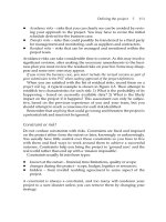
Achalasia
Diagnostic Imaging
Lab Evaluation
Best Test(s)
Best Test(s)
• Barium swallow with fluoroscopy
• None
Ancillary Tests
Ancillary Tests
• Esophageal manometry if barium
swallow is inconclusive
• Upper endoscopy
• CBC
• Serum albumin for nutritional
assessment
Diagnostic Algorithm
Diagnostic
Suspected
achalasia
Esophageal
manometry
Barium
swallow
Inconclusive
Upper endoscopy to
rule out malignancy
and strictures
Normal
Achalasia
Megaesophagus
GEJ
Bird's beak
Figure 3-1 Achalasia. (From Weissleder R, Wittenberg J,
Harisinghani MG, Chen JW: Primer of Diagnostic Imaging, ed 4,
St. Louis, Mosby, 2007.)
4.
Acoustic Neuroma
Diagnostic Imaging
Lab Evaluation
Best Test(s)
Best Test(s)
• MRI with gadolinium of brain and
auditory canal
• None
Ancillary Tests
• None
Ancillary Tests
• CT of brain and auditory canal with
IV contrast if MRI is contraindicated
Diagnostic Algorithm
Suspected acoustic
neuroma (hearing
loss, unilateral
tinnitus, balance
problems, facial
pain)
Detailed neurologic
exam with special
attention to the
cranial nerves
MRI of brain and
auditory canals
with gadolinium
5. Acquired Immunodeficiency Syndrome
Diagnostic Imaging
Lab Evaluation
Best Test(s)
Best Test(s)
• None
• HIV antibody test
Ancillary Tests
Ancillary Tests
• MRI or CT of brain for encephalopathy or focal CNS complications
• Pulmonary gallium scan in suspected
Pneumocystis pneumonia
• T-lymphocyte subset analysis to determine degree of immunodeficiency
• Viral load assay to plan long-term
antiviral therapy
Diagnostic Algorithm
Negative
Suspected
infection
with HIV
HIV antibody
test
Repeat in
4–6 weeks
T-lymphocyte
subset analysis
Positive
Viral load
169
170
6.
Acromegaly
Diagnostic Imaging
Lab Evaluation
Best Test(s)
Best Test(s)
• MRI of pituitary and hypothalamus
with contrast
• Serum insulin-like growth factor
(IGF)-I level
Ancillary Tests
Ancillary Tests
• CT of pituitary and hypothalamus if
MRI is contraindicated
• Suppression test with oral glucose
• Serum phosphate (increased)
• Serum calcium (increased)
Diagnostic Algorithm
Diagnosis
unlikely
Normal
Suspected
acromegaly
Serum
IGF-I
Elevated
Suppression
test with oral
glucose
(failure to
suppress GH
to <2 ng/mL
after 100 g
of oral glucose
is considered
conclusive)
MRI of
pituitary and
hypothalamus
7. Actinomycosis
Diagnostic Imaging
Lab Evaluation
Best Test(s)
Best Test(s)
• Chest radiograph
• Isolation of “sulfur granules” (nests
of Actinomyces species) from tissue
specimens or draining sinuses
Ancillary Tests
• CT of head, chest, abdomen, and
pelvis
Ancillary Tests
• CBC
Diagnostic Algorithm
Abnormal
CT of chest, head,
abdomen and pelvis
Chest
x-ray
Negative
Consider other diagnosis
Suspected
actinomycosis
Identification
of abscess,
sinus tracts
I&D of
abscesses,
excision
of sinus tract
Stain and
culture
of isolated
“sulfur granules”
8.
Acute Respiratory Distress Syndrome
Diagnostic Imaging
Lab Evaluation
Best Test(s)
Best Test(s)
• Chest radiograph
• ABGs
Ancillary Tests
Ancillary Tests
• CT of chest when lymphangitic
carcinomatosis is suspected
• CBC with differential
• Blood and urine cultures
• Bronchoalveolar lavage (in selected patients who respond poorly to therapy)
Diagnostic Algorithm
Chest x-ray
CBC with
differential,
blood and
urine cultures
Suspected
ARDS
Hemodynamic
monitoring
ABGs
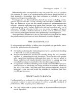
Figure 3-2 ARDS due to extrapulmonary disease. Chest radiograph 21⁄2 days
after postoperative hemorrhage. There is diffuse ground-glass opacification,
slightly greater on the right than the left. For unknown reasons, the left apex
is spared. Incidentally noted are signs of barotraumas—pneumomediastinum
and subcutaneous air in the neck. (From Grainger RG, Allison DJ, Adam A,
Dixon AK, eds: Grainger & Allison’s Diagnostic Radiology, ed 4, Churchill
Livingstone, Philadelphia, 2001.)
171
172
9.
Addison’s Disease
Diagnostic Imaging
Lab Evaluation
Best Test(s)
Best Test(s)
• None
• IV cosyntropin test, serial
measurement of cortisol
Ancillary Tests
Ancillary Tests
• CT or MRI of adrenals with contrast
• Chest X-ray
• Serum electrolytes (hyponatremia,
hyperkalemia)
• FBS, BUN, creatinine
• CBC (anemia)
Diagnostic Algorithm
Suspected
Addison’s
disease
IV cosyntropin; measure
cortisol level at baseline,
30 min, 60 min
Elevated cortisol level
(>18 mcg/dL)
Low cortisol level
(<18 mcg/dL)
ACTH level
No adrenal insufficiency
Normal/decreased
Elevated
Adrenal insufficiency
secondary to pituitary
insufficiency
Primary adrenal
insufficiency
10. Adrenal Mass
Diagnostic Imaging
Lab Evaluation
Best Test(s)
Best Test(s)
• MRI of adrenal gland with contrast
• Serum electrolytes
Ancillary Tests
Ancillary Tests
• CT of adrenal gland with and
without contrast if MRI is
contraindicated
• If symptoms of pheochromocytoma,
obtain plasma-free metanephrine
level, 24-hour urine collection for
metanephrines
• If cushingoid appearance, obtain
overnight dexamethasone suppression test
• If signs of virilization or feminization,
order 24-hour urine for 17-ketosteroids
and plasma dehydroepiandrosterone
sulfate (DHEAS)
• If hypertension is present with
associated hypokalemia, evaluate for
aldosteronism
Diagnostic Algorithm
Cystic
appearance
Suspected
adrenal mass
MRI or CT
of adrenal
gland
Homogeneous
appearance
Nonhomogeneous
or large mass
(>6 cm)
Ultrasound
Ancillary
lab tests
Surgical
resection
Simple
cyst
173
174
11.
Aldosteronism
Diagnostic Imaging
Lab Evaluation
Best Test(s)
Best Test(s)
• None
• Plasma aldosterone concentration
(PAC)
• Plasma renin activity (PRA)
Ancillary Tests
• MRI with contrast or CT scan of
adrenals with contrast to localize
neoplasm
• Adrenal scan with iodocholesterol
(NP-59) or 6-beta-iodomethyl-19norcholesterol
Ancillary Tests
• Serum electrolytes
• Aldosterone suppression test
Diagnostic Algorithm
Hypertension,
hypokalemia
Suspected
hyperaldosteronism
PAC/PRA
≥20 and
PAC >15 ng/dL
Positive
Aldosterone suppression test
(2 L of normal saline infusion
over 4 hours followed by
measurement of plasma
aldosterone level)
Plasma aldosterone
level >10 ng/dL
Primary aldosteronism
confirmed
Negative
Diagnosis excluded
12.
Alkaline Phosphatase Elevation
Diagnostic Imaging
Lab Evaluation
Best Test(s)
Best Test(s)
• CT of liver
• GGT
Ancillary Tests
Ancillary Tests
• Ultrasound of liver
• Radiograph of pelvis or Paget’s
disease of bone is suspected
• Serum calcium, phosphate
• ALT, AST
Diagnostic Algorithm
Elevated alkaline phosphatase (ALP), adult patient
Repeat ALP, obtain serum GGT
Elevated ALP
Elevated GGT
Elevated ALP
Normal GGT
Probable hepatic source of elevation
Probable bone source of elevation
CT or ultrasound of liver, ancillary
lab tests
Diagnostic
X-ray of pelvis, serum calcium, phosphate
Consider Paget’s disease of bone, osteomalacia,
neoplasm, hyperparathyroidism
Inconclusive
Consider liver biopsy
175
176
13.
Alpha1-Antitrypsin Deficiency
Diagnostic Imaging
Lab Evaluation
Best Test(s)
Best Test(s)
• None
• Serum protein alpha1-antitrypsin level
Ancillary Tests
Ancillary Tests
• Chest radiograph (usually reveals
emphysematous changes)
• Pulmonary function tests (PFTs)
• C-reactive protein (CRP)
Diagnostic Algorithm
Suspected
alpha1-antitrypsin
deficiency
Serum alpha-1 antitrypsin
level, C-reative protein
Low serum alpha-1 antitrypsin
level, Normal C-reative protein
Chest
x-ray
High Resolution Chest CT
Abnormal
Genotype identification
Pulmonary function tests
Diagnosis confirmed
14.
ALT/AST Elevation
Diagnostic Imaging
Lab Evaluation
Best Test(s)
Best Test(s)
• CT of liver
• None
Ancillary Tests
Ancillary Tests
• Ultrasound of liver
• Ferritin/transferrin saturation
• Viral hepatitis serology
• GGT, alkaline phosphatase, bilirubin
• Antimitochondrial antibody (AMA),
anti–smooth muscle antibody
(ASMA), antinuclear antibody (ANA)
Diagnostic Algorithm
ALT/AST Elevation
• Consider fatty liver in obese patient (urge weight loss)
• Stop potential hepatotoxins (e.g., alcohol, statins, niacin, acetaminophen)
• Examine for stigmata of liver disease (e.g., jaundice, heptomegaly, nodular liver, ascites)
Physical exam unremarkable
Stigmata of liver disease
present
Discontinue potential offending agents,
order ancillary tests, repeat ALT/AST in 4-6 weeks
Repeat ALT/AST normal or
significantly improved
No further evaluation
at this time, repeat in
3−6 months
CT or ultrasound of liver,
ancillary lab tests
Diagnostic
Non-diagnostic
Consider liver
biopsy
Persistent ALT/AST
elevation
177
178
15.
Alzheimers’s Disease
Diagnostic Imaging
Lab Evaluation
Best Test(s)
Best Test(s)
• None
• None
Ancillary Tests
Ancillary Tests
• PET scan or HMPAO SPECT of
brain (selected cases only)
• CT or MRI of brain to rule out (r/o)
hydrocephalus or mass lesion and
document atrophy (selected cases)
• TSH, B12 level, methylmalonic acid
• VDRL, HIV (selected patients)
• Basic metabolic profile
Diagnostic Algorithm
Score >23
Suspected
cognitive
defects
Diagnosis
unlikely
Repeat in
6 months
Folstein’s
Mini-Mental
Status Exam
Score <23
TSH, B12
level, VDRL,
basic metabolic
profile
CT/PET of brain
(selected cases)
16.
Amaurosis Fugax
Diagnostic Imaging
Lab Evaluation
Best Test(s)
Best Test(s)
• Carotid Doppler is best initial test
• Lipid panel
• ESR (r/o temporal arteritis)
Ancillary Tests
• MRA of cerebral circulation
• Echocardiogram (r/o embolic source)
• MRI of brain with diffusion-weighted
imaging (P/O INFARCT)
Ancillary Tests
• CBC
• PT, PTT, platelet count
• VDRL, toxicology (based on patient’s
history and age)
• Coagulopathy screening in young
patient or with family history (hx) of
coagulopathy (e.g., protein C, protein S,
anticardiolipin Ab, fibrinogen level)
• ANA
Diagnostic Algorithm
Normal
Carotid
Doppler
Amaurosis
fugax
Lipid panel ESR
Significant
stenosis
Ancillary lab tests
Brain
MRA
17. Amebiasis
Diagnostic Imaging
Lab Evaluation
Best Test(s)
Best Test(s)
• CT of liver with IV contrast when
amebic abscess is suspected
• Stool exam for ova and parasites
(O&P)
Ancillary Tests
Ancillary Tests
• Ultrasound of liver if CT not readily
available
• Serum antibody for Entamoeba
histolytica
Diagnostic Algorithm
Positive
Suspected
amebiasis
CT of liver
Stool exam
for O+P × 3
Negative
Serum antibody for
Entamoeba histolytica
if suspecting extraintestinal
infection
CT of liver if liver
abscess is suspected
179
180
18.
Amenorrhea, Primary
Diagnostic Imaging
Lab Evaluation
Best Test(s)
Best Test(s)
• MRI of pituitary/hypothalamus with
gadolinium when hypothalamic/
pituitary lesion is suspected
• FSH
• Prolactin
• TSH
Ancillary Tests
Ancillary Tests
• Pelvic ultrasound
• Serum hCG
Diagnostic Algorithm
Normal/decreased
MRI of pituitary gland,
prolactin level
FSH
Elevated
Infantile
Elevated
Primary gonadal
disease
Hypothyroidism
(primary)
TSH
Decreased/normal
Hypothyroidism
(secondary)
Sexual
development
Pelvic
ultrasound
Normal sexual
development
MRI of brain
Serum prolactin
level
Elevated
Serum hCG
Positive
(pregnancy)
MRI of pituitary
MRI of pituitary
Normal/decreased FSH,
elevated prolactin
Elevated Testosterone
Normal DHEAS
• Serum hCG
• Prolactin
• FSH
Ancillary Tests
Ancillary Tests
• Pelvic ultrasound
• CT or MRI of adrenals
• LH
• Testosterone, DHEAS
• TSH
Polycystic Ovary Syndrome
Best Test(s)
• MRI of pituitary/hypothalamus with
gadolinium when hypothalamic/
pituitary lesion is suspected
Ovarian neoplasm
Elevated LH
Elevated DHEAS
Normal or decreased FSH
FSH, LH,
testosterone,
DHEAS
Pregnancy
Lab Evaluation
Best Test(s)
Pelvic ultrasound or CT of adrenals
to evaluate for PCO, ovarian
neoplasm
Ovarian failure
Elevated FSH,
normal prolactin
Yes
Positive
Diagnostic Imaging
Primary
hypothyroidism
Elevated
TSH
Prolactin
FSH
No
Virilized patient
hCG
19.
Amenorrhea, Secondary
181
Diagnostic Algorithm
182
20.
Amyloidosis
Diagnostic Imaging
Lab Evaluation
Best Test(s)
Best Test(s)
• None
• Subcutaneous fat aspiration and
Congo red staining
• Rectal biopsy (positive in Ͼ 60%
of cases) to demonstrate amyloid
deposits in tissue
Ancillary Tests
• Chest radiograph
• Echocardiogram
• Serum amyloid P scintigraphy
Ancillary Tests
• Serum and urine immunoelectrophoresis (IEP)
• CBC, TSH, creatinine, ALT
• Urinalysis
Diagnostic Algorithm
Suspected
amyloidosis
Ancillary
lab tests
Subcutaneous fat
aspiration and
Congo red staining
Positive
Negative
Rectal biopsy
Diagnosis confirmed
Positive
Diagnosis confirmed
Negative
Diagnosis unlikely, seek
alternate diagnosis
21.
Amyotrophic Lateral Sclerosis
Diagnostic Imaging
Lab Evaluation
Best Test(s)
Best Test(s)
• None
• None
Ancillary Tests
Ancillary Tests
• Chest radiograph
• MRI of brain and spinal cord
• Modified Barium swallow to evaluate
aspiration risk
• Lumbar puncture (LP) and CSF
analysis
• B12 level, TSH, HIV, lead level
• Serum protein IEP
• Muscle biopsy in selected patients to
rule out myopathy
Diagnostic Algorithm
EMG and nerve
conduction
studies
Suspected ALS
(diagnosis is generally
made on clinical grounds)
Ancillary tests
MRI of brain and
spinal cord
Bone marrow exam
(selected cases)
to rule out multiple
myeloma or other
lymphoproliferative
disorder if CSF
protein >75 mg/dL
183
184
22.
Anemia, Macrocytic
Diagnostic Imaging
Lab Evaluation
Best Test(s)
Best Test(s)
• None
• Reticulocyte count
Ancillary Tests
Ancillary Tests
• None
• Serum B12 level, RBC folate level
• ALT, AST, gammaglutamyl
transpeptidase (GGTP)
• TSH
Diagnostic Algorithm
Macrocytic anemia
Reticulocyte count
Normal
Increased
Ancillary labs
Inconclusive
Rule out alcohol
abuse
Rule out blood loss
Rule out hemolysis
Stool for OB x 3
Coombs’ test
Diagnostic
Bone marrow
exam
23.
Anemia, Microcytic
Diagnostic Imaging
Lab Evaluation
Best Test(s)
Best Test(s)
• None
• Reticulocyte count
• Stool for occult blood test ϫ 3
Ancillary Tests
Ancillary Tests
• None
• Ferritin level
• TIBC, serum iron
• Hemoglobin electrophoresis
• Serum lead level
Diagnostic Algorithm
Anemia
with low
MCV
Measure
serum iron,
TIBC, ferritin
level
Low serum
iron, low TIBC,
elevated ferritin
Anemia of
chronic disease
Low serum
iron, high
TIBC, low
ferritin
Iron deficiency
anemia
Normal serum
iron, normal
TIBC, normal
ferritin
Normal
Hemoglobin
electrophoresis
Abnormal
Basophilic
stippling noted
on RBCs
Elevated
hemoglobin
A2
Low
hemoglobin
A2
Lead poisoning
or sideroblastic
anemia
Betathalassemia
trait/disease
Alphathalassemia
trait/disease
185