Ebook Atlas on X-ray and angiographic anatomy: Part 2
Bạn đang xem bản rút gọn của tài liệu. Xem và tải ngay bản đầy đủ của tài liệu tại đây (15.5 MB, 80 trang )
7
CHAPTER
Angiograms
CEREBRAL CIRCULATION
Normal Intracranial Arterial System
Branches of the aortic arch: Brachiocephalic
artery, the left common carotid artery, and left
subclavian artery (Flow chart 7.1).
The extracranial carotid arteries: The right
common carotid artery usually arises from the
bifurcation of the brachiocephalic artery. The
left common carotid artery arises from the aortic
arch distal to the origin of brachiocephalic artery.
Both the right and left common carotid arteries
bifurcate into the external and internal carotid
arteries on either side at C4- C5 level.
Branches of the external carotid artery: Superior
thyroidal artery, ascending pharyngeal artery,
lingual artery, occipital artery, facial artery,
posterior auricular artery, internal maxillary
artery and superficial temporal artery.
The internal maxillary artery branches are
superficial temporal artery, middle meningeal
artery, accessory meningeal artery and anterior
deep temporal artery.
The superior thyroid artery supplies the thyroid
and larynx. The ascending pharyngeal artery
supplies the nasopharynx and tympanic cavity. The
lingual artery supplies the tongue, floor of the mouth
and submandibular gland. The occipital artery
supplies the scalp and upper cervical musculature.
Facial artery branches supply the palate, pharynx,
orbit, face and important anastomosis with other
external carotid artery branches.
The superficial temporal artery and posterior
auricular arteries supply the scalp, buccal region
and ear structures. The internal maxillary artery
gives vascular supply to temporalis muscles,
meninges, paranasal sinuses and mandible.
While traversing the foramen spinosum, the
middle meningeal artery may supply a branch,
through the petrous bone, to the facial nerve.
Internal carotid artery: The intracranial portions
are petrous and cavernous portions.
Petrous portion of internal carotid artery: The ICA
while passing through the carotid canal, gives of
the Vidian artery which anastomoses with the
basilar artery of posterior circulation.
Cavernous
portion
of
internal
carotid
artery: It gives off the following branches—
Meningohypophyseal trunk, inferolateral trunk,
ophthalmic artery, posterior communicating
artery, anterior choroidal artery, anterior and
middle cerebral arteries.
The ophthalmic artery is the first branch of the
supraclinoid portion of the ICA and thus serves as
a demarcation between the intracavernous and
subarachnoid segments of the ICA.
The posterior communicating artery (PCOM)
connects the ICA with vertebrobasilar circulation
68
Atlas on X-ray and Angiographic Anatomy
Flow chart 7.1: Cerebral circulation
Flow chart 7.2: Internal carotid artery branches
(P1 segment of ipsilateral posterior cerebral
artery). The posterior communicating artery
supplies the thalamus, hypothalamus and optic
chiasm.
The anterior choroidal artery originates
from ICA, it supplies the choroid plexus of
lateral ventricle and anastomoses with lateral
posterior choroidal artery. The occlusion of
anterior choroidal artery can cause hemiplegia,
hemiparesis, homonymous hemianopia as its
minute perforators supply the internal capsule,
thalamus, basal ganglia (Flow chart 7.2).
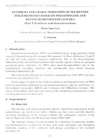
Circle of Willis: It is an important collateral
system at the base of the brain surrounding the
optic chiasm and pituitary stalk. It comprises
of—the basilar artery bifurcation (basilar tip), P1
segments of posterior cerebral artery proximal
Angiograms
segments, paired distal ICA’s, paired posterior
communicating arteries (PCOM), paired
proximal A1 segments of ACA’s and the anterior
communicating artery (ACOM). This vascular ring
is complete only in about 25 percent of cases (Fig.
7.1). Perforating vessels arising from the circle of
Willis include branches to the thalamus, limbic
system, reticular activating system, cerebral
peduncles, posterior limb of internal capsule
and oculomotor nerve nucleus. The recurrent
artery of Heubner originates from the A1 segment
to supply the anterior limb of internal capsule,
portion of the globus pallidus and head of the
caudate nucleus.
The anterior cerebral artery: The most proximal
segment is the A1 segment, its origin at the
terminal ICA to the anterior communicating
artery (ACOM). A2 segment is the portion distal
69
to the ACOM and extends into the distal ACA.
The A2 segment supplies the head of the caudate
nucleus, portions of the globus pallidus, anterior
limb of the internal capsule, anterior two-thirds of
medial cerebral cortex. The main branches of the
A2 segment are the orbitofrontal and frontopolar
arteries. The ACA bifurcates into the pericallosal
and callosomarginal arteries (Figs 7.2 to 7.6).
The middle cerebral artery: The most proximal
segment is M1 segment. It extends from ICA
bifurcation to the insular cortex (island of Reil).
M2 segment is the course of the artery in the
insular cortex and sylvian fissure and it bifurcates
into anterior and posterior cortical branches. The
branches of the anterior cortical M2 segment
are lateral orbitofrontal, operculofrontal and
central sulcus arteries. The central sulcus arteries
are called precentral (prerolandic) and central
(rolandic) branches which supply motor and
sensory cortical strips. The branches of posterior
cortical M2 segment are the anterior and posterior
parietal, angular and posterior temporal arteries
(Figs 7.2 to 7.6).
The Vertebrobasilar Circulation
Fig. 7.1: Circle of Willis
Abbreviations: ACA: Anterior cerebral artery; ACom: Anterior
communicating artery; MCA: Middle cerebral artery; ICA:
Internal carotid artery; PCom: Posterior communicating
artery; PCA: Posterior cerebral artery; SCA: Superior- internal
carotid artery; Basilar: Basilar artery; AICA: Anterior cerebral
artery; VA: Vertebral artery; ASA: Anterior spinal artery
Vertebral arteries: The vertebral arteries originate
from the subclavian arteries. One of the vertebral
arteries may be dominant in size as compared to
the other. Each vertebral artery passes through the
transverse foramen of C6 and passes superiorly
through the transverse foramina of C5 to C1, then
it courses posteriorly around the atlanto-occipital
joint and ascends through the foramen magnum,
penetrating the atlanto-occipital membrane and
dura. It gives off the posterior-inferior cerebellar
artery and the anterior spinal arteries. It then
travels superiorly around the lateral aspect of
medulla to join with the contralateral vertebral
artery to form the basilar artery at pontomedullary
junction.
The posterior inferior cerebellar artery (PICA)
provides branches to the medulla, the occlusion of
which can cause the lateral medullary syndrome
or pyramidal tract ischemia. Lateral medullary
70
Atlas on X-ray and Angiographic Anatomy
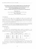
Fig. 7.2: Angiogram of right anterior cerebral circulation arterial phase—AP view
Fig. 7.3: Angiogram of right anterior cerebral circulation arterial phase—Lateral view
Angiograms
Fig. 7.4: Angiogram of right anterior cerebral circulation arterial phase—Lateral view
Fig. 7.5: Angiogram right anterior cerebral circulation capillary phase—AP view
71
72
Atlas on X-ray and Angiographic Anatomy
Fig. 7.6: Angiogram of right anterior cerebral circulation capillary phase—Lateral view
Fig. 7.7: Angiogram of right anterior cerebral circulation venous phase—AP view
Angiograms
73
Fig. 7.8: Angiogram of right anterior cerebral circulation venous phase—Lateral view
syndrome consists of ipsilateral Horner’s
syndrome, facial sensory loss, pharyngeal/
laryngeal paralysis, contralateral pain and
temperature sensory loss in the limbs and trunk.
Superior cerebellar artery provides vascular
supply to the cerebellar peduncles, vermis,
dentate nucleus, lateral pontine structures,
spinothalamic tracts and sympathetic.
Anterior spinal arteries: It originates from the
vertebral arteries distal to the posteroinferior
cerebellar artery origin, they course inferomedially
to join with their contralateral artery along the
anterior cord.
Posterior cerebral arteries: Arise from the basilar
artery at the level of pontomesencephalic junction, superior to the oculomotor nerve and
tentorium. The proximal PCA is divided into P1
and P2 segments at the junction of the PCA with the
posterior communicating artery. A filling defect is
frequently seen at the transition between P1 and
P2 during frontal vertebral artery angiograms
due to the inflow of unopacified blood from the
ipsilateral posterior communicating artery. The
proximal P2 segment gives rise to the posterior
thalamoperforating and thalamogeniculate
arteries which supply the posterior portions
of the thalamus, geniculate bodies, choroid
plexus of third and lateral ventricles, posterior
limb of internal capsule, optic tract and small
Basilar artery: The two vertebral arteries
join together to form the basilar artery at the
pontomedullary junction. The basilar artery
courses anterosuperiorly over the ventral pons. It
gives off small pontine perforating branches which
supply the pyramidal tracts, medial lemnisci, red
nuclei, respiratory centers and nuclei for cranial
nerves (III, VI, XII). The basilar artery gives off the
anterior inferior cerebellar artery and the superior
cerebellar artery. The labyrinthine artery is a
branch of the anterior inferior cerebellar artery.
74
Atlas on X-ray and Angiographic Anatomy
branches to the cerebral peduncles. The other
branches of posterior cerebral artery are the
splenial artery, anterior and posterior temporal
branches, parietooccipital artery. The distal PCA
courses posteriorly around the brainstem in the
ambient cistern, travelling more medially in the
quadrigeminal plate cistern. The distal calcarine
cortical branches converge towards the midline
but are separated by falx, on Townes projection
vertebral angiogram (Figs 7.9 to 7.12).
NORMAL INTRACRANIAL
VENOUS SYSTEM
Cerebral cortical veins: Multiple cortical veins
drain towards the superior sagittal sinus. The
superficial middle cerebral vein which lies
in the sylvian fissure may have anastomotic
communication with the deep cerebral venous
system, the facial veins and the extracranial
pterygoid venous plexus. Posteriorly the
superficial middle cerebral vein communicates
with the veins of Trolard and Labbe towards the
ipsilateral transverse sinus. The veins of Trolard
and Labbe cross the subdural space to enter the
dural sinuses.
Deep cerebral veins: These are the paired septal
veins which run close to midline beside septum
pellucidum. The paired thalamostriate veins pass
along the floor of the lateral ventricles between
the body of caudate nucleus and thalamus. The
internal cerebral veins run posteriorly in the
roof of third ventricle. The paired basal veins of
Rosenthal are formed by the confluence of deep
middle and anterior cerebral veins on the ventral
surface of brain. The basal veins then coalese
posteriorly with the internal cerebral veins to
form the vein of Galen (Figs 7.7 and 7.8). This vein
of Galen travels in the midline for about 1–2 cm
under the splenium of corpus callosum, it then
joins the inferior sagittal sinus in the posterior
fossa to form the straight sinus at the junction of
falx and tentorial incisura (Flow chart 7.3).
The posterior fossa veins: These are the anterior
pontomesencephalic veins, the precentral veins,
superior and inferior vermian veins. The anterior
Fig. 7.9: Angiogram of posterior cerebral circulation arterial phase—AP view
Angiograms
Fig. 7.10: Angiogram of posterior cerebral circulation arterial phase—Lateral view
Fig. 7.11: Angiogram of posterior cerebral circulation capillary phase—AP view
75
76
Atlas on X-ray and Angiographic Anatomy
Fig. 7.12: Angiogram of posterior cerebral circulation capillary phase—Lateral view
pontomesencephalic vein runs along the ventral
surface of pons, it drains either into the basal
vein of Rosenthal or posterior mesencephalic
vein (Figs 7.13 and 7.14). The precentral veins run
along the posteriorly in the roof of fourth ventricle
and drains into the vein of Galen (Flow chart 7.4).
Dural sinuses: The dura mater which envelops the
central nervous system has two layers that form
the reflections like the falx cerebri, tentorium and
falx cerebelli. The layers of dura separate to form
venous drainage channels or dural sinuses for the
brain. Some of them anastomose with the veins of
scalp through the emissary veins. The main dural
sinuses found are the superior sagittal sinus,
inferior sagittal sinus, occipital sinuses, paired
transverse sinuses and paired cavernous sinuses
(Figs 7.7 and 7.8).
The superior sagittal sinus travels along the
superior margin of falx cerebri, it continues
posteriorly and inferiorly in a cresenteric course to
the junction point between the falx and tentorium
containing the confluence of sinuses—The torcular
Herophili near the occipital protuberance.
The inferior sagittal sinus is found within
the lower edge of falx between the cerebral
hemispheres. It drains posteriorly to join with
the vein of Galen forming the straight sinus. The
straight sinus drains posteriorly in midline into
the torcular herophili.
The occipital sinuses are of variable size, are
seen to course superomedially within the dura of
the posterior fossa, just lateral to foramen magnum
and drains towards the torcular herophili.
The paired transverse sinus follows a
cresenteric course within the periphery of the
tentorium, laterally and anteriorly from the
torcula. The transverse sinuses receive drainage
from the inferior cerebral veins and vein of Labbe,
it communicates with the cavernous sinuses via
Angiograms
77
Fig. 7.13: Angiogram of posterior cerebral circulation venous phase—AP view
Fig. 7.14: Angiogram of posterior cerebral circulation venous phase—Lateral view
the superior petrosal sinuses, which run along the
petrous bone and as it nears the tentorium it is
called the sigmoid sinus which later empties into
the internal jugular vein (Flow chart 7.5).
78
Atlas on X-ray and Angiographic Anatomy
Flow chart 7.3: Normal venous anatomy of the brain
Flow chart 7.4: Posterior fossa veins and jugular bulb
Angiograms
79
Flow chart 7.5: Dural sinuses
The paired cavernous sinuses receive venous
drainage from the orbits through the superior
and inferior ophthalmic veins. The jugular bulbs
communicate with the cavernous sinuses by
means of the paired inferior petrosal sinuses. The
inferior petrosal sinuses also interconnect with
those on the opposite side through a clival venous
plexus.
THE THORACIC AORTA
The ascending aorta arises at the aortic root,
from the left ventricle. Immediately above the
aortic root, the ascending aorta bulges to form
the aortic sinuses, the aortic sinuses give rise
to right and left coronary arteries to supply the
heart. The ascending aorta the courses upwards
and continues as the aortic arch. The main
branches of the aortic arch (arch of aorta) are
the brachiocephalic trunk, left common carotid
artery and the left subclavian artery (Figs 7.15
and 7.16). Sometimes the thyroidea ima artery
may arise from the aortic arch. These branches
of aortic arch supply the head, neck, brain and
upper limbs (Flow chart 7.6).
The aortic arch on plain chest X-ray appears
behind the mediastinal structures in midline.
The aortic knuckle or arch at the level of sternal
angle (angle of Louis). Sometimes age-related
calcification may be noted at this site. The arch
of aorta passes above the left bronchus and to
80
Atlas on X-ray and Angiographic Anatomy
Fig. 7.15: Outline of the thoracic aorta on chest X-ray—PA view. (A) Ascending thoracic aorta curves upwards and at the level
of sternal angle continues as arch of aorta; (B) Arch of aorta curves above the left main bronchus and descends into posterior
mediastinum. It gives off the: 1. Brachiocephalic trunk; 2. Left common carotid artery; 3. Left subclavian artery; (C) At the level
of 4th thoracic vertebra, the arch of aorta becomes the descending thoracic aorta; (D) Descending thoracic aorta in posterior
mediastinum enters the abdominal cavity through the aortic hiatus (12th dorsal vertebra level)
Fig. 7.16: Angiogram showing the thoracic aorta
Angiograms
81
Flow chart 7.6: Thoracic aorta
the left of trachea and esophagus. At the level of
4th thoracic vertebra the arch of aorta courses
downwards as the descending thoracic aorta in
the posterior mediastinum.
The descending thoracic aorta gives off posterior intercostal arteries, 9 in number on either
side. These intercostal arteries pass laterally into
the intercostal spaces. At the level of the aortic
hiatus in diaphragm (at 12th thoracic vertebra),
the descending aorta passes into the abdominal
cavity and continues in the abdomen as the
abdominal aorta.
ABDOMINAL ANGIOGRAPHY
ABDOMINAL AORTA
The abdominal aorta is the continuation of the
thoracic aorta below the diaphragm at T12 vertebral
level. In the abdomen aorta is retroperitoneal in its
course and travels downwards to its bifurcation
at the level of L4 vertebral body. The abdominal
aorta supplies the viscera, peritoneum, gonads
and spine during its course. Its anterior branches
are the celiac arterial trunk, superior mesenteric
artery, inferior mesenteric artery (Fig. 7.17).
Its lateral branches are inferior phrenic artery,
suprarenal arteries, gonadal arteries, lumbar
arteries. Its terminal branches at L4 vertebral level
are the common iliac arteries and the median
sacral artery (Flow chart 7.7).
CELIAC TRUNK
The celiac trunk is the main vascular supply
of the foregut supplying the lower part of the
esophagus to the duodenum; it also supplies the
liver, pancreas and spleen. The celiac trunk arises
at the level of T12 vertebra from the abdominal
aorta and courses forwards until the upper border
of pancreas and terminates into: the left gastric
artery, splenic artery, common hepatic artery (Fig.
7.18). The left gastric artery gives off esophageal
branches, then courses to the right along the lesser
curvature of stomach and gives of branches to the
stomach. The splenic artery courses to the left, is
tortuous and runs in the splenorenal ligament to
the hilum of the spleen. Before giving off terminal
splenic branches it gives off 6-7 short gastric arteries
which course in gastrosplenic ligament and the left
gastroepiploic artery (which supplies the stomach
and omentum).The splenic artery also gives off the
posterior gastric artery during its course to splenic
hilum. The common hepatic artery courses
over the upper border of the pancreas, the main
branches are: right gastric artery, gastroduodenal
artery, small supraduodenal arteries and terminal
branch—The hepatic artery. The right gastric artery
runs forwards in the lesser omentum and to the
left in lesser curvature of stomach to anastomose
with the left gastric artery. The gastroduodenal
artery passes behind the 1st part of duodenum
82
Atlas on X-ray and Angiographic Anatomy
Fig. 7.17: Angiogram of abdominal aorta
Flow chart 7.7: Abdominal aorta branches
and at the lower border of duodenum divides
into the right gastroepiploic artery and superior
pancreaticoduodenal arteries. The supraduodenal
arteries are smaller branches arise from the
common hepatic artery. The common hepatic
artery at the porta hepatis divides into the right
and left hepatic arteries to supply the liver (Flow
chart 7.8).
Angiograms
83
Fig. 7.18: Angiogram of celiac arterial trunk
Flow chart 7.8: Celiac arterial trunk (artery of foregut)
SUPERIOR MESENTERIC ARTERY
The superior mesenteric artery is the artery of
mid- gut and supplies the gut from the bile duct
entrance to the splenic flexure of colon. This
artery arises from the abdominal aorta at the level
of lower border of L1 vertebra. It courses behind
the body of pancreas, later it lies anterior to the
left renal vein, uncinate process of pancreas and
third part of duodenum. Its main branches are
84
Atlas on X-ray and Angiographic Anatomy
Fig. 7.19: Angiogram of superior mesenteric artery
the inferior pancreaticoduodenal artery, jejunal
and ileal branches, ileocolic artery, right colic
artery, middle colic artery (Fig. 7.19). The inferior
pancreaticoduodenal artery is the first branch of
superior mesenteric artery. It further divides into
anterior and posterior branches to supply the
head of pancreas and adjacent duodenum. The
jejunal and ileal branches pass between the two
layers of the mesentery and create a network of
arteries along the jejunum and ileum to supply
the same. The ileocolic artery courses down to
the base of mesentery into the right iliac fossa
and divides into superior and inferior branches.
The superior branch courses along the ascending
colon to anastomose with the right colic artery.
The inferior branch courses down to the ileocolic
junction and gives off the anterior and posterior
cecal arteries, an appendicular artery and an
ileal artery that anastomoses with the terminal
branches of superior mesenteric artery. The
right colic artery course downwards into the
right infracolic compartment and divides into
the ascending and descending branches. The
ascending branch courses along the ascending
colon upwards to anastomose with a branch from
middle colic artery at hepatic flexure of colon. The
descending branch courses downwards along the
ascending colon to anastomose with a branch
from the ileocolic artery. The middle colic artery
arises from the superior mesenteric artery at the
lower border of neck of pancreas. It courses into
the transverse mesocolon and on the right side
of transverse colon divides into two branches
– The right and left branches. The right branch
anastomoses with the ascending branch of right
colic artery. The left branch anastomoses with a
branch of the left colic artery (Flow chart 7.9).
INFERIOR MESENTERIC ARTERY
It is also called as the artery of hindgut. It arises as
an anterior branch of abdominal aorta at the level
of L3 vertebra and courses downwards in lower
abdomen. Its branches are the left colic artery,
Angiograms
sigmoidal arteries and superior rectal artery. These
branches supply the descending colon, sigmoid
colon and upper rectum. The marginal artery
85
of Drummond is formed by an interconnecting
anastomotic network of the branches along the
mesenteric border of large bowel. The marginal
Flow chart 7.9: Superior mesenteric arteriogram (artery of midgut)
Fig. 7.20: Angiogram of right renal artery early arterial phase
86
Atlas on X-ray and Angiographic Anatomy
Fig. 7.21: Angiogram of right renal artery late arterial phase
Fig. 7.22: Angiogram of right renal artery nephrogram phase
Angiograms
Fig. 7.23: Angiogram of renal arteries in pyeloureterogram phase
Flow chart 7.10: Renal artery angiogram
87
88
Atlas on X-ray and Angiographic Anatomy
artery of Drummond is crucial to maintain the
vascular supply of large bowel.
RENAL ARTERY
Both the renal arteries arise at right angles to the
abdominal aorta at the level of L2 vertebra. The left
artery is shorter than the right. Each renal artery gives
off small suprarenal and ureteric branches. The renal
arteries course behind the pancreas and the renal vein
to reach the hilum of the kidney on either side (Figs
7.20 to 7.23). At the hilum the renal artery branches
into anterior and posterior divisions. Each kidney is
subdivided into five segments based on arterial supply.
The anterior arterial division supplies the apical,
upper, middle and lower segments while the posterior
arterial division supplies the posterior segment (Flow
chart 7.10). There is no collateral circulation between
these segmental arteries. The segmental arteries are
accompanied by their corresponding veins. Each
segmental artery divides into lobar artery, interlobar
artery, arcuate artery and finally into interlobular
arteries. The segmental veins communicate with each
other and at the hilum they join to form the renal vein.
At the hilum of each kidney the structures from front
to back are vein, artery and ureter.
UPPER LIMB ANGIOGRAPHY
ARTERIAL SYSTEM
The axillary artery is the main artery supplying the
upper extremity. It is a continuation of the third part
of the subclavian artery. The axillary artery begins
at the outer border of the first rib and continues
until the lower border of teres major muscle (Fig.
7.24). Beyond the teres major muscle the axillary
artery continues into the arm as the brachial
artery (Flow chart 7.11 and 7.12). The axillary
artery for description purposes is subdivided into
three parts by the pectoralis minor muscle which
crosses middle 1/3rd the axillary artery. The 1st
part of axillary is proximal to pectoralis muscle; it
gives off the superior thoracic artery. The 2nd part
of axillary artery is beneath the pectoralis minor
muscle, it gives off the lateral thoracic artery and
the thoracoacromial artery. The 3rd part of axillary
artery is distal to the pectoralis minor muscle; it
gives off the subscapular artery, anterior humeral
circumflex artery and the posterior circumflex
artery.
The brachial artery is continuation of axillary
artery in arm. The artery is superficial in its
course and lies beneath the deep fascia in the
anteromedial aspect of arm. Its branches are: the
profunda brachii artery, middle collateral artery,
radial collateral artery, superior ulnar collateral
artery, inferior ulnar collateral artery, muscular
branches to flexor muscles and nutrient artery to
humerus (Figs 7.25 and 7.26).
The radial artery originates as a terminal
branch of the brachial artery at the cubital fossa.
It runs deep to the brachioradialis muscle on the
lateral aspect of forearm and at the wrist joint it
courses in the anatomical snuff box and forms
the deep palmar arch. The radial artery gives
small muscular branches in forearm, the radial
recurrent artery and a superficial branch near the
radiocarpal joint (Flow chart 7.13). The princeps
pollicis artery is a branch of radial artery in hand,
it divides into two smaller branches that run
laterally along the thumb (Figs 7.27 and 7.28).
The ulnar artery arises as a terminal branch of
the brachial artery at cubital fossa. It courses on
the medial aspect of forearm deep to the flexor
muscles. The ulnar artery gives off the anterior
and posterior ulnar recurrent arteries in proximal
forearm and also a few muscular branches along
its course in forearm. The ulnar artery passes
superficial to the flexor retinaculum at the wrist
joint and continues as the superficial palmar arch
Angiograms
Fig. 7.24: Angiogram showing subclavian artery and axillary artery
Fig. 7.25: Angiogram showing brachial artery
89
90
Atlas on X-ray and Angiographic Anatomy
Fig. 7.26: Angiogram showing radial and ulnar arteries
Fig. 7.27: Angiogram showing ulnar artery and anterior interosseous artery
Angiograms
in the hand. A deep branch of the ulnar artery in
hand anastomoses with the deep palmar arch to
maintain collateral circulation.
The common interosseous artery is a branch
of the ulnar artery close to cubital fossa. It divides
into the anterior and posterior interosseous
branches distal to the radial tubercle and supplies
the muscles of the forearm (Figs 7.27 and 7.28).
The superficial palmar arch is a direct continuation of the ulnar artery in the hand, it is joined
on its lateral side by the superficial branch of
radial artery to complete the superficial palmar
arch.
The deep palmar arch is a direct continuation
of the radial artery, it is joined on its medial side
by the deep branch of ulnar artery to complete the
deep palmar arch (Fig. 7.29).
The dorsal carpal arch is formed by both the
radial and ulnar arteries within the fascia on
dorsum of hand.
91
Venous System
The veins of the upper extremity can be classified
into the superficial veins and the deep veins. The
superficial veins are digital veins, metacarpal
veins, cephalic veins, basilic vein and median
vein. The deep veins are the venae comitantes of
radial and ulnar arteries, volar arches of hand,
brachial vein, axillary vein and subclavian vein.
Superficial Veins
The digital veins are subclassified into dorsal and
volar digital veins. The dorsal digital veins pass
along the sides of the fingers and are joined to one
another by oblique communicating branches.
They have an ulnar and radial network of veins on
either side. A communicating branch frequently
connects the dorsal venous network with the
cephalic vein about the middle of the forearm. The
volar digital veins on each finger are connected to
Fig. 7.28: Angiogram showing superficial palmar arch