Ebook Ferri''s fast facts in dermatology: Part 2
Bạn đang xem bản rút gọn của tài liệu. Xem và tải ngay bản đầy đủ của tài liệu tại đây (19.37 MB, 394 trang )
CHAPTER 3
DISEASES AND DISORDERS
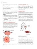
1. ACANTHOSIS NIGRICANS (AN)
FIGURE 03-001. Velvety thickening of skin in
flexural areas such as the axilla with brown-black
hyperpigmentation. Because of excess chafing,
skin tags appear occasionally, as seen in this
obese patient.
FIGURE 03-002. Darkened, “dirty” appearing thick
skin with prominent lighter-colored deep skin
lines. Several overlying warty or papillomatous
growths are common in this condition.
General Comments
Definition
■ Acanthosis nigricans refers to the presence of symmetrical brown velvety or
verrucous plaques with a predilection for intertriginous sites as the back of the
neck, groin, and axillae (Fig. 03-001).
Etiology
It is most commonly seen in obese individuals with insulin resistance or an
internal malignancy and in those taking certain medications (nicotinic acid,
glucocorticoids, contraceptives, and diethylstilbestrol).
■
Keys to Diagnosis
Clinical Manifestation(s)
■ Asymptomatic. The axilla and neck are the most commonly involved. In
obese females who are hyperandrogenic, the vulva is the most commonly
affected site.
Physical Examination
Symmetrical hyperpigmented velvety plaques of the major flexures (axilla, groin),
neck (Fig. 03-002), nipples, and vulva.
■
42
Acne Keloidalis
Diagnostic Tests
■ Laboratory evaluation often reveals elevated glucose levels. Additional useful
laboratory tests are thyroid-stimulating hormone (TSH) and follicle-stimulating
hormone (FSH)/luteinizing hormone (LH).
Differential Diagnosis
■
■
■
■
■
Seborrheic keratosis
Hyperpigmented nevus (Becker nevus), linear epidermal nevus
Pemphigus vegetans
Lichen simplex chronicus
Confluent and reticulated papillomatosis
Treatment
First Line
■ Therapy for underlying cause (weight loss in obese, discontinuation of offending
drugs, treatment of malignancy if present)
Second Line
■ Topical tretinoin, dermabrasion, ammonium lactate, carbon dioxide laser
Third Line
Oral contraceptives, cyproheptadine, oral isotretinoin
■
Clinical Pearl(s)
■
■
■
The sudden onset of acanthosis nigricans should be followed by investigation
for internal malignancy (e.g., upper endoscopy to rule out gastric cancer and
computed tomography [CT] of abdomen and pelvis).
Skin changes precede the malignancy diagnosis (usually neoplasm of abdominal
cavity) in one third of cases.
Consider drug use as a cause and review new medications (e.g., nicotinic acid,
contraceptives, glucocorticoids).
2. ACNE KELOIDALIS
FIGURE 03-003. A papular, pustular eruption
found on the nape of the neck and often associated with keloidal scarring. A typical finding as
seen here is the emergence of hair follicles from
the center of the lesion.
DISEASES AND DISORDERS
43
General Comments
Definition
■ Acne keloidalis is an idiopathic chronic inflammatory eruption of the nape of
the neck occurring most commonly in dark-skinned men. Also known as acne
keloidalis nuchae, acne keloid, and folliculitis keloidalis. These are misnomers
because there is no family history of keloids, no presence of keloids at other sites,
and no development of keloid formation following excision. Despite the name,
acne vulgaris also is not associated.
Etiology
Unknown. Close shaving of the hair, picking by patients, and chronic rubbing by
collars have been suggested as possible contributing factors.
■
Keys to Diagnosis
Clinical Manifestation(s)
■ Onset is usually after puberty and before age 50.
■ Clinical presentation consists of a follicular pustular eruption on the nape of the
neck (Fig. 03-003).
Physical Examination
Hard papules with hair emerging from the center are seen on the nape of neck
and occipital scalp. Comedomes are not seen.
■ Papules coalesce into sclerotic plaques.
■ Pustules, crusting, and drainage may occur with secondary infections.
■
Diagnostic Tests
Pustule swab
■ Deep biopsy
■
Differential Diagnosis
■
■
■
■
■
■
■
Folliculitis
Simple ingrowing hairs (pili incurvatorum)
Nevus sebaceous
Traumatic causes of keloid
Acne vulgaris
Pseudofolliculitis
Pediculosis capitis
Treatment
First Line
■ Dissuade close cutting. Allow hair to grow long in affected areas.
■ Limit mechanical irritation by a tight collar.
44
■
■
Acne Vulgaris
Encourage patient not to pick or squeeze lesions.
Administer topical antibiotics (clindamycin or erythromycin).
Second Line
Oral doxycycline, tetracycline, or minocycline
■
Third Line
Intralesional triamcinolone alone or following use of CO2 laser vaporization
■ Oral isotretinoin
■ Surgery: punch biopsy for small papular lesions, surgical debulking for larger
lesions. Any excision must be carried out to the subfollicular depth. If any of
the hair follicle is left, recurrence is common.
■
Clinical Pearl(s)
■
Most cultures are sterile, but when a bacterium is found it is usually Staphylococcus
aureus
3. ACNE VULGARIS
FIGURE 03-004. Erythematous nodulocystic
acne vulgaris found on the back, often resulting
in scarring.
FIGURE 03-005. Acne vulgaris consisting of open
comedones (“blackheads”) and closed comedones
(“whiteheads”).
General Comments
Definition
■ Acne vulgaris is a chronic disorder of the pilosebaceous apparatus caused by abnormal
desquamation of follicular epithelium leading to obstruction of the pilosebaceous
canal, resulting in inflammation and subsequent formation of papules, pustules,
nodules, comedones, and scarring (Fig. 03-004). Acne can be classified by the type
of lesion (comedonal, papulopustular, and nodulocystic). The American Academy
of Dermatology classification scheme for acne denotes the following three levels:
1. Mild acne: characterized by the presence of comedomes (noninflammatory
lesions), few papules and pustules (generally Ͻ10), but no nodules.
DISEASES AND DISORDERS
FIGURE 03-006. Acne vulgaris is often found on
the face. Other common areas include the chest
and the back.
45
FIGURE 03-007. Presence of scars and comedones.
2. Moderate acne: presence of several to many papules and pustules (10-40)
along with comedomes (10-40). The presence of more than 40 papules and
pustules along with larger, deeper nodular inflamed lesions (up to 5) denotes
moderately severe acne.
3. Severe acne: presence of numerous or extensive papules and pustules as well as
many nodular lesions.
Etiology
■ Overactivity of the sebaceous glands and blockage in the ducts result in acne
vulgaris. The obstruction leads to the formation of comedones, which can
become inflamed because of overgrowth of Propionibacterium acnes.
■ The condition is exacerbated by environmental factors (hot, humid, tropical
climate), medications (e.g., iodine in cough mixtures, hair greases), and industrial
exposure to halogenated hydrocarbons.
Keys to Diagnosis
Clinical Manifestation(s)
■ Various stages of development and severity may be present concomitantly.
■ Common distribution of acne is on the face, back, and upper chest.
46
Acne Vulgaris
Physical Examination
■ Open comedones (blackheads), closed comedones (whiteheads)
(Fig. 03-005)
■ Greasiness (oily skin) (Fig. 03-006)
■ Presence of scars from prior acne cysts (Fig. 03-007)
■ Inflammatory papules, pustules, and ectatic pores
Diagnostic Tests
Laboratory evaluation is generally not helpful.
■ Patients who are candidates for therapy with isotretinoin (Accutane) should
have baseline liver enzymes, cholesterol, and triglycerides checked, because this
medication may result in elevation of lipids and liver enzymes.
■ A negative serum pregnancy test or two negative urine pregnancy tests should
also be obtained in female patients 1 week before initiation of isotretinoin; it is
also imperative to maintain effective contraception during and 1 month after
therapy with isotretinoin ends because of its teratogenic effects. Pregnancy status
should be rechecked at monthly visits.
■ In female patients, if hyperandrogenism is suspected, levels of dehydroepiandrosterone sulfate (DHEAS), testosterone (total and free), and androstenedione
should be measured. Generally for women with regular menstrual cycles, serum
androgen measurements are not necessary.
■
Differential Diagnosis
■
■
■
■
■
■
■
■
■
Gram-negative folliculitis
Staphylococcal pyoderma
Acne rosacea
Drug eruption
Sebaceous hyperplasia
Angiofibromas, basal cell carcinomas, osteoma cutis
Occupational exposures to oils or grease
Steroid acne
Flat warts
Treatment
First Line
Treatment generally varies with the type of lesions (comedones, papules, pustules,
cystic lesions) and the severity of acne.
■ Comedones (noninflammatory acne) can be treated with retinoids or retinoid
analogs. Topical retinoids are comedolytic and work by normalizing follicular
keratinization. Commonly available agents are adapalene (0.1% gel or cream,
applied once or twice daily), tazarotene (0.1% cream or gel applied daily),
tretinoin (0.1% cream or 0.025% gel applied once nightly), tretinoin microsphere (0.1% gel, applied at bedtime). Tretinoin is inactivated by UV light and
DISEASES AND DISORDERS
■
■
■
■
47
oxydized by benzoyl peroxide; therefore, it should only be applied at night and
not used concomitantly with benzoyl peroxide.
Tretinoin is pregnancy category C; tazarotene is pregnancy category X. Salicylic
acid preparations (e.g., 2% wash) have keratolytic and antiinflammatory
properties and are also useful in the treatment of comedones. Large open
comedones (blackheads) should be expressed.
Patients should be reevaluated after 4 to 6 weeks. Benzoyl peroxide gel (2.5% or
5%) may be added if the comedones become inflamed or form pustules. The most
common adverse effects are dryness, erythema, and peeling. Topical antibiotics
(erythromycin, clindamycin lotions or pads) can also be used in patients with
significant inflammation. They reduce P. acnes in the pilosebaceous follicle and have
some antiinflammatory effects. The combinations of 5% benzoyl peroxide and 3%
erythromycin or 1% clindamycin with 5% benzoyl peroxide are highly effective in
patients who have a mixture of comedonal and inflammatory acne lesions.
Pustular acne can be treated with tretinoin and benzoyl peroxide gel applied on
alternate evenings; drying agents (e.g., sulfacetamide/sulfur lotions) are also
effective when used in combination with benzoyl peroxide.
Azelaic acid, a bacteriostatic dicarboxylic acid, is used to normalize keratinization
and reduce inflammation.
Second Line
Oral antibiotics (doxycycline 100 mg QD or erythromycin 1 g QD in 2-3 divided
doses) are effective in patients with moderate to severe pustular acne; patients not
responding well to these antibiotics can be switched to minocycline 50 to 100 mg
BID; however, this medication is more expensive.
■ Patients with nodular cystic acne can be treated with systemic agents: antibiotics
(erythromycin, tetracycline, doxycycline, minocycline), isotretinoin, or oral
contraceptives. Periodic intralesional triamcinolone injections are also effective.
The possibility of endocrinopathy should be considered in patients responding
poorly to therapy.
■ Oral contraceptives reduce androgen levels and therefore sebum production.
They represent a useful adjunctive therapy for all types of acne in women and
adolescent girls. Commonly used agents are norgestimate/ethinyl estradiol and
drospirenone/ethinyl estradiol.
■ Spironolactone 100 to 200 mg/day can be administered to women only.
■ Blue light can be used for treatment of moderate inflammatory acne vulgaris.
Light in the violet/blue range can cause bacterial death by a photoreaction in
which porphyrins react with oxygen to generate reactive oxygen species, which
damage the cell membranes of P. acnes. Treatment usually consists of 15-minutes
of exposure twice weekly for 4 weeks.
■
Third Line
Isotretinoin is indicated for acne resistant to antibiotic therapy and severe acne;
dosage is 0.5 to 1 mg/kg/day in 2 divided doses (maximum of 2 mg/kg/day);
duration of therapy is generally 20 weeks for a cumulative dose 120 mg/kg
■
48
Acrochordon
or more for severe cystic acne; before using this medication, patients should
undergo baseline laboratory evaluation. This drug is absolutely contraindicated
during pregnancy because of its teratogenicity. It should be used with caution in
patients with a history of depression. In order to prescribe this drug, physicians
must be registered members of the manufacturer’s System to Manage AccutaneRelated Teratogenicity (SMART) program.
Clinical Pearl(s)
■
■
■
■
■
■
■
■
■
Gram-negative folliculitis should be suspected if inflammatory acne worsens after
several months of oral antibiotic therapy.
Acne may worsen during the first 3 to 4 weeks of retinoid therapy before
improving.
Indications for systemic therapy of acne are painful deep papules or nodules,
extensive lesions, active acne with severe scarring or hyperpigmentation, and
patient’s morale.
Erythromycin has a high incidence of early drug resistance.
Doxycycline has a high incidence of sun sensitivity.
Benzoyl peroxide will cause bleaching of clothes.
Spironolactone can produce menstrual irregularity.
Tetracyclines are contraindicated in children and pregnant women.
Isotretinoin is contraindicated in patients with depression.
4. ACROCHORDON
FIGURE 03-008. Acrochordons are soft, fleshcolored, pedunculated papules that are commonly
located on the neck and axilla.
General Comments
Definition
■ Acrochordons are benign outgrowths of the skin, also known as skin tags or
fibroepithelial polyps.
DISEASES AND DISORDERS
49
Etiology
■ Unknown. They are more prevalent in obese individuals and in women.
Acrochordon may be associated with pregnancy and acanthosis nigricans.
Keys to Diagnosis
Clinical Manifestation(s)
■ This condition is asymptomatic unless irritated by clothing, jewelry, or friction.
It is most common in middle-aged and elderly persons.
Physical Examination
Skin-colored or brown fleshy outgrowths (Fig. 03-008) are usually seen on the
side of the neck and around the axillae and groin.
■
Diagnostic Tests
None necessary. A shave/snip biopsy can be done when diagnosis is unclear.
■
Differential Diagnosis
■
■
■
■
■
■
Wart
Seborrheic keratosis
Melanocytic nevus
Dermatosis papulosa nigra
Neurofibroma
Melanoma
Treatment
First Line
■ No treatment is needed.
■ Scissor excision with or without local anesthesia may be done for cosmetic
reasons or when the skin tag is irritated.
Second Line
Electrodessication
■
Third Line
Liquid nitrogen cryosurgery
■
Clinical Pearl(s)
■
■
Skin tags in periorbital area are often confused with neoplastic skin lesions.
Freezing of a skin tag in dark-skinned patients may result in a white spot.
50
Actinic Keratosis
5. ACTINIC KERATOSIS
FIGURE 03-009. Several scaly, adherent, yellowbrown lesions on the sun-exposed dorsum of
the hand.
FIGURE 03-010. An actinic keratosis located on
this patient’s forehead is often best appreciated
by its rough, tactile quality, similar to that of
sandpaper.
FIGURE 03-011. Scaly, raised lesion on sunexposed back. Pain was elicited when scraping
this lesion.
FIGURE 03-012. Raised, rough, gritty actinic
keratosis on the anterior thigh of an outdoorsman.
General Comments
Definition
■ Actinic keratosis is a common skin lesion usually presenting as multiple,
erythematous or yellow-brown, dry, scaly lesions in the middle aged or elderly.
It is also known as solar keratosis or senile keratosis.
Etiology
Sun exposure, ionizing radiation
■
DISEASES AND DISORDERS
51
Keys to Diagnosis
Clinical Manifestation(s)
■ Typical lesions occur on sun-damaged skin usually on the face and neck and the
dorsal aspects of hands (Fig. 03-009) and forearms.
■ Actinic keratosis is more common in males than females, especially in those with
fair complexions who burn rather than tan following sun exposure.
Physical Examination
Advanced lesions are characterized by a hard spiky scale (Fig. 03-010) and
usually measure 1 cm in diameter or less. Early lesions manifest with redness and
minimal scale. With progression scales become thicker and yellow (Fig. 03-011)
and may resemble a small squamous cell carcinoma. On examinations lesions are
rough and gritty (Fig. 03-012)
■ The surrounding skin often shows additional features of sun damage, including
atrophy, pigmentary changes, and telengiectasia.
■
Diagnostic Tests
Skin biopsy can be performed for recurrent lesions or when diagnosis is unclear
to rule out squamous cell or basal cell carcinoma.
■
Differential Diagnosis
■
■
■
■
■
■
■
■
Lentigo maligna (heavily pigmented variants may be clinically mistaken for this
condition)
Basal cell or squamous cell carcinoma
Seborrheic keratosis
Eczema
Bowen’s disease (intraepithelial carcinoma)
Wart
Lichenoid keratosis
Cutaneous lupus
Treatment
First Line
■ Avoidance of sun exposure, use of sunscreens
■ Cryosurgery with liquid nitrogen
Second Line
Topical 5-fluorouracil BID for 3 to 6 weeks
■ Topical diclofenac
■ Carbon dioxide laser
■ Dermabrasion
■ Curettage
■
52
Alopecia Areata
Third Line
■ Excision
■ Photodynamic therapy with aminolevulinic acid and blue light
■ Imiquimod 5% cream BID for 3 to 4 months
■ Oral retinoids
Clinical Pearl(s)
■
■
■
Actinic keratoses are of particular importance because they are a sensitive indicator of exposure to ultraviolet (UV) light and strongly predict the likelihood of
developing cutaneous squamous cell carcinoma.
The cumulative probability of development of invasive squamous cell carcinoma
in patients with 10 or more actinic keratoses has been estimated at 14% in a
5-year period.
It is estimated that up to 10% of actinic keratoses tend to progress to invasive
carcinoma.
6. ALOPECIA AREATA
FIGURE 03-013. Round, well-demarcated area of
hair loss is characteristic of alopecia areata.
FIGURE 03-014. Alopecia areata presenting as
an annular band of hair loss anterior to the right
ear in this case with no erythema or scarring.
General Comments
Definition
■ Alopecia areata is a variant of alopecia in which large numbers of hair follicles
undergo progression into catagen and telogen while smaller numbers enter
an abnormal anagen stage. Alopecia areata is also known as autoimmune
alopecia.
■ Alopecia areata affects up to 1% of the population and is more common between
15 and 40 years of age.
DISEASES AND DISORDERS
53
Etiology
■ Alopecia areata is basically a disease driven by cellular immunity with autoantibody
production representing a secondary phenomenon.
■ The increased frequency of this disorder in genetically related individuals suggests
that there is a genetic link to the disease.
■ Histologically, alopecia areata is characterized by normal numbers of follicular
units and hair follicles, an increase in the number of catagen and telogen follicles,
and a lymphocytic infiltrate affecting the bulbs of the anagen.
Keys to Diagnosis
Clinical Manifestation(s)
■ Typically patients present with an abrupt development of patches of nonscarring
alopecia in different patterns: circumscribed (Fig. 03-013), bandlike (Fig. 03-014),
and reticular. The degree of involvement is highly variable and can range from very
mild disease to diffuse hair loss that may affect the entire scalp (alopecia totalis).
Physical Examination
Examination of the involved scalp generally reveals that except for the absence of
hair, the skin appears normal.
■
Diagnostic Tests
Laboratory evaluation is generally not helpful.
■ Antinuclear antibody (ANA), TSH, complete blood cell count (CBC), and B12
level should be considered in patients with a family history of the disease or other
manifestations of autoimmune diseases.
■ VDRL can be performed in selected patients.
■
Differential Diagnosis
■
■
■
■
■
Androgenic alopecia
Trichotillomania
Secondary syphilis
Telogen effluvium
Tinea capitis
Treatment
First Line
■ Intralesional corticosteroids (triamcinolone acetonide, 5 to 10 mg/mL, raising a
small bleb within the affected patch)
Second Line
Topical corticosteroids such as clobetasol 0.05% cream BID, cycled (2 weeks on,
1 week off)
■ Topical minoxidil
■
54
■
■
Amalgam Tattoo
Topical sensitizing agent or irritants (dithranol, diphencyprone)
Phototherapy (ultraviolet radiation or psoralen with ultraviolet A [PUVA])
Third Line
Systemic immune modulators (e.g., cyclosporine)
■ Oral minoxidil
■
Clinical Pearl(s)
■
■
Fifty percent of cases resolve spontaneously without treatment within 1 year.
Ten percent evolve to chronic disease.
7. AMALGAM TATTOO
FIGURE 03-015. Amalgam tattoo is a benign
hyperpigmented area of the gingival mucosa
adjacent to teeth with amalgam fillings.
FIGURE 03-016. Amalgam tattoo, occasionally
mistaken for melanoma, results from the local
absorption of amalgam particles (mercury, silver,
or copper) used to fill carious teeth.
General Comments
Definition
■ Amalgam tattoo is characterized by painless gray, bluish, black, or slate-colored
macules that generally occur on the gingival/alveolar ridge or buccal mucosa.
Etiology
Particles of amalgam restorations may be traumatically implanted into the mucosa
by the dentist during placement or removal of a restoration, by the patient from
bite injury, from leakage and disintegration of a restoration (or root canal filling
material), or from a restoration falling into a tooth socket after extraction.
■
Keys to Diagnosis
Clinical Manifestation(s)
■ This condition is asymptomatic, generally noted by dentist during routine dental
examination.
DISEASES AND DISORDERS
55
Physical Examination
■ Gray, bluish, black or slate-colored macules can be seen on the gingival/alveolar
ridge or buccal mucosa (Fig. 03-015).
Diagnostic Tests
None necessary. Biopsy only when diagnosis is uncertain and neoplasm is being
considered.
■
Differential Diagnosis
■
■
Melanoma or mucosal melanosis
Nevus
Peutz-Jeghers
Hemangioma or venous lake
■
No treatment is necessary.
■
The only significance of this lesion is that its appearance can be mistaken for
melanoma (Fig. 03-016).
■
■
Treatment
Clinical Pearl(s)
8. ANAGEN EFFLUVIUM
FIGURE 03-017. Generalized hair loss, thinning
of hair shafts, and normal appearing scalp
secondary to chemotherapy for breast cancer
in this patient.
General Comments
Definition
■ Anagen effluvium is nonscarring hair loss of the scalp following a toxic insult to
growing hair (in anagen phase).
Etiology
Cancer chemotherapy (e.g., cyclophosphamide, nitrosoureas, doxorubicin) is the
most common cause.
■
56
Androgenic Alopecia
Keys to Diagnosis
Clinical Manifestation(s)
■ Hair loss occurs usually within 2 weeks of cancer chemotherapy.
Physical Examination
Hair loss may be slight but is often extensive.
■ Alopecia is noninflammatory and nonscarring (Fig. 03-017).
■
Diagnostic Tests
None necessary.
■
Differential Diagnosis
■
■
Iron deficiency
Malnutrition
Androgenic alopecia
Telogen effluvium
Trichotillomania
Traction alopecia
■
No treatment is necessary; the disorder is self-limited.
■
Be sympathetic, even if hair loss seems trivial to you. Reassure patient that hair
loss is only temporary.
■
■
■
■
Treatment
Clinical Pearl(s)
9. ANDROGENIC ALOPECIA
FIGURE 03-018. Frontal recession of hairline
typical of early androgenic alopecia.
FIGURE 03-019. Progressive androgenic alopecia
with loss of hair extending from frontal to vertex
regions.
DISEASES AND DISORDERS
57
FIGURE 03-020. The loss of hair in advanced
androgenic alopecia leaves the scalp smooth,
shiny, and devoid of hair follicles.
General Comments
Definition
■ Androgenic alopecia is characterized by progressive patterned hair loss of the
scalp due to androgens in genetically susceptible men.
Etiology
Androgens are the main regulators of hair growth. After puberty, they promote
transformation of vellus hair follicles, resulting in production of either tiny,
nonpigmented hairs or large pigmented terminal hairs. However, androgens may
also reverse this process, resulting in the gradual replacement of terminal hairs
with vellus hairs and the onset of androgenetic alopecia. This phenomenon is the
direct result of 5-alpha-reductase activity, which is mainly found on the external
root sheath and the hair bulb papilla. The enzyme converts testosterone into
dihydrotestosterone, which has a great affinity for the androgen receptors in the
hair follicle.
■
Keys to Diagnosis
Clinical Manifestation(s)
■ In males the condition usually starts early after puberty, mainly affecting the
crown, vertex, frontal (Fig. 03-018), central, and temporal areas of the scalp
(Hamilton’s male pattern). There is usually no involvement of the occipital and
lower parietal regions.
■ In females the hair loss is patterned and characterized by progressive thinning
over the frontal/parietal scalp, retention of the frontal hairline (Ludwig’s female
pattern), and the presence of miniaturized hairs. The hair loss often starts around
the onset of menopause.
Physical Examination
Noninflammatory, nonscarring alopecia is seen in defined patterns often resulting
in a smooth, shiny scalp devoid of hair follicles (Fig. 03-019, Fig. 03-020).
■
58
Androgenic Alopecia
Diagnostic Tests
■ Ferritin and iron studies, TSH, serum testosterone and dihydrotestosterone
levels, ANA
■ Scalp biopsy if diagnosis is unclear
Differential Diagnosis
■
■
■
■
■
■
■
■
■
Iron deficiency
Malnutrition
Hypothyroidism
Telogen effluvium
Trichotillomania
Traction alopecia
Alopecia areata
Anagen effluvium
Tinea capitis
Treatment
First Line
■ Topical minoxidil 5% applied BID
■ Finasteride 1 mg PO QD (men only)
Second Line
Hair transplant from occipital scalp
■ Hair weaves, wigs
■
Third Line
Spironolactone 100 mg BID (women only)
■
Clinical Pearl(s)
■
■
■
Androgenetic alopecia affects more than 50% of males over age 50 and 40% of
females by age 70. There is usually a familial history of baldness.
Patients with low iron and alopecia are rarely anemic (the hair is sacrificed before
the blood).
At least 6 months are needed to assess a response to minoxidil and nearly
12 months for finasteride.
DISEASES AND DISORDERS
59
10. ANGIOEDEMA
FIGURE 03-021. Angioedema is a hivelike swelling
of the mucosa that can involve the tongue, lips, or
larynx and at times can encroach on the airway.
General Comments
Definition
■ Mucocutaneous swelling caused by the release of vasoactive mediators. The hivelike
swelling involves the deep layers of the dermis and the subcutaneous tissue.
■ Angioedema is classified as acquired (allergic or idiopathic) or hereditary.
Etiology
Angioedema is primarily due to mast cell activation and degranulation with release
of vasoactive mediators (e.g., histamine, serotonin, bradykinins) resulting in
postcapillary venule inflammation, vascular leakage, and edema in the deep layers
of the dermis and subcutaneous tissue.
■ Hereditary angioedema is an autosomal dominant disease caused by a deficiency
of C1 esterase inhibitor (C1-INH). C1-INH is a protease inhibitor that is
normally present in high concentrations in the plasma.
■ Other causes of angioedema include infection (e.g., herpes simplex, hepatitis B, and
coxsackie A and B viruses; Streptococcus, Candida, Ascaris, and Strongyloides bacteria),
insect bites and stings, stress, physical factors (e.g., cold, exercise, pressure, and
vibration), connective tissue diseases (e.g., systemic lupus erythematosis (SLE),
Henoch-Schönlein purpura), and idiopathic causes. Angiotensin-converting enzyme
(ACE) inhibitors can increase kinin activity and lead to angioedema.
■
Keys to Diagnosis
Clinical Manifestation(s)
■ This condition is characterized by poorly demarcated, nonpruritic, burning-like
edema, often involving the eyelids, lips (Fig. 03-021), tongue, and extremities,
which resolves slowly.
■ It can involve the upper airway, causing respiratory distress, and can involve the
gastrointestinal (GI) tract, leading to cyclic abdominal pain.
60
Angioedema
Physical Examination
■ Edema of the subcutaneous tissues, often resulting in temporary disfigurement,
is seen.
Diagnostic Tests
A detailed history and physical examination usually establish the diagnosis of
angioedema.
■ Extensive laboratory testing is of limited value.
■ CBC, erythrocyte sedimentation rate (ESR), and urinalysis are sometimes helpful
as part of the initial evaluation.
■ Stool testing can be done to detect ova and parasites.
■ Serology testing can be performed.
■ C4 levels are reduced in acquired and hereditary angioedema (occuring without
urticaria). If C4 levels are low, C1-INH levels and activity should be obtained.
■ Skin and radioallergosorbent (RAST) testing may be done if food allergies are
suspected.
■ Skin biopsy is usually done in patients with chronic angioedema refractory to
corticosteroid treatment.
■
Differential Diagnosis
■
■
■
■
■
■
■
■
■
■
■
■
■
Cellulitis
Arthropod bite
Hypothyroidism
Contact dermatitis
Atopic dermatitis
Mastocytosis
Granulomatous cheilitis
Bullous pemphigoid
Urticaria pigmentosa
Anaphylaxis
Erythema multiforme
Epiglottitis
Peritonsillar abscess
Treatment
First Line
■ Acute life-threatening angioedema involving the larynx is treated with:
● Epinephrine 0.3 mg in a solution of 1:1000 given SC
● Diphenhydramine 25 to 50 mg intravenously (IV) or intramuscularly (IM)
● Cimetidine 300 mg IV or ranitidine 50 mg IV
● Methylprednisolone 125 mg IV
■ Mainstay therapy in angioedema is H1 antihistamines.
■ H2 antihistamines can be added to H1 antihistamines.
DISEASES AND DISORDERS
61
Second Line
■ Corticosteroids are rarely required for symptomatic relief of acute angioedema
and are used more often in chronic angioedema. Prednisone 1 mg/kg/day is
generally given for 5 days and then tapered over a period of weeks.
Third Line
Tricyclic antidepressants (Doxepin 25-50 mg QD) can be used.
■ Androgens (danazol, stanozolol, oxandrolone, methyltestosterone) are used
for the treatment of hereditary angioedema which does not respond to
antihistamines or corticosteroids. C1-INH replacement therapy is available
in some countries.
■
Clinical Pearl(s)
■
■
ACE inhibitors can cause angioedema months after initiation.
Acquired angioedema is usually associated with other diseases, most commonly
B-cell lymphoproliferative disorders, but may also result from the formation of
autoantibodies directed against C1 inhibitor protein.
11. ANGIOMA (CHERRY ANGIOMA)
FIGURE 03-022. Example of numerous erythema- FIGURE 03-023. Raised, dark, violaceous angiotous to violaceous papules found primarily on the mas such as this can sometimes be confused
trunk and upper extremities.
with nodular melanomas.
General Comments
Definition
■ Cherry angiomas (also known as Campbell de Morgan spots and senile angiomas) are very common tiny red papules on the trunk (Fig. 03-022) and upper
limbs of the middle aged and elderly.
62
Angioma (Cherry Angioma)
Etiology
■ Etiology is unknown. Histologically a cherry angioma is a small polypoid lesion
with an epidermal collarette and multiple lobules of dilated and congested
capillaries in the papillary dermis.
Keys to Diagnosis
Clinical Manifestation(s)
■ Asymptomatic lesions appear most often in middle age and increase in size and
number with age.
Physical Examination
Smooth, cherry-red lesions with shape variable from dome to polypoid papules
(Fig. 03-023).
■
Diagnostic Tests
None necessary. Skin biopsy is done only when the diagnosis is unclear.
■
Differential Diagnosis
■
■
■
■
■
■
■
■
Petechiae
Telengiectasia
Bacillary angiomatosis
Melanoma
Benign pigmented purpura
Insect bite
Pyogenic granuloma
Angiokeratoma
Treatment
First Line
■ None necessary
Second Line
Electrodesiccation and curettage
■
Third Line
Liquid nitrogen therapy
■ Laser surgery
■
Clinical Pearl(s)
■
There is no known association with malignancy.
DISEASES AND DISORDERS
63
12. ANGULAR CHEILITIS (PERLECHE)
FIGURE 03-024. Angular cheilitis in the elderly
is characterized by moist, overlapping skin at
the angles of the mouth, which often becomes
inflamed and fissured as a result of nocturnal
drooling of saliva.
FIGURE 03-025. Chronic inflammation at the
corners of the mouth caused by angular cheilitis,
which predisposes the skin to secondary bacterial
and yeast infections.
General Comments
Definition
■ Chronic inflammation of the commissures of the lips, also commonly known as
perleche.
Etiology
Most unilateral lesions are due to trauma (mechanical irritation from dental
flossing, excessive salivation, lip licking, mouth breathing, braces, tongue studs).
Bilateral lesions are often due to infection (most often Candida albicans or
S. aureus) or nutritional deficiencies (iron deficiency, riboflavin deficiency).
■
Keys to Diagnosis
Clinical Manifestation(s)
■ Burning and discomfort are felt at the corners of the mouth.
■ Symptoms made worse by attempts of patients to moisten the area by licking it.
Physical Examination
Erythema, fissures (Fig. 13-024), scales, and crust may be present at the angles
of the mouth (Fig. 13-025).
■ Area of fissure may be surrounded by papules and pustules.
■
Diagnostic Tests
■ Culture for candidiasis and bacteria, potassium hydroxide preparation (KOH)
preparation
■ Human immunodeficiency virus (HIV) testing in patients with risk factors
64
Antiphospholipid Syndrome
Differential Diagnosis
■
■
■
Impetigo
Contact dermatitis (lip balms, mouthwash, toothpaste)
Lip smacking dermatitis
Treatment
First Line
■ Elimination of risk factors (e.g., poorly fitting dentures, repeated attempts by
patients to lick and moisten area)
■ Topical miconazole or nystatin cream after meals and at bedtime
Second Line
Topical mupirocin if microbiology swabs reveal Staphylococcus colonization
■ Protective lip balms or ointments at bedtime
■
Third Line
Injection of collagen in the commisures when mechanical factors are
causative
■
Clinical Pearl(s)
■
Angular cheilitis is often present in HIV-positive patients (Ͼ10% may have
localized candidiasis).
13. ANTIPHOSPHOLIPID SYNDROME
FIGURE 03-026. Bluish, netlike reticular pattern
of discoloration involving the lower extremities
associated with circulating antiphospholipid
antibodies in this patient.
FIGURE 03-027. Lacelike appearance of the skin
with blue mottling in this patient with evidence of
superficial dermal scarring from prior thrombus
formation and infarction.
DISEASES AND DISORDERS
65
General Comments
Definition
Antiphospholipid antibody syndrome (APS) is characterized by arterial or venous
thrombosis and/or pregnancy loss and the presence of antiphospholipid antibodies
(aPL). aPL are antibodies directed against either phospholipids or proteins bound
to anionic phospholipids. Three types of aPL have been characterized:
■ Lupus anticoagulants
■ Anticardiolipin antibodies
■ Anti-b2 glycoprotein 1 antibodies
Etiology
APS is an autoimmune disorder.
■
Keys to Diagnosis
Clinical Manifestation(s)
■ The syndrome is referred to as primary APS when it occurs alone and as
secondary APS when in association with systemic lupus erythematosus (SLE),
other rheumatic disorders, or certain infections or medications. APS can affect
all organ systems and includes venous and arterial thrombosis, recurrent fetal
losses, and thrombocytopenia.
Physical Examination
Cutaneous: livedo reticularis (Fig. 03-026, Fig. 03-027), cutaneous necrosis,
skin ulcerations, gangrene of digits
■
Diagnostic Tests
Diagnostic criteria for APS include at least one clinical criterion and at least one
laboratory criterion.
■ Clinical:
1. Venous, arterial, or small vessel thrombosis or
2. Morbidity with pregnancy, defined as:
Fetal death at more than 10 weeks gestation or
More than one premature births before 34 weeks gestation secondary to
eclampsia, preeclampsia, or severe placental insufficiency or
More than three unexplained consecutive spontaneous abortions at less than
10 weeks gestation
■ Laboratory:
1. IgG and/or IgM anticardiolipin antibody in medium or high titers or
2. Lupus anticoagulant activity found or
3. Anti-b2 glycoprotein-1 IgM or IgG antibodies found on more than two
occasions, at least 12 weeks apart
■