Ebook Essentials of Kumar and Clark''s clinical medicine (5th edition): Part 2
Bạn đang xem bản rút gọn của tài liệu. Xem và tải ngay bản đầy đủ của tài liệu tại đây (30.11 MB, 466 trang )
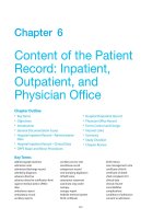
10
Cardiovascular
disease
COMMON PRESENTING SYMPTOMS OF
HEART DISEASE
The common symptoms of heart disease are chest pain, breathlessness,
palpitations, syncope, fatigue and peripheral oedema, but none are specific
for cardiovascular disease. The severity of anginal pain, dyspnoea, palpitations or fatigue may be classified according to the New York Heart Association
grading of ‘cardiac status’ (Table 10.1).
Chest pain
Chest pain or discomfort is a common presenting symptom of cardiovascular
disease and must be differentiated from non-cardiac causes. The site of pain,
its character, radiation and associated symptoms will often point to the cause
(Table 10.2)
Dyspnoea
Causes are discussed on page 407. Left heart failure is the most common
cardiac cause of exertional dyspnoea and may also cause orthopnoea and
paroxysmal nocturnal dyspnoea (p. 407).
Palpitations
Palpitations are an awareness of the heartbeat. The normal heartbeat is
sensed when the patient is anxious, excited, exercising or lying on the left
side. In other circumstances it usually indicates a cardiac arrhythmia, commonly ectopic beats or a paroxysmal tachycardia (p. 420).
Syncope
This is a temporary impairment of consciousness due to inadequate cerebral
blood flow. There are many causes and the most common is a simple faint
©
2011 Elsevier Ltd, Inc, BV
10 408 • Cardiovascular disease
Table 10.1 The New York Heart Association grading of ‘cardiac status’
(modified)
Grade 1
Uncompromised (no breathlessness)
Grade 2
Slightly compromised (on severe exertion)
Grade 3
Moderately compromised (on mild exertion)
Grade 4
Severely compromised (breathless at rest)
Table 10.2 Common causes of chest pain
Central
Angina pectoris
Crushing pain on exercise, relieved by rest. May
radiate to jaw or arms
ACS
Similar in character to angina but more severe, occurs
at rest, lasts longer
Pericarditis
Sharp pain aggravated by movement, respiration and
changes in posture
Aortic dissection
Severe tearing chest pain radiating through to the
back
Massive PE
With dyspnoea, tachycardia and hypotension
Musculoskeletal
Tender to palpate over affected area
GORD
May be exacerbated by bending or lying down (at
night). Pain may radiate into the neck
Lateral/peripheral
Pulmonary infarct
Pneumonia
Pneumothorax
}
Musculoskeletal
Sharp, well-localized pain with a tender area on
palpation
Lung carcinoma
Constant dull pain
Herpes zoster
Burning unilateral pain corresponding to a dermatome
that appears 2 to 3 days before the typical rash
Pleuritic pain, i.e. sharp, well-localized, aggravated
by inspiration, coughing and movement
ACS, acute coronary syndrome; PE, pulmonary embolus; GORD, gastro-oesophageal
reflux disease.
or vasovagal attack (p. 717). The cardiac causes of syncope are the result of
either very fast (e.g. ventricular tachycardia) or very slow heart rates (e.g.
complete heart block) which are unable to maintain an adequate cardiac
output. Attacks occur suddenly and without warning. They last only 1 or 2
minutes, with complete recovery in seconds (compare with epilepsy, where
Cardiovascular disease • 409
Other symptoms
Tiredness and lethargy occur with heart failure and result from poor perfusion
of brain and skeletal muscle, poor sleep, side-effects of medication particularly β-blockers, and electrolyte imbalance due to diuretic therapy. Heart
failure also causes salt and water retention leading to oedema, which in
ambulant patients is most prominent over the ankles. In severe cases it may
involve the genitalia and thighs.
INVESTIGATIONS IN CARDIAC DISEASE
The chest X-ray
This is usually taken in the postero-anterior (PA) direction at maximum
inspiration (p. 509). A PA chest film can aid the identification of cardiomegaly,
pericardial effusions, dissection or dilatation of the aorta, and calcification
of the pericardium or heart valves. A cardiothoracic ratio (p. 510) of greater
than 50% on a PA film is abnormal and normally indicates cardiac dilatation
or pericardial effusion. Examination of the lung fields may show signs
of left ventricular failure (Fig. 10.1), valvular heart disease (e.g. markedly
enlarged left atrium in mitral valve disease) or pulmonary oligaemia
(reduction of vascular markings) associated with pulmonary embolic
disease.
The electrocardiogram
The electrocardiogram (ECG) is a recording from the body surface of the
electrical activity of the heart. Each cardiac cell generates an action potential
as it becomes depolarized and then repolarized during a normal cycle. Normally, depolarization of cardiac cells proceeds in an orderly fashion beginning
in the sinus node (lying in the junction between superior vena cava and right
atrium) and spreading sequentially through the atria, AV node (lying beneath
the right atrial endocardium within the lower inter-atrial septum), and the His
bundle in the interventricular septum, which divides into right and left bundle
branches (Fig. 10.2). The right and left bundle branches continue down the
right and left side of the interventricular septum and supply the Purkinje
network which spreads through the subendocardial surface of the right
ventricle and left ventricle respectively. The main left bundle divides into an
anterior superior division (the anterior hemi-bundle) and a posterior inferior
division (the posterior hemi-bundle).
Cardiovascular disease
complete recovery may be delayed for some hours). Obstruction to ventricular
outflow also causes syncope (e.g. aortic stenosis, hypertrophic cardiomyo
pathy), which typically occurs on exercise when the requirements for
increased cardiac output cannot be met.
10 410 • Cardiovascular disease
Fig. 10.1 The chest X-ray in acute left ventricular failure demonstrating
cardiomegaly, hilar haziness, Kerley B lines, upper lobe venous blood
engorgement and fluid in the right horizontal fissure. Hilar haziness and Kerley
B lines (thin linear horizontal pulmonary opacities at the base of the lung
periphery) indicate interstitial pulmonary oedema.
The standard ECG has 12 leads:
� Chest leads, V1–V6, look at the heart in a horizontal plane (Fig. 10.3).
� Limb leads look at the heart in a vertical plane (Fig. 10.4). Limb leads
are unipolar (AVR, AVL and AVF) or bipolar (I, II, III).
The ECG machine is arranged so that when a depolarization wave spreads
towards a lead the needle moves upwards on the trace (i.e. a positive deflection), and when it spreads away from the lead the needle moves
downwards.
Cardiovascular disease • 411
Cardiovascular disease
Sinoatrial node
Atrioventricular
node
His bundle
Left posterior
bundle
Right bundle
Purkinje
fibres
Fig. 10.2 The conducting system of the heart. In normal circumstances only
the specialized conducting tissues of the heart undergo spontaneous
depolarization (automaticity) which initiates an action potential. The sinus (SA)
node discharges more rapidly than the other cells and is the normal pacemaker
of the heart. The impulse generated by the sinus node spreads first through the
atria, producing atrial systole, and then through the atrioventricular (AV) node to
the His-Purkinje system, producing ventricular systole.
ECG waveform and definitions (Fig. 10.5)
Heart rate At normal paper speed (usually 25 mm/s) each ‘big square’
measures 5 mm wide and is equivalent to 0.2 s. The heart rate (if the rhythm
is regular) is calculated by counting the number of big squares between two
consecutive R waves and dividing into 300.
P wave is the first deflection and is caused by atrial depolarization. When
abnormal it may be:
� Broad and notched (> 0.12 s, i.e. 3 small squares) in left atrial enlargement (‘P mitrale’, e.g. mitral stenosis)
� Tall and peaked (> 2.5 mm) in right atrial enlargement (‘P pulmonale’,
e.g. pulmonary hypertension)
� Replaced by flutter or fibrillation waves (p. 429)
� Absent in sinoatrial block (p. 421).
The QRS complex represents ventricular activation or depolarization:
� A negative (downward) deflection preceding an R wave is called a Q wave.
Normal Q waves are small and narrow; deep (> 2 mm), wide (> 1 mm) Q
waves (except in AVR and V1) indicate myocardial infarction (p. 453).
10 412 • Cardiovascular disease
(A)
V1
V 2 V 3 V4
V5 V6
(B)
LA
RA
V1
V6
LV
RV
V5
V2
V3
V4
Fig. 10.3 ECG chest leads. (A) The V leads are attached to the chest wall
overlying the intercostal spaces as shown: V4 in the mid-clavicular line, V5 in the
anterior axillary line, V6 in the mid-axillary line. (B) Leads V1 and V2 look at the
right ventricle, V3 and V4 at the interventricular septum, and V5 and V6 at the left
ventricle. The normal QRS complex in each lead is shown. The R wave in the
chest (precordial) leads steadily increases in amplitude from lead V1 to V6 with a
corresponding decrease in S wave depth, culminating in a predominantly
positive complex in V6.
Cardiovascular disease • 413
The augmented unipolar leads
AVR
I
AVL
III
II
AVF
Fig. 10.4 ECG limb leads. Lead I is derived from electrodes on the right arm
(negative pole) and left arm (positive pole), lead II is derived from electrodes on
the right arm (negative pole) and left leg (positive pole), and lead III from
electrodes on the left arm (negative pole) and the left leg (positive pole).
� A deflection upwards is called an R wave whether or not it is preceded
by a Q wave.
� A negative deflection following an R wave is termed an S wave.
Ventricular depolarization starts in the septum and spreads from left to right
(Fig. 10.2). Subsequently the main free walls of the ventricles are depolarized.
Thus, in the right ventricular leads (V1 and V2) the first deflection is upwards
(R wave) as the septal depolarization wave spreads towards those leads. The
second deflection is downwards (S wave) as the bigger left ventricle (in which
depolarization is spreading away) outweighs the effect of the right ventricle
(see Fig. 10.3). The opposite pattern is seen in the left ventricular leads (V5
and V6), with an initial downwards deflection (small Q wave reflecting septal
depolarization) followed by a large R wave caused by left ventricular
depolarization.
Cardiovascular disease
The bipolar leads
10 414 • Cardiovascular disease
R
ST segment
T
P
Voltage (mV)
10 mm = 1 mV
PR
interval
P
U
QS
Isoelectric line
QRS interval
QT interval
0.04 s
1 mm
0
0.2
0.4
0.6
Time (s)
0.8
Fig. 10.5 The waves and elaboration of the normal ECG. (From Goldman MJ
(1976) Principles of Clinical Electrocardiography, 9th edn. Los Altos: Lange.)
Left ventricular hypertrophy with increased bulk of the left ventricular
myocardium (e.g. with systemic hypertension) increases the voltage-induced
depolarization of the free wall of the left ventricle. This gives rise to tall R
waves (> 25 mm) in the left ventricular leads (V5, V6) and/or deep S waves
(> 30 mm) in the right ventricular leads (V1, V2). The sum of the R wave in
the left ventricular leads and the S wave in the right ventricular leads exceeds
40 mm. In addition to these changes there may also be ST-segment depression and T wave flattening or inversion in the left ventricular leads.
Right ventricular hypertrophy (e.g. in pulmonary hypertension) causes tall
R waves in the right ventricular leads.
The QRS duration reflects the time that excitation takes to spread through
the ventricle. A wide QRS complex (> 0.10 s, 2.5 small squares) occurs if
conduction is delayed, e.g. with right or left bundle branch block, or if conduction is through a pathway other than the right and left bundle branches,
e.g. an impulse generated by an abnormal focus of activity in the ventricle
(ventricular ectopic).
T waves result from ventricular repolarization. In general the direction of
the T wave is the same as that of the QRS complex. Inverted T waves occur
in many conditions and, although usually abnormal, they are a non-specific
finding.
The PR interval is measured from the start of the P wave to the start of
the QRS complex whether this is a Q wave or an R wave. It is the time taken
Cardiovascular disease • 415
Exercise electrocardiography
This assesses the cardiac response to exercise. The 12-lead ECG and blood
pressure is recorded whilst the patient walks or runs on a motorized treadmill.
The test is performed according to a standardized method (e.g. the Bruce
protocol). Myocardial ischaemia provoked by exertion results in ST segment
depression (> 1 mm) in leads facing the affected area of ischaemic cardiac
muscle. Exercise normally causes an increase in heart rate and blood pressure. A sustained fall in blood pressure usually indicates severe coronary
artery disease. A slow recovery of the heart rate to basal levels has also been
reported to be a predictor of mortality. Contraindications include unstable
angina, severe hypertrophic cardiomyopathy, severe aortic stenosis and
malignant hypertension. A submaximal exercise test can be performed within
4 days of a myocardial infarction. A positive test and indications for stopping
the test are:
Cardiovascular disease
for excitation to pass from the sinus node, through the atrium, atrioventricular
node and His-Purkinje system to the ventricle. A prolonged PR interval
(> 0.22 s) indicates heart block (p. 422).
The ST segment is the period between the end of the QRS complex and
the start of the T wave. ST elevation (> 1 mm above the isoelectric line)
occurs in the early stages of myocardial infarction (p. 453) and with acute
pericarditis (p. 479). ST segment depression (> 0.5 mm below the isoelectric
line) indicates myocardial ischaemia.
The QT interval extends from the start of the QRS complex to the end of
the T wave. It is primarily a measure of the time taken for repolarization of
the ventricular myocardium, which is dependent on heart rate (shorter at
faster heart rates). The QT interval, corrected for heart rate (QTc = QT/√2(R-R)),
is normally ≤ 0.44 s in males and ≤ 0.46 s in females. Long QT syndrome
(p. 432) is associated with an increased risk of torsades de pointes ventricular
tachycardia and sudden death.
The cardiac axis refers to the overall direction of the wave of ventricular
depolarization in the vertical plane measured from a zero reference point
(Fig. 10.6). The normal range for the cardiac axis is between –30° and +90°.
An axis more negative than –30° is termed left axis deviation and an axis
more positive than +90° is termed right axis deviation. A simple method to
calculate the axis is by inspection of the QRS complex in leads I, II and III.
The axis is normal if leads I and II are positive; there is right axis deviation
if lead I is negative and lead III positive, and left axis deviation if lead I is
positive and leads II and III negative. Left axis deviation occurs due to a block
of the anterior bundle of the main left bundle conducting system (p. 409),
inferior myocardial infarction and the Wolff–Parkinson–White syndrome.
Right axis deviation may be normal and occurs in conditions in which there
is right ventricular overload, dextrocardia, Wolff–Parkinson–White syndrome
and left posterior hemiblock.
10 416 • Cardiovascular disease
(A)
Left axis
deviation
AVR
–150°
AVL
–30°
I 0°
Normal
axis
II
+60°
III
+120°
Right axis
deviation
(B)
Normal
axis
AVF
+90°
Left axis
deviation
Right axis
deviation
Lead I
Lead II
Lead III
Fig. 10.6 Cardiac vectors. (A) The hexaxial reference system, illustrating the
six leads in the frontal plane, e.g. lead I is 0°, lead II is +60°, lead III is 120°.
(B) ECG leads showing the predominant positive and negative deflection with
axis deviation.
Cardiovascular disease • 417
Chest pain
ST segment depression or elevation > 1 mm
Fall in systolic blood pressure > 20 mmHg
Fall in heart rate despite an increase in workload
BP > 240/110
Significant arrhythmias or increased frequency of ventricular ectopics.
24-Hour ambulatory taped electrocardiography
A 12-lead ECG is recorded continuously over a 24-hour period and is used
to record transient changes such as a brief paroxysm of tachycardia, an
occasional pause in rhythm or intermittent ST segment shifts. It is also called
‘Holter’ electrocardiography after its inventor. Event recording is used to
record less frequent arrhythmias in which the patient triggers ECG recording
at the time of symptoms. They are both outpatient investigations.
Tilt testing
This is performed to investigate unexplained syncope when cardiac (usually
echocardiogram and 24-hour ECG) and other tests have not provided a
diagnosis. It is specifically used to diagnose neurocardiogenic (vasovagal)
syncope in which patients give a history of repeated episodes of syncope
which occur without warning and are followed by a rapid recovery. The
patient lies on a swivel motorized table in a flat position with safety straps
applied across the chest and legs to hold them in position. Blood pressure,
heart rate, symptoms and ECG are recorded after the table is tilted +60O to
the vertical for 10–60 minutes – thus simulating going from a flat to an
upright position. Reproduction of symptoms, bradycardia or hypotension indicates a positive test.
Echocardiography
This is an ultrasound examination of the heart (Fig. 10.7). Different modalities
(e.g. M mode, two- and three-dimensional) are used to provide information
about cardiac structure and function. The examination is performed in two
ways:
� Transthoracic echo is the most common method and involves the placement of a handheld transducer on the chest wall. Ultrasound pulses are
emitted through various body tissues, and reflected waves are detected
by the transducer as an echo. The commonest reasons for undertaking
an echocardiogram are to assess ventricular function in patients with
symptoms suggestive of heart failure, or to assess valvular disease. Left
ventricular function is assessed by the ejection fraction (percentage of
Cardiovascular disease
�
�
�
�
�
�
10 418 • Cardiovascular disease
Recorder
Chest wall
LV
Aorta
LA
RVOT
IVS
Aorta
AMVC
LA
PMVC
(A)
RV
LV
Ao
MV
LA
(B)
Fig. 10.7 Echocardiogram: an example of a two-dimensional long-axis view.
(A) Diagram showing the anatomy of the area scanned and a diagrammatic
representation of the echocardiogram. (B) Two-dimensional long-axis view.
Cardiovascular disease • 419
Further refinements of the echocardiogram are Doppler and stress echo
cardiography. Doppler echocardiography uses the Doppler principle (in this
case, the frequency of ultrasonic waves reflected from blood cells is related
to their velocity and direction of flow) to identify and assess the severity of
valve lesions, estimate cardiac output and assess coronary blood flow. Stress
(exercise or pharmacological) echocardiography is used to assess myocardial
wall motion as a surrogate for coronary artery perfusion. It is used in the
detection of coronary artery disease, assessment of risk post-myocardial
infarction and perioperatively, and in patients in whom routine exercise
ECG testing is non-diagnostic. For those who cannot exercise, pharmaco
logical intervention with dobutamine is used to increase myocardial oxygen
demand.
Cardiac nuclear imaging
This is used to detect myocardial infarction or to measure myocardial function, perfusion or viability, depending on the radiopharmaceutical used and
the technique of imaging. A variety of radiotracers can be injected intra
venously and these diffuse freely into myocardial tissue or attach to red blood
cells.
Thallium-201 is taken up by cardiac myocytes. Ischaemic areas (produced
by exercising the patient) with reduced tracer uptake are seen as ‘cold spots’
when imaged with a γ camera.
Technetium-99m is used to label red blood cells and produce images of
the left ventricle during systole and diastole.
Cardiac computed tomography (CT)
CT is useful for the assessment of the thoracic aorta and mediastinum and
multidetector thin slice scanners can assess calcium content of coronary
arteries as an indicator of the presence and severity of coronary artery
stenoses. CT coronary angiography has high sensitivity for the detection of
coronary artery diseases and may become part of the assessment of patients
presenting with acute chest pain to look for aortic dissection, pulmonary
embolism as well as coronary artery disease.
Cardiovascular magnetic resonance (CMR)
CMR is a non-invasive imaging technique that does not involve harmful
radiation. It is increasingly utilized in the investigation of cardiovascular
Cardiovascular disease
blood ejected from the left ventricle with each heartbeat) – normally
> 50%.
� Transoesophageal echo uses miniaturized transducers incorporated into
special endoscopes. It allows better visualization of some structures and
pathology, e.g. aortic dissection, prosthetic valve endocarditis.
10 420 • Cardiovascular disease
disease to provide both anatomical and functional information. Contra
indications are permanent pacemaker or difibrillator, intracerebral clips
and significant claustrophobia. Coronary stents and prosthetic valves are
not a contraindication.
Cardiac catheterization
A small catheter is passed through a peripheral vein (for study of right-sided
heart structures) or artery (for study of left-sided heart structures) into the
heart, permitting the securing of blood samples, measurement of intracardiac
pressures and determination of cardiac anomalies. Specially designed catheters are then used to selectively engage the left and right coronary arteries,
and contrast cine-angiograms are taken in order to define the coronary circulation and identify the presence and severity of any coronary artery disease.
Coronary artery stenoses can be dilated (angioplasty) and metal stents also
placed to reduce the rate of restenosis – this is referred to as percutaneous
coronary intervention (PCI). A further development is the introduction of stents
coated with drugs (sirolimus or paclitaxel) to reduce cellular proliferation and
restenosis rates still further. However, there is a risk of late-stent
thrombosis.
CARDIAC ARRHYTHMIAS
An abnormality of cardiac rhythm is called a cardiac arrhythmia. Arrhythmia
may cause sudden death, syncope, dizziness, palpitations or no symptoms
at all. Paroxysmal arrhythmias may not be detected on a single ECG recording. Twenty-four-hour ambulatory ECG monitoring and event recorders
(p. 417) are often used to detect arrhythmias causing intermittent symptoms.
There are two main types of arrhythmia:
� Bradycardia: the heart rate is slow (< 60 beats/min). Slower heart rates
are more likely to cause symptomatic arrhythmias.
� Tachycardia: the heart rate is fast (> 100 beats/min). Tachycardias are
more likely to be symptomatic when the arrhythmia is fast and sustained.
They are subdivided into supraventricular tachycardias (SVTs), which
arise from the atrium or the atrioventricular junction, and ventricular
tachycardias, which arise from the ventricles.
Arrhythmias and conduction disturbances complicating acute myocardial
infarction are discussed on page 456.
General principles of management of arrhythmias
Patients with adverse symptoms and signs (low cardiac output with cold
clammy extremities, hypotension, impaired consciousness or severe pulmonary oedema) require urgent treatment of their arrhythmia. Oxygen is given
to all patients, intravenous access established and serum electrolyte abnormalities (potassium, magnesium, calcium) are corrected.
Cardiovascular disease • 421
The normal cardiac pacemaker is the sinus node (p. 409) with the rate of
sinus node discharge under control of the autonomic nervous system with
parasympathetic predominating (resulting in slowing of the spontaneous
discharge rate).
Sinus arrhythmia
Fluctuations of autonomic tone result in phasic changes in the sinus discharge rate. During inspiration parasympathetic tone falls and the heart rate
quickens, and on expiration the heart rate falls. This variation is normal,
particularly in children and young adults, and typically results in predictable
irregularities of the pulse.
Bradycardia
Sinus bradycardia
Sinus bradycardia is normal during sleep and in well-trained athletes. Causes
are:
� Extrinsic to the heart: drug therapy (β-blockers, digitalis and other
antiarrhythmic drugs), hypothyroidism, hypothermia, cholestatic jaundice, raised intracranial pressure. Treatment of symptomatic bradycardia
is that of the underlying cause.
� Intrinsic to the heart: acute ischaemia and infarction of the sinus
node (as a complication of myocardial infarction) and chronic degenerative changes such as fibrosis of the atrium and sinus node (sick sinus
syndrome) occurring in elderly people. Patients with persistent symptomatic bradycardia are treated with a permanent cardiac pacemaker.
First-line treatment in the acute situation with adverse signs (p. 420), is
atropine (500 µg intravenously repeated to a maximum of 3 mg, but
contraindicated in myasthenia gravis and paralytic ileus). Temporary
pacing (transcutaneous, or transvenous if expertise available) is an
alternative.
� Sick sinus syndrome. Bradycardia is caused by intermittent failure of
sinus node depolarization (sinus arrest) or failure of the sinus impulse
to propagate through the perinodal tissue to the atria (sinoatrial
block). The slow heart rate predisposes to ectopic pacemaker activity
and tachyarrhythmias are common (tachy–brady syndrome). The ECG
shows severe sinus bradycardia or intermittent long pauses between
consecutive P waves (> 2 s, dropped P waves). Permanent pacemaker insertion is indicated in symptomatic patients. Antiarrhythmic
drugs are used to treat tachycardias. Thromboembolism is common
in sinus node dysfunction and patients are anticoagulated unless
there is a contraindication.
Cardiovascular disease
Sinus rhythms
10 422 • Cardiovascular disease
� Neutrally mediated, e.g. carotid sinus syndrome and vasovagal attacks,
resulting in bradycardia and syncope.
Heart block
The common causes of heart block are coronary artery disease, cardio
myopathy and, particularly in elderly people, fibrosis of the conducting tissue.
Block in either the atrioventricular (AV) node or the His bundle results in AV
block, whereas block lower in the conduction system (p. 411) produces right
or left bundle branch block.
Atrioventricular block
There are three forms:
First-degree AV block This is the result of delayed atrioventricular conduction and reflected by a prolonged PR interval (> 0.22 s) on the ECG. No
change in heart rate occurs and treatment is unnecessary.
Second-degree AV block This occurs when some atrial impulses fail to
reach the ventricles.
There are several forms (Fig. 10.8):
� Mobitz type 1 block (Wenckebach block phenomenon) is progressive PR
interval prolongation until a P wave fails to conduct, i.e. absent QRS after
the P wave. The PR interval then returns to normal and the cycle repeats
itself.
� Mobitz type II block occurs when a dropped QRS complex is not preceded
by progressive PR prolongation. Usually the QRS complex is wide.
� 2 : 1 or 3 : 1 (advanced) block occurs when only every second or third P
wave conducts to the ventricles.
Progression from second-degree AV block to complete heart block occurs
more frequently following acute anterior myocardial infarction and in Mobitz
type II block, and treatment is with a cardiac pacemaker. Patients with
Wenckebach AV block or those with second-degree block following acute
inferior infarction are usually monitored.
Third-degree AV block Complete heart block occurs when all atrial
activity fails to conduct to the ventricles. There is no association between
atrial and ventricular activity; P waves and QRS complexes occur independently of one another on the ECG. Ventricular contractions are maintained by
a spontaneous escape rhythm originating below the site of the block in the:
� His bundle (p. 411) – which gives rise to a narrow complex QRS (< 0.12 s)
at a rate of 50–60 b.p.m. and is relatively reliable. Recent onset block
due to transient causes, e.g. ischaemia, may respond to intravenous
atropine (p. 421) without the need for pacing. Chronic narrow-complex
AV block usually requires permanent pacing.
� His-Purkinje system (i.e. distally) – gives rise to a broad QRS complex
(> 0.12 s), is slow (< 40 b.p.m.), unreliable and often associated with
Cardiovascular disease • 423
Cardiovascular disease
(A)
(B)
(C)
Fig. 10.8 Three varieties of second-degree atrioventricular (AV) block. (A)
Wenckebach (Mobitz type I) AV block. The PR interval gradually prolongs until
the P wave does not conduct to the ventricles (arrows). (B) Mobitz type II AV
block. The P waves that do not conduct to the ventricles (arrows) are not
preceded by gradual PR interval prolongation. (C) Two P waves to each QRS
complex. The PR interval prior to the dropped P wave is always the same. It is
not possible to define this type of AV block as type I or type II Mobitz block and
it is, therefore, a third variety of second-degree AV block (arrows show P
waves), not conducted to the ventricles.
dizziness and blackouts (Stokes–Adams attacks). Permanent pacemaker
insertion is indicated.
Bundle branch block
Complete block of a bundle branch (Fig. 10.2) is associated with a wide QRS
complex (≥ 0.12 s) with an abnormal pattern and is usually asymptomatic.
The shape of the QRS depends on whether the right or the left bundle is
blocked (Fig 10.9):
� Right bundle branch block (RBBB) – the right bundle branch no longer
conducts an impulse and the two ventricles do not receive an impulse
10 424 • Cardiovascular disease
I
V1
AVR
V4
R’
r
s
s
II
V2
AVL
V5
s
III
V3
AVF
V6
s
(A)
(B)
I
II
III
AVR
AVL
AVF
V1
V2
V3
V4
V5
V6
Cardiovascular disease • 425
simultaneously. There is sequential spread of an impulse (i.e. first the left
ventricle and then the right) resulting in a secondary R wave (RSR′) in V1
and a slurred S wave in V5 and V6. RBBB occurs in normal healthy
individuals, pulmonary embolus, right ventricular hypertrophy, ischaemic
heart disease and congenital heart disease, e.g. atrial and ventricular
septal defect and Fallot’s tetralogy.
� Left bundle branch block (LBBB) – the opposite occurs with an RSR′
pattern in the left ventricular leads (I, AVL, V4–V6) and deep slurred S
waves in V1 and V2. LBBB indicates underlying cardiac pathology and
occurs in ischaemic heart disease, left ventricular hypertrophy, aortic
valve disease and following cardiac surgery.
Supraventricular tachycardias
These arise from the atrium or the atrioventricular junction. Conduction is via
the His-Purkinje system and the QRS shape during tachycardia is usually
similar to that seen in the same patient during baseline rhythm.
Sinus tachycardia
Sinus tachycardia is a physiological response during exercise and excitement.
It also occurs with fever, anaemia, heart failure, thyrotoxicosis, acute pulmonary embolism, hypovolaemia and drugs (e.g. catecholamines and atropine).
Treatment is aimed at correction of the underlying cause. If necessary,
β-blockers may be used to slow the sinus rate, e.g. in hyperthyroidism.
Atrioventricular junctional tachycardias
Tachycardia arises as a result of re-entry circuits in which there are two
separate pathways for impulse conduction. They are usually referred to as
paroxysmal SVTs and are often seen in young patients with no evidence of
structural heart disease.
Atrioventricular nodal re-entry tachycardia (AVNRT) is the commonest
type of SVT. It is due to the presence of a ‘ring’ of conducting pathway in the
atrioventricular (AV) node of which the ‘limbs’ have differing conduction times
and refractory periods. This allows a re-entry circuit and an impulse to
produce a circus movement tachycardia. On the ECG, the P waves are either
not visible or are seen immediately before or after the QRS complex
(Fig. 10.10). The QRS complex is usually of normal shape because the ventricles are activated in the normal way, down the bundle of His. Occasionally
Cardiovascular disease
Fig 10.9 A 12-lead ECG showing (A) right bundle branch block.Note an rsR
pattern with the tall R in lead V1–V2 and the broad S waves in leads I and V5 and
V6. (B) left bundle branch block. The QRS duration is greater than 0.12 s. Note
the broad notched R waves with ST depression in leads I, AVL and V6, and the
broad QRS waves in V1–V3.
10 426 • Cardiovascular disease
(A)
(B)
p’
p’
p’
p’
δ
(C)
(D)
Fig. 10.10 Atrioventricular junctional tachycardia. (A) Atrioventricular nodal
re-entry tachycardia. The QRS complexes are narrow and the P waves cannot
be seen. (B) Atrioventricular re-entry tachycardia (Wolff–Parkinson–White
syndrome). The tachycardia P waves (arrows) are clearly seen after narrow QRS
complexes. (C) An electrocardiogram taken in a patient with Wolff–Parkinson–
White (WPW) syndrome during sinus rhythm. Note the short PR interval and the
δ wave (arrow). (D) Atrial fibrillation in the WPW syndrome. Note tachycardia
with broad QRS complexes with fast and irregular ventricular rate.
Cardiovascular disease • 427
Ventricular tachycardia is more likely than supraventricular tachycardia
with:
History of ischaemic heart disease
QRS interval > 140 ms
Atrioventricular dissociation – P waves have no relationship to the QRS
complexes
Capture complexes – intermittent normal QRS complex
RS interval > 100 ms
Bifid, upright QRS complex with a taller first peak in V1
Deep S wave in V6
Concordant QRS direction in leads V1–V6, i.e. all positive or all negative
complexes
the QRS complex is wide, because of a rate-related bundle branch block, and
it may be difficult to distinguish from ventricular tachycardia (Table 10.3).
Atrioventricular reciprocating tachycardia (AVRT) is due to the presence of an accessory pathway that connects the atria and ventricles and is
capable of antegrade or retrograde conduction, or in some cases both. Wolff–
Parkinson–White syndrome is the best-known type of AVRT in which there
is an accessory pathway (bundle of Kent) between atria and ventricles. The
resting ECG in Wolff–Parkinson–White syndrome shows evidence of the
pathway’s existence if the path allows some of the atrial depolarization to
pass quickly to the ventricle before it gets though the AV node. The early
depolarization of part of the ventricle leads to a shortened PR interval and a
slurred start to the QRS (delta wave). The QRS is narrow (Fig. 10.10). These
patients are also prone to atrial and occasionally ventricular fibrillation.
Symptoms
The usual history is of rapid regular palpitations usually with abrupt onset
and sudden termination. Other symptoms are dizziness, dyspnoea, central
chest pain and syncope. Exertion, coffee, tea or alcohol may aggravate the
arrhythmia.
Acute management
The aim of treatment is to restore and maintain sinus rhythm:
� Unstable patient – emergency cardioversion is required in patients whose
arrhythmia is accompanied by adverse symptoms and signs (p. 420).
Cardiovascular disease
Table 10.3 Clinical indicators for the identification of sustained
ventricular tachycardia (12-lead ECG) in a patient presenting with wide
complex tachycardia
10 428 • Cardiovascular disease
� Haemodynamically stable patient:
� Increase vagal stimulation of the sinus node by the Valsalva man
oeuvre (ask the patient to blow into a 20-mL syringe with enough
force to push back the plunger) or right carotid sinus massage (contraindicated in the presence of a carotid bruit).
� Adenosine (p. 490) is a very short-acting AV nodal-blocking drug that
will terminate most junctional tachycardias. Other treatments are
intravenous verapamil (p. 501) or β-blockers, e.g. metoprolol. Verapamil is contraindicated with β-blockers, if the QRS is wide and
therefore differentiation from VT difficult or if there is AF and an
accessory pathway.
Long-term management
Radiofrequency ablation of the accessory pathway via a cardiac catheter is
successful in about 95% of cases. Flecainide, verapamil, sotalol and amiodarone are the drugs most commonly used.
Atrial tachyarrhythmias
Atrial fibrillation, flutter, tachycardia and ectopic beats all arise from the atrial
myocardium. In some cases automaticity is acquired by damaged atrial cells.
They share common aetiologies (Table 10.4). Baseline investigations in a
patient with an atrial arrhythmia include an ECG, thyroid function tests and
transthoracic echocardiogram.
Table 10.4 Causes of atrial arrhythmias
Ischaemic heart disease
Rheumatic heart disease
Thyrotoxicosis
Cardiomyopathy
Lone atrial fibrillation (i.e. no cause discovered)
Wolff–Parkinson–White syndrome
Pneumonia
Atrial septal defect
Carcinoma of the bronchus
Pericarditis
Pulmonary embolus
Acute and chronic alcohol use
Cardiac surgery
Cardiovascular disease • 429
This is the most common arrhythmia and occurs in 5–10% of patients over
65 years of age. It also occurs, particularly in a paroxysmal form, in younger
patients. Atrial activity is chaotic and mechanically ineffective. The AV node
conducts a proportion of the atrial impulses to produce an irregular ventricular
response – giving rise to an irregularly irregular pulse. In some patients it is
an incidental finding; in others symptoms range from palpitations and fatigue
to acute heart failure. AF is associated with a five-fold increased risk of
stroke, primarily as a result of embolism of a thrombus that has formed in
the atrium. There are no clear P waves on the ECG (Fig. 10.11), only a fine
oscillation of the baseline (so-called fibrillation or f waves).
Management
When AF is caused by an acute precipitating event, such as alcohol
toxicity, chest infection or hyperthyroidism, the underlying cause should be
treated:
� Haemodynamically unstable patient (p. 420) – immediate heparinization
and attempted cardioversion with a synchronized DC shock (p. 489). If
cardioversion fails or AF recurs, intravenous amiodarone is given (p. 491)
before a further attempt at cardioversion. A second dose of amiodarone
can be given.
III
V1
F
F
F
F
F
(A)
f
f
(B)
Fig. 10.11 (A) Atrial flutter. The flutter waves are marked with an F, only half
of which are transmitted to the ventricles. (B) Atrial fibrillation. There are no P
waves; the ventricular response is fast and irregular.
Cardiovascular disease
Atrial fibrillation (AF)
10 430 • Cardiovascular disease
� Stable patient – two strategies are available for the long-term management of AF: rate control or rhythm control (i.e. conversion to, and maintaining sinus rhythm):
� Rate control aims to reduce heart rate at rest and during exercise but
the patient remains in AF. β-blockers (p. 495) or calcium antagonists
(verapamil, diltiazem, p. 501) are the preferred treatment except in
predominantly sedentary people where digoxin (p. 493) is used.
� Rhythm control is generally appropriate in younger patients (i.e. < 65
years of age), patients who are highly symptomatic, patients who also
have congestive heart failure, and individuals with recent onset AF
(< 48 h). Conversion to sinus rhythm is achieved by electrical DC
cardioversion (p. 489) and then administration of β-blockers to suppress the arrhythmia. Other agents used depend on the presence (use
amiodarone) or absence (sotalol, flecainide, propafenone) of under
lying heart disease. Catheter ablation techniques such as pulmonary
vein isolation are used in patients who do not respond to antiarrhythmic drugs. Patients with infrequent symptomatic paroxysms of AF
(< 1/month) which are haemodynamically well tolerated and whom
have little underlying heart disease, are treated on an as-needed basis
(‘pill in the pocket’) with oral flecainide (p. 492) or propafenone.
Assessment for anticoagulation
AF is associated with an increased risk of thromboembolism, and anticoagulation with warfarin should be given for at least 3 weeks before (with the
exception of those who require emergency cardioversion or new onset AF
< 48 h duration) and 4 weeks after cardioversion. Most patients should also
be anticoagulated (INR 2.0–3.0) long term; the exception being young
patients (< 65 years) with lone AF, i.e. in the absence of demonstrable cardiac
disease, diabetes or hypertension. This latter group has a low incidence of
thromboembolism and is treated with aspirin alone.
Atrial flutter
Atrial flutter is often associated with AF. The atrial rate is typically 300 beats/
min and the AV node usually conducts every second flutter beat, giving a
ventricular rate of 150 beats/min. The ECG (Fig. 10.11) characteristically
shows ‘sawtooth’ flutter waves (F waves), which are most clearly seen when
AV conduction is transiently impaired by carotid sinus massage or drugs. The
treatment of atrial flutter is similar to AF except that most cases of flutter can
be cured by radiofrequency catheter ablation of the re-entry circuit.
Atrial ectopic beats
These are caused by premature discharge of an ectopic atrial focus. On the
ECG this produces an early and abnormal P wave, usually followed by a
normal QRS complex. Treatment is not usually required unless they cause
Cardiovascular disease • 431
Ventricular tachyarrhythmias
Ventricular ectopic premature beats (extrasystoles)
These are asymptomatic or patients complain of extra beats, missed beats
or heavy beats. The ectopic electrical activity is not conducted to the ventricles through the normal conducting tissue and thus the QRS complex on the
ECG is widened, with a bizarre configuration (Fig. 10.12). Treatment is with
β-blockers if symptomatic.
Sustained ventricular tachycardia
Ventricular tachycardia (VT) and ventricular fibrillation (VF) are usually associated with underlying heart disease. The ECG in sustained VT (> 30 s) shows
a rapid ventricular rhythm with broad abnormal QRS complexes. Supraventricular tachycardia with bundle branch block also produces a broad complex
tachycardia which can usually be differentiated from VT on ECG critera (Table
10.3). However, the majority of broad complex tachycardias are VT and if in
doubt treat at such. Urgent DC cardioversion is necessary if the patient is
haemodynamically compromised (p. 420). If there is no haemodynamic compromise, treatment of VT is usually with intravenous lidocaine (p. 492) or
amiodarone (p. 491). Recurrence is prevented with β-blockers or an implantable cardioverter–defibrillator (ICD). This is a small device implanted behind
the rectus abdominis and connected to the heart; it recognizes VT or VF and
automatically delivers a defibrillation shock to the heart.
Non-sustained ventricular tachycardia
This is defined as VT ≥ 5 consecutive beats but lasting < 30 s. It is common
in patients with heart disease (and in a few individuals with normal hearts).
The treatments indicated are β-blockers in symptomatic patients or an ICD
Fig. 10.12 A rhythm strip demonstrating two ventricular ectopic beats of
different morphology (multimorphological).
Cardiovascular disease
troublesome palpitations or are responsible for provoking more significant
arrhythmias when β-blockers may be effective.