Ebook Surgical care of major newborn malformations: Part 2
Bạn đang xem bản rút gọn của tài liệu. Xem và tải ngay bản đầy đủ của tài liệu tại đây (5.16 MB, 199 trang )
b1319 Surgical Care of Major Newborn Malformations
CHAPTER 9
ESOPHAGEAL ATRESIA
Frederick Alexander, M.D.*
Joseph M. Sanzari Children’s Hospital,
Hackensack, New Jersey
INTRODUCTION
Esophageal atresia has remained the defining challenge for several generations of
pediatric surgeons. Coveted by pediatric surgical fellows and featured in the
commercial hit movie “M*A*S*H”, these cases have epitomized a pediatric surgeon’s ability to repair a catastrophic embryologic anomaly and restore normal
life. Today, thanks to the contributions of many brilliant pediatric surgeons, anesthesiologists, and neonatologists, children with esophageal atresia cannot only
survive but thrive as they head into early childhood. As a result of this work,
associated anomalies have now surpassed esophageal atresia as the greatest barrier
to survival and a rich quality of life.
Nearly one third of all infants with esophageal atresia have associated anomalies connected with the VACTERL syndrome. Hence, once the diagnosis is made
it is critically important to perform a complete physical examination and obtain
an echocardiogram as well as appropriate radiologic imaging to rule out associated Vertebral, Anal or Intestinal, Cardiac, Renal, and Limb/Lung anomalies.
Additionally, genetic and neurologic screening should be done in every case.
Moreover, since many infants are now diagnosed prenatally, it is often important
to consult with the maternal fetal medical specialist or fetal radiologist who may
have pertinent information to share concerning associated anomalies.
Historically, the incidence of esophageal atresia has been 1 in 5000 live births,
although it does appear that, in developing countries, this rate may be declining
*Address: Pediatric Surgical Associates, 30 West Century Road, Suite 235, Paramus, NJ
07652. E-mail:
189
B1319_Ch-09.indd 189
5/17/2012 3:05:14 PM
b1319 Surgical Care of Major Newborn Malformations
190
F. Alexander
as the result of selective termination used in conjunction with prenatal screening.
This is difficult to assess due to regional differences in prenatal management and
referral patterns. This emphasizes the need for pediatric surgical involvement in
fetal care programs to educate prospective parents and healthcare providers about
treatment options, risks, benefits, and expected outcomes for esophageal atresia.
History
The history of esophageal atresia is relevant to the many treatment options available
to the pediatric surgeon today. Every case of esophageal atresia is different in terms
of anatomy, gestational age, and associated anomalies; and surgical treatment has
evolved through many iterations over the past six decades.
Esophageal atresia was uniformly fatal until 1939 when Leven1 salvaged one
patient using a three-staged repair including gastrostomy, extra-pleural division of
an associated tracheoesophageal fistula, and cervical esophagostomy. Two years
later, the first successful primary repair of esophageal atresia with fistula was performed by Haight2 who very clearly described the technical innovations that would
pave the way for future success, including meticulous mobilization of the proximal
and distal ends of esophagus, an interrupted two layer closure, and attentive perioperative care including initial fluid restriction and esophagram prior to feeding.
Twenty years later, Waterston3 proposed a risk classification (Table 1) based
upon a large series of patients with esophageal atresia and tracheoesophageal fistula that demonstrated greater than 90% survival in infants greater than 2000 g
without congenital anomalies compared to less than 50% survival in premature
infants with congenital anomalies, especially cardiac.
At about the same time, Holder et al.4 reported significantly improved survival
in high-risk premature infants with esophageal atresia and tracheoesophageal fistula
using a staged repair, including gastrostomy followed by fistula ligation, and then
Table 1. Waterston Risk Classification for infants with esophageal atresia and tracheoesophageal fistula.
Group A:
Over 5.5 lb birth weight and well.
Group B:
1. Birth weight 4–5.5 lb and well.
2. Higher birth weight, moderate pneumonia and congenital anomaly.
Group C:
1. Birth weight under 4 lb.
2. Higher birth weight and severe pneumonia and severe congenital
anomaly.
B1319_Ch-09.indd 190
5/17/2012 3:05:14 PM
b1319 Surgical Care of Major Newborn Malformations
Esophageal Atresia
191
delayed primary anastomosis when the infant’s condition would safely permit.
Using this strategy of repair, he achieved 66% survival in high-risk Waterston C
infants. His strategy became the standard of care for high-risk infants with esophageal atresia and tracheoesophageal fistula and continues to be selectively used today
by many surgeons both to maximize survival and minimize complications.5 In that
era, survival of high-risk infants with esophageal atresia and tracheoesophageal
fistula ranged between 30% and 70% depending upon associated risk factors, while
survival of contemporaneous low-risk infants approached 100%.
With improved technical support, surgical techniques, and perioperative
care, surgeons began to have increasing success with primary repair in select highrisk infants. In 1972, Abrahamson and Shandling at the Hospital for Sick Children
in Toronto6 reported equivalent survival in groups of high-risk infants treated by
primary versus staged repair. Although the study groups were not really comparable, the authors concluded that most high-risk infants could be safely treated by
primary repair irrespective of weight, even when other complications were present. Ten years later, Louhimo and Lindahl at the University of Helsinki7 reported
similar findings and suggested a modification of the Waterston classification to
exclude pneumonia and general condition from the criteria. Like the surgeons in
Toronto, they reserved staged repair for infants who were desperately ill with
severe respiratory problems or associated gross anomalies, any one of which were
life-threatening. Using their modified risk classification, they reported incrementally increased survival in all categories: 100% survival in group A patients, 95%
survival in group B patients, and 57% survival in group C patients. As these outcomes were emulated in many centers throughout North America, it became clear
that some high-risk infants with esophageal atresia would not survive even with a
repaired esophagus, and esophageal atresia was no longer a limiting factor in the
survival of infants with prematurity or associated congenital anomalies.
One of the great unresolved technical challenges concerning esophageal atresia is long-gap atresia and so-called ultra-long-gap atresia, defined as a separation
of greater than 6 cm between proximal and distal esophageal ends. Long-gap
atresia may be found in association with tracheoesophageal fistula, but most commonly occurs in the absence of fistula, sometimes referred to as “pure” esophageal
atresia. As discussed below, this anomaly is encountered much less frequently than
esophageal atresia and tracheoesophageal fistula and may be temporized in virtually all infants by placement of a gastrostomy tube for feeding along with intermittent nasopharyngeal suction to prevent aspiration pneumonia.
Initial attempts at early primary repair of pure, long-gap atresia invariably failed.
Attention soon turned to use of esophageal replacement procedures including the
following: gastric pull-up, first performed at the Hospital for Sick Children in
B1319_Ch-09.indd 191
5/17/2012 3:05:14 PM
b1319 Surgical Care of Major Newborn Malformations
192
F. Alexander
Toronto in 1952 and later modified by Spitz;8 colon replacement, first reported by
Dale and Sherman in 19559 and subsequently advocated by Grosfeld,10 Hendren11
and others; and finally gastric tube reconstruction, first reported by Burrington and
Stevens12 in 1968 and later advocated by Anderson13 and Ein.14 These three procedures have been used concurrently for nearly 50 years and continue to be selectively
used by some surgeons with variable success. Each of these procedures has its advocates and each its complications including leak, stricture, gastroesophageal reflux,
and dysphagia requiring surgical revision in as many as 50% of all patients.15
In experienced hands, esophageal replacement may serve as an excellent substitute for the real thing, although even its staunchest advocates have admitted
that the patient’s own esophagus works best. In this regard, several early contributions set the stage for delayed primary repair of pure long-gap atresia that has
become not only feasible but achievable in most cases. In 1965, Howard and
Myers16 reported a successful technique for elongating the proximal pouch using
daily bougienage and delayed primary repair. Several years later, Livaditis17
reported the ingenious technique of circumferential esophagomyotomy of the
proximal pouch. Using these techniques to construct a well-vascularized single
layer anastomosis under tension, a number of surgeons demonstrated excellent
results using delayed primary repair in the 1980s.18–20 Since then, the trend in
North America has been toward delayed primary repair for long-gap atresia, utilizing esophageal replacement only for failures or extremely complicated cases.
ANATOMY AND EMBRYOLOGY
The normal esophagus arises from the proximal end of the foregut. The respiratory diverticulum emerges from the laryngeal groove on the ventral surface of the
proximal foregut by the end of the fourth postconceptual week. A septating process occurs between the evolving ventral trachea and dorsal esophagus. The trachea and esophagus, therefore, share a common ancestry, the primitive foregut.
The etiology of esophageal atresia is unknown but involves a mesenchymal field
defect that results in several distinct anatomical patterns. Occurring in nearly 90%
of cases, the most common form is a blind proximal esophageal pouch that ends
at the 2nd–4th vertebral body and a tracheoesophageal fistula that leads to the
distal esophagus and stomach. The blood supply to the proximal pouch originates
from the thyrocervical trunk and reaches the caudal aspect of the blind ending
pouch through the submucosal plexus allowing circumferential dissection and
circular myotomy without interruption of the circulation. The distal tracheoesophageal fistula usually joins the back wall of the trachea just proximal to the
carina but may enter the trachea cranial or caudal to that site and may even enter
the right or left main stem bronchus below the carina. The blood supply of the
B1319_Ch-09.indd 192
5/17/2012 3:05:14 PM
b1319 Surgical Care of Major Newborn Malformations
Esophageal Atresia
193
distal esophagus consists of fragile vessels that originate from the aorta and are
easily damaged by surgical dissection.
In most cases, separation of the proximal pouch and distal tracheoesophageal
fistula does not exceed three vertebral bodies, although it can be as many as six. In
contrast, esophageal atresia without fistula (pure esophageal atresia) typically
involves a separation of at least six vertebral bodies. Although pure esophageal atresia is the second most common type of major esophageal malformation, it occurs
in only 5–7% of cases, and has a similar profile of associated anomalies compared
to the more common form of esophageal atresia (with distal TEF). It is important
to recognize that both forms of esophageal atresia may be associated with a right
descending aorta which significantly affects exposure, mobilization, and ultimate
reconstruction of the esophagus. Thus, it is critical to review aortic position on the
preoperative echocardiogram before proceeding with operative repair.
Other forms of esophageal atresia and/or fistula are rare. For example, H-type
fistula without atresia occurs in 2% of cases. Usually, this is a single fistula which is
slanted cranially from the esophagus to the trachea and is located close to the 2nd–3rd
vertebral body allowing exposure through the neck. Even less common is a double
fistula with atresia, in which a fistula connects the blind proximal pouch to the trachea
in addition to a distal tracheoesophageal fistula. Some surgeons have recommended
routine preoperative endoscopy in order to rule out a possible proximal fistula prior
to thoracotomy. An alternate approach is to perform a complete circumferential dissection of the proximal pouch extending to the thoracic inlet in order to rule out the
possibility of a proximal fistula. Least common of all is a proximal tracheoesophageal
fistula with distal esophageal atresia. This deformity is thought to be incompatible
with life and has been diagnosed primarily at autopsy.
CLINICAL PRESENTATION
Those infants with esophageal atresia who are not diagnosed prenatally present
shortly after birth with acute onset of respiratory distress. Within the first few hours,
the infants usually develop coughing, excessive salivation, and drooling. If fed by
mouth, they begin to choke and gag leading to a drop in oxygen saturation and cyanosis. If not treated, they will eventually develop acidosis, heart failure, and death.
Infants with proximal atresia and distal tracheoesophageal fistula may present with a more precipitous course than those with pure atresia. As their respiratory distress worsens, they often force air into their stomach which in turn causes
gastric distension and reflux of acidic gastric contents through the fistula and into
the endobronchial tree. This does not occur in infants with pure atresia who are
not as sick as those with tracheoesophageal fistula and may survive longer without
treatment.
B1319_Ch-09.indd 193
5/17/2012 3:05:14 PM
b1319 Surgical Care of Major Newborn Malformations
194
F. Alexander
Infants with H-type fistula (TE fistula with no atresia) usually do not present
at birth but weeks to months later with upper airway congestion, gastroesophageal reflux, and recurrent pneumonia. Their symptoms are subtle and may be
difficult to recognize initially; but most will present with a pattern of choking,
coughing, and gagging with feeds or with persistent upper airway congestion or
gas bloating minutes after feeding. Whether infants present in the newborn
period or months later, these symptoms should provoke in the surgeon a high
index of suspicion and low threshold for diagnostic investigation.
DIAGNOSIS
The diagnosis of esophageal atresia may be made with a 6–8 French nasogastric tube. If the tube can not be passed beyond 9–11 cm from the mouth or
nose of an infant under suspicion, then the diagnosis is verifiably esophageal
atresia. A “babygram” (chest and abdominal radiograph) will often show an air
esophagram with the tube stopped or coiled in the blind end of the proximal
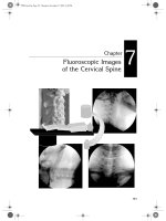
esophageal pouch near the 2nd–4th vertebral body (Figure 1). More
Figure 1. Babygram of infant with esophageal atresia and tracheoesophageal fistula.
Note coiled Replogle tube in blind ending upper esophagus at thoracic inlet.
B1319_Ch-09.indd 194
5/17/2012 3:05:14 PM
b1319 Surgical Care of Major Newborn Malformations
Esophageal Atresia
195
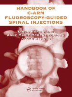
Figure 2. Babygram of infant with pure esophageal atresia. Note absence of gas below the
diaphgram and Replogle tube in upper esophagus.
importantly, the radiograph will demonstrate the presence or absence of intestinal gas beneath the diaphragm. If gas is present, the patient has a distal tracheoesophageal fistula. On the contrary, if there is no intestinal gas beneath the
diaphragm (a gasless abdomen), the patient has pure esophageal atresia
(Figure 2).
In addition to careful physical examination, a number of noninvasive studies should be obtained prior to definitive repair. On examination, particular
attention should be paid to the possibility of a cardiac murmur (that may indicate a structural cardiac defect), genitourinary anomalies, and/or anorectal
malformation and limb abnormalities. Following a complete physical examination and babygram as previously mentioned, several noninvasive studies should
be performed prior to definitive repair. Most important of these is an echocardiogram to detect a possible associated structural cardiac defect (23%) as well
as a possible right aortic arch (5%). Other studies that should be done prior to
definitive repair of the esophageal atresia include renal ultrasonography as
well as AP and lateral radiographs of the lumbosacral spine. Contrast esophagram is generally not required and, in fact, may be harmful. For example,
B1319_Ch-09.indd 195
5/17/2012 3:05:19 PM
b1319 Surgical Care of Major Newborn Malformations
196
F. Alexander
administration of oral barium to an infant with esophageal atresia could lead to
barium aspiration and death.
Some surgeons recommend routine triple endoscopy including laryngoscopy
to rule out a laryngoesophgaeal cleft, rigid esophagoscopy in order to rule out a
proximal tracheoesophageal fistula, and rigid bronchoscopy in order to indentify
the location of the tracheoesophageal fistula(s), especially in the new era of thoracoscopic repair. These procedures are not routinely performed by all surgeons
prior to definitive repair, yet may provide important and relevant information.
They require general anesthesia which may pose significant risk, especially to
infants with proximal atresia and distal tracheoesophageal fistula, and thus are
deferred until the time of anticipated repair.
On the contrary, contrast studies can be quite helpful in diagnosing an
H-type fistula. Routine barium swallow in an upright position is unlikely to detect
this deformity because of the variable size as well as slant of the fistula from cranial to caudal between the trachea and esophagus. More reliable results may be
obtained from a tube esophagram performed with the patient in a prone position.
In this study, an orogastric tube is passed into the stomach and contrast is injected
as the tube is withdrawn. Fluoroscopy is used to immediately visualize any contrast material that extravasates through the fistula and into the trachea. This
technique may be cumbersome but is the most effective radiographic study to
diagnose H-type fistula. The prone esophagram may also be used to detect recurrent fistulas that are equally difficult to diagnose and often require repeated
studies.
Whether H-type fistula is diagnosed or not by radiographic studies, most
surgeons advocate endoscopy to confirm and localize this type of fistula when it
is strongly suspected. Usually, the fistula is visualized through the bronchoscope
in the membranous portion of the trachea close to the thoracic inlet. A ureteral
catheter may be passed through the fistula to aid in operative identification.
Alternatively, several drops of methylene blue dye may be instilled into an
endotracheal tube of a fully anesthetized patient, and with positive pressure, blue
dye may be seen by esophagoscopy draining through the fistula and into the
esophagus. Diagnosis of tracheoesophageal fistula whether congenital or recurrent can be difficult and requires persistence as well as a high index of suspicion.
Thus, if radiographic studies are negative, endoscopy should be seriously
entertained.
Lastly, many cases of esophageal atresia may now be diagnosed prenatally.
Depending upon geographic region and socioeconomic status, nearly 85% of all
pregnant women now undergo prenatal ultrasound. Although ultrasound cannot
accurately diagnose esophageal atresia, it is an excellent screening tool for
B1319_Ch-09.indd 196
5/17/2012 3:05:23 PM
b1319 Surgical Care of Major Newborn Malformations
Esophageal Atresia
197
polyhydramnios which can be a marker of foregut obstruction. The finding of
polyhydramnios should serve as an indication for prenatal MRI, which in the case
of esophageal atresia will often demonstrate a dilated proximal esophageal pouch,
sometimes associated with microgastria, as well as other associated anomalies.
Because of their intimate knowledge about esophageal atresia, pediatric surgeons
should play an important role in the process of prenatal diagnosis and counseling.
TREATMENT
Once the diagnosis of esophageal atresia has been made, treatment should begin
immediately. A 10-French double lumen oropharyngeal tube (known as a
Replogle tube) should be placed to intermittent suction, and the infant should be
positioned with head up at a 20–30-degree angle to prevent aspiration. Broadspectrum antibiotics should be administered and intravenous fluids should be
carefully modulated in order to prevent fluid overload. Of course, careful monitoring by pulse oximetry is critical.
Some infants will require immediate intubation and ventilation due to either
respiratory distress syndrome associated with prematurity and/or aspiration
related to esophageal atresia and tracheoesophageal fistula. Infants with esophageal atresia aspirate saliva from their proximal blind pouch and have tracheomalacia due to distension of the proximal pouch, but infants with distal
tracheoesophageal fistula are often much more compromised as a result of reflux
of gastric content into the bronchial tree. It is preferable to manage these infants
with oxygen supplementation as needed via nasal prongs or facial mask, since
intubation entails positive pressure which may exacerbate gastric distension and
reflux aspiration by forcing gas through the distal tracheoesophageal fistula into
the stomach. If intubation is required, infants should be ventilated with the lowest
possible peak inspiratory pressure.
Premature infants with esophageal atresia and tracheoesophageal fistula who
develop respiratory distress syndrome are at greatest risk due to stiff, noncompliant lungs. As mentioned above, they may shunt greater minute ventilation
through the fistula and into the stomach, causing distension and even rupture
requiring immediate repair and gastrostomy tube placement. Even with a gastrostomy tube placed to 5–10-mm water seal, continued loss of minute ventilation
may destabilize the patient and require immediate thoracotomy and ligation of
the fistula as a life-saving procedure. This scenario illustrates a decision that must
sometimes be made by the pediatric surgeon, whether or not to proceed with
primary repair in critically ill infants with respiratory compromise. Ultimately,
this judgment must be made intraoperatively in conjunction with the
B1319_Ch-09.indd 197
5/17/2012 3:05:23 PM
b1319 Surgical Care of Major Newborn Malformations
198
F. Alexander
anesthesiologist depending upon the infant’s physiologic status once the fistula
has been closed. It sometimes may be advantageous to proceed with definitive
repair if the infant’s physiology improves with fistula ligation, otherwise staged
repair remains the safest alternative for infants who need further resuscitation.
But what about infants who are physiologically stable? Today, few would disagree
that physiologically stable infants should undergo primary repair without gastrostomy tube placement.
Esopahgeal Atresia with Tracheoesophageal Fistula
Definitive repair of esophageal atresia with tracheoesophageal fistula is typically
performed through a right thoracotomy; alternatively, a left thoracotomy is used
when there is a right aortic arch. Frequently, the latissimus dorsi muscle can be
mobilized along its anterior border and retracted posteriorly, preserving muscle
function; however, the muscle should be divided if a muscle-sparing technique
would compromise exposure. Either an intra-pleural or extra-pleural approach to
the posterior mediastinum can be used depending upon the status of the infant.
An extra-pleural approach takes longer and is more tedious; thus, it would not be
appropriate in an unstable infant. Yet this approach offers a number of advantages: it affords excellent exposure of the posterior mediastinal structures, protects
the lung from retraction injury, and prevents empyema in the event of anastomotic leak.
Next, the azygos vein usually is divided, whereupon the vagus nerves are
identified and scrupulously preserved. Dividing the azygous vein typically exposes
the tracheoesophageal fistula. If the fistula is not clearly visualized, the distal
esophagus can be identified just above the hiatus and followed proximally to the
fistula’s origin. The tracheoesophageal fistula is carefully mobilized from surrounding structures, taking care to preserve the medial perforating vessels originating from the aorta. The site of the fistula is sequentially divided as the trachea
is repaired with interrupted 5.0 permanent sutures into the small remnant of
fistula left on the trachea. To make sure that this closure is secure, saline can be
poured into the mediastinum and the anesthesiologist asked to inflate the lungs
as the surgeon looks for bubbles from the suture line that would indicate a leak.
Then, the proximal pouch is usually easy to find with the use of a nasopharyngeal tube, and a traction stitch is placed in the muscular tip of the proximal
pouch to aid dissection. Some surgeons incorporate the tip of the tube in the
traction stitch to minimize injury to the proximal esophagus with manipulation.
Circumferential dissection is performed up to and above the thoracic inlet taking
care to stay close to the esophageal wall so as not to damage the trachea, thoracic
B1319_Ch-09.indd 198
5/17/2012 3:05:23 PM
b1319 Surgical Care of Major Newborn Malformations
Esophageal Atresia
199
duct, or recurrent laryngeal nerves. Electrocautery is used sparingly as dissection
nears the thoracic inlet for the same reason. Careful attention is paid to the tracheoesophageal septum in order to ensure a proximal fistula is recognized if
present.
Finally, if the decision is made to perform a primary repair, the proximal
esophageal pouch is opened, the fistula is trimmed back to pink, well-vascularized
esophageal tissue, and a single-layer anastomosis is performed. Prior to anastomosis a soft catheter, such as an 8-French red rubber tube, should be passed distally into the stomach to guard against an unrecognized distal stricture. The
anastomosis is done by laying interrupted full-thickness sutures across the back
wall leaving the knots on the luminal side. Many pediatric surgeons use 5.0 monofilament or braided nylon permanent sutures, but the choice of suture depends
upon surgeon preference. The two ends of esophagus are gently held with noncrushing forceps while the back wall sutures are tied diffusing tension across the
posterior aspect of the anastomosis. The front wall of the anastomosis is then
completed in an interrupted fashion, again taking care to place full-thickness
sutures. This is particularly important and challenging on the distal esophagus
where the mucosa tends to retract from the cut edge of the esophageal wall. These
sutures are tied leaving the knots on the extra luminal aspect of the esophagus,
and the anastomosis is completed (Figure 3). If available, mediastinal fat or lymphatic tissue is placed between the trachea and esophagus to buttress the anastomosis and guard against fistula recurrence. A 10–12-French chest tube is inserted
Figure 3. Completed trans-thoracic repair of esophageal atresia and tracheoesophageal
fistula.
B1319_Ch-09.indd 199
5/17/2012 3:05:23 PM
b1319 Surgical Care of Major Newborn Malformations
200
F. Alexander
through a separate stab incision, and the thoracotomy is closed in layers. Sutures
around the ribs are not made tight, to limit future thoracic deformity. Esophageal
stents in the form of nasogastric tubes are still used by many surgeons; however,
there is no data to show they reduce the incidence of anastomotic stricture.
However, trans-anastomotic nasogastric tubes can be useful to initiate continuous
gastric feeds early in the postoperative period and reduce dependence upon parenteral nutrition. They can also help decompress the stomach.
As discussed above, it can be difficult to decide which infants in the modified
Waterston C category may be safely treated by primary versus staged repair.
Moreover, many high-risk infants may be safely temporized by gastrostomy tube
drainage only without preliminary ligation of the distal tracheoesophageal fistula,
followed by subsequent delayed primary repair. All these options must be considered in light of the patient’s weight and physiologic status and with recognition
that ligation of the fistula may potentially render subsequent primary repair more
difficult. If the infant is not deemed a candidate for immediate primary repair, a
gastrostomy may be placed to prevent reflux of gastric contents until the infant
can be optimized for surgery. The gastrostomy tube should be at least 14-French
in diameter to prevent occlusion and limit reflux of gastric contents into the bronchial tree. In addition, the infant should be positioned head up at 20–30 degrees
with intermittent oropharyngeal suction to prevent antegrade aspiration. Delayed
repair in premature infants is supported by studies which demonstrate that the
two ends of esophagus continue to grow in length following delivery19 and postoperative anastomotic complications related to tension including leak, stricture,
and reflux may be minimized5. Weight alone does not preclude primary repair.
However, if the infant weighs less than 1300–1500 g, most surgeons prefer to place
a gastrostomy tube for drainage and parenterally feed the infant via a PICC line
or Broviac® catheter until the infant reaches this general weight range when
delayed primary repair may be more safely performed.
Thoracoscopic Repair
In the future, thoracoscopy may be more widely used to repair esophageal atresia.
This was accomplished as a surgical first in 1999,21 and several groups in the
United States and Europe are routinely using this technique for esophageal atresia
and tracheoesophageal fistula when the two ends are in close proximity. At this
point, it is unclear whether the end result will justify the learning curve, and
already there have been reported leaks and some fatalities using this technique.22
As with any other technique, the surgeon must determine his/her level of comfort
and ultimately choose the technique which yields the optimal long- and shortterm outcomes in each individual case.
B1319_Ch-09.indd 200
5/17/2012 3:05:25 PM
b1319 Surgical Care of Major Newborn Malformations
Esophageal Atresia
201
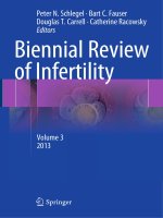
Figure 4. Gastrostomy tube contrast injection study demonstrating long-gap pure
esophageal atresia.
Pure Esophageal Atresia
Infants with pure esophageal atresia are not nearly as sick as those with distal tracheoesophageal fistula yet require the same initial treatment as those with a fistula.
Once the VACTERL evaluation has been completed, a gastrostomy tube should be
placed for enteral feedings that may be given without risk of reflux. Shortly after
this is done, a limited upper GI series may be performed via the gastrostomy with
a Bougie placed in the upper pouch to assess the gap between the blind ends of the
esophagus (Figure 4). Distance is usually measured using the vertebral bodies as a
reference, and, in most cases, the separation is at least six vertebral bodies equating
to a distance of 6–10 cm. At this point, there are several options. As previously
discussed, historically there had been considerable enthusiasm for esophageal
replacement procedures using a vascularized colonic segment (Figure 5), gastric
tube, or gastric transposition. In many cases, these procedures were performed
using a staged approach including cervical esophagostomy performed in the newborn period followed by colon replacement or gastric tube performed at one year
B1319_Ch-09.indd 201
5/17/2012 3:05:25 PM
b1319 Surgical Care of Major Newborn Malformations
202
F. Alexander
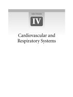
Figure 5. Tortuous colon replacement 2 years following repair in a child with dysphagia
and reflux.
of age. On which side to place the cervical esophagostomy is purely surgeon preference: a left-sided esophagostomy may simplify subsequent replacement while
thoracic duct injury may be avoided with right-sided placement.
More recently, these procedures have been successfully performed without
cervical esophagostomy even in premature infants at 1–3 months of age.23 The
gastric tube is constructed using a GIA stapler on the greater curvature of the
stomach based on the left gastroepiploic vessels with division of the short gastric
vessels as necessary. The optimal colon conduit is constructed from the right and
transverse colon based on the middle colic and arcuate vessels, preserving the
ileocecal valve.10 Both the gastric tube and colon conduit are often placed in the
straight, substernal position with a proximal anastomosis in the neck. Both of
these procedures require pyloroplasty to facilitate gastric emptying and minimize
gastroesophageal reflux. In addition, the cervical anastomosis should be drained
following either of these procedures until a postoperative esophagram demonstrates an intact anastomosis. Some surgeons prefer placing the conduit in the
esophageal bed, a shorter route to the neck.
B1319_Ch-09.indd 202
5/17/2012 3:05:27 PM
b1319 Surgical Care of Major Newborn Malformations
Esophageal Atresia
203
Another option for esophageal replacement in the newborn period is the
gastric transposition or pull-up modified by Spitz.24 This procedure involves the
mobilization of the stomach, preserving the epiploic vascular arcades and ligation
of the short gastric vessels and left gastric artery and vein. The stomach is then
mobilized into a posterior mediastinal tunnel created from the neck above to
hiatus below without thoracotomy. Then, anastomosis is performed between the
proximal fundus and cervical esophagus using a single layer of fine polyglycolic
acid sutures which is drained internally using a nasogastric tube. Once again, this
procedure requires pyloroplasty and, in addition, feeding jejunostomy tube to
allow enteral feedings in the first few weeks after repair.
The options discussed above have yielded reasonably good results in the past
but have lost their appeal for all but the most complicated cases. There are a number of reasons for this including the desire to avoid a prolonged staged repair
involving cervical esophagostomy, reluctance to invade the abdominal cavity,
recognition of a 5–15% morbidity and mortality rate even in the best of hands,
and realization that at least 50% of these patients will develop long-term dysphagia and reflux requiring remedial surgery.15 Instead, most surgeons today embrace
the concept that the infant’s best esophagus is his/her own esophagus. For all these
reasons, delayed primary repair has become the most popular and frequently
performed reconstructive procedure for pure long-gap atresia.
The first step of delayed primary repair is to place a gastrostomy tube for
feeding. After the gastrostomy tube is placed, the esophagus is lengthened. There
have been a number of proposed techniques for this, but the most practical and
least invasive of these is twice daily bougienage using a 20- or 22-French leadweighted bougie. After not less than 2 weeks, the patient is taken to the operating
room where, using a brief anesthetic, the gastrostomy tube is removed and a
pediatric cystoscope is inserted under direct visualization into the gastrostomy
tube site through the GE junction and into the blind distal esophagus. At the same
time, a Bougie is passed into the proximal pouch, and the separation of the two
ends is measured in terms of the number of intervening vertebral bodies. This
procedure is performed intermittently for at least 2–6 weeks or until the separation has narrowed to two vertebral bodies (Figure 6). At that point, a definitive
repair may be performed (Figure 7).
Another interesting approach to long-gap esophageal atresia, advocated by
Dr. John Foker,25,26 is to perform a mini-thoracotomy at the time of gastrostomy
tube insertion in order to identify and hitch the upper and lower esophageal segments to pledgeted traction sutures. These sutures are then brought out of the
chest above and below the incision. Tension on the external sutures is increased
1–3 times each day inducing “growth” of the esophageal ends over a period of
B1319_Ch-09.indd 203
5/17/2012 3:05:29 PM
b1319 Surgical Care of Major Newborn Malformations
204
F. Alexander
Figure 6. Same patient as in Figure 4 with long-gap atresia after 4 weeks of bougienage.
Figure 7. Same patient with long-gap atresia after completion of repair.
6–10 days allowing a true primary repair. Dr. Foker describes the preliminary
thoracotomy performed through a 3-cm incision and postulates the future application of thoracoscopy to accomplish this preliminary operation.
B1319_Ch-09.indd 204
5/17/2012 3:05:29 PM
b1319 Surgical Care of Major Newborn Malformations
Esophageal Atresia
205
Definitive repair begins with extensive mobilization of the proximal pouch to
the level of the thoracic inlet with circular myotomy as needed to add length. The
distal esophagus is usually found lying just above the diaphragm as a small nubbin
protruding above the hiatus. This is carefully mobilized taking care not to divide
or injure the perforating vessels from the aorta. If there is not enough distal
length, then the gastroesophageal junction is mobilized above the diaphragm taking care so as not to injure the vagal nerves. A back row of full-thickness interrupted fine permanent sutures is placed and then tied with the knots on the
intraluminal side while holding both ends of the esophagus together using atraumatic ring forceps. The front wall of the anastomosis is completed, again taking
care to include both the muscular and mucosal components of the esophageal
wall, especially on the distal esophagus where the mucosa often retracts. The anastomosis is usually completed with a fair amount of tension which will nevertheless heal well as long as there is good tissue apposition and the cut ends of the
esophagus are pink. Again a 10–12-French chest tube is placed through a separate
stab incision, and the thoracotomy is closed in layers.
Postoperative Care
Regardless of which repair strategy is used, it is wise to leave the infant intubated
at least 24 hours or until completely recovered from the anesthetic, whereupon
the infant may be safely extubated. As in the preoperative period, the infant
should be positioned at least 20–30-degrees in the upright position. Nasopharyngeal
suctioning may be performed but should be done with a tube that has been clearly
marked at the time of surgery to limit the depth of suctioning to at least 2 cm or
more above the level of anastomosis.
The chest tube is left for drainage and not removed until an esophagram has
been performed 5–7 days later. If the esophagram shows no evidence of a leak,
oral feedings are cautiously started. Then the chest tube is removed. Infants with
a gastrostomy tube may also be supplemented with tube feedings. If a small leak
is found, oral feedings are withheld for several weeks, during which time the
infant is supported with parenteral nutrition or fed by gastrostomy tube when
present. Once oral feedings are initiated and advanced to sustainable levels, the
infant is carefully followed for signs of esophageal stenosis which include choking,
coughing, gagging, or excessive drooling. Once the infant has reached full feeds by
mouth, supplemental parenteral or enteral nutrition is discontinued. When a
gastrostomy tube is present, it is usually left in place for 6–12 months.
Any suspicion of esophageal stenosis should be investigated by esophagoscopy
and treated by gentle esophageal dilatation. This can be easily and safely
B1319_Ch-09.indd 205
5/17/2012 3:05:34 PM
b1319 Surgical Care of Major Newborn Malformations
206
F. Alexander
accomplished using filiform and follower dilators passed through the esophagoscope
under direct visualization, followed by Maloney dilators sized appropriately for the
infant. Alternatively, balloon dilators may be passed through the esophagoscope, but
extreme care should be taken to avoid esophageal perforation. It is not unusual for
infants to require several dilatations within the first 3–6 months following primary
repair; however, the need for further dilatation gradually diminishes. If stenosis persists despite multiple dilatations and the infant has clinical and radiologic evidence
of severe gastroesophageal reflux, then fundoplication must be considered.
Infants with long-gap pure esophageal atresia who undergo preliminary
esophageal lengthening via bougienage or Foker’s technique of externalized traction sutures followed by delayed primary repair often will require repeated
esophageal dilatation and many, if not most, will require fundoplication.
Fundoplication can be relatively challenging in this group since the esophagus is
short and many of these infants have microgastria which may be more amenable
to Thal fundoplication as opposed to 360-degree Nissen fundoplication.
ISOLATED (H-TYPE) TRACHEOESOPHAGEAL FISTULA
As mentioned previously, isolated, or H-type, tracheoesophageal fistula has a single orifice typically located close to the 2nd–3rd vertebral body allowing exposure
through the neck. A low right cervical incision is made along the anterior border
of the sternocleidomastoid muscle, which is retracted posteriorly or divided as
necessary to facilitate exposure. Dissection continues medial to the carotid sheath
and often the inferior thyroid artery and middle thyroid vein will need to be
divided to adequately expose the underlying tracheoesophageal groove. The
recurrent laryngeal nerve must be carefully identified and preserved since it as
well the contralateral recurrent laryngeal nerve branch may be damaged with
circumferential dissection of the fistula.
Once the fistula is isolated, traction sutures are placed at the proximal and distal
extents of the fistula on the tracheal side. The fistula is now divided close to the
esophagus and the trachea closed with fine non-absorbable sutures. In this repair, as
opposed to an esophageal anastomosis, a two-layer plicated repair of the esophagus
is usually performed with fine absorbable sutures. Some surgeons buttress the repair
with mediastinal fat or alveolar tissue to prevent recurrence. After this repair is performed, prompt return to regular diet for age and a relatively short hospital stay is
expected. Rarely, the H-type fistula is located in the upper thorax and is best
approached through a right thoracotomy.
B1319_Ch-09.indd 206
5/17/2012 3:05:35 PM
b1319 Surgical Care of Major Newborn Malformations
Esophageal Atresia
207
OUTCOME
The pediatric surgical literature is replete with long-term retrospective outcome
reports on esophageal atresia. Because of the relative rarity of cases, many of these
reports span several technologic eras and include cases of variable risk, acuity, and
surgical treatment making it difficult to assess current best practice. However,
there is no question that improved technology, intensive neonatal care, and judicious use of primary or delayed primary repair has lead to vastly improved outcomes. For several decades, survival has been 100% for infants in Waterston and
modified-Waterston groups A and B. Within the past decade, survival of higherrisk patients, modified-Waterston group C category, has caught up with their
lower risk counterparts, approaching 100% in infants without chromosomal
defects or other anomalies incompatible with life.27 Delayed primary repair using
temporary gastrostomy tube drainage still plays an important role in low weight
micropremature infants (less than 1300 g) and those with life-threatening cardiac
defects, but the role of staged division of distal tracheoesophageal fistula has all
but been eliminated except in rare cases involving extremely unstable or potentially non-viable patients.
There is an emerging consensus that delayed primary repair is the treatment of
choice for long-gap and ultra–long-gap pure esophageal atresia. Whatever the
means of esophageal lengthening, the definitive repair requires patience and is technically challenging but produces superior functional results with significantly lower
long-term requirement for surgical revision compared to esophageal replacement
procedures. In the author’s (unpublished) experience, bougienage followed by
delayed primary repair was successful in 100% of 14 patients, all of whom had
ultra–long-gap atresia with initial separation of greater than 6 cm. One patient had
a small anastomotic leak found by postoperative contrast study that healed spontaneously within two weeks, and only three patients required subsequent fundoplication, although every patient developed symptomatic stenosis requiring repeated
esophagoscopy and dilatation in the first 6 months following repair. All but one of
these patients are alive and well and feeding normally for age without dysphagia.
Using traction sutures to induce growth, Foker26 achieved sufficient lengthening
within 6–10 days to allow true primary repair with no discernible leaks by postoperative esophagram and 100% survival. Similar to the author’s experience with
bougienage, all patients in Foker’s series required at least 2–4 esophageal dilatations
within the first year following primary repair. However, all patients in this series
required fundoplication compared to only 3 of 14 patients undergoing bougienage.
In either series, all patients had good to excellent long-term function.
B1319_Ch-09.indd 207
5/17/2012 3:05:35 PM
b1319 Surgical Care of Major Newborn Malformations
208
F. Alexander
Esophageal replacement procedures are still used in cases where primary
repair has failed, but, again, there is no consensus as to which procedure is best.
To some extent, choice of replacement procedure depends upon training and
experience of the surgeon. Nevertheless, most surgeons would agree that best
results are obtained when gastric tubes and colon conduits are placed in the substernal or posterior mediastinal position and when gastric transposition is placed
in the posterior mediastinum rather than the thorax. In addition, there is consensus that the proximal anastomosis should always be done in the neck rather than
the thorax and that pyloroplasty should be done in all cases to facilitate gastric
emptying and prevent gastric bloating and reflux.
Finally, H-type fistula repair results in 100% survival with minimal complications when properly performed. Esophageal leak and recurrent fistula are rarely
reported.
COMPLICATIONS
Now that survival of infants with esophageal atresia has reached nearly 100%,
most surgeons have turned their attention to reducing complications and hospital
length of stay. Some complications such as gastroesophageal reflux and esophageal stricture are unavoidable, whereas others, including esophageal leak and
disruption, are preventable. In any case, it is fair to say that the key to shortened
length of stay is reduction or amelioration of complications.
Gastroesophageal reflux is the most common complication following repair of
esophageal atresia occurring in virtually all infants, but becomes clinically relevant
in 40–60%.28 It is due in part to esophageal dysmotility and also esophageal mobilization and/or placement of a gastrostomy tube with disruption of the angle of HIS.
In most cases, it responds well to medical management including H2 receptor blockers, proton pump inhibitors, and the prokinetic agent metoclopramide (Reglan®).
Approximately 20–30% of infants with esophageal atresia and tracheoesophageal
fistula require fundoplication; however, as previously discussed, fundoplication may
be required in a higher percentage of infants with ultra–long-gap atresia depending
upon the strategy used for delayed primary repair. Fundoplication provides effective
treatment for gastroesophageal reflux unresponsive to medical management but,
unfortunately, may slip or disrupt in as many as 30% of all infants.29 Another issue
is the potential for dysphagia after fundoplication since all of these infants have an
inborn dysmotility disorder. It is for this reason that some surgeons prefer to use a
Thal technique or 270-degree fundoplication.
Reflux is often associated with and, in some cases, heralded by anastomotic
strictures that occur in 30–50% of all cases of esophageal atresia.30 Anastomotic
B1319_Ch-09.indd 208
5/17/2012 3:05:35 PM
b1319 Surgical Care of Major Newborn Malformations
Esophageal Atresia
209
stricture is defined as a narrowing that impairs normal swallowing and usually
requires esophagoscopy and dilatation. Dilatation has traditionally been done
using progressive filiform and follower and Maloney dilators passed through the
esophagoscope. Another option is to use balloon dilators passed through the
esophagoscope and deployed by a pressure control device. With the addition of
flouroscopy, contrast can be instilled into the balloon, demonstrating a waist seen
at the site of the stricture when the balloon is dilated. Initially, strictures may be
caused by local ischemia, foreign body reaction, and leak but are certainly aggravated by gastroesophageal reflux and usually resolve as the reflux abates. In some
cases, persistent or recurrent strictures may require fundoplication for resolution
and, along with cyanotic episodes and aspiration pneumonia, is an important
indication for fundoplication.
Anastomotic leak usually indicates a technical problem. In the past, most
leaks were assumed to be due to anastomotic tension, but recent results of primary
and delayed primary repair of long-gap and ultra–long-gap atresia have demonstrated that this is not necessarily the case. In fact, we have learned that it is better
to anastomose two pink esophageal ends under tension than two slightly cyanotic
ends without tension. The keys to success are gentle tissue handling, meticulous
dissection, proximal circular esophagomyotomy as necessary, and distal preservation of aortic perforators to the distal esophagus. A single-layer anastomosis
should be employed consisting of properly placed full-thickness interrupted
sutures. The back wall sutures are placed first, tied firmly but not tightly leaving
the knots on the intraluminal surface, to diffuse tension across the anastomosis.
Then the front wall sutures are carefully placed and tied as a unit leaving the knots
on the extra luminal surface of the anastomosis. The suture line should be reinforced only sparingly with adjacent tissue as necessary and available.
Recurrent tracheoesophageal fistula occurs in up to 15% of cases30 and presents similarly to an H-type fistula with coughing, choking, and congestion (especially after feeding) with or without recurrent pneumonia. In most cases, it
presents within the first 6–8 weeks following initial repair and is more prevalent
in patients who have developed an anastomotic leak. In some cases, recurrent
fistula may occur months after initial repair in conjunction with esophageal dilatation for anastomotic stricture. Again, similar to H-type fistula, it may be difficult to establish the diagnosis. Prone esophagram through a tube in the proximal
esophagus is helpful if positive; however, if the clinical index of suspicion is high
and the esophagram is negative, then bronchoscopy and esophagoscopy with
attempted passage of a catheter through the fistula site is essential for diagnosis.
This technique can also be used for treatment with passage of a Bugbee electrode
for cauterization of the fistula followed by injection of fibrin glue. This technique
B1319_Ch-09.indd 209
5/17/2012 3:05:35 PM
b1319 Surgical Care of Major Newborn Malformations
210
F. Alexander
may be successful, but if symptoms persist and/or follow-up esophagram remains
positive for a fistula, then thoracotomy and operative repair is warranted. This is
a delicate procedure which poses some risk for damage to the recurrent laryngeal
nerve and requires the surgeon to confine his/her dissection of the cervical esophagus to the tracheoesophageal groove.
There are a number of other issues that may need to be addressed following
definitive repair of esophageal atresia. Recurrent pneumonias and reactive airway
raise the suspicion for tracheomalacia and gastroesophageal reflux. All infants have
some degree of tracheomalacia which usually resolves spontaneously between 24
and 48 months of life. In some patients, it may never completely resolve and may
manifest by persistent, barking cough classically described as a seal bark cough,
often exacerbated by upper respiratory tract infection. Much less commonly, tracheomalacia may be associated with stridor due to innominate artery compression.
In most infants, this may be placated by medical treatment including aerosols. If
not, then it may require transthoracic aortopexy performed via a left lateral thoracotomy or thoracoscopic technique.
As already discussed, pneumonia and swallowing difficulties may result from
gastroesophageal reflux, esophageal stricture, and dysmotility. Gastroesophageal
reflux and esophageal strictures are treatable, but dysmotility is not and may
persist for years. The etiology of esophageal dysmotility in this setting is unknown
but may be related to an intrinsic abnormality versus disruption of vagal innervation of the esophagus. As long as esophageal continuity is reestablished within the
first few months of life, these problems rarely interfere with an infant’s avidity or
ability to feed. On the other hand, if an infant incurs a serious complication such
as a major leak or anastomotic disruption, then these problems may contribute to
a possible feeding aversion requiring years of remedial feeding therapy and supplementation by gastrostomy tube feedings. Feeding aversion is far less common
now that primary and delayed primary repair has largely replaced staged repair
and esophageal replacement as the treatment of choice. Recent clinical and psychologic studies have demonstrated that most children with esophageal atresia
grow up to be healthy, well-adjusted adults who live normal lives.31,32
REFERENCES
1. Leven NL. (1941) Congenital atresia of the esophagus with tracheoesophageal fistula.
J Thorac Cardiovasc Surg 10: 648–657.
2. Haight C, Townsley HA. (1943) Congenital atresia of the esophagus with tracheoesophageal fistula. Surg Gynecol Obstet 76: 672–688.
B1319_Ch-09.indd 210
5/17/2012 3:05:35 PM
b1319 Surgical Care of Major Newborn Malformations
Esophageal Atresia
211
3. Waterston DJ, Carter RE, Aberdeen E. (1962) Oesophageal atresia: Tracheo-oesophageal
fistula. A study of survival in 218 infants. Lancet 1: 819–822.
4. Holder TM, Mc, Jr. DV, Woolley MM. (1962) The premature or critically ill infant
with esophageal atresia: Increased success with a staged approach. J Thorac Cardiovasc
Surg 44: 344–358.
5. Alexander F, Johanningman J, Martin LW. (1993) Staged repair improves outcome of
high-risk premature infants with esophageal atresia and tracheoesophageal fistula.
J Pediatr Surg 28: 151–154.
6. Abrahamson J, Shandling B. (1972) Esophageal atresia in the underweight baby:
A challenge. J Pediatr Surg 7: 608–613.
7. Louhimo I, Lindahl H (1983) Esophageal atresia: Primary results of 500 consecutively
treated patients. J Pediatr Surg 18: 217–229.
8. Spitz L. (1992) Gastric transposition for esophageal substitution in children. J Pediatr
Surg 27: 252–257; discussion 257–259.
9. Dale WA, Sherman Jr. CD. (1955) Late reconstruction of congenital esophageal atresia
by intrathoracic colon transplantation. J Thorac Surg 29: 344–356.
10. West KW, Vane DW, Grosfeld JL. (1986) Esophageal replacement in children:
Experience with thirty-one cases. Surgery 100: 751–757.
11. Hendren WH, Hendren WG. (1985) Colon interposition for esophagus in children.
J Pediatr Surg 20: 829–839.
12. Burrington JD, Stephens CA. (1968) Esophageal replacement with a gastric tube in
infants and children. J Pediatr Surg 3: 24–52.
13. Anderson KD, Randolph JG. (1973) The gastric tube for esophageal replacement in
children. J Thorac Cardiovasc Surg 66: 333–342.
14. Ein SH, Shandling B, Stephens CA. (1987) Twenty-one year experience with the pediatric gastric tube. J Pediatr Surg 22: 77–81.
15. Anderson KD, et al. (1992) Long-term follow-up of children with colon and gastric
tube interposition for esophageal atresia. Surgery 111: 131–136.
16. Howard R, Myers NA. (1965) Esophageal atresia: A technique for elongating the upper
pouch. Surgery 58: 725–727.
17. Livaditis A, Radberg L, Odensjo G. (1972) Esophageal end-to-end anastomosis.
Reduction of anastomotic tension by circular myotomy. Scand J Thorac Cardiovasc
Surg 6: 206–214.
18. Ricketts RR, Luck SR, Raffensperger JG. (1981) Circular esophagomyotomy for
primary repair of long-gap esophageal atresia. J Pediatr Surg 16: 365–369.
19. Puri P, et al. (1981) Delayed primary anastomosis following spontaneous growth of
esophageal segments in esophageal atresia. J Pediatr Surg 16: 180–183.
20. Boyle Jr. EM, Irwin ED, Foker JE. (1994) Primary repair of ultra–long-gap esophageal
atresia: Results without a lengthening procedure. Ann Thorac Surg 57: 576–579.
21. Lobe TE, et al. (1999) Thoracoscopic repair of esophageal atresia in an infant:
A surgical first. Pediatr Endo Surg Innov Tech 3: 141–148.
B1319_Ch-09.indd 211
5/17/2012 3:05:35 PM
b1319 Surgical Care of Major Newborn Malformations
212
F. Alexander
22. MacKinlay GA. (2009) Esophageal atresia surgery in the 21st century. Semin Pediatr
Surg 18: 20–22.
23. Pedersen JC, Klein RL, Andrews DA. (1996) Gastric tube as the primary procedure for
pure esophageal atresia. J Pediatr Surg 31: 1233–1235.
24. Spitz L. (2009) Gastric transposition in children. Semin Pediatr Surg 18: 30–33.
25. Foker JE, et al. (1997) Development of a true primary repair for the full spectrum of
esophageal atresia. Ann Surg 226: 533–541; discussion 541–533.
26. Foker JE, et al. (2009) Long-gap esophageal atresia treated by growth induction: The
biological potential and early follow-up results. Semin Pediatr Surg 18: 23–29.
27. Mortell AE, Azizkhan RG. (2009) Esophageal atresia repair with thoracotomy: The
Cincinnati contemporary experience. Semin Pediatr Surg 18: 12–19.
28. Jolley SG, et al. (1980) Patterns of gastroesophageal reflux in children following repair
of esophageal atresia and distal tracheoesophageal fistula. J Pediatr Surg 15: 857–862.
29. Spitz L. (2006) Esophageal atresia. Lessons I have learned in a 40-year experience.
J Pediatr Surg 41: 1635–1640.
30. Engum SA, et al. (1995) Analysis of morbidity and mortality in 227 cases of esophageal
atresia and/or tracheoesophageal fistula over two decades. Arch Surg 130: 502–508;
discussion 508–509.
31. Deurloo JA, et al. (2005) Quality of life in adult survivors of correction of esophageal
atresia. Arch Surg 140: 976–980.
32. Ure BM, et al. (1998) Quality of life more than 20 years after repair of esophageal
atresia. J Pediatr Surg 33: 511–515.
B1319_Ch-09.indd 212
5/17/2012 3:05:35 PM
b1319 Surgical Care of Major Newborn Malformations
CHAPTER 10
ABDOMINAL WALL DEFECTS
Benedict C. Nwomeh, M.D., M.P.H.*
Nationwide Children’s Hospital,
Columbus, Ohio
INTRODUCTION
Of the two major types of abdominal wall defects (AWD), omphaloceles were
historically easier to manage because an intact sac afforded some protection to
the viscera. When the occasional application of a topical escharotic agent to the
omphalocele sac caused it to thicken and become epithelized, and if sepsis did not
supervene, some infants were able to survive. In contrast, gastroschisis was almost
uniformly fatal. Without the ability to contain the dehydration, heat loss, sepsis,
and other consequences of exposed viscera, few infants with gastroschisis survived
until relatively recently.
Until the 1940s, definitive surgical repair was only possible for small omphaloceles. Rupture of the sac invariably produced sepsis from which the infant often
succumbed. As antibiotics became available, the prognosis from the once dreaded
rupture of the omphalocele sac improved. The modern surgical management of
omphaloceles began in the 1940s when Robert Gross first described a surgical
procedure that permitted the repair of even large omphaloceles. The Gross procedure was done in two stages: first, he raised skin flaps to cover the defect and
protect the viscera, and at a later stage he repaired the ventral hernia.1
As the Gross procedure was refined, mortality from omphaloceles dramatically
declined, and was often due to associated cardiac and other malformations.
Gastroschisis, however, remained a dismal challenge that was accepted with some
resignation among pediatric surgeons. In fact, the textbook of pediatric surgery
*Address: Nationwide Children’s Hospital, Columbus, Ohio. Tel: 614-722-3972. E-mail:
213
B1319_Ch-10.indd 213
5/17/2012 3:05:41 PM