Ebook Neurointensive care: Part 2
Bạn đang xem bản rút gọn của tài liệu. Xem và tải ngay bản đầy đủ của tài liệu tại đây (6.64 MB, 192 trang )
Chapter 9
Intracerebral Hemorrhage
Moon Ku Han
Introduction
Spontaneous or nontraumatic intracerebral hemorrhage (ICH) is associated with
poor outcome, a higher case fatality than ischemic stroke, and is one of the leading
causes of death. Patients with ICH are among the highest number of admissions to
the neurocritical intensive care unit (NICU) [1].
ICH represents 10–15 % of all strokes, but the median 1 month case fatality is
40–50 % with only 38 % surviving the first year [2]. The Oxfordshire Community
Stroke Project estimated that about 60 % of the patients with ICH do not survive
beyond one year [3]. Outcome is determined by the initial severity of the bleeding,
and treatment regimens are limited [4].
The most common etiology of ICH is microangiopathy caused by arterial
hypertension, which is estimated to constitute around 80 % of all causes. Since high
blood pressure (BP) by itself often causes no symptoms, many people with ICH are
not aware that they have high BP, or that their BP needs to be treated. Less common
causes of ICH include amyloid angiopathy, trauma, infections, intracranial neoplasm, coagulopathy (either inherent or drug induced, such as chronic vitamin K
antagonist therapy and thrombolytic therapy), cerebral venous thrombosis, and
abnormalities of blood vessels (such as arteriovenous malformations, cavernous
angioma, venous angioma). Other risk factors for ICH appeared to be advanced age,
male sex, and high alcohol intake. High cholesterol tends to be associated with a
lower risk of ICH [5].
M.K. Han, MD, PhD
Department of Neurology, Seoul National University Bundang Hospital,
Seongnam, South Korea
e-mail:
© Springer International Publishing Switzerland 2015
K.E. Wartenberg et al. (eds.), Neurointensive Care: A Clinical Guide
to Patient Safety, DOI 10.1007/978-3-319-17293-4_9
145
146
M.K. Han
Case
A 63-year-old Korean man with a history of hypertension and alcohol abuse was
admitted to the hospital with sudden onset of nausea, vomiting, speech disturbance,
and right hemiparesis. He was on amlodipine 5 mg and irbesartan 150 mg every
morning for hypertension. The time of onset of symptoms was approximately
50 min ago. On arrival at the emergency department, the patient was found to be
somnolent and responsive to painful stimuli. His Glasgow Coma Scale (GCS) score
was 8. Vital signs were taken: BP: 180/100 mmHg, heart rate (HR): 98 bpm, respiratory rate (RR): 26, blood sugar by fingerstick: 160 mg/dL (8.8 mmol/L). Initial
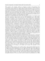
computed tomography (CT) scan showed a left basal ganglia ICH with intraventricular hemorrhage (IVH) into the left lateral ventricle (Fig. 9.1). Early intensive
BP lowering (systolic BP ≤ 140 mmHg) was achieved and intraventricular administration of 1 mg tissue plasminogen activator (tPA) every 8 h via external ventricular
drainage (EVD) was applied to reduce IVH volume and ICP.
Risks of Patient Safety and Management
Outcomes with ICH are significantly worse than with ischemic stroke, with up to
50 % mortality at 30 days. Morbidity and mortality in spontaneous ICH are correlated with low GCS score (≤8), hematoma volume, the presence of IVH, advanced
age (≥80 years), and infratentorial hematoma [6]. Almost 40 % of patients with
brain imaging obtained in the first 3 h after onset of symptoms of ICH experience
hematoma expansion and this is highly associated with the increase of ICP and
neurological deterioration [7]. The sudden increase in pressure within the brain can
cause damage to the brain cells surrounding the hemorrhage. If the amount of blood
increases rapidly, the sudden buildup in ICP can lead to unconsciousness or death.
Expanding hematoma results from persistent and/or secondary bleeding at the
periphery of an existing clot. Recent studies showed a strong association between
contrast extravasation (“spot sign”) on computed tomography angiography (CTA)
and hematoma expansion and worse outcome [8].
Initial goals of treatment include stabilization of airway, breathing, and circulation, followed by preventing hemorrhage extension, as well as the prevention and
management of elevated intracranial pressure along with other neurologic and medical complications. The patients should be monitored and treated in an NICU.
Blood Pressure
In general, the American Heart Association guidelines indicate that systolic BP
exceeding 180 mmHg or mean arterial pressure (MAP) exceeding 130 mmHg
should be managed with continuous-infusion antihypertensive agents (Table 9.1)
[9]. There was concern about a reduction of cerebral blood flow surrounding the
9
Intracerebral Hemorrhage
Fig. 9.1 CT scan showing
left basal ganglia
intracerebral hemorrhage
with extravasation into the
left lateral ventricle
147
a
b
hemorrhage with aggressive BP reduction. However, despite a peri-hematomal
reduction of cerebral metabolism, an ischemic zone was not found on several
radiographic cerebral metabolism studies.
The use of nitroprusside has drawbacks since this agent may exacerbate cerebral
edema and intracranial pressure, and sublingual agents are not preferred because of
148
M.K. Han
Table 9.1 Intravenous anti-hypertensive agents for blood pressure reduction in ICH
Drug
Labetalol
Esmolol
Nicardipine
Enalapril
Mechanism
α-1, β-1, β-2
receptor
antagonist
β-1 receptor
antagonist
L-type calcium
channel blocker
(dihydropyridine)
ACE inhibitor
Fenoldopam
Dopamine-1
receptor agonist
Nitroprusside
Nitrovasodilator
(arterial and
venous)
Dose
10–80 mg bolus every
10 min, up to 300 mg;
0.5–2.0 mg/min infusion
0.5 mg/kg bolus;
50–300 μg/kg/min
5–15 mg/h infusion
0.625 mg bolus;
1.25–5 mg every 6 h
0.1–0.3 μg/kg/min
0.25–10 μg/kg/min
Contraindications
Bradycardia, congestive heart
failure, bronchospasm
Bradycardia, congestive heart
failure, bronchospasm
Severe aortic stenosis,
myocardial ischaemia
Variable response, sudden in
BP with high-renin states
Tachycardia, headache,
nausea, flushing, glaucoma,
portal hypertension
Increased ICP, variable
response, myocardial
ischemia, thiocyanate and
cyanide toxicity
Abbreviations: ACE angiotension-converting enzyme, BP blood pressure
the need for precise BP control [10]. Therefore, nitroprusside should not be the first
agent for BP reduction in patients with ICH. In general, no matter how high the BP
is, the MAP should not be reduced beyond 15–30 % over the first 24 h [11].
Early elevation of BP is very common after ICH and is strongly associated with
poor outcomes [12]. The adverse effects of high BP levels on outcomes in ICH are
likely to involve a number of different mechanisms: elevated hydrostatic pressure in
the region of the ICH is likely to result in a larger initial hemorrhage with more
rapid increase of hematoma volume, whereas elevated BP may increase the likelihood of surrounding cerebral edema [13].
Current guidelines for the acute management of ICH provide an indication of
perceived harm associated with “very high” BP levels. Early intensive BP lowering
(systolic BP ≤ 140 mmHg) was feasible, well tolerated, and appeared to reduce
hematoma growth over 72 h, which may translate into beneficial effects in patients
treated within 6 h after acute ICH [14]. Early intensive lowering of BP (systolic
BP ≤ 140 mmHg) with any agent did not result in a significant reduction in the rate
of the death or major disability, but intensive treatment may improve functional
outcomes and areas of perceived quality of life. The intensive treatment was not
associated with an increase in the rates of death or serious adverse events [15].
Therefore, the guidelines for management of ICH by the European Stroke
Organization recommend reduction of the systolic BP to less than 140 mmHg within
6 h of symptom onset which was shown to be safe [16].
Seizures
Clinical seizures should be treated with anti-epileptic drugs as recurrent seizures may increase mass effect and midline shift. Continuous EEG monitoring is
9
Intracerebral Hemorrhage
149
indicated in ICH patients with depressed mental status out of proportion to the
degree of brain injury. Patients with a change in mental status who are found to have
electrographic seizures on EEG should be treated with anti-epileptic drugs.
Prophylactic anticonvulsant medication should not be used [9, 16].
Treatment of Intraventricular Hemorrhage
Intraventricular extension of ICH that occurs in 45 % of cases is a known independent predictor of poor outcome. Several studies have demonstrated a direct
relationship between IVH volume and poor outcome or mortality [17–19].
Another study showed that IVH volume predicts mortality independent of the
GCS [20]. The mechanisms by which IVH volume affects outcome likely include
increased intracranial pressure with reduced cerebral perfusion, mechanical disruption, ventricular wall distension, and possibly an inflammatory response [21–
23]. Total volume of IVH in itself is associated with poor outcome and a
“poor-outcome threshold” of 50 mL above which 100 % of patients had a poor
outcome [18]. An IVH volume >60 mL was associated with a mortality rate of
60 %. Low-dose recombinant tissue plasminogen activator (r-tPA) administered
via extraventricular drainage catheter in the treatment of ICH with IVH has an
acceptable safety profile compared to placebo and historical controls of the natural history [24]. A dose of 1 mg of r-tPA every 8 h (followed by clamping of the
EVD for 1 h) is reasonable until clearance of blood from the third or fourth ventricle has been achieved (CLEAR INTRAVENTRICULAR HEMORRHAGE TRIAL
study protocol). However, prior to administration of r-tPA further hematoma
expansion and the possible presence of EVD-associated hemorrhage should be
excluded by repeat head CT. This treatment is currently under investigation in a
phase III trial.
Intracranial Hypertension
Patients with a GCS score of 8 or less, or those with significant IVH or hydrocephalus, might be considered for ICP monitoring and treatment. Ventricular drainage as
treatment for hydrocephalus is reasonable in patients with decreased level of consciousness [9].
The head of the bed should be elevated to 30°. Hyperosmolar therapy of mannitol
or hypertonic saline is indicated in patients with intracranial hypertension and with
impending herniation. Hypertonic saline was found to have a longer duration of
effect. Safety concerns are renal failure with the use of mannitol and worsening of
preexisting congestive heart failure with administration of hypertonic saline. In
patients with renal failure, the osmolar gap should be followed instead of serum
osmolarity to monitor the effect of mannitol.
Surgery has the greater potential to reduce the volume of ICH and there is clinical and experimental evidence that mass removal might reduce nervous tissue
150
M.K. Han
damage, possibly by relieving local ischemia or removal of noxious chemicals [25,
26]. Large, surgically accessible clots exerting a mass effect might benefit from
early surgery, especially in younger patients; whereas, inaccessible clots with surgical approach paths that cross eloquent speech and motor regions probably do not.
Most neurosurgeons would remove a large frontopolar or temporal ICH after recent
deterioration of consciousness, an ICH of deeper location is not amendable to surgical removal. Minimally invasive techniques might be more beneficial for deeper
clots and IVH.
In several prospective randomized controlled trials, the patient outcome early
surgery for spontaneous supratentorial ICH was unchanged compared to controls.
Some patients did worse with surgery (e.g., those with deep-seated bleeds or with
IVH and hydrocephalus) and some had better results (e.g., patients with superficial
lobar hematomas without IVH) [25]. The same effect was noted in a meta-analysis
of other studies and in a large randomized trial: a benefit for mortality and functional from early surgery for ICH was not seen, there was a trend to better outcome
with surgery of superficially located ICH [26, 27]. The results of STICH II showed
no benefit for early surgery for patients with lobar ICH within 1 cm of the surface
[28]. Therefore, the indication for surgical clot removal should be discussed individually and be based on the patient’s age, the size and location of the hemorrhage,
and the presence of mass effect.
For patient’s safety, early aggressive BP lowering along with neuromonitoring, treatment of seizures, and early recognition of signs of intracranial hypertension followed by initiation of ICP reducing management are the most important
steps.
Safety Barriers and Risk–Benefit Assessment
During all treatment steps discussed the patient must be monitored closely. The
overall aim is to stop hemorrhage expansion and to limit the additional brain tissue
reduction by mass effect and seizures. Intensive BP reduction is reasonable [15, 16].
The indication for craniotomy and clot removal needs to be carefully evaluated as
hematoma evacuation may cause further tissue destruction and may be followed by
rebleeding. In lobar ICH and younger patients, a CT angiogram upon presentation
may help to exclude sources of bleeding which may be unmasked during hematoma
evacuation and to identify patients at risk for hematoma expansion by demonstrating a “spot sign.” Hemicraniectomy may be a reasonable alternative to hematoma
evacuation, especially in younger patients.
All patients with ICH should be screened for coagulopathies, and anticoagulant
medication effects antagonized emergently, especially before undergoing a neurosurgical procedure (see Table 9.2) [9].
Protamine sulfate
Prothrombin complex concentrate
50 g charcoal if Xa inhibitor
ingested within 2 h
Hemodialysis for dabigatran
overdose or renal insufficiency
Direct thrombin inhibitors
(argatroban, hirudin,
dabigatran) or inhibitors of
factor Xa (apixaban,
rivaroxaban, endoxaban)
Agent
Fresh frozen plasma (FFP)
or
Prothrombin complex
concentrate (Factor II, IV, IX, X,
protein C, S)
and
IV Vitamin K
Above plus consider
Recombinant factor VIIa
Unfractionated or lowmolecular-weight heparin
Target PTT 25–35 s
Warfarin and emergency
neurosurgical intervention
Scenario
Warfarin
Target: INR < 1.4
Table 9.2 Emergency management of ICH due to coagulopathy
Can take up to 24 h to normalize INR
Contraindicated in acute
thromboembolic disease, increased
risk of ischemic stroke and myocardial
infarction
Slowly: less than 20 mg per min
Maximum 50 mg
Can cause flushing, bradycardia, or
hypotension.
More effective for tinzaparin than for
dalteparin or enoxaparin
Minimal efficacy against danaparoid
or fondaparinux
Carries risk of DIC, thrombosis,
infection, anaphylaxis
10 mg
20–80 μg/kg
1–1.5/0.5–0.75/0.25–
0.375 mg per 100
units of heparin
(<30 min/
30–120 min/ 2 h)
1 mg per 100 anti-Xa
units LMWH if given
within last 8 h
30–60 U/kg
10–50 U/kg
Comments
Usually 4–6 units (200 mL) each are
given, risk of volume overload
Works faster than FFP, but carries risk
of DIC, thrombosis, infection,
anaphylaxis
Dose
10–15 mL/kg
(continued)
Very low
Very low
Low
Very low
Low
Level of evidencea
Low
9
Intracerebral Hemorrhage
151
Cryoprepicitate
Agent
Platelet transfusion
and/or
Desmopressin (DDAVP)
6–8 U
Dose
6 units or 1 single
donor apheresis unit
0.3 μg/kg
Mostly 4–6 units
Comments
Range 4–8 units based on size; within
12 h of symptom onset
Single dose required
Very low
Low
Level of evidencea
Low
Abbreviations: DIC disseminated intravascular coagulation, INR international normalized ratio, LMWH low molecular weight heparin, PTT prothrombin time
a
According to the GRADE criteria: Low quality of evidence = The authors are not confident in the effect estimate and the true value may be substantially different; very low quality evidence = The authors do not have any confidence in the estimate and it is likely that the true value is substantially different from it
Scenario
Platelet dysfunction or
thrombocytopenia
Target platelets > 100,000/μL
If planned for neurosurgical
procedure and documented
platelet dysfunction
Thrombolysis Complication
Table 9.2 (continued)
152
M.K. Han
9
Intracerebral Hemorrhage
153
Summary
In management of ICH, acute severe hypertension should be aggressively, but
carefully, controlled with IV medications to reduce systolic blood pressure to less
than 140 mmHg. Coagulopathies need to be antagonized aggressively to prevent
hematoma expansion. Suspected ICP elevation and symptomatic intracranial mass
effect should be treated with head elevation, mannitol or hypertonic saline, surgical
treatment should be considered for individual patients. Observation in a neurocritical care unit is strongly recommended for at least the first 24 h based on the risk of
neurologic deterioration.
Dos and Don’ts
Dos
• Stabilize airway, breathing and circulation
• Observation in the NICU is strongly recommended for at least 24 h based on
neurologic status and hemodynamics
• Prevention of extension of hemorrhage by BP control and antagonization of
coagulopathy
• Patients with GCS of 8 or less with significant ICH or hydrocephalus should be
considered for ICP monitoring
• Early intensive BP reduction of systolic BP to less than 140 mmHg within
first 6 h
• Use continuous EEG monitoring with patients with depressed mental status out
of proportion to brain injury
• Monitor for early signs and symptoms of intracranial hypertension
• Hypertonic saline is indicated for intracranial hypertension and impending
herniation
• Indication for surgical clot removal depends on individual case
• In selected cases with right skills and resources, r-TPA administered via extraventricular drainage can be effective
Don’ts
• Reduction of the MAP beyond 15–30 % over the first 24 h
• Use nitroprusside IV as a first line agent to control BP in ICH
• Prophylactic anticonvulsant should not be used
154
M.K. Han
References
1. Anderson RN, Smith BL. Deaths: leading causes for 2002. Natl Vital Stat Rep. 2005;53:
1–89.
2. Qureshi AI, Tuhrim S, Broderick JP, Batjer HH, Hondo H, Hanley DF. Spontaneous intracerebral hemorrhage. N Engl J Med. 2001;344:1450–60.
3. Dennis MS, Burn JP, Sandercock PA, Bamford JM, Wade DT, Warlow CP. Long-term survival
after first-ever stroke: the Oxfordshire Community Stroke Project. Stroke. 1993;24:796–800.
4. Gebel JM, Broderick JP. Intracerebral hemorrhage. Neurol Clin. 2000;18:419–38.
5. Ariesen MJ, Claus SP, Rinkel GJ, Algra A. Risk factors for intracerebral hemorrhage in the
general population: a systematic review. Stroke. 2003;34:2060–5.
6. Hemphill JC, Bonovich DC, Besmertis L, Manley GT, Johnston SC. The ICH score: a simple,
reliable grading scale for intracerebral hemorrhage. Stroke. 2001;32:891–7.
7. Brott T, Broderick J, Kothari R, et al. Early hemorrhage growth in patients with intracerebral
hemorrhage. Stroke. 1997;28:1–5.
8. Delgado Almandoz JE, Yoo AJ, Stone MJ, et al. The spot sign score in primary intracerebral
hemorrhage identifies patients at highest risk of in-hospital mortality and poor outcome among
survivors. Stroke. 2010;41:54–60.
9. Morgenstern LB, Hemphill 3rd JC, Anderson C, Becker K, Broderick JP, Connolly Jr ES, et al.
Guidelines for the management of spontaneous intracerebral hemorrhage: a guideline for
healthcare professionals from the American Heart Association/American Stroke Association.
Stroke. 2010;41(9):2108–29.
10. Rose JC, Mayer SA. Optimizing blood pressure in neurological emergencies. Neurocrit Care.
2004;1:287–99.
11. Powers WJ, Asams RF, Yundt KD. Acute pharmacological hypotension after intracerebral
hemorrhage does not change cerebral blood flow. Stroke. 1999;30:242.
12. Vemmos KN, Tsivgoulis G, Spengos K, Zakopoulos N, Synetos A, Manios E, Konstantopoulou
P, Mavrikakis M. U-shaped relationship between mortality and admission blood pressure in
patients with acute stroke. J Intern Med. 2004;255:257–65.
13. Kazui S, Minematsu K, Yamamoto H, Sawada T, Yamaguchi T. Predisposing factors to
enlargement of spontaneous intracerebral hematoma. Stroke. 1997;28:2370–5.
14. Anderson CS, Huang Y, Arima H, et al. Effects of early intensive blood pressure lowering
treatment on the growth of hematoma and perihematomal edema in acute intracerebral hemorrhage: the Intensive Blood Pressure Reduction in Acute Cerebral Haemorrhage Trial
(INTERACT). Stroke. 2010;41:307–12.
15. Anderson CS, Heeley E, Huang Y, Wang J, Stapf C, Delcourt C, et al. INTERACT2
Investigators. Rapid blood-pressure lowering in patients with acute intracerebral hemorrhage.
N Engl J Med. 2013;368:2355–65.
16. Steiner T, Al-Shahi Salman R, Beer R, Christensen H, Cordonnier C, Csiba L, Forsting M,
Harnof S, Klijn CJ, Krieger D, Mendelow AD, Molina C, Montaner J, Overgaard K, Petersson
J, Roine RO, Schmutzhard E, Schwerdtfeger K, Stapf C, Tatlisumak T, Thomas BM, Toni D,
Unterberg A, Wagner M. European Stroke Organisation (ESO) guidelines for the management
of spontaneous intracerebral hemorrhage. Int J Stroke. 2014;9(7):840–55.
17. Hallevi H, Albright K, Aronowski J, et al. Intraventricular hemorrhage: anatomic relationships
and clinical implications. Neurology. 2008;70:848–52.
18. Young WB, Lee KP, Pessin MS, et al. Prognostic significance of ventricular blood in supratentorial hemorrhage: a volumetric study. Neurology. 1990;40:616–9.
19. Steiner T, Diringer MN, Schneider D, et al. Dynamics of intraventricular hemorrhage in
patients with spontaneous intracerebral hemorrhage: risk factors, clinical impact, and effect of
hemostatic therapy with recombinant activated factor VII. Neurosurgery. 2006;59:767–73.
20. Tuhrim S, Horowitz DR, Sacher M, et al. Volume of ventricular blood is an important
determinant of outcome in supratentorial intracerebral hemorrhage. Crit Care Med. 1999;27:
617–21.
9
Intracerebral Hemorrhage
155
21. Mayer SA, Thomas CE, Diamond BE. Asymmetry of intracranial hemodynamics as an
indicator of mass effect in acute intracerebral hemorrhage. A transcranial Doppler study.
Stroke. 1996;27:1788–92.
22. Mayfrank L, Kissler J, Raoofi R, et al. Ventricular dilatation in experimental intraventricular
hemorrhage in pigs characterization of cerebrospinal fluid dynamics and the effects of
fibrinolytic treatment. Stroke. 1997;28:141–8.
23. Wasserman JK, Zhu X, Schlichter LC. Evolution of the inflammatory response in the brain
following intracerebral hemorrhage and effects of delayed minocycline treatment. Brain Res.
2007;1180:140–54.
24. Naff N, Williams MA, Keyl PM, et al. Low-dose recombinant tissue-type plasminogen
activator enhances clot resolution in brain hemorrhage: the intraventricular hemorrhage
thrombolysis trial. Stroke. 2011;42:3009–16.
25. Xi G, Keep RF, Hoff JT. Mechanisms of brain injury after intracerebral haemorrhage. Lancet
Neurol. 2006;5:53–63.
26. Keep RF, Xi G, Hua Y, Hoff JT. The deleterious or beneficial effects of different agents in
intracerebral hemorrhage: think big, think small, or is hematoma size important? Stroke.
2005;36:1594–6.
27. Bhattathiri PS, Gregson B, Prasad KS, Mendelow AD, STICH Investigators. Intraventricular
hemorrhage and hydrocephalus after spontaneous intracerebral hemorrhage: results from the
STICH trial. Acta Neurochir Suppl. 2006;96:65–8.
28. Mendelow AD, Gregson BA, Rowan EN, Murray GD, Gholkar A, Mitchell PM, STICH II
Investigators. Early surgery versus initial conservative treatment in patients with spontaneous
supratentorial lobar intracerebral haematomas (STICH II): a randomised trial. Lancet.
2013;382:397–408.
Chapter 10
Patient Safety in Acute Ischemic Stroke
Ivan Rocha Ferreira da Silva and Bernardo Liberato
Introduction
Patient safety has been an increasing concern in modern medicine worldwide, and
recent discussions about quality of care, safety precautions and performance
measures of stroke care have gained growing interest. Healthcare systems
throughout the world face the vexing problem of improving healthcare quality
while at the same time confronted with ever-increasing costs and greater demands
for accountability [1].
Stroke is a common and serious disorder. Each year, approximately 750,000
individuals have a new or recurrent stroke in the United States [2]. Also, stroke
patients occupy 20 % of acute medical beds in the British National Health System
[3]. Safety is a major issue in this population, as medical complications are frequent
among individuals who have had a stroke, increasing the length of hospitalization as
well as the costs of care [4]. Moreover, many of the complications described are
potentially preventable or treatable if promptly recognized [5], and patients at risk
for or who have had a stroke often do not receive medical care consistent with
current evidence-based standards [6].
The aim of this chapter is to introduce the importance of structured stroke care,
minimizing complications and risks, as well as promoting safe, effective, and
durable care interventions.
I.R.F. da Silva, MD
Department of Neurocritical Care, Hospital Copa D’Or, Rio de Janeiro, Brazil
B. Liberato, MD (*)
Department of Neurology, Hospital Copa D’Or, Rio de Janeiro, Brazil
e-mail:
© Springer International Publishing Switzerland 2015
K.E. Wartenberg et al. (eds.), Neurointensive Care: A Clinical Guide
to Patient Safety, DOI 10.1007/978-3-319-17293-4_10
157
158
I.R.F. da Silva and B. Liberato
Case Scenario
A 75 year-old lady, with history of diabetes mellitus and hypertension, is brought to
the emergency department by her son after a sudden onset of weakness on her right
side and difficulty speaking. He mentioned that she was last seen normal approximately 45 min ago, and an immediate neurological exam discloses dense paresis of
her right side, with severe aphasia and a left gaze deviation, with a National Institute
of Health Stroke Scale (NIHSS) score of 18. An emergency computed tomography
(CT) scan of the head was unremarkable, and so the decision was to proceed with
intravenous thrombolysis with recombinant tissue plasminogen activator (r-tPA).
The per-protocol bolus of the medication was uneventful, but during the first half of
the infusion, the bedside nurse noticed a blood pressure of 200/115 mmHg and a
finger test showed a capillary glucose of 210 mg/dL (11.6 mmol/L). Soon after, her
level of consciousness declined suddenly, and a repeat CT disclosed a 35 mL intraparenchymal hemorrhage in the area of the left basal ganglia, with a 5 mm midline
shift and intraventricular blood (Fig. 10.1). The r-tPA infusion was held, freshfrozen plasma and cryoprecipitate were given and she was admitted to the neurocritical care unit (NICU). No surgical intervention was indicated at that point.
During the first week in the NICU she was treated for aspiration pneumonia, not
Fig. 10.1 CT scan of the
75-year-old patient with acute
right sided hemiparesis and
aphasia receiving intravenous
thrombolysis showing the
intraventricular hemorrhage
originating from a left basal
ganglia intracerebral
hemorrhage and a 5 mm
midline shift
10
Patient Safety in Acute Ischemic Stroke
159
requiring mechanical ventilation. Two weeks later the patient was moved to the
neurology ward with some improvement of the right-sided weakness, but still with
severe aphasia. During the following night, the patient was found on the ground by
the on-call nurse, likely a consequence of the bed side-rail being down. She suffered
no neurological insults, but a wrist fracture was noticed, prolonging her hospital
stay and transfer to a rehabilitation facility.
Risks of Patient Safety
Stroke patients are exposed to several possible complications, which can occur at
any time during the disease process, as early as a hemorrhagic transformation or
intracerebral hemorrhage in the first few hours after thrombolysis, or later in the
shape of aspiration pneumonia secondary to some degree of dysphagia, fall risk,
deep venous thrombosis (DVT), and pressure ulcers during rehabilitation. Previous
studies have shown that complications are common, with estimates of frequencies
ranging from 40 to 96 % of patients [7–11]. As is true for long-term neurological
recovery and overall mortality, age and stroke severity are associated with the development of complications, which most commonly occur in the first 4 days [12].
Several studies retrospectively analyzed the incidence and timing of medical
complications in stroke patients. Davenport et al. [7] found that seizures and chest
infections occurred early, whereas depression and painful shoulder were later problems. Dromerick et al. [8] noticed that the mean number of medical and neurological complications per patient were 3.6 and 0.6, respectively, and complications were
independently related to both the severity of functional disability as judged by
Barthel score and length of rehabilitation hospital stay. Finally, Johnston et al. [9]
reported a 3-month mortality of 14 % in stroke patients, with 51 % of these deaths
were attributed primarily to medical complications. Outcome was significantly
worse in patients with serious medical complications [9].
A prospective cohort Scottish study [5] found that the most frequent complications during hospital stay were confusion (56 %), pain (34 %), falls (25 %), infections (24 %, mostly respiratory and urinary tract infections), depression (16 %,) and
recurrent stroke (9 %), but during follow-up as outpatient, infections, falls, “blackouts,” pain, and symptoms of depression and anxiety remained common.
Pneumonia, which is usually associated with immobility, ineffective cough and
difficulty of airway protection, is an important cause of death after stroke [13–15].
Moreover, stroke-associated pneumonia increases length of stay, mortality, and hospital costs [16]. Early mobility and good pulmonary care can help to prevent pneumonia [16], as well as preventive measures in intubated patients, including
ventilation in a semi-recumbent position, frequent suctioning, mouth hygiene, and
early extubation. A retrospective study disclosed that patients with brain-stem
stroke were more likely to develop early pneumonia. The incidence was higher in
patients who failed swallowing evaluation and in those who were intubated [17].
160
I.R.F. da Silva and B. Liberato
Urinary tract infections are quite common, occurring in 15–60 % of stroke
patients, independently predicting worse outcomes [5, 13, 18, 19]. Patients with
major impairments as well as use of indwelling catheters are associated with urinary tract infections [20]. Early removal of indwelling catheters, bladder training
and use of intermittent catheterization are strategies to lessen the risk of such
infections [21].
Pulmonary embolism accounts for 10 % of deaths after stroke, and the complication may be detected in 1 % of stroke patients [22]. The risk of DVT is highest
amongst immobilized and older patients with severe stroke [23–25], and is more
frequent in the first 3 months after the stroke [12]. Besides being associated with a
pulmonary embolism, symptomatic DVT also delays recovery and rehabilitation
after stroke [21]. The alternatives for mitigating the risk of DVT include early mobilization, administration of antithrombotic agents, and the use of external compression devices. In patients with acute ischemic stroke, there is strong evidence that the
use of low-molecular weight heparin is the therapy of choice to prevent DVT [26].
The late introduction of DVT prophylaxis with low-molecular weight heparin in
hospitalized stroke patients, based on the unfounded concern for hemorrhagic transformation, adds to this problem, especially in patients with large hemispheric
strokes who happen to be the most susceptible to thrombotic complications. The
misconception that patients with a large hemispheric ischemic stroke should have
the low-molecular weight heparin withheld for a few days only adds to the medical
morbidity in such patients and is not supported by either anecdotal or evidencebased experience. Early introduction of DVT prophylaxis, even in the presence of
small petechial bleeds should be the rule in all stroke patients.
Swallowing impairments are associated with an increased risk of death and
pneumonia [14, 27]. Mann et al. [27] have shown that at presentation, a swallowing
abnormality was detected clinically in 51 % of acute stroke patients and videofluoroscopically in 64 %, with 20 % having developed respiratory infections. An abnormal gag reflex, impaired voluntary cough, dysphonia, incomplete oral-labial
closure, a high NIHSS score, or cranial nerve palsies should alert the care team to
the risk of dysphagia [21]. A formal speech and swallow evaluation should be
obtained early on in all stroke patients for detection of subtle signs of microaspiration. When such evaluation is not readily available, a water swallow test performed
at the bedside is a useful screening tool [21], and dysphagia screening protocols
have shown to lessen the risk of pneumonia in different settings [28, 29]. Although
caution should be exerted to orally feed stroke patients, nutrition should be started
as soon as possible, usually through nasogastric tubes, as it is associated with
improved outcomes [30].
Stroke, as many neurological disorders, is associated with a high risk of falls
[31]. It has been shown that up to 21 % of patients after an acute stroke might experience falls within the first 6 weeks [11], and studies investigating falls in the later
phase report an incidence of up to 73 % in the first year post-stroke [32]. Falls can
lead to a variety of consequences, such as traumatic brain injuries, fractures, fear of
falling, reduced activity and death, and involve both personal suffering and economic costs for the community [33–35]. Exercises and physical therapy are
recommended to improve gait stability, and assessment tools of fall risk on
10
Patient Safety in Acute Ischemic Stroke
161
admission, both in the acute and subacute settings, seem to decrease the incidence
of this complication [36, 37]. Professional advice with prescription of orthotic
devices when appropriate and counseling regarding improvement in home safety
measures might mitigate this problem.
Decubitus ulcers are an often neglected problem in hospitalized patients with
stroke, and are considered a quality metric for many hospitals. Decubiti were
reported in up to 21 % of acute stroke patients in a prospective study [5], and the
Center for Medicare Services (CMS) does not reimburse wound care if the patient
develops decubiti while hospitalized due to the potentially preventable nature of this
complication [38]. This and other reinforcement tools might decrease its occurrence. Well known risk factors include immobility, lack of turning by nursing
personnel, poor nutrition, and urinary incontinence.
Safety Barriers and Structured Stroke Care
Safety in healthcare is an essential part of modern medicine, and vast evidence has
been produced recently on the matter. It is well known that protocol bundles might
improve outcomes in critical care, such as decreasing mortality in severe sepsis
[39], mitigating the incidence of central line associated infections [40, 41] and
ventilator-associated pneumonia [42]. Furthermore, protocol bundles also optimize
cost-effectiveness of care [43, 44]. A “bundle” is a group of evidence-based care
components for a given disease that, when executed together, may result in better
outcomes than when implemented individually, according to the Institute for
Healthcare Improvement [45].
Unfortunately, no studies so far have assessed the implementation of safety bundles in patients with stroke. Important strategies such as DVT prophylaxis, dysphagia screening, fall prevention, blood pressure and serum glucose management are
intuitive measures and are cited in guidelines [21, 46], but the impact of those
actions taken together is unknown. At least in theory, all the benefits of the bundled
care for the stroke patients can be found when they are admitted to a separate physical unit where attention is given to the specific needs of this patient population, e.g.
the Stroke Unit. Also the presence of a team, experienced in the care of stroke
patients offers a greater chance of protocol compliance, increased surveillance for
potential medical complications and expedited discharge to an acute or subacute
rehabilitation unit. Even more evident is the level of care for the severe stroke
patients in a dedicated neurological ICU, where close attention and familiarity with
the unstable neurological patient often make a difference in the outcome.
Recently, several attempts have been made to protocolize stroke care, with the
aim of improving outcomes and minimizing complications. The Get With The
Guidelines–Stroke is an ongoing voluntary, continuous quality-improvement initiative involving hospitals mainly in the United States and Canada that collects patient
level data on characteristics, treatments, in-hospital outcomes, and adherence to
quality measures in stroke, including ischemic stroke and hemorrhagic stroke. The
initiative has been successful so far, with studies showing significant improvement
162
I.R.F. da Silva and B. Liberato
of quality of care [47, 48]. Performance measures of quality of care in stroke are
essential tools to assess what is offered to stroke patients, and several studies have
been conducted in Germany [49, 50], Denmark [51], the United States [52–55],
Chile [56], the Netherlands [57], and Austria [58] to better understand the gaps
between the guidelines and bedside care. Recently, a study compared the performance measures used in several centers in Europe, and found significant differences
in benchmarks, quality indicators and data documentation, suggesting that an equalization of such measurements should be done urgently [59].
The implementation of safety barriers is an important part of organized stroke
care. Safe care can be promoted with patient and family’s education, strict observance of established protocols, continuous feedback on performance measures and
frequent training of healthcare personnel. Structured care, through establishment of
neurocritical care and stroke units, can definitely change the outcomes in critically
ill neurological patients [60–63]. The American Heart Association/American Stroke
Association and the Joint Commission on Accreditation of Healthcare Organizations
have merged efforts to create standards on stroke care, and recently started accrediting hospitals in the United States as Primary Stroke Centers or Comprehensive
Stroke Centers, if strict criteria are fulfilled. A comparable accreditation process is
available for regional, hyperregional, and comprehensive stroke centers in Germany
and other countries in Europe through regional stroke societies and associations.
Some actions to prevent complications in stroke patients are well recognized,
and will be discussed later in this chapter. The daily assessment of patient’s needs,
including measures to avoid complications, structured plan of care and fluid communication through all levels of the care team are essential to promote safety. The
implementation of multidisciplinary rounds and the use of “check lists” to remind
of important preventive measures are well established in critical care units [64, 65],
and this successful model should be thoroughly used in the stroke population as
well.
Risks and Benefits of Systemic Thrombolysis in Acute
Ischemic Stroke
To this date, systemic thrombolysis with r-tPA is the only evidence-based treatment
able to improve outcomes in patients with acute ischemic stroke. The landmark
NINDS trial in 1995 randomized 624 patients, and produced clinical and statistical
benefit over placebo for patients treated within 3 h of evaluation [66]. It showed that
patients treated with r-tPA were at least 30 % more likely to have minimal or no
disability at 3 months, with a 6.4 % risk of intracerebral hemorrhage [66]. Based on
these results, r-tPA was approved for treatment of acute ischemic stroke in the
United States in 1996 [67] for use within 3 h of onset of symptoms, but not without
controversy. At that time, some authors [68–70], as well as associations of emergency medicine physicians [71, 72], criticized the study’s methodology, and did not
endorse its use by emergency specialists, as previous trials on thrombolysis in acute
stroke were negative, such as the ECASS [73], ECASS II [74], and ATLANTIS
10
Patient Safety in Acute Ischemic Stroke
163
[75]. Worth mentioning, these studies used longer time windows (>3 h) and in
ECASS a different dose of r-tPA was used. However, a Cochrane review using
meta-analysis of various types of thrombolysis, including r-tPA, urokinase and
streptokinase, concluded that this therapy was effective [76]. In a response to the
emergency physicians, a re-analysis of the NINDS trial, now dividing into subgroups regarding the NIH stroke scale upon arrival and time of onset, was published
in the Annals of Emergency Medicine, showing that the results were still similar
after balancing again both groups [77].
The controversy finally settled almost a decade later. In order to have alteplase
(r-tPA) approved under European Union regulations, the SITS-MOST study was
conducted to assess the safety profile of alteplase in clinical practice by comparison
with results in randomized controlled trials [78]. A total of 6,483 patients were
recruited from 285 centers (50 % with little previous experience in stroke thrombolysis) in 14 countries between 2002 and 2006 for this prospective, open, monitored, observational study. Results showed that intravenous alteplase is safe and
effective in routine clinical use when used within 3 h of stroke onset, even by centers with little previous experience in thrombolytic therapy for acute stroke. Two
years later, the ECASS III trial found similarly positive results, now with patients
with acute stroke within a time window within 3–4.5 h, leaving no doubt about the
effectiveness and safety of this therapy [79].
A more elegant way of understanding the risk versus benefit in patients receiving
r-tPA, and hence its safety implications, is to use the number needed to treat (NNT)
and the number needed to harm (NNTH). The number needed to treat for benefit
(NNT) is an effect measure that indicates how many patients need to be treated with
an intervention for one patient to experience a benefit, with the opposite being the
NNTH. The post-hoc analysis of the NINDS trial showed that for different dichotomized global functional end-points, the number needed to harm as a result of r-tPArelated cerebral hemorrhages ranges widely, from 36.5 to 707 [80]. To better refine,
a reasonable key dichotomization for NNTH estimation is the number needed to
treat for one additional patient to end up severely disabled or dead, with a calculated
NNTH of 126 in the NINDS trial [80]. Finally, for every 100 patients treated with
rtPA, across all levels of final global disability, approximately 32 will benefit and
approximately three will be harmed, with odds of better results ten times higher
than for harm [80]. Two years later another post-hoc analysis was conducted, now
including the pooled data set of the first six major randomized acute stroke trials of
intravenous r-tPA [81]. The results found that the NNT for benefit was 3.6 for
patients treated between 0 to 90 min, 4.3 for 91 to 180 min, 5.9 for 181 to 270 min,
and 19.3 with treatment between 271 and 360 min. This underscores not only the
effectiveness of the therapy but also its strong time-dependency.
Some studies have shown that most complications related to r-tPA in acute
ischemic stroke are derived from protocol violations (e.g., blood pressure control,
patient selection, etc.) [82–84], and from a medico-legal standpoint, physicians
have a much higher chance of being sued for not offering r-tPA for eligible candidates than for drug-related complications [85, 86]. Alteplase is the only Class I,
Level of Evidence A treatment for acute ischemic stroke accordingly to the current American Heart Association and European Stroke Organization Guidelines
164
I.R.F. da Silva and B. Liberato
[21, 46]. It is recommended that institutions adhere to strict protocols in order to
ensure minimal safety requirements. Continuous auditing and case-by-case
discussion should be encouraged and regarded as basic safety measures, especially in centers with little experience (less than 5 cases/year). As is true in medical complications after an acute stroke the presence of checklists pre and post
intravenous thrombolysis should be the rule with special emphasis on blood
pressure monitoring per protocol, adequate glucose control, and frequent
neurological assessments.
Finally, some case series and observational studies have shown that r-tPA can be
probably safely administered in off label situations, such as in very elderly patients
[87], patients with prior stroke and diabetes mellitus [88], stroke mimics [89], presence of unruptured aneurysm or arteriovenous malformation [90] and in pregnant
patients [91–93].
Solutions to Potential Risks
Table 10.1 summarizes the most important and thoroughly studied actions to prevent and/or minimize the most frequent medical complications encountered in
stroke patients.
Table 10.1 Solutions to potential risks
Risks
Aspiration
Falls
Urinary infections
Pressure ulcers
Deep venous
thrombosis
Delirium
Secondary stroke
prevention
Possible solutions/prevention
Early screening for dysphagia per protocol, elevate head –of-bed 30°
Education of patients and family, elevate bed rails, assisted walking,
early physical therapy, encourage use of corrective lenses, use of
walking devices (e.g. cane, walker), assessment of fall risk on admission
with clear identification of patients at risk
Early removal of urinary catheters, bladder training, use of intermittent
catheterization instead of indwelling catheters if needed later in the
hospital stay
Avoid immobility, aggressive skin care, frequent turning per protocol,
adequate nutritional support, and control of urinary incontinence
Avoid immobility, early use of low-molecular weight heparin (preferred)
or unfractionated heparin, use of sequential compression devices in the
first 24 h after systemic thrombolysis
Support family at the bedside, early move to wards with windows and
sunlight, encourage use of corrective lenses and hearing devices,
mitigate the use of benzodiazepines and physical restraints, minimize
metabolic derangements, use of orienting techniques, day-night structure
Education of patients and family (blood pressure control, diabetes, stroke
prevention, smoking cessation, nutrition), assure use of anti-platelet
aggregation agents and statins upon discharge, referral for follow up
soon after discharge
10
Patient Safety in Acute Ischemic Stroke
165
Summary
Patient safety has been an increasing worldwide concern in modern medicine, and
recent discussions about quality of care, safety precautions and performance
measures of stroke care have gained growing interest. Stroke patients are exposed to
several possible complications, which can occur at any time during the disease
process, and can potentially worsen their prognosis.
Unfortunately, no studies so far have studied the implementation of safety
bundles in patients with stroke. Important strategies such as DVT prophylaxis,
dysphagia screening, fall prevention, blood pressure and serum glucose management are cited in guidelines, but the impact of those actions taken together is
unknown. The auditing for implementation of secondary prevention measures upon
discharge, as suggested by the accrediting agencies and the American Heart
Association – Get with the Guidelines, are also instrumental in maximizing the
benefit and reducing the harm associated with early and late stroke recurrence.
The implementation of safety barriers is an important part of organized stroke
care. Safe care can be promoted with patient and family’s education, strict
observance of established protocols, continuous feedback on performance measures
and frequent training of healthcare personnel.
Dos and Don’ts
Dos
•
•
•
•
•
Stroke patients should preferably be admitted to specialized units, e.g. stroke units
Early physical/occupational therapy and speech/swallow evaluation
Multidisciplinary teams are essential for adequate stroke care
Check-lists should be used to remind of important preventive actions
Clear identification (wrist band) of anticoagulated or recently thrombolized
stroke patients
• Considered stroke a priority in your ER, with the same classification of urgency
as trauma or acute myocardial infarction
• Education and training of healthcare personnel, as well as patients and their relatives is important.
• Adhere to approved guidelines for IV thrombolysis, especially in centers with
little experience
Don’ts
• Do not underestimate medical complications in stroke patients
• Do not leave stroke patients with severe disabilities unattended
166
I.R.F. da Silva and B. Liberato
• Do not underestimate the aid of family members in stroke care
• Avoid protocol deviations in stroke care, especially utilizing systemic
thrombolysis
• Do not admit stroke patients to your facility if stroke is not considered to be a
priority in your ER, and receive the classification of urgency as trauma or acute
myocardial infarction
• Do not admit stroke patients to your facility if it’s unsure that quality care can be
provided
References
1. Reeves MJ, Parker C, Fonarow GC, Smith EE, Schwamm LH. Development of stroke performance measures: definitions, methods, and current measures. Stroke. 2010;41(7):1573–8.
2. Alberts MJ, Latchaw RE, Selman WR, et al. Recommendations for comprehensive stroke
centers: a consensus statement from the Brain Attack Coalition. Stroke. 2005;36(7):
1597–616.
3. Walsh K, Gompertz PH, Rudd AG. Stroke care: how do we measure quality? Postgrad Med
J. 2002;78(920):322–6.
4. Kumar S, Selim MH, Caplan LR. Medical complications after stroke. Lancet Neurol.
2010;9(1):105–18.
5. Langhorne P, Stott DJ, Robertson L, et al. Medical complications after stroke: a multicenter
study. Stroke. 2000;31(6):1223–9.
6. Holloway RG, Benesch C, Rush SR. Stroke prevention: narrowing the evidence-practice gap.
Neurology. 2000;54(10):1899–906.
7. Davenport RJ, Dennis MS, Wellwood I, Warlow CP. Complications after acute stroke. Stroke.
1996;27(3):415–20.
8. Dromerick A, Reding M. Medical and neurological complications during inpatient stroke
rehabilitation. Stroke. 1994;25(2):358–61.
9. Johnston KC, Li JY, Lyden PD, et al. Medical and neurological complications of ischemic
stroke: experience from the RANTTAS trial. RANTTAS Investigators. Stroke.
1998;29(2):447–53.
10. Kalra L, Yu G, Wilson K, Roots P. Medical complications during stroke rehabilitation. Stroke.
1995;26(6):990–4.
11. McClatchie G. Survey of the rehabilitation outcome of strokes. Med J Aust. 1980;1(13):
649–51.
12. Indredavik B, Rohweder G, Naalsund E, Lydersen S. Medical complications in a comprehensive stroke unit and an early supported discharge service. Stroke. 2008;39(2):414–20.
13. Aslanyan S, Weir CJ, Diener HC, Kaste M, Lees KR. Pneumonia and urinary tract infection
after acute ischaemic stroke: a tertiary analysis of the GAIN International trial. Eur J Neurol.
2004;11(1):49–53.
14. Martino R, Foley N, Bhogal S, Diamant N, Speechley M, Teasell R. Dysphagia after stroke:
incidence, diagnosis, and pulmonary complications. Stroke. 2005;36(12):2756–63.
15. van der Worp HB, Kappelle LJ. Complications of acute ischaemic stroke. Cerebrovasc Dis
(Basel, Switzerland). 1998;8(2):124–32.
16. Hilker R, Poetter C, Findeisen N, et al. Nosocomial pneumonia after acute stroke: implications
for neurological intensive care medicine. Stroke. 2003;34(4):975–81.
17. Upadya A, Thorevska N, Sena KN, Manthous C, Amoateng-Adjepong Y. Predictors and consequences of pneumonia in critically ill patients with stroke. J Crit Care. 2004;19(1):16–22.
18. Kong KH, Young S. Incidence and outcome of poststroke urinary retention: a prospective
study. Arch Phys Med Rehabil. 2000;81(11):1464–7.
10
Patient Safety in Acute Ischemic Stroke
167
19. Roth EJ, Lovell L, Harvey RL, Heinemann AW, Semik P, Diaz S. Incidence of and risk factors
for medical complications during stroke rehabilitation. Stroke. 2001;32(2):523–9.
20. Ween JE, Alexander MP, D’Esposito M, Roberts M. Incontinence after stroke in a rehabilitation setting: outcome associations and predictive factors. Neurology. 1996;47(3):659–63.
21. Jauch EC, Saver JL, Adams Jr HP, et al. Guidelines for the early management of patients with
acute ischemic stroke: a guideline for healthcare professionals from the American Heart
Association/American Stroke Association. Stroke. 2013;44(3):870–947.
22. Wijdicks EF, Scott JP. Pulmonary embolism associated with acute stroke. Mayo Clin Proc.
1997;72(4):297–300.
23. Desmukh M, Bisignani M, Landau P, Orchard TJ. Deep vein thrombosis in rehabilitating
stroke patients. Incidence, risk factors and prophylaxis. Am J Phys Med Rehabil. 1991;70(6):
313–6.
24. Kelly J, Rudd A, Lewis RR, Coshall C, Moody A, Hunt BJ. Venous thromboembolism after
acute ischemic stroke: a prospective study using magnetic resonance direct thrombus imaging.
Stroke. 2004;35(10):2320–5.
25. Warlow C, Ogston D, Douglas AS. Deep venous thrombosis of the legs after strokes. Part I–
incidence and predisposing factors. Br Med J. 1976;1(6019):1178–81.
26. Sherman DG, Albers GW, Bladin C, et al. The efficacy and safety of enoxaparin versus unfractionated heparin for the prevention of venous thromboembolism after acute ischaemic stroke
(PREVAIL Study): an open-label randomised comparison. Lancet. 2007;369(9570):1347–55.
27. Mann G, Hankey GJ, Cameron D. Swallowing function after stroke: prognosis and prognostic
factors at 6 months. Stroke. 1999;30(4):744–8.
28. Hinchey JA, Shephard T, Furie K, Smith D, Wang D, Tonn S. Formal dysphagia screening
protocols prevent pneumonia. Stroke. 2005;36(9):1972–6.
29. Martino R, Silver F, Teasell R, et al. The Toronto Bedside Swallowing Screening Test
(TOR-BSST): development and validation of a dysphagia screening tool for patients with
stroke. Stroke. 2009;40(2):555–61.
30. Dennis MS, Lewis SC, Warlow C. Effect of timing and method of enteral tube feeding for
dysphagic stroke patients (FOOD): a multicentre randomised controlled trial. Lancet.
2005;365(9461):764–72.
31. Homann B, Plaschg A, Grundner M, et al. The impact of neurological disorders on the risk for
falls in the community dwelling elderly: a case-controlled study. BMJ Open. 2013;3(11):
e003367.
32. Verheyden GS, Weerdesteyn V, Pickering RM, et al. Interventions for preventing falls in
people after stroke. Cochrane Database Syst Rev. 2013;(5):CD008728.
33. Batchelor FA, Mackintosh SF, Said CM, Hill KD. Falls after stroke. Int J Stroke.
2012;7(6):482–90.
34. Friedman SM, Munoz B, West SK, Rubin GS, Fried LP. Falls and fear of falling: which comes
first? A longitudinal prediction model suggests strategies for primary and secondary prevention. J Am Geriatr Soc. 2002;50(8):1329–35.
35. Nystrom A, Hellstrom K. Fall risk six weeks from onset of stroke and the ability of the
Prediction of Falls in Rehabilitation Settings Tool and motor function to predict falls. Clin
Rehabil. 2013;27(5):473–9.
36. Rosario ER, Kaplan SE, Khonsari S, Patterson D. Predicting and assessing fall risk in an acute
inpatient rehabilitation facility. Rehabil Nurs. 2014;39:86–93.
37. Smith J, Forster A, Young J. Use of the ‘STRATIFY’ falls risk assessment in patients recovering from acute stroke. Age Ageing. 2006;35(2):138–43.
38. Freeman WD, Dawson SB, Flemming KD. The ABC’s of stroke complications. Semin Neurol.
2010;30(5):501–10.
39. Marwick C, Davey P. Care bundles: the holy grail of infectious risk management in hospital?
Curr Opin Infect Dis. 2009;22(4):364–9.
40. Berenholtz SM, Lubomski LH, Weeks K, et al. Eliminating central line-associated bloodstream infections: a national patient safety imperative. Infect Control Hosp Epidemiol.
2014;35(1):56–62.
168
I.R.F. da Silva and B. Liberato
41. Hsu E, Lin D, Evans SJ, et al. Doing well by doing good: assessing the cost savings of an
intervention to reduce central line-associated bloodstream infections in a Hawaii hospital. Am
J Med Qual. 2014;29(1):13–9.
42. Morris AC, Hay AW, Swann DG, et al. Reducing ventilator-associated pneumonia in intensive
care: impact of implementing a care bundle. Crit Care Med. 2011;39(10):2218–24.
43. Moller AH, Hansen L, Jensen MS, Ehlers LH. A cost-effectiveness analysis of reducing
ventilator-associated pneumonia at a Danish ICU with ventilator bundle. J Med Econ.
2012;15(2):285–92.
44. Muscedere JG, Martin CM, Heyland DK. The impact of ventilator-associated pneumonia on
the Canadian health care system. J Crit Care. 2008;23(1):5–10.
45. Institute for Healthcare Improvement. 2014. Accessed 6 Feb 2014.
46. European Stroke Organisation (ESO) Executive Committee; ESO Writing Committee.
Guidelines for management of ischaemic stroke and transient ischaemic attack 2008.
Cerebrovasc Dis (Basel, Switzerland). 2008;25(5):457–507.
47. Fonarow GC, Reeves MJ, Smith EE, et al. Characteristics, performance measures, and inhospital outcomes of the first one million stroke and transient ischemic attack admissions in
get with the guidelines-stroke. Circ Cardiovasc Qual Outcomes. 2010;3(3):291–302.
48. Schwamm LH, Fonarow GC, Reeves MJ, et al. Get With The Guidelines-Stroke is associated
with sustained improvement in care for patients hospitalized with acute stroke or transient
ischemic attack. Circulation. 2009;119(1):107–15.
49. Heuschmann PU, Biegler MK, Busse O, et al. Development and implementation of evidencebased indicators for measuring quality of acute stroke care: the Quality Indicator Board of the
German Stroke Registers Study Group (ADSR). Stroke. 2006;37(10):2573–8.
50. Nimptsch U, Mansky T. Quality measurement combined with peer review improved German
in-hospital mortality rates for four diseases. Health Aff (Project Hope). 2013;32(9):1616–23.
51. Ingeman A, Andersen G, Hundborg HH, Svendsen ML, Johnsen SP. Processes of care and
medical complications in patients with stroke. Stroke. 2011;42(1):167–72.
52. Jacobs BS, Baker PL, Roychoudhury C, Mehta RH, Levine SR. Improved quality of stroke
care for hospitalized Medicare beneficiaries in Michigan. Stroke. 2005;36(6):1227–31.
53. Lakshminarayan K, Borbas C, McLaughlin B, et al. A cluster-randomized trial to improve
stroke care in hospitals. Neurology. 2010;74(20):1634–42.
54. Pandey DK, Cursio JF. Data feedback for quality improvement of stroke care: CAPTURE
Stroke experience. Am J Prev Med. 2006;31(6 Suppl 2):S224–9.
55. Roychoudhury C, Jacobs BS, Baker PL, Schultz D, Mehta RH, Levine SR. Acute ischemic
stroke in hospitalized medicare patients: evaluation and treatment. Stroke. 2004;35(1):e22–3.
56. Hoffmeister L, Lavados PM, Comas M, Vidal C, Cabello R, Castells X. Performance measures
for in-hospital care of acute ischemic stroke in public hospitals in Chile. BMC Neurol.
2013;13:23.
57. Lingsma HF, Dippel DW, Hoeks SE, et al. Variation between hospitals in patient outcome after
stroke is only partly explained by differences in quality of care: results from the Netherlands
Stroke Survey. J Neurol Neurosurg Psychiatry. 2008;79(8):888–94.
58. Steiner MM, Brainin M. The quality of acute stroke units on a nation-wide level: the Austrian
Stroke Registry for acute stroke units. Eur J Neurol. 2003;10(4):353–60.
59. Wiedmann S, Norrving B, Nowe T, et al. Variations in quality indicators of acute stroke care
in 6 European countries: the European Implementation Score (EIS) Collaboration. Stroke.
2012;43(2):458–63.
60. Josephson SA, Douglas VC, Lawton MT, English JD, Smith WS, Ko NU. Improvement in
intensive care unit outcomes in patients with subarachnoid hemorrhage after initiation of neurointensivist co-management. J Neurosurg. 2010;112(3):626–30.
61. Knopf L, Staff I, Gomes J, McCullough L. Impact of a neurointensivist on outcomes in critically ill stroke patients. Neurocrit Care. 2012;16(1):63–71.
62. Suarez JI, Zaidat OO, Suri MF, et al. Length of stay and mortality in neurocritically ill patients:
impact of a specialized neurocritical care team. Crit Care Med. 2004;32(11):2311–7.
10
Patient Safety in Acute Ischemic Stroke
169
63. Varelas PN, Eastwood D, Yun HJ, et al. Impact of a neurointensivist on outcomes in patients
with head trauma treated in a neurosciences intensive care unit. J Neurosurg. 2006;104(5):
713–9.
64. Irwin RS, Flaherty HM, French CT, et al. Interdisciplinary collaboration: the slogan that must
be achieved for models of delivering critical care to be successful. Chest. 2012;142(6):
1611–9.
65. Vincent JL. Give your patient a fast hug (at least) once a day. Crit Care Med. 2005;33(6):
1225–9.
66. Tissue plasminogen activator for acute ischemic stroke. The National Institute of Neurological
Disorders and Stroke rt-PA Stroke Study Group. N Engl J Med. 1995;333(24):1581–7.
67. Nightingale SL. From the food and drug administration. JAMA. 1996;276(6):443.
68. Lenzer J. Alteplase for stroke: money and optimistic claims buttress the “brain attack”
campaign. BMJ (Clinical research ed). 2002;324(7339):723–9.
69. Lenzer J. Controversial stroke trial is under review following BMJ report. BMJ (Clinical
research ed). 2002;325(7373):1131.
70. Li J. Questioning thrombolytic use for cerebrovascular accidents. J Emerg Med. 1998;16(5):
757–8.
71. Adams JG, Chisholm CD. The Society for Academic Emergency Medicine position on
optimizing care of the stroke patient. Acad Emerg Med. 2003;10(7):805.
72. Brown DL, Barsan WG, Lisabeth LD, Gallery ME, Morgenstern LB. Survey of emergency
physicians about recombinant tissue plasminogen activator for acute ischemic stroke. Ann
Emerg Med. 2005;46(1):56–60.
73. Hacke W, Kaste M, Fieschi C, et al. Intravenous thrombolysis with recombinant tissue
plasminogen activator for acute hemispheric stroke. The European Cooperative Acute Stroke
Study (ECASS). JAMA. 1995;274(13):1017–25.
74. Hacke W, Kaste M, Fieschi C, et al. Randomised double-blind placebo-controlled trial
of thrombolytic therapy with intravenous alteplase in acute ischaemic stroke (ECASS II).
Second European-Australasian Acute Stroke Study Investigators. Lancet. 1998;352(9136):
1245–51.
75. Clark WM, Wissman S, Albers GW, Jhamandas JH, Madden KP, Hamilton S. Recombinant
tissue-type plasminogen activator (Alteplase) for ischemic stroke 3 to 5 hours after symptom
onset. The ATLANTIS Study: a randomized controlled trial. Alteplase Thrombolysis for
Acute Noninterventional Therapy in Ischemic Stroke. JAMA. 1999;282(21):2019–26.
76. Wardlaw JM, Zoppo G, Yamaguchi T, Berge E. Thrombolysis for acute ischaemic stroke.
Cochrane Database Syst Rev. 2003;(3):CD000213.
77. Kwiatkowski T, Libman R, Tilley BC, et al. The impact of imbalances in baseline stroke
severity on outcome in the National Institute of Neurological Disorders and Stroke Recombinant
Tissue Plasminogen Activator Stroke Study. Ann Emerg Med. 2005;45(4):377–84.
78. Wahlgren N, Ahmed N, Davalos A, et al. Thrombolysis with alteplase for acute
ischaemic stroke in the Safe Implementation of Thrombolysis in Stroke-Monitoring Study
(SITS-MOST): an observational study. Lancet. 2007;369(9558):275–82.
79. Hacke W, Kaste M, Bluhmki E, et al. Thrombolysis with alteplase 3 to 4.5 hours after acute
ischemic stroke. N Engl J Med. 2008;359(13):1317–29.
80. Saver JL. Hemorrhage after thrombolytic therapy for stroke: the clinically relevant number
needed to harm. Stroke. 2007;38(8):2279–83.
81. Lansberg MG, Schrooten M, Bluhmki E, Thijs VN, Saver JL. Treatment time-specific number
needed to treat estimates for tissue plasminogen activator therapy in acute stroke based on
shifts over the entire range of the modified Rankin Scale. Stroke. 2009;40(6):2079–84.
82. Katzan IL, Furlan AJ, Lloyd LE, et al. Use of tissue-type plasminogen activator for acute
ischemic stroke: the Cleveland area experience. JAMA. 2000;283(9):1151–8.
83. Lopez-Yunez AM, Bruno A, Williams LS, Yilmaz E, Zurru C, Biller J. Protocol violations in
community-based rTPA stroke treatment are associated with symptomatic intracerebral
hemorrhage. Stroke. 2001;32(1):12–6.
170
I.R.F. da Silva and B. Liberato
84. Tsivgoulis G, Frey JL, Flaster M, et al. Pre-tissue plasminogen activator blood pressure levels
and risk of symptomatic intracerebral hemorrhage. Stroke. 2009;40(11):3631–4.
85. Bambauer KZ, Johnston SC, Bambauer DE, Zivin JA. Reasons why few patients with acute
stroke receive tissue plasminogen activator. Arch Neurol. 2006;63(5):661–4.
86. Weintraub MI. Thrombolysis (tissue plasminogen activator) in stroke: a medicolegal quagmire. Stroke. 2006;37(7):1917–22.
87. Mishra NK, Ahmed N, Andersen G, et al. Thrombolysis in very elderly people: controlled
comparison of SITS International Stroke Thrombolysis Registry and Virtual International
Stroke Trials Archive. BMJ (Clinical research ed). 2010;341:c6046.
88. Mishra NK, Ahmed N, Davalos A, et al. Thrombolysis outcomes in acute ischemic stroke
patients with prior stroke and diabetes mellitus. Neurology. 2011;77(21):1866–72.
89. Scott PA, Silbergleit R. Misdiagnosis of stroke in tissue plasminogen activator-treated patients:
characteristics and outcomes. Ann Emerg Med. 2003;42(5):611–8.
90. Aleu A, Mellado P, Lichy C, Kohrmann M, Schellinger PD. Hemorrhagic complications after
off-label thrombolysis for ischemic stroke. Stroke. 2007;38(2):417–22.
91. Del Zotto E, Giossi A, Volonghi I, Costa P, Padovani A, Pezzini A. Ischemic stroke during
pregnancy and puerperium. Stroke Res Treat. 2011;2011:606780.
92. Selim MH, Molina CA. The use of tissue plasminogen-activator in pregnancy: a taboo
treatment or a time to think out of the box. Stroke. 2013;44(3):868–9.
93. Tassi R, Acampa M, Marotta G, et al. Systemic thrombolysis for stroke in pregnancy. Am
J Emerg Med. 2013;31(2):448.e1–3.