Ebook Surgical pathology of the head and neck (Vol 3 - 3/E): Part 1
Bạn đang xem bản rút gọn của tài liệu. Xem và tải ngay bản đầy đủ của tài liệu tại đây (12.39 MB, 284 trang )
Volu m e
3
Surgical
Pathology
of the
Head and Neck
Third Edition
EDITED BY
LEON BAR NES
Surgical
Pathology
of the
Head and Neck
Volu m e
3
Surgical
Pathology
of the
Head and Neck
Third Edition
EDITED BY
LEON BARNES
University of Pittsburgh Medical Center
Presbyterian-University Hospital
Pittsburgh, Pennsylvania, USA
Printed in India by Replika Press Pvt. Ltd.
Preface to Third Edition
Seven years have elapsed since the second edition of Surgical Pathology of the Head
and Neck was published. During this interval there has been an enormous amount
of new information that impacts on the daily practice of surgical pathology.
Nowhere is this more evident than in the area of molecular biology and genetics.
Data derived from this new discipline, once considered to be of research interest
only, have revolutionized the evaluation of hematolymphoid neoplasms and are
now being applied, to a lesser extent, to the assessment of mesenchymal and
epithelial tumors. While immunohistochemistry has been available for almost
30 years, it has not remained static. New antibodies are constantly being
developed that expand our diagnostic and prognostic capabilities.
Although these new technologies are exciting, they only supplement and do
not replace the ‘‘H&E slide,’’ which is, and will continue to be, the foundation of
surgical pathology and this book particularly. This edition has been revised to
incorporate some of these new technologies that further our understanding of the
pathobiology of disease and improve our diagnostic acumen, while at the same
time retaining clinical and pathological features that are not new but remain
useful and important.
Due to constraints of time and the expanse of new knowledge, it is almost
impossible for a single individual to produce a book that adequately covers the
pathology of the head and neck. I have been fortunate, however, to secure the aid
of several new outstanding collaborators to assist in this endeavor and wish to
extend to them my sincere thanks and appreciation for lending their time and
expertise. In addition to new contributors, the illustrations have also been
changed from black and white to color to enhance clarity and emphasize
important features.
This edition has also witnessed changes in the publishing industry. The two
previous editions were published by Marcel Dekker, Inc., which was subsequently acquired by Informa Healthcare, the current publisher. At Informa
Healthcare, I have had the pleasure of working with many talented individuals,
including Geoffrey Greenwood, Sandra Beberman, Alyssa Fried, Vanessa Sanchez, Mary Araneo, Daniel Falatko, and Joseph Stubenrauch. I am especially
indebted to them for their guidance and patience.
I also wish to acknowledge the contributions of my secretary, Mrs. Donna
Bowen, and my summer student, Ms. Shayna Cornell, for secretarial support and
Ms. Linda Shab and Mr. Thomas Bauer for my illustrations. Lastly, this book
would not have been possible without the continued unwavering support of my
family, Carol, Christy, and Lori, who have endured yet another edition!
Leon Barnes
Contents
Preface to Third Edition . . . . iii
Contributors . . . . vii
Volume 1
1. Fine Needle Aspiration of the Head and Neck . . . . . . . . . . . . . . . . . . . . . . . . . . . . . 1
Tarik M. Elsheikh, Harsharan K. Singh, Reda S. Saad, and Jan F. Silverman
2. Uses, Abuses, and Pitfalls of Frozen-Section Diagnoses of Diseases
of the Head and Neck . . . . . . . . . . . . . . . . . . . . . . . . . . . . . . . . . . . . . . . . . . . . . . . . . . . . . . . . 95
Mario A. Luna
3. Diseases of the Larynx, Hypopharynx, and Trachea . . . . . . . . . . . . . . . . . . . . . . 109
Leon Barnes
4. Benign and Nonneoplastic Diseases of the Oral Cavity
and Oropharynx . . . . . . . . . . . . . . . . . . . . . . . . . . . . . . . . . . . . . . . . . . . . . . . . . . . . . . . . . . . . . 201
Robert A. Robinson and Steven D. Vincent
5. Noninfectious Vesiculoerosive and Ulcerative
Lesions of the Oral Mucosa . . . . . . . . . . . . . . . . . . . . . . . . . . . . . . . . . . . . . . . . . . . . . . . . . 243
Susan M€
uller
6. Premalignant Lesions of the Oral Cavity . . . . . . . . . . . . . . . . . . . . . . . . . . . . . . . . . . 267
Pieter J. Slootweg and Thijs A.W. Merkx
7. Cancer of the Oral Cavity and Oropharynx
Samir K. El-Mofty and James S. Lewis, Jr.
. . . . . . . . . . . . . . . . . . . . . . . . . . . . . . . 285
8. Diseases of the Nasal Cavity, Paranasal Sinuses,
and Nasopharynx . . . . . . . . . . . . . . . . . . . . . . . . . . . . . . . . . . . . . . . . . . . . . . . . . . . . . . . . . . . . 343
Leon Barnes
9. Diseases of the External Ear, Middle Ear, and Temporal Bone . . . . . . . . . . 423
Bruce M. Wenig
10. Diseases of the Salivary Glands . . . . . . . . . . . . . . . . . . . . . . . . . . . . . . . . . . . . . . . . . . . . 475
John Wallace Eveson and Toshitaka Nagao
Volume 2
11. Midfacial Destructive Diseases . . . . . . . . . . . . . . . . . . . . . . . . . . . . . . . . . . . . . . . . . . . . . 649
Leon Barnes
12. Tumors of the Nervous System . . . . . . . . . . . . . . . . . . . . . . . . . . . . . . . . . . . . . . . . . . . . . 669
Beverly Y. Wang, David Zagzag, and Daisuke Nonaka
13. Tumors and Tumor-Like Lesions of the Soft Tissues . . . . . . . . . . . . . . . . . . . . 773
Julie C. Fanburg-Smith, Jerzy Lasota, Aaron Auerbach, Robert D. Foss,
William B. Laskin, and Mark D. Murphey
14. Diseases of the Bones and Joints . . . . . . . . . . . . . . . . . . . . . . . . . . . . . . . . . . . . . . . . . . . 951
Kristen A. Atkins and Stacey E. Mills
vi
Contents
15. Hematolymphoid Lesions of the Head and Neck . . . . . . . . . . . . . . . . . . . . . . . . . 997
Alexander C. L. Chan and John K. C. Chan
16. Pathology of Neck Dissections
Mario A. Luna
...........................................
1135
17. The Occult Primary and Metastases to and
from the Head and Neck . . . . . . . . . . . . . . . . . . . . . . . . . . . . . . . . . . . . . . . . . . . . . . . . . . 1147
Mario A. Luna
18. Cysts and Cyst-like Lesions of the Oral Cavity,
Jaws, and Neck . . . . . . . . . . . . . . . . . . . . . . . . . . . . . . . . . . . . . . . . . . . . . . . . . . . . . . . . . . . . 1163
Steven D. Budnick and Leon Barnes
Volume 3
19. Odontogenic Tumors . . . . . . . . . . . . . . . . . . . . . . . . . . . . . . . . . . . . . . . . . . . . . . . . . . . . . . 1201
Finn Prætorius
20. Maldevelopmental, Inflammatory, and Neoplastic
Pathology in Children . . . . . . . . . . . . . . . . . . . . . . . . . . . . . . . . . . . . . . . . . . . . . . . . . . . . . 1339
Louis P. Dehner and Samir K. El-Mofty
21. Pathology of the Thyroid Gland . . . . . . . . . . . . . . . . . . . . . . . . . . . . . . . . . . . . . . . . . 1385
Lori A. Erickson and Ricardo V. Lloyd
22. Pathology of the Parathyroid Glands . . . . . . . . . . . . . . . . . . . . . . . . . . . . . . . . . . . . 1429
Raja R. Seethala, Mohamed A. Virji, and Jennifer B. Ogilvie
23. Pathology of Selected Skin Lesions of the Head and Neck . . . . . . . . . . . . 1475
Kim M. Hiatt, Shayestah Pashaei, and Bruce R. Smoller
24. Diseases of the Eye and Ocular Adnexa . . . . . . . . . . . . . . . . . . . . . . . . . . . . . . . . . 1551
Harry H. Brown
25. Infectious Diseases of the Head and Neck . . . . . . . . . . . . . . . . . . . . . . . . . . . . . . 1609
Panna Mahadevia and Margaret Brandwein-Gensler
26. Miscellaneous Disorders of the Head and Neck . . . . . . . . . . . . . . . . . . . . . . . . 1717
Leon Barnes
Index . . . . I-1
Contributors
Kristen A. Atkins Department of Pathology, University of Virginia Health
System, Charlottesville, Virginia, U.S.A.
Aaron Auerbach Department of Hematopathology, Armed Forces Institute of
Pathology, Washington D.C., U.S.A.
Leon Barnes Department of Pathology, University of Pittsburgh Medical
Center, Presbyterian-University Hospital, Pittsburgh, Pennsylvania, U.S.A.
Margaret Brandwein-Gensler Department of Pathology, Albert Einstein
College of Medicine, Montefiore Medical Center—Moses Division, Bronx,
New York, U.S.A.
Harry H. Brown Departments of Pathology and Ophthalmology, Harvey and
Bernice Jones Eye Institute, University of Arkansas for Medical Sciences, Little
Rock, Arkansas, U.S.A.
Steven D. Budnick Emory University School of Medicine Atlanta, Georgia, U.S.A.
Alexander C. L. Chan
Hong Kong
Department of Pathology, Queen Elizabeth Hospital,
Department of Pathology, Queen Elizabeth Hospital,
John K. C. Chan
Hong Kong
Louis P. Dehner Lauren V. Ackerman Laboratory of Surgical Pathology,
Barnes-Jewish and St. Louis Children’s Hospitals, Washington University
Medical Center, Department of Pathology and Immunology, St. Louis, Missouri,
U.S.A.
Samir K. El-Mofty Department of Pathology and Immunology, Washington
University, St. Louis, Missouri, U.S.A.
Samir K. El-Mofty Lauren V. Ackerman Laboratory of Surgical Pathology,
Barnes-Jewish and St. Louis Children’s Hospitals, Washington University
Medical Center, Department of Pathology and Immunology, St. Louis, Missouri,
U.S.A.
Tarik M. Elsheikh
Lori A. Erickson
PA Labs, Ball Memorial Hospital, Muncie, Indiana, U.S.A.
Mayo Clinic College of Medicine, Rochester, Minnesota, U.S.A.
John Wallace Eveson Department of Oral and Dental Science, Bristol Dental
Hospital and School, Bristol, U.K.
Julie C. Fanburg-Smith Department of Orthopaedic and Soft Tissue Pathology,
Armed Forces Institute of Pathology, Washington D.C., U.S.A.
Robert D. Foss Department of Oral and Maxillofacial Pathology, Armed Forces
Institute of Pathology, Washington D.C., U.S.A.
Kim M. Hiatt Department of Pathology, University of Arkansas for Medical
Sciences, Little Rock, Arkansas, U.S.A.
William B. Laskin Surgical Pathology, Northwestern Memorial Hospital,
Feinberg School of Medicine, Northwestern University, Chicago, Illinois, U.S.A.
viii
Contributors
Jerzy Lasota Department of Orthopaedic and Soft Tissue Pathology, Armed
Forces Institute of Pathology, Washington D.C., U.S.A.
James S. Lewis, Jr. Department of Pathology and Immunology, Washington
University, St. Louis, Missouri, U.S.A.
Ricardo V. Lloyd
U.S.A.
Mayo Clinic College of Medicine, Rochester, Minnesota,
Mario A. Luna Department of Pathology, The University of Texas,
M.D. Anderson Cancer Center, Houston, Texas, U.S.A.
Susan Mu¨ller Department of Pathology and Laboratory Medicine and
Department of Otolaryngology-Head & Neck Surgery, Emory University School
of Medicine, Atlanta, Georgia, U.S.A.
Panna Mahadevia Department of Pathology, Albert Einstein College of
Medicine, Montefiore Medical Center—Moses Division, Bronx, New York, U.S.A.
Thijs A.W. Merkx Department of Oral and Maxillofacial Surgery, Radboud
University Nijmegen Medical Center, Nijmegen, The Netherlands
Stacey E. Mills Department of Pathology, University of Virginia Health System,
Charlottesville, Virginia, U.S.A.
Mark D. Murphey Department of Radiologic Pathology, Armed Forces
Institute of Pathology, Washington D.C., U.S.A.
Toshitaka Nagao Department of Diagnostic Pathology, Tokyo Medical
University, Tokyo, Japan
Daisuke Nonaka Department of Pathology, New York University School of
Medicine, New York University Langone Medical Center, New York, New York,
U.S.A.
Jennifer B. Ogilvie University of Pittsburgh Medical Center, Pittsburgh,
Pennsylvania, U.S.A.
Shayesteh Pashaei Department of Pathology, University of Arkansas for
Medical Sciences, Little Rock, Arkansas, U.S.A.
Finn Prætorius Department of Oral Pathology, University of Copenhagen,
Copenhagen, Denmark
Robert A. Robinson Department of Pathology, The University of Iowa, Roy
J. and Lucille A. Carver College of Medicine, Iowa City, Iowa, U.S.A.
Reda S. Saad
Canada
Sunnybrook Hospital, University of Toronto, Toronto, Ontario,
Raja R. Seethala University of Pittsburgh Medical Center, Pittsburgh,
Pennsylvania, U.S.A.
Jan F. Silverman Department of Pathology and Laboratory Medicine,
Allegheny General Hospital, and Drexel University College of Medicine,
Pittsburgh, Pennsylvania, U.S.A.
Harsharan K. Singh University of North Carolina-Chapel Hill School of
Medicine, Chapel Hill, North Carolina, U.S.A.
Pieter J. Slootweg Department of Pathology, Radboud University Nijmegen
Medical Center, Nijmegen, The Netherlands
Bruce R. Smoller Department of Pathology, University of Arkansas for Medical
Sciences, Little Rock, Arkansas, U.S.A.
Contributors
Steven D. Vincent Department of Oral Pathology, Oral Radiology and Oral
Medicine, The University of Iowa College of Dentistry, Iowa City, Iowa, U.S.A.
Mohamed A. Virji University of Pittsburgh Medical Center, Pittsburgh,
Pennsylvania, U.S.A.
Beverly Y. Wang Departments of Pathology and Otolaryngology, New York
University School of Medicine, New York University Langone Medical Center,
New York, New York, U.S.A.
Bruce M. Wenig Department of Pathology and Laboratory Medicine,
Beth Israel Medical Center, St. Luke’s and Roosevelt Hospitals, New York,
New York, U.S.A.
David Zagzag Department of Neuropathology, New York University School of
Medicine, Bellevue Hospital, New York, New York, U.S.A.
ix
19
Odontogenic Tumors
Finn Prætorius
Department of Oral Pathology, University of Copenhagen, Copenhagen, Denmark
INTRODUCTION
The term ‘‘odontogenic tumors’’ comprises a group of
neoplasms and hamartomatous lesions derived from
cells of tissues involved in the formation of teeth or
remnants of tissues that has been involved in the
odontogenesis. Few of them are odontogenic in the
sense that the formation of dental hard tissues takes
place in them; it is primarily the case in the ameloblastic fibro-odontoma (AFOD), the odontomas, and
the cementoblastoma (CEMBLA).
The tumors occur exclusively in three locations
(i) intraosseous (centrally) in the jaws, (ii) extraosseous
(peripherally) in the gingiva or alveolar mucosa overlying tooth bearing areas, and (iii) in the cranial base,
as one of the variants of the craniopharyngioma, a
tumor arising from cell rest derived from the hypophyseal stalk or Rathke’s pouch. The craniopharyngi o m a o c c u r s a s s u b t y p e s , w hi c h r e s e m b l e s
ameloblastoma, calcifying odontogenic cyst (COC) or
AFOD with intracranial formation of tooth-like elements (1–4). The craniopharyngiomas are not further
described in this chapter.
Odontogenic tumors are rare, with some of them
being exceedingly rare. Our knowledge of these
tumors is primarily based on published reports of
cases, reviews of such cases, and reviews of cases
from files from institutions. In the later years, the
use of electron microscopy, immunohistochemistry,
and molecular biological techniques has increased
our knowledge of the biology of the tumors considerably (5). Development of experimental models of
odontogenic tumors in animals have been tried, but
with limited success; although it has been possible to
breed animals that develop tumors resembling, e.g.,
ameloblastomas and odontomes (6,7), they are not
true equivalents to odontogenic tumors in humans—
their histology is similar, but their biological behavior
is different (8). Tissue culture has been more successful and has primarily been used in studies of the
molecular biology of the tumors.
The accumulated knowledge has led to numerous attempts at classification of odontogenic tumors,
reviews of older classifications have been written by
Gorlin et al. (9) and Baden (10), and valuable information about older references is found in these articles. A
short, but more recent review, including the classifications issued by World Health Organization (WHO)
in 1971, 1992, and 2005 has been published by Philipsen et al. (11). The description of the tumors in the
present chapter in based on the WHO 2005 classification (12) (Table 1), apart from a diverging conception
of the odontogenic ghost cell lesions and the inclusion
of some very rare tumors, which were left out of the
2005 WHO classification as they were considered
insufficiently defined.
The etiology of the odontogenic tumors is essentially unknown, apart from indications that genetic
factors play a role as cofactor in some cases. The
pathogenesis is incompletely understood, the subject
has been discussed in several articles (13–17).
Since odontogenic tumors appear to develop
from remnants of odontogenic tissues and many of
the histomorphological and other biological features
of the normal odontogenesis are retrieved in odontogenic tumors, particularly in the group consisting of
odontogenic epithelium and odontogenic ectomesenchyme, with or without hard tissue formation, a
certain knowledge of the normal odontogenesis is
required to identify and understand the tissue
changes observed. Apart from chapters in textbooks
like Oral Cells and Tissues by Garant (18), shorter
reviews have been published by Theslaff et al. (19),
Peters et al. (20), Coubourne et al. (21), and Philipsen
et al. (16).
The histomorphological variants of odontogenic
tumors are numerous and cannot be fully illustrated
in a single treatise. Additional photos in colors are
accessible in the three publications by WHO
(12,22,23), in Sciubba et al. (24) and Reichart et al. (25).
I. BENIGN ODONTOGENIC TUMORS
1. Tumors of Odontogenic Epithelium with
Mature, Fibrous Stroma Without
Odontogenic Ectomesenchyme
This group of tumors covers the following recognized
entities: ameloblastoma, squamous odontogenic
tumor (SOT), calcifying epithelial odontogenic tumor
(CEOT), and adenomatoid odontogenic tumor (AOT).
1202
Prætorius
Table 1 WHO Histological Classification of Odontogenic Tumors (2005)
Malignant Tumors
Odontogenic carcinomas
Metastasizing (malignant) ameloblastoma
Ameloblastic carcinoma: primary type
Ameloblastic carcinoma: secondary type (dedifferentiated), intraosseous
Ameloblastic carcinoma: secondary type (dedifferentiated), peripheral
Primary intraosseous squamous cell carcinoma: solid type
Primary intraosseous squamous cell carcinoma derived from keratocystic odontogenic tumor
Primary intraosseous squamous cell carcinoma derived from odontogenic cysts
Clear cell odontogenic carcinoma
Ghost cell odontogenic carcinoma
Odontogenic sarcomas
Ameloblastic fibrosarcoma
Ameloblastic fibrodentino- and fibro-odontosarcoma
Benign Tumors
Odontogenic epithelium with mature, fibrous stroma without odontogenic ectomesenchyme
Ameloblastoma solid/multicystic type
Ameloblastoma, extraosseous (peripheral) type
Ameloblastoma, desmoplastic type
Ameloblastoma, unicystic type
Squamous odontogenic tumor
Calcifying epithelial odontogenic tumor
Adenomatoid odontogenic tumor
Keratocystic odontogenic tumor
Odontogenic epithelium with odontogenic ectomesenchyme, with or without hard tissue formation
Ameloblastic fibroma
Ameloblastic fibrodentinoma
Ameloblastic fibro-odontoma
Odontoma
Odontoma, complex type
Odontoma, compound type
Odonto-ameloblastoma
Calcifying cystic odontogenic tumor
Dentinogenic ghost cell tumor
Mesenchyme and/or odontogenic ectomesenchyme, with or without odontogenic epithelium
Odontogenic fibroma
Odontogenic myxoma/myxofibroma
Cementoblastoma
9310/3
9270/3
9270/3
9270/3
9279/3
9270/3
9270/3
9341/3
9302/3
9330/3
9290/3
9310/0
9310/0
9310/0
9310/0
9312/0
9340/0
9300/0
9270/0
9330/0
9271/0
9290/0
9280/0
9282/0
9281/0
9311/0
9301/0
9302/0
9321/0
9320/0
9273/0
Note: The numbers indicate the morphology code of the International Classification of Diseases for Oncology (ICD-O) and the Systematized Nomenclature
of Medicine ().
Behavior is coded /0 for benign tumors, /3 for malignant tumors, and /1 for borderline or uncertain behavior.
Source: From Ref. 12.
1.1
Ameloblastoma
1.1.1.1 Solid/Multicystic Ameloblastoma–Central.
Introduction. The central solid/multicystic ameloblastoma (s/mAM) is a slowly growing, locally invasive epithelial odontogenic neoplasm of the jaws with a
high rate of recurrence but with a very low tendency to
metastasize (26).
ICD—O 9310/0
Synonyms: Conventional ameloblastoma; classical intraosseous ameloblastoma.
Clinical Features. The prevalence and incidence
of the s/mAM is unknown apart from two studies, both
of which comprised all variants of ameloblastoma, not
only the s/m. Shear et al. (27) calculated age-standardized incidence rates of the tumor in the population of
the Witwatersrand region of South Africa from 1965 to
1974. The annual incidence rates, standardized against
the standard world population, for all variants of
ameloblastomas per million populations were 1.96,
1.20, 0.18, and 0.44 for black males, black females,
white males, and white females, respectively. The figures show that ameloblastoma is very much more
common in blacks than in whites in the population at
risk. Gardner (28) recalculated the figures without
separating the two genders and found the incidence
rates to be 2.29 new cases each year per one million
people for blacks and 0.31 for whites. It is unknown
whether this marked difference is caused by genetic or
environmental factors.
Another valuable study of the incidence of ameloblastomas was published by Larsson et al. (29). All
cases of ameloblastoma reported to the Swedish Cancer Register in the period 1958–1971 (except the years
1966 and 1969) were reexamined histologically with
criteria indicated in the 1971 WHO classification (22);
31 cases of ameloblastoma (peripheral and unicystic
included) were accepted. The number of annual cases
varied between 1 and 5, corresponding to 0.13 to 0.63
Chapter 19: Odontogenic Tumors
annual cases per one million people, and an average
of 0.3 annual case per one million inhabitants. On the
basis of the study of the files of two major hospitals,
the authors estimated an under registration of about
50%. The true incidence was thus close to 0.6 cases
each year per one million people, a figure which can
be accepted as a reasonable estimate of the incidence
of ameloblastoma in a Caucasian population.
The relative frequency of the tumor is known
from several studies, it is the second most common
odontogenic tumor after the odontomas. The relative
frequency of the tumor in material received for histological diagnosis in services of diagnostic pathology in
various countries for various amounts of years ranges
from 11.0% to 73.3% in studies comprising more than
300 samples of odontogenic tumors. Except for one
study [Buchner et al. (30)] subdivision in ameloblastoma variants (s/m, peripheral, desmoplastic, and
unicystic) have not been made in these studies. The
results are indicated as follows: number of odontogenic tumors/number of ameloblastomas/percentage.
Regezzi et al., Michigan, U.S.A. (31): 706/78/11.0%,
Gu¨nhan et al., Turkey (32): 409/149/36.4%, Daley
et al., Canada (33): 392/53/13.5%, Mosqueda-Taylor
et al., Mexico (34): 349/83/23.7%, Ochsenius et al.,
Chile (35): 362/74/20.4%, Adebayo et al., Nigeria (36):
318/233/73.3%, Fernandes et al., Brazil (37): 340/154/
45.3%, Ladeinde et al., Nigeria (38): 319/201/63.0%,
Buchner et al., California (30): 1088/127/11.7% [unicystic ameloblastoma (UNAM) 5.3%, solid/multicystic (s/m) 6.3%], Jones et al., England (2006, pooled
figures from two studies)(39,40): 523/111/21.2%,
Olgac et al., Turkey (41): 527/133/25.2%, and Jing et
al., China (42): 1642/661/40.3%. The data are skewed,
however, the figures reflect regional differences in
type of lesions sent for histopathological confirmation
rather than effects of genetical or environmental
factors.
The most comprehensive review of ameloblastomas has been published by Reichart et al. (43) who
evaluated 3677 cases published in various languages
between 1960 and 1993, including 693 case reports and
2984 cases from reviews.
In this review, figures were reported for occurrence in the three major racial groups (Caucasoid,
Mongoloid, Negroid), no conclusions can be drawn
from this information. As pointed out by Gardner (28)
the numbers do not reflect the occurrence of ameloblastomas in the three major racial groups but rather
the number of published cases in those groups, and
the number of published cases does not reflect the
actual prevalence in a population.
Details for age (including peripheral and unicystic variants) were retrieved from 2280 cases (1630
from reviews, 650 from case reports) the age range at
time of diagnosis was 4 to 92 years, and the median
age was 35 years. The mean age from case reports was
37.4 years and from reviews 35.4 years. The figures for
the individual variants were ‘‘hidden’’ in the review,
but recalculated by Gardner (28) who estimated a
mean age of 39 years for s/mAM, 51 years for peripheral, and 22 years for UNAMs. In comparison
Ledesma-Montes (44) found (N ¼ 163) that the mean
1203
age was 41.4 years for s/mAM and 26.3 years for
UNAM (p < 0.001).
The majority of ameloblastomas in Caucasian
children, but not in African are unicystic. Ord et al.
(45) reported 11 own cases of ameloblastoma in children (2 s/m AM and 9 unicystic) and reviewed the
literature on ameloblastoma in children in Western
reports (85 children) and reports from Africa (77
children). The mean age was 15.5, 14.3, and 14.7
years, respectively. UNAMs accounted for 76.5% of
the Western and only for 19.5% of the African children. The pattern in African children seems to resemble the pattern of adults. These findings were
confirmed by Arotiba et al. (46).
Reichart et al. (43) found the mean age of patients
with tumors of the maxilla to be 47.0 years compared
with tumors of the mandible with a mean age of 35.2
years. The difference may at least partly be explained
by the fact that UNAMs are rare in the maxilla and
about 30% of solid/multicystic ameloblastomasperipheral (PERAMs) occur in the maxilla.
The gender distribution has varied in different
reviews but is often close to 50:50; in the review by
Reichart et al.(43) 53.5% were males and 46.7% were
females (N ¼ 3677).
The location of the tumor was recorded in the
same review, but only for all variants combined. The
ratio between maxillary (N ¼ 185) and mandibular
(N ¼ 404) ameloblastomas was 1:2.2 when case reports
were evaluated. If, however case reports and reviews
were considered together (N ¼ 1932) the ratio between
maxillary and mandibular tumors was 1:5.8. The
difference is presumably because ameloblastomas, as
they are more unusual, are reported more often in
case reports. The incisor region and ramus of the
mandible were affected more often in females than
in males. The premolar region and the maxillary sinus
were affected more often in males than in females,
whereas the molar region was affected equally in both
genders. The predilection site is the posterior part of
the mandible in which 44.4% of the tumors (all variants) were located. In the study by Ledesma-Montes
et al. (44) 79.3% of the s/mAM were located in the
mandible and 20.7% in the maxilla (N ¼ 163). Forty
percent were located in the mandibular molar area,
26.2% in the mandibular angle.
The tumor is slowly growing and with few
symptoms apart from the swelling. Some published
cases of mandibular ameloblastomas have been
extremely large (25 cm or more), a huge tumor
reported by Carlson et al. (47) had been present for
16 years. The duration of symptoms varied from half a
year to 40 years (for all variants, N ¼ 198) in the
review by Reichart et al (43); the median duration was
six-and-a-half months, and the mean duration time
was 27 months. Ledesma-Montes et al. (44) reported a
range of duration time from 1 to 39 years for s/mAM
(N ¼ 163), with a mean of 4.5 years. In this review, the
most common clinical findings were swelling (97%),
pain (34.4%), ulceration (12.5%), and tooth displacement (12.5%). Delayed tooth eruption and mobility of
teeth has also been reported (43). In large tumors with
expansion and resorption of the jawbone a crepitation
1204
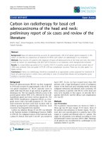
Prætorius
Figure 1 Radiogram of an ameloblastoma with soap bubble
appearance in the right side of the mandible of a 25-year-old
woman. There was a swelling of the mandible, which was noticed
six months earlier and had reached the size of 3.5 cm. No other
symptoms. Note the partial resorption of the roots of the first
molar and the second premolar.
may be elicited, perforation of the cortical bone is a
late feature, however. Paresthesia of the lower lip is a
rare symptom (48).
Imaging. A radiolucent, often well-demarcated,
sometimes corticated, multilocular radiolucency is a
characteristic radiological appearance of the s/mAM,
but it is not diagnostic (Fig. 1). The radiographic image
may vary considerably. Among 55 cases reviewed by
Ledesma-Montes et al. (44) 88.1% were radiolucent,
66.7% were unilocular, and 66.7% were well defined.
The radiographic descriptions of 1234 cases (377 case
reports and 857 cases from reviews) were evaluated by
Reichart et al. (43), but were only reported for all four
variants combined, 102 were of the unicystic type. The
appearance was unilocular in 51.1%, and multilocular
(‘‘soap-bubble-like’’) in 48.9%. Embedded teeth were
detected in 8.7%, root resorption of neighboring teeth
in 3.8%, and undefined borderline in 3.6%. Embedded
teeth were not surprisingly seen more often in younger
patients. The size of the tumor was stated in 129 cases,
the maximum size was 24 cm. The mean size was 4.3
cm, and the median size 3.0 cm. Ledesma-Montes et al.
(44) reported (N ¼ 55) a mean size of 6.7 cm for
mandibular s/mAM and a mean size of 4.6 cm for
the maxillary tumors.
Some s/mAM particularly those with a plexiform growth pattern show a highly vascular stroma,
this feature may have an impact on the radiographic
image making the lesion resemble a poorly-defined
fibro-osseous lesion (49). In such cases, and in the
diagnosis of ameloblastomas in general the use of
computed tomography (CT) and magnetic resonance
imaging (MRI) is highly recommended (47). Asaumi
(50) demonstrated the quality of MRI and dynamic
contrast-enhanced MRI in the study of 10 ameloblastomas. Solid and cystic portions of the tumor could be
identified, mural nodules and thick walls could be
detected, and solid and fluid areas could be distinguished. No differences in the signal intensities
between primary and recurrent cases were found.
Pathology. The etiology of the s/mAM is
unknown. The pathogenesis is insufficiently understood. The tumor is believed to arise in remnants of
odontogenic epithelium, primarily rests of the dental
lamina, which however have been found primarily in
the overlying gingiva or oral mucosa (14). The remnants of the epithelial root sheet (islands of Malassez)
are usually not considered a likely source of ameloblastomas although some cases of early ameloblastoma in the periodontal area might suggest this as a
possibility (51,52). Dentigerous cysts as a source of
ameloblastoma cannot be excluded but it seems
unlikely as discussed in the section on UNAM. It
has some times been suggested that an ameloblastoma
could develop from the basal cells of the overlying
surface epithelium; it is well known that intraosseous
ameloblastomas, which progress through the cortical
bone and reaches contact with the surface epithelium
may cause induction of the surface epithelium to
produce ameloblastomatous proliferations. Since
benign PERAMs do not invade the underlying bone,
it is difficult to envision that intraosseous ameloblastomas should develop from the surface epithelium.
Studies of cytokeratins (CK) (53) have also supported
the hypothesis that ameloblastomas are of odontogenic origin and not direct derivates of basal cells of
oral epithelium.
The macroscopical appearance of the operation
specimen depends on the size of the tumor and the
treatment modality. Resected tumors are surrounded
by normal bone and may contain teeth. The tumor
area is grayish and does not contain hard tissue apart
from the border areas, it usually presents as a mixture
of solid and multicystic areas, but some lesions are
completely solid, and others are dominated by formation of cysts. The cysts are of varying size, usually
most of them are small some are microscopic, but in
large tumors several may be quite conspicuous. They
are filled with a brownish fluid, which often is of low
viscosity, but may be more gelatinous.
Microscopically the tumor consists of odontogenic epithelium growing in a relatively cell-poor
collagenous stroma. Two growth patterns and four
main cell types are recognized within the histopathological range of the entity (Table 2). The two growth
patterns are named follicular and plexiform.
In the follicular pattern the tumor epithelium
(Figs. 2, 3) primarily presents as islands of various
size and shape (23,54). They usually consist of a
Table 2 Ameloblastoma Growth Patterns and Cell Types
Growth patterns
Follicular growth pattern
Plexiform growth pattern
Cell types
Stellate reticulum-like cell type
Acanthomatous (squamous cell) cell type
Granular cell type
Basal cell type
Chapter 19: Odontogenic Tumors
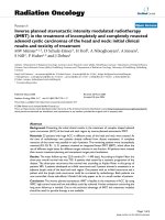
1205
Figure 2 Solid/multicystic ameloblastoma with follicular growth
pattern and stellate reticulum-like cells in the islands. Squamous
metaplasia is seen in a few islands. Minor cysts are seen in the
islands, as well as in the stroma. H&E stain.
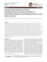
Figure 4 Ameloblastoma. Peripheral cells of a tumor island.
The basal cells are palisaded and columnar with reverse polarity
of the nucleus and show some morphological similarity to preameloblasts. The suprabasal cells are stellate reticulum-like. van
Gieson stain.
Figure 3 Solid/multicystic ameloblastoma with follicular growth
pattern in a stroma consisting of narrow strands of collagenous
connective tissue. H&E stain.
central mass of polyhedral or angular cells with
prominent intercellular contact and conspicuous intercellular spaces. The morphology has some resemblance to the stellate reticulum of the normal enamel
organ, but many details are different. The peripheral
cells are palisaded, columnar, or cuboidal with dark
nuclei. The columnar cells contain elongated nuclei,
which may show reverse polarity and have a histomorphological likeness to preameloblasts (Fig. 4).
Mitoses are absent or very infrequent. The term follicular alludes to a certain resemblance of the structure
of the epithelial islands to enamel organs. The stellate
reticulum-like cells may be replaced by squamous
cells, granular cells, or basal cells (vide infra). If cysts
develop, they arise in the center of the islands.
Figure 5 Solid/multicystic ameloblastoma with a plexiform
growth pattern. van Gieson stain.
In the plexiform growth pattern (Fig. 5) the
tumor epithelium is arranged as a network (plexus),
which is bounded by a layer of cuboidal to columnar
cells and includes stellate reticulum-like cells (23). The
width of the epithelial cords in the network may vary
considerably. Sometimes double row of columnar or
cuboidal cells are lined up back to back. The peripheral cells are similar to those seen in the follicular
pattern, although they are more often cuboidal and
may even be squamous. In the plexiform type as well,
but more rarely, the stellate reticulum-like cells may
be replaced by squamous cells, granular cells, or basal
1206
Prætorius
Figure 6 Islands of an ameloblastoma with follicular growth
pattern. Squamous metaplasia is seen in the central areas.
Ameloblastomas with extensive squamous metaplasia are
termed acanthomatous. H&E stain.
cells. The stroma is generally looser than in the
follicular pattern, and if cyst formation occurs, it is
usually due to stromal degeneration rather than to a
cystic change within the epithelium.
Each of the two growth patterns may be dominating in a s/mAM, but often both patterns are
present in the same tumor. It is generally believed
that the growth pattern is unrelated to the clinical
behavior of the tumor, but some reports have suggested a higher tendency for recurrence in follicular
than in plexiform ameloblastomas (55) and many
molecular biological findings are different (5).
Squamous cell metaplasia of the central areas of
the tumor epithelium is not unusual (Fig. 6), and is
particularly seen in tumors with a follicular growth
pattern. When extensive squamous metaplasia is seen,
sometimes with keratin formation the term acanthomatous ameloblastoma is applied. This variant accounted
for 12.1% of 397 cases reviewed by Reichart et al. (43).
When cysts are formed in the epithelium, they are
lined by squamous cells. The squamous cells are
sometimes plump or fusiform and may exhibit few
junctions.
Rarely an s/mAM shows formation of orthokeratinized or more often parakeratinized horn pearls in
central areas of the tumor epithelium. It may even be
seen in areas, which are not dominated by squamous
cell metaplasia (Fig. 7). Very rarely calcifications are
seen in these horn pearls (56).
The central stellate cells may be replaced by large
eosinophilic rounded or polyhedral granular cells. The
granules may be diastase resistant period acid–Schiff
(PAS)-positive and they represent lysosomes. Most
nuclei in these cells are placed at the periphery of the
cells (Fig. 8). The granular cells may take up a complete
epithelial island and then even the basal cells are
granular. When a conspicuous part of the tumor or
the entire tumor is composed of granular cells, the
tumor is usually called a granular cell ameloblastoma.
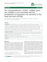
Figure 7 Unusual ameloblastoma with keratinization and calcification without conspicuous squamous metaplasia. The case
was published by Pindborg et al. in 1958 (56). Periodic acid–
Schiff stain. Source: Ref. 56.
Figure 8 Granular cell ameloblastoma. The nuclei are placed in
the periphery in most of the rounded cells. A few cuboidal basal
cells are still seen. H&E stain.
Such tumors are infrequent, particularly those with a
plexiform growth pattern (Fig. 9) (57,58).
Hartman (59) studied 20 cases of granular cell
ameloblastom, which accounted for 5% of all ameloblastomas in their file and stated that they occurred
predominantly in the posterior regions of the mandible (which is a predilection site for all s/mAMs). He
observed that they had a marked tendency to recur
after conservative treatment, but this behavior seems
related to the treatment modality and not to the
histology of the tumor.
Rarely, an ameloblastoma may show a predominantly basaloid pattern (Fig. 10), and this tumor is
referred to as a basal cell ameloblastoma, or basaloid
ameloblastoma (23). It is the least common of the
cytological variants and accounted for 2% of the case
reports reviewed by Reichart et al. (43). The epithelial
Chapter 19: Odontogenic Tumors
Figure 9 Granular cell ameloblastoma with plexiform growth
pattern. H&E stain.
Figure 10 Basal cell type ameloblastoma from the posterior
part of the maxilla of an 85-year-old man. The peripheral cells are
primarily cuboidal. The tumor cell plates and islands show a
highly increased cellular density. The cells are small with dark
nuclei, and an elevated number of mitotic figures were found.
H&E stain.
elements are composed almost exclusively of islands
of plump cells with a high nucleus to cytoplasm ratio,
and reticulum-like cells are few or absent (54). The
periphery is dominated by cuboidal rather than
columnar cells. Cystic changes in the epithelial component are infrequent.
Mucous cell metaplasia may be seen in the
tumor epithelium, but is very rare (60,61).
Clear cells may be found in an s/mAM; if they
occur in more than a few areas, a clear cell odontogenic carcinoma (CCOC) should be considered. The
significance of clear cells is discussed in the section on
that tumor.
Tumor cells containing melanin granules may be
observed.
1207
Figure 11 Ameloblastoma. Conspicuous stromal hyalinization
is seen adjacent to a tumor island. H&E stain.
Various amounts of ghost cells may be seen, but
they are not frequent (62). A dentinogenic ghost cell
tumor (DGCT) should be considered, but the diagnosis requires that the tumor has formed dentinoid in the
stroma adjacent to the epithelial tumor component.
The connective tissue stroma varies in amount,
vascularity, and collagen content. No dental hard
tissue is formed. The basement membrane may be
thick and hyalinized, and this juxtaepithelial hyalinization may be conspicuous (Fig. 11). Few cells, if any
are seen in the hyalinized zone. Scattered lymphocytes
may be observed, but there is no inflammation, except
caused by secondary factors.
In ameloblastomas with a plexiform growth pattern, a highly vascular stroma may be seen, and it may
be in terms of several highly dilated vessels. The
pattern should be considered within the spectrum of
appearances of an ameloblastoma. Previously such
cases were called hemangioameloblastoma (63).
Cystic degeneration of the stroma is not unusual
in s/mAMs with a plexiform growth pattern. Residual
capillaries may be found in these stromal cysts and
cellular debris is a common finding in the cysts.
In a study of 31 cases of s/mAM Mu¨ller et al.
(64) observed that infiltration of the surrounding
spongy bone is frequent, but there was little tendency
to invade cortical bone. They also found that periosteum largely prevented extension of the tumor. Gortzak
et al. (65) studied five voluminous mandibular ameloblastomas after resection and confirmed the invasive
growth pattern. Small tumor nests were found in the
cancellous bone at a maximum distance of 5 mm from
the bulk of the tumor (Fig. 12). Expansive and invasive
growth in the Haversian canals was observed, but
there was no invasion of the inferior alveolar nerve.
The mucoperiosteal layer was invaded but not perforated, and no invasion was observed in the surrounding soft tissues of the periosteum and in the skin
tissues. The authors stated that when the tumor is
radiologically closer than 1 cm to the inferior border of
the mandible, a continuity resection is mandatory.
1208
Prætorius
Figure 12 Solid/multicystic ameloblastoma. Invasion by tumor
islands of the bone surrounding the tumor is the reason for
excision with a margin of 1 to 1.5 cm. H&E stain.
Immunohistochemistry. Because ameloblastoma is one of the more common odontogenic tumors
and because of the florid development of the immunohistochemical technique, the literature concerning
immunohistochemical investigations of ameloblastomas is very extensive. Several investigators have used
immunohistochemistry together with molecular biological methods to study a special subject, many of
these reports published before the middle of 2005
were reviewed by Kumamoto in 2006 (5). These studies will primarily be reviewed in the section on
molecular-genetic data.
The following summaries comprise primarily
reports regarding cytofilaments, extracellular matrix
proteins, basement associated molecules, protein kinases, and cell proliferation markers.
Heikinheimo et al. (53) studied the presence of
CKs and vimentin in nine s/mAMs and three fetal
human tooth germs at bell stage. They used eight
antibodies against CKs, which individually or in combination could detect CK-4, CK-5, CK-6, CK-8, CK-10,
CK-11, CK-13, CK-16, CK-17, CK-18, and CK-19. Most,
but not all ameloblastomas lacked CKs typical of
keratinization. CK-8 and CK-19 were expressed in
all, and CK-18 in the epithelial component of most
of the ameloblastomas, including the granular cell
type, which expressed CK-8, CK-18, and CK-19 very
distinctly. Vimentin was detected in the epithelial cells
of all ameloblastomas except the granular cell type.
The ameloblastomas and the human tooth germ epithelia shared a complex pattern of CK polypeptides
together with the expression of vimentin. The authors
concluded that the findings strongly supported that
ameloblastomas are of odontogenic origin and not
derived from basal cells of the gingiva or oral mucosa.
Crivelini et al. (66) performed a similar study on
10 ameloblastomas and four other types of odontogenic tumors. They used monoclonal antibodies
against single CK types CK-7, CK-8, CK-10, CK-13,
CK-14, CK-18, CK-19 and against vimentin. The
results differed somewhat from those of Heikinheimo
et al.; all ameloblastomas were CK-8, CK-18, and
vimentin negative. They were all, including the granular cell type immunoreactive to CK-14. They also
reacted to CK-13 and CK-19, but only in metaplastic
squamous cells, central stellate cells and in the lining
of cystic structures.
Extracellular matrix proteins and basement
membrane associated molecules have been studied.
Ito et al. (67) detected versican, a large aggregating chondroitin sulfate proteoglycans in 17 ameloblastomas. All samples showed a positive reaction for
versican in the connective tissues, whereas positive
staining of epithelial nests was observed in only some
samples.
Tenascin, an extracellular matrix glycoprotein
was detected by Heikinheimo et al. (68) in the stromal
component of all of 11 ameloblastomas. The epithelial
component was negative. Nagai et al. (69) got very
variable results in the study of 10 ameloblastomas.
Hyalinized stroma was both positive and negative.
Cystic stroma was negative. The basement membranes showed an irregular linear positive reaction
with focal accumulation of tenascin. Mori et al. (70) on
the other hand detected a strong reaction to tenascin
in the interface around the epithelial component,
although with frequent breaks. A positive reaction
was found in stellate reticulum-like cells and granular
epithelial cells as well.
Nadimi et al. (71) studied laminin in 29 ameloblastomas. An intense linear deposit was found in the
basement areas of all of them. Heikinheimo et al. (68)
confirmed these results.
Nadimi et al. (71) were unable to detect fibronectin except in areas with inflammation. Nagai et al.
(69) got very variable results but detected an irregular
linear immunoreaction in basement areas. Heikinheimo et al. (68) detected an extra domain sequenceA-containing form of fibronectin in the extracellular
matrix of all ameloblastomas (N ¼ 11), and an oncofetal domain containing form of fibronectin in most
ameloblastomas. They studied collagen type VII as
well; the immunoreaction was very similar to that of
laminin: most ameloblastomas exhibited a continuous
staining of the basement membranes.
Parikka et al. (72) detected collagen XVII, a
hemidesmosomes transmembrane adhesion molecule,
in the cytoplasm of basal and suprabasal cells in 11 s/
mAMs and 2 UNAM using immunohistochemistry
and in situ hybridization (ISH).
Poomsawat et al. (73) used antibodies against
laminins 1 and 5, collagen type IV, and fibronectin on
14 ameloblastomas. An intense staining of laminin 1
and a weak to moderate intensity of laminin 5 were
seen as continuous linear deposits at the basement
membrane zone surrounding tumor islands. Collagen
type IV showed irregular patterns; focal loss of staining was observed. A weak to moderate staining for
fibronectin was occasionally present; fibronectin was
also present in the fibrous stroma. The tumor cells also
showed reaction to laminin 1 and 5, collagen type IV,
and fibronectin. In general, laminin 1 showed moderate to strong intensity in the cytoplasm of both central
Chapter 19: Odontogenic Tumors
and peripheral cells; collagen type IV was rarely
observed. Laminin 5 was expressed in peripheral
cells, but less often.
Collagen type IV was also studied by Nakano
et al. (74) and Nagatsuka et al. (75). Nakano et al.
found that ameloblastoma (N ¼ 2) basement membranes expressed five of six genetically distinct forms
of collagen IV: a1(IV), a2(IV), a5(IV), and a6(IV)—
chains occurred as intense linear stainings without
disruption around neoplastic epithelium. A similar
study of 5 ameloblastomas by Nagatsuka et al. (75)
gave the same results.
Integrin, a plasma membrane protein, which
plays a role in the attachment of cell to cell and cell
to the extracellular matrix, and as a signal transductor
has been studied by Souza Andrada et al. (76). Integrin a2b1, a3b1, and a5b1 were detected in 20 s/mAMs,
10 UNAMs, and 12 AOT. The labeling intensity was
considerably stronger in the ameloblastomas than in
the AOTs, but no significant differences were found
between the two variants of ameloblastoma. In s/
mAMs the immunoreaction was detected in intercellular contacts and at the connective tissue interface.
Using immunohistochemistry, in situ hybridization, immunoprecipitation, and reverse transcriptase
polymerase chain reaction (RT-PCR), Ida-Yonemochi
et al. (77) detected basement-type heparan sulfate
proteoglycan (HSPG), also known as Perlecan in the
intercellular spaces of the epithelial component and in
the stroma of 20 ameloblastomas and cultured ameloblastoma cells. The studies indicate that ameloblastoma cells synthesize HSPG.
The roles of mitogen-activated protein kinases
(MAPKs) in oncogenesis and cytodifferentiation of
odontogenic tumors were investigated by Kumamoto
et al. (78), using antibodies against phosphorylated
c-Jun NH2-terminal kinase (p-JNK), phosphorylated
p38 mitogen-activated protein kinases (p-p38 MAPK),
and phosphorylated extracellular signal-regulated
kinase 5 (p-ERK5) on 47 ameloblastomas (including
4 desmoplastic), 2 metastasizing ameloblastomas
(METAMs), 3 ameloblastic carcinomas (AMCAs),
and 10 human third molar tooth germs. Almost all s/
mAMs were p-JNK negative. From 84% to 91% of the
various histological types of ameloblastomas were moderately p-p38 MAPK positive. The basal cell ameloblastomas (N ¼ 3), however, and the desmoplastic
ameloblastomas (DESAMs) (N ¼ 4) were 100% positive,
three of six granular cell ameloblastomas were positive.
Between 64% and 66% of the histological types of
ameloblastoma were p-ERK5 positive, except basal cell
and DESAM, which were 100% positive. The authors
suggested that these MAPK signaling pathways contribute to cell proliferation, differentiation, or apoptosis
in both normal and neoplastic odontogenic tissues.
Cell proliferation markers have been studied by
several investigators. The results have been somewhat
contradictory. Kim et al. (79) used antibodies against
proliferating cell nuclear antigen (PCNA) on 25 s/
mAMs and 13 unicystic types and a case of AMCA.
There was no significant difference between the proliferating activities of the different histological types of
s/mAM, but a recurrent ameloblastoma and the
1209
AMCA showed remarkably higher PCNA activity.
Funaoka et al. (80) measured the PCNA index in 23
s/mAMs, they found a higher, but not significantly
higher index in follicular than in plexiform ameloblastomas. Interestingly, they found a remarkable difference in the index of biopsies of the same tumor taken
at different times. Ong’uti et al. (81) measured the Ki67 index in 54 s/mAMs, 24 follicular, and 30 plexiform. They found a significantly higher labeling index
(L.I.) in ameloblastomas with a follicular growth pattern than in those with a plexiform pattern. They did
not find any significant correlation between the Ki-67
L.I. and clinical features like age, gender, and tumor
size.
Piattelli et al. (82) evaluated the proliferative
activity of 22 ameloblastoma among which 13 were
s/mAM by measuring the immunoreactivity of
PCNA. Recurrent ameloblastoma (N ¼ 4) presented
higher PCNA positive cell counts than other types of
ameloblastoma.
Sandra et al. (83) used antibodies against PCNA
and Ki-67 on 25 s/mAMs, 5 unicystic, and 3 DESAMs,
and measured the indices. There was a strong correlation between the PCNA and the Ki-67 labeling indices.
Positively stained cells were primarily found in the
peripheral layers. The basal cell types of ameloblastomas showed the highest L.I., but it was not significantly higher than that of follicular, plexiform, and
acanthomatous types. It was significantly higher,
however, than the labeling indices measured in unicystic and DESAMs. On the contrary, Meer et al. (84)
found a statistically significantly higher PCNA and
Ki-67 L.I. in unicystic (N ¼ 10) than in the s/m variant
(N ¼ 10).
Thosaporn et al. (85) used antibodies against a
novel cell proliferation marker, IPO-38 (N-L 116) on 10
ameloblastomas, 10 keratocystic odontogenic tumors
(KCOTs), 7 orthokeratinized odontogenic cysts, and
8 dentigerous cysts. Positive nuclei were found in the
peripheral cell layers of the ameloblastomas. The L.I.
was similar to that of the KCOTs, but twice as high as
that of the orthokeratinized odontogenic cysts and 14
times higher than that of the dentigerous cysts.
Payeras et al. (86) evaluated the proliferation
activity in 11 cases of s/mAM by means of quantification of the argyrophilic nuclear organizer regions
(AgNORs) and the pattern of immunohistochemical
expression of the epidermal growth factor receptor
(EGF-R). There was no significant statistical difference
as per quantification of the AgNORs, the expression of
the EGF-R on the epithelial islands of ameloblastoma
was not uniform, and the location of the expression
was also variable. The authors concluded that the
tumor presents an irregular growth, and that smaller
epithelial islands could be responsible for tumor infiltration since they are associated with a higher proliferation activity.
Granular cell ameloblastoma has been studied in
particular by Kumamoto et al. (87). Granular cells
were positive for CK, CD68, lysozyme, and alpha-1antichymotrypsin, but negative for vimentin, desmin,
S-100 protein, neuron-specific enolase (NSE) and CD
15, indicating epithelial origin and lysosomal
1210
Prætorius
aggregation. The authors suggested that the cytoplasmic granularity in granular cell ameloblastomas
might be caused by increased apoptotic cell death of
neoplastic cells and associated phagocytosis by neighboring neoplastic cells.
Electron Microscopy. Several studies have
reported the ultrastructure of the ameloblastoma,
Moe et al. (88), Sujaku et al. (89), Csiba et al. (90),
Navarrette et al. (91) Lee et al. (92), Mincer et al. (93),
Cutler et al. (94), Tandler et al. (95), Kim et al. (96),
Matthiessen et al. (97), Rothouse et al. (98), Chomette
et al. (99), Nasu et al. (100), Takeda et al. (101), Smith
et al. (102), and Farman et al. (103). Some of the earlier
studies concentrated on ultrastructural similarities
between the columnar peripheral epithelial cells of
the s/mAM and the preameloblasts of the normal
enamel organ (88,89,92,93). Kim et al. (96) and
Matthiessen et al. (97) confirmed this similarity and
further observed that the stellate cells of the tumor
epithelium were in many respects similar to the
stellate reticulum of the normal enamel organ. They
were joined by desmosomes and the nucleus occupied
a central position within the cell. The perinuclear
cytoplasm contained mitochondria, tonofilaments,
endoplasmic reticulum, and dense granules. Some
epithelial cells contained numerous lipid granules
and mitochondria formed a network of cords. Matthiessen et al. (97) found that the low peripheral cells
in s/mAM were very similar to the external enamel
epithelium cells. The central cells of the islands had a
certain resemblance to the stellate reticulum and stratum intermedium cells. The high peripheral cells of
the s/mAM had no counterpart in the enamel organ.
Unlike the enamel organ the ameloblastoma showed
extremely few and small gap junctions.
The ultrastructural features of squamous epithelial cells were similar to those described for basal cells
and lower prickle cells of the oral mucosa. The granular cells in particular were studied by Navarrette et al.
(91) and Tandler et al. (95) and Nasu et al. (104). The
granular cells commonly occur in the islands of ameloblastomas with a follicular growth pattern, in one of
the cases reported by Nasu et al. (104), they were in a
plexiform pattern. The cytoplasmic granules were
identified as lysosomes, supported by the fact that
they were intensively stained for acid phosphatase; no
cytoplasmic components were found in the numerous
lysosomes, they do not seem to be engaged in autophagy, their function is unknown. The occurrence of
intracytoplasmic desmosomes was described by
described by Cutler et al. (94) in an ameloblastom
from the maxilla. Hyaline bodies, a structure that is
relatively common in odontogenic cysts were
observed by Takeda et al. (101), ultrastructurally
they did not differ from those found in the epithelium
of the wall of odontogenic cysts. Farman et al. (103)
studied the interface between the tumor component
and the stroma in seven ameloblastomas. All showed
differing degrees of thickening of lamina densa by a
granulofilamentous material having a range of width
of approximately 80 to 800 nm. Fragmentation of the
granulofilamentous material was seen in several
instances. The resulting defects were less linear and
had more of a soap bubble appearance. The hyaline
cell free zone, which may be seen adjacent to the
epithelium, comprised relatively cell-free, normally
banded, mature collagen. The stroma contains fibroblasts and collagen fibers. Multinucleated giant cells
near the epithelial component were described by Kim
et al. (96). Rothouse et al. (98) detected myofibroblasts
in the stroma, a finding that was confirmed by Smith
et al. (102) in a case of recurrent s/mAM.
Molecular-Genetic Data. It is not possible
within the frame of this chapter to review all studies
of the molecular pathology of the ameloblastoma. A
comprehensive review of the molecular pathology of
odontogenic tumors covering the literature till the
middle of 2005 was published by Kumamoto (5), the
majority of the studies deals with ameloblastomas. For
the following summaries the same subheadings as
used by Kumamoto have been used; the majority of
articles have been selected because they were not
mentioned in Kumamoto’s review or were published
subsequently.
1. Molecules Involved in Tumorigenesis and/or
Cell Differentiation of Ameloblastomas.
a. Oncogenes. In ameloblastomas, p21Ras is
expressed in the epithelial cells and overexpression
has been detected (105). c-Myc oncoprotein is
expressed predominantly in the tumor cells neighboring the basement membrane (106). On cDNA microarray and subsequent real-time reverse transcriptase
RT-PCR overexpression of Fos has been detected (107).
b. Gene Modifications. Ja¨a¨skela¨inen et al. (108)
used immunocytochemical staining with MIB-1 antibodies and comparative genomic hybridization (CGH)
to study cell proliferation and chromosomal imbalances
in 20 cases of ameloblastoma. CGH involved hybridization of FITC-dUTP-labeled tumor DNA with Texasred-labeled normal DNA. The MIB-1 index was low for
all tumors and was not correlated to the tendency to
recur; it does not seem helpful in assessing future
clinical behavior of the tumor. Chromosomal aberrations were only detected in 2 of 17 cases.
Carinci et al. (109) compared the expression
profiles of three ameloblastomas and three malignant
odontogenic tumors by hybridization to microarrays
containing 19,200 cDNAs to identify genes, which
were significantly differentially regulated when compared with nonneoplastic tissues. The investigators
detected 43 cDNAs, which differentiated the three
malignant tumors from the three ameloblastomas.
The cancer specific genes included a range of functional activities like transcription, signaling transduction, cell-cycle regulation, apoptosis, differentiation,
and angiogenesis. The authors suggested that the
identified genes might help to better classify borderline odontogenic tumors.
A study for loss of heterozygosity of tumor
suppressor genes in 12 ameloblastomas revealed that
DNA damage in ameloblastomas seems to be sporadic
and cumulative (110). The frequency of allelic loss and
intratumoral heterogeneity did not correlate with age,
gender, histological subtype, or prognosis.
Chapter 19: Odontogenic Tumors
1211
expression of RB than follicular ameloblastomas.
Expression of RB, E2F-1, and phosphorylated RB
was considered to be involved in cell proliferation
and differentiation of odontogenic epithelium via
control of the cell cycle.
In a study performed to identify possible genes
involved in the development and progression of ameloblastomas the investigators used microarray analysis,
semiquantitative RT-PCR and immunohistochemistry
on selected genes (111). Tissue from dentigerous cysts
was used as control. Overexpression of 73 genes was
detected and 49 genes were underexpressed.
Mutations in microsatellite sequences have been
studied in 24 ameloblastomas by DNA sequencing
analysis (112) and supplied with an evaluation of the
Ki67 L.I. of the tumors. The occurrence and the pattern
of microsatellite alterations, in form of loss or length
variation, was evaluated and correlated with the Ki67
L.I. and with other clinicopathological parameters.
Alterations of at least one of the selected loci were
observed in all (100%) the ameloblastomas with a
mean of four altered microsatellites for each tumor.
Microsatellite alterations were more frequent in
tumors displaying a high Ki67 L.I., and in a univariate
analysis, their occurrence was found to be a predictor
of increased risk of recurrence, but no correlation was
found to the patient’s age or gender, or to tumor size,
location and histology.
d. DNA-Repair Genes. Errors during DNA
replication or repair are maintained by DNA-repair
genes belonging to the human DNA mismatch repair
(hMMR) system. It is composed of at least six genes.
The protein expression of two of the genes, hMSH2
and hMLH1 was studied by means of antibodies in 25
cases of ameloblastoma, including three peripheral
and three unicystic (124). All ameloblastomas showed
a nuclear expression of the proteins in the peripheral
layers of the epithelial component. These data suggest
that the development and progression of these tumors
do not depend on a defect in the hMMR system.
c. Tumor Suppressor Genes. Increased immunohistochemical reactivity for p53 has been detected
in ameloblastomas (113,114), although it has been
shown in several studies that p53 mutations are
infrequent in ameloblastomas (115–117). Regulators
of p53, murine double minute 2 (MDM2), and p14
(ARF), are also expressed in ameloblastomas, and
overexpression has been detected (118,119).
Two members of theTP53 gene family, named
p73 and p63, have been identified and analyzed by
immunohistochemistry and RT-PCR in ameloblastomas. They seem to function differently from p53 in
odontogenic tissue (120). Immunohistochemical reactivity for p63 was detected by Lo Muzio et al. (121) in
26 s/mAMs and several other benign and malignant
odontogenic tumors. Benign odontogenic, locally
aggressive tumors with a high risk of recurrence
exhibited statistically higher p63 expression than
benign odontogenic, nonaggressive tumors with a
low risk of recurrence.
The immunohistochemical reactivity for the APC
gene that inhibits cell proliferation was found to be
lower in benign and malignant ameloblastomas than
in tooth germs (122).
Retinoblastoma protein (RB) is a product of the
retinoblastoma (RB) tumor suppressor gene, which acts
as a signal transducer connecting the cell cycle with
the transcription machinery. Kumamoto et al. (123)
used antibodies against RB, E-2 promotor-bindingfactor-1(E2F-1), and phosphorylated RB on 40 ameloblastomas (including 4 desmoplastic), 2 METAMs,
3 AMCAs, and 10 human tooth germs to clarify their
roles in cell-cycle regulation in oncogenesis and cytodifferentiation of odontogenic tumors. Ki-67 antibody
was used as a marker of cell proliferation. The levels
of immunoreactivity for RB, E2F-1, phosphorylated
RB, and Ki-67 were slightly higher in benign and
malignant ameloblastomas than in tooth germs. Plexiform ameloblastomas showed significantly higher
f. Growth Factors. Using ISH Heikinheimo et
al. (130) detected EGF-R and transforming growth
factor alpha (TGF-a) mRNA in 4 ameloblastomas;
EGF transcripts was not found. The findings have
been confirmed (131,132). The growth factors seem
to be involved in the tumogenesis.
Transforming growth factor beta (TGF-b), a multifunctional growth factor has been demonstrated in
ameloblastomas and has been attributed an important
role in cell differentiation and matrix formation
(133,134).
Hepatocyte growth factor (HGF), which has
mitogenic, motogenic, and morphogenic functions,
has been found in ameloblastomas (134).
Various types of fibroblast growth factors (FGF)
and their receptors (FGFR) have been studied. FGF-1
and FGF-2 are mitogenic polypeptides that have been
demonstrated to enhance cell growth in a dose dependent manner of cultered ameloblastoma epithelial cells
(135). In tissue specimens, FGF-1 was localized in the
epithelial component, whereas FGF-2 was primarily
found in the basement membranes. In another study
(136), ameloblastomas showed a weak and focal reaction
for FGF-1 and FGFR3 in the tumor epithelium, while
FGF-2 and FGFR2 exhibited significant cytoplasmic
staining of all layers of the neoplastic epithelium.
Expression of platelet-derived endothelial cell
growth factor/thymidine phosphorylase (PD-ECGF/
TP) and of angiopoietins have been detected immunohistochemically in the stroma of ameloblastomas
and in the ectomesenchymal cells of human tooth
germs (137). The level of PD-ECGF/TP reactivity
was significantly higher in ameloblastomas than in
tooth germs. Granular cell ameloblastoma showed
PD-ECGF/TP reactivity in granular neoplastic cells
as well as in stromal cells. Immunoreactivity for
angiopoietins-1 and -2 was detected predominantly
in odontogenic epithelial cells near the basement
membrane in tooth germs and in the ameloblastomas.
e. Oncoviruses. Although several investigators have reported detection of human papillomavirus
(HPV) (125–128) and Epstein–Barr virus (EBV) (129) in
ameloblastomas the etiological role of the viruses
remains controversial.
1212
Prætorius
The authors suggested that these angiogenic factors
participate in tooth development and odontogenic
tumor progression by regulating angiogenesis.
The immunohistochemical expression of insulinlike growths factors (IGFs), platelet-derived growth
factor (PDGF), and their receptors has been analyzed
in 47 ameloblastomas and 10 human tooth germs (138)
by use of antibodies against IGF-I, IGF-II, IGF-I receptor (IGF-IR), PDGF A-chain, PDGF B-chain, PDGF
a-receptor, and PDGF b-receptor. The reactivity for
IGFs, PDGF chains, and their receptors was detected
predominantly in odontogenic epithelial cells near the
basement membrane in tooth germs as well as in
ameloblastomas. The expression levels of IGF-II and
PDGF chains were significantly higher in the tumors
than in the tooth germs, and the expression level of
PDGF chains were significantly higher in follicular
ameloblastomas than in plexiform ameloblastomas.
DESAMs showed higher expression of IGFs and
IGFIR when compared with other ameloblastoma
subtypes. These growth factor signals thus contribute
to cell proliferation or survival in both normal and
neoplastic odontogenic tissues.
g. Telomerase. Ameloblastomas have been
consistently positive for telomerase activity suggesting
that telomerase activation is associated with the tumorigenesis of the neoplastic epithelium (139,140). Telomerase is a specialized reverse transcriptase that
synthesizes telomeric DNA at the ends of chromosomes
and compensates for its loss with each cell division, and
is thus a participant in cell immortalization. The immunoreactivity for telomerase in ameloblastomas shows a
similar distribution pattern to that of the c-Myc oncoprotein. This oncogenic protein is known to activate
telomerase transcription directly, so it possibly induces
telomerase activity in ameloblastomas.
h. Cell Cycle Regulators. The immunoreaction
of cell cycle-related factors were examined by Kumamoto et al. (141) in 8 human tooth germs and 31
ameloblastomas by means of antibodies against
cyclin D1, p16INK4a, p2WAF1/Cip1, p27Kip1, and DNA
topoisomerase IIa and by ISH of histoneH3 mRNA.
Cyclin D1, p16 protein, p21, and p27 were all
expressed in the epithelium of tooth germs and ameloblastomas, although p21 was not expressed in granular epithelial cells and keratinizing cells. It is
suggested that the odontogenic epithelium is strictly
controlled by these cell cycle regulators.
i. Apoptosis-Related Factors. Physiological cell
death, apoptosis is mediated by two alternative apoptotic pathways, death by receptors or death by
mitochondria. A commonly used method to detect
apoptosis is called TUNEL (Terminal deoxynucleotidyl transferase biotin-dUTP-nick-end labeling).
Other ways of detection of apoptotic cells and specific
parts of the apoptotic pathway are detection of
caspase, fas-ligand, and annexin V activity. TUNEL
and single-stranded DNA (ssDNA), fas-ligand, and
caspase-3 antibodies have been used to detect apoptotic cells in ameloblastomas and ghost cell odontogenic
carcinoma (GCOC) (87,114,142–145). Death receptors
such as fas, tumor necrosis factor (TNF) receptor I,
and TNF-related apoptosis-related ligand (TRAIL) 1
and 2 have been demonstrated in ameloblastomas, but
expression of caspase-8, an apoptosis initiator has
been extremely limited, suggesting that apoptotic
cell death in ameloblastomas is minimally affected
by signaling of death factors (144,146).
Bcl-2 and inhibitor of apoptosis (IAP) family
proteins are modulators of the mitochondrial apoptotic pathway. In ameloblastomas, apoptosis inhibitory
factors, such as Bcl-2, Bcl-x, surviving, and X chromosome–linked IAP (XIAP) are predominantly expressed, which may indicate that these apoptosis
modulators are associated with survival and neoplastic transformation of the odontogenic epithelial cells
(147–149).
Factors involved in the apoptosis signaling pathways mediated by mitochondria have been investigated in ameloblastomas and normal human tooth germs
(150). Tissue specimens were examined by RT-PCR and
antibodies against cytochrome c, apoptotic proteaseactivating factor-1 (APAF-1), caspase-9, and apoptosisinducing factor (AIF). The mRNA expression of APAP1, caspase-9, and AIF was detected in all samples and
immunoreactivity for cytochrome c, APAP-1, caspase9, and AIF was positive in all samples. The results
suggest that the mitochondria-mediated apoptotic
pathway has a role in apoptotic cell death of normal
and neoplastic odontogenic epithelium.
Expressions of tumor-necrosis-factor-related
apoptosis-inducing ligand (TRAIL/Apo2L), a potent
ligand in inducing apoptosis, has been studied in 32
ameloblastomas and in AM-1 cells (an HPV-16
infected ameloblastoma cell line) together with death
receptor 4 (DR4) and 5 (DR5). It was observed that
TRAIL cleaved caspase-8, -9, and -3, lowered mitochondrial membrane potential and markedly induced
apoptosis in AM-1 cells. The results suggested that
TRAIL is a potent apoptosis-inducing ligand in ameloblastoma (151). Osteoprotegerin (OPG) is a receptor
that is capable in inhibiting receptor activator of
nuclear factor-kB ligand (RANKL) in inducing osteoclastogenesis. As mentioned above TRAIL is a potent
apoptosis-inducing ligand in ameloblastomas. The
expression of OPG in ameloblastomas has been investigated by immunohistochemistry, immunofluorescense,
and Western blot (152), and was observed in tissue
samples from 20 ameloblastomas as well as in cultured
ameloblastoma cells (AM-1). An apoptosis assay was
performed to investigate the potential of TNF-a, TRAIL,
and RANKL in inducing apoptosis. It was found that
TRAIL had the highest potential in inducing apoptosis
compared with TNF-a and RANKL. A binding assay
revealed that OPG preferably binds with RANKL,
rather than with TRAIL. The results suggest that the
binding of OPG to TRAIL might cause TRAIL to induce
apoptosis in ameloblastomas.
TNF-a is involved in inducing cell survival,
proliferation, differentiation, and apoptosis. Its
expression has been studied in 24 ameloblastomas
and in AM-1 cells, and TNF-a as well as its receptors
(TNFR1 and TNFR2) were clearly observed in
all ameloblastoma samples and in AM-1 cells.
Chapter 19: Odontogenic Tumors
TNF-a-induced Akt (protein kinase) and MAPK signals
were studied as well (153). The results suggested that
TNF-a can induce Akt and p44/42 MAPK activation
through PI3K (phosphatidylinositol-3-OH kinase),
which might later induce cell survival and proliferation
in ameloblastoma. In a subsequent study (154), it was
observed that prolonged treatment of AM-1 cells with
TNF-a induced the cells into apoptosis.
j. Regulators of Tooth Development. Underexpression of Sonic Hedgehog (SHH) gene and of Patched
(PTCH), a cell-surface transmembrane protein has
been shown in ameloblastomas on cDNA microarray
(107). SHH is involved in the morphogenesis and
cytodifferentiation of teeth. SHH signals control cellto-cell interactions and cell proliferation in tissue
patterning of various organs, including teeth.
By means of RT-PCR and immunohistochemistry,
Kumamoto et al. (155) detected expressions of SHH,
PTCH, Smoothened (SMO), a membrane bounded
protein, and GLI1 (a zinc finger DNA–binding protein) in ameloblastomas. Expression of SHH, PTCH,
and GLI1 was more evident in epithelial than in
mesenchymal cells, whereas SMO reactivity was
marked in both components. Keratinizing and granular cells showed no or little reactivity.
The Wnt signaling pathway is a complex network
of proteins involved in embryogenesis (including
odontogenesis) and oncogenesis. Wnt signaling is regulated by the levels of the protein b-catenin. Mutations
of b-catenin are detected frequently in COCs but are
rare in ameloblastomas (156,157). The b-catenin protein
is expressed in the nuclei of the ameloblastomas (122).
The transmembrane heparan sulfate proteoglycan, Syndecan-1 (SDC-1), also known as CD 138 and
Wingless type 1 glycoprotein (Wnt1), which belongs to
a large family of 19 secreted signal transducers and
promotes cell proliferation has been detected in 29 s/
mAMs, but not consistently (158). Immunostaining of
SDC-1 was observed in the epithelial component
as well as in the stroma cells. Wnt1 was almost exclusively seen in the epithelial tumor cells. The authors
suggested that SDC-1 is a critical factor for Wntinduced carcinogenesis in the odontogenic epithelium.
k. Hard Tissue-Related Proteins. Immunohistochemical expression of enamel proteins, such as
enamelin, enamelysin, and sheathlin could not be
detected in ameloblastomas (159–161). Amelogenin,
however has been demonstrated immunohistochemically (162,163) and by mRNA phenotyping in combination with Northern blot analysis and ISH analysis of
mRNA (164). Ameloblastin (AMBN) gene mutations
were detected in two s/mAMs, an exon 11 mutation
in a follicular ameloblastoma and a compound exon 4
mutation in a follicular ameloblastoma (165). The
expression pattern of X and Y amelogenin genes
(AMGX and AMGY) was studied in 19 ameloblastomas (9 male and 10 female) by RT-PCR, ISH, immunohistochemistry, and restriction enzyme digestion
(166). All tumor samples expressed amelogenin
mRNA. An increased level of AMGY expression,
higher than that of AMGX was detected in all male
samples, in contrast to normal male tooth development,
1213
where expression of AMGY is very much lower than
that of AMGX.
Bone sialoprotein (BSP) has been detected in the
neoplastic epithelial component of ameloblastoma,
but not in the stroma using cRNA ISH and immunohistochemistry (167). BSP is synthesized and secreted
by bone- dentine- and cementum-forming cells and is
implicated in de novo formation of bone formation
and mineralization, but seems also involved in oncogenesis.
Gao et al. (168) were unable to detect bone
morphogenetic protein (BMP) in 20 ameloblastomas
by means of antibodies. On the contrary, Kumamoto
et al. (169) demonstrated BMP, bone morphogenetic
protein receptor (BMPR), core-binding factor a1
(CBFA1) [also known as run-related protein 2
(RUNX2)], and osterix, a zinc finger–containing transcription factor in the epithelial component as well as
in the stroma cells of 31 ameloblastomas; 6 granular
cell ameloblastomas, however showed no reaction in
the granular cells. Acanthomatous ameloblastomas
exhibited increased reactivity of BMP-7 in keratinizing
cells. The investigators used RT-PCR and immunohistochemistry.
2. Molecules Involved in Progression of Ameloblastomas.
l. Cell Adhesion Molecules. Ameloblastomas
express vascular endothelium cell adhesion molecules
such as the cellular adhesion receptors ICAM-1,
E-selectin, and VCAM-1 suggesting that stromal blood
vessels are activated in these tumors (170).
E-cadherin and its undercoat protein a-catenin
were detected in 24 ameloblastomas by means of
monoclonal antibodies (171). There was a loss of
expression in keratinizing areas and reduction in granular cell clusters. Several integrin subunits, a2, a3, and
b4 and CD 44 exhibited immunoreaction in 22 ameloblastomas that were studied to clarify the role of these
cell adhesion molecules in epithelial odontogenic
tumors (172). CD 44 showed decreased expression in
keratinizing areas in acanthomatous ameloblastomas.
Integrins and CD 44 are both families of cell surface
glycoproteins that mediate cell-cell and cell–extracellular matrix adhesion. In an immunohistochemical study
of 14 ameloblastomas with antibodies against integrin
subunits, a2, a3, a5, av, b1, b3, and b4 all integrins were
detected. The immunoreaction showed variations in
distribution and staining intensity(173).
m. Matrix-Degrading Proteinases. The role of
proteolytic enzymes in extracellular matrix degradation
has been studied by several investigators (174–178).
Matrix metalloproteinases (MMPs) and their tissue
inhibitors (TIMPs) were found in 22 ameloblastomas
by means of antibodies against MMP-1, MMP-2 and
MMP-9, and TIMP-1, and TIMP-2 (174). Intense reactivity for these antibodies was found in the cytoplasm of
stromal fibroblasts, a weak reaction for MMP-2, MMP-9,
and TIMP-1was found in the tumor cells of some s/
mAMs. A strong expression of TIMP-2 was found on
the basement membrane and in the stromal cells. These
results were essentially confirmed by Pinheiro et al.
(175) using immunohistochemistry, zymography, and
1214
Prætorius
Western blotting. They observed expression of latent
and active forms of MMP-1, -2 and -9, and compared
the results with AgNOR analysis, which was used
simultaneously. They found a strong reaction for the
MMPs in granular cells of ameloblastomas. The MMPs
might digest bone matrix and release mitogenic factors.
The hypothesis was supported by the finding of an
increased proliferation index in tumor cells in the
vicinity of the bone. In a study of matrix-degrading
proteinases regulators the immunohistochemical
expression of MMP, membrane type 1-matrix metalloproteinase (MT1-MMP), MMP inhibitor RECK (reversion-inducing cysteine-rich protein with Kazal motifs),
and EMMPRIN (extracellular matrix metalloproteinase
inducer) were detected in the majority of 40 ameloblastomas (176). The reactivity was seen predominantly in
tumor cells near the basement membrane. Follicular
ameloblastomas showed significantly lower expression
of RECK than plexiform ameloblastomas.
Heparanase, an endo-glucuronidase enzyme that
specifically cleaves heparan sulfate has been detected
by immunohistochemistry and mRNA ISH in 23 ameloblastomas (177). The enzyme was strongly
expressed in the tumor epithelium of all samples. A
weak reaction was seen in stromal cells adjacent to
tumor cells, a stronger reaction was seen in inflammatory cells and endothelial cells of small blood capillaries. Heparanase is believed to contribute in the local
invasiveness of the tumor.
The roles of EMC-degrading serine proteinases
in progression of ameloblastomas has been evaluated
by studying the immunoexpression of urokinase-type
plasminogen activator (uPA), uPA receptor (uPAR),
plasminogen activator inhibitor 1 (PAI-1), and maspin
(a serine proteinase inhibitor) in 45 ameloblastomas
(178). The uPA was recognized predominantly in
mesenchymal cells, uPAR was evident in epithelial
cells, PAI-1 was found in both epithelial and mesenchymal cells, and maspin was expressed only in
epithelial cells. The findings suggest that interactions
among these molecules contribute to EMC degradation and cell migration during tumor progression.
n. Angiogenic Factors.
The association
between vascular endothelial growth factor (VEGF)
immunohistochemical expression and tumor angiogenesis has been studied in 35 ameloblastomas (179).
Increased expression of VEGF, which enhances angiogenesis and vascular permeability, was found in
peripheral tumor cells and in stromal cells adjacent to
these cells, which suggests that VEGF is an important
mediator of tumor angiogenesis in ameloblastomas.
Granular cell clusters in granular cell ameloblastomas
showed low reactivity.
o. Osteolytic Cytokines. The balance between
bone formation and bone resorption is regulated by a
wide variety of hormones, growth factors, and cytokines. Synthesizing of inflammatory cytokines with
osteolytic activity such as interleukin-1 (IL-1), interleukin-6 (IL-6), and TNF-a in ameloblastomas has been
demonstrated by several investigators (146,170,180,181).
Osteoclast differentiation and activation is stimulated by binding of receptor activator of RANKL to
its receptor RANK, which is expressed on osteoclast
precursors. Osteoprotegerin (OPG) functions as a
decoy receptor for RANKL and inhibits osteoclastogenesis and osteoclast activation. RANKL and OPG
have been detected in ameloblastomas predominantly
in the stromal cells rather than in the neoplastic cells
(182,183). The secretion of RANKL and TNF-a in
ameloblastomas and its role in osteoclastogenesis has
been confirmed (184).
Differential Diagnosis. Ameloblastomas with
a plexiform growth pattern may be difficult to distinguish from hyperplastic odontogenic epithelium so
commonly seen in the walls of odontogenic cysts. At
low-power microscopy, a network of epithelial
strands embracing islands of loose connective tissue
is seen in both cases. If the basal cells are cuboidal or
squamous in stead of columnar this criteria is not very
helpful, and if the suprabasal epithelial cells are
squamous rather than reticulum cell-like it may lead
to diagnostic confusion (54). Inflammation is usually
seen in the cystic environment and is rare in ameloblastomas and may be a useful feature, and the clinical
and radiographic features should be included in the
diagnostic decision.
The acanthomatous ameloblastoma should be
distinguished from the SOT. In the latter, the stroma
is more abundant; in the tumor component all cells are
squamous cells, no stellate reticulum-like cells are
seen, cyst formation is absent, and the peripheral
cells are flattened.
The granular cell type of the s/mAM may be
confused with the granular cell odontogenic tumor
(GCOT). The main difference is that the s/mAM is an
epithelial tumor and that the granular cells are epithelial, while the tumor component of the granular cell
tumor is ectomesenchymal and the granular cells of
the same origin. Cords and islands of odontogenic
epithelium are seen, but they are quite different from
the proliferating epithelium of an ameloblastoma.
A dental papilla–like connective tissue is never
seen in an ameloblastoma, if it is observed in the
tumor together with odontogenic epithelium with
the morphology of an ameloblastoma, the extremely
rare odonto-ameloblastoma (O-A) should be considered. If dental hard tissue has been produced in the
dental papilla–like areas, the diagnosis is more
straightforward.
The intraosseous basal cell ameloblastoma
should be differentiated from the AMCA. Although
hypercellularity and hyperchromatic nuclei may be
seen in a basal cell ameloblastoma, numerous mitoses,
nuclear and cellular pleomorphia, vascular and neural
invasion are signs of malignancy and not a feature of
this tumor.
Treatment and Prognosis. There has been
some difference of opinion about the preferable methods of treatment of the s/mAM, and there is still no
consensus. Nakamura et al. (185) reported on a longterm follow-up of treatment of 27 unicystic, 21 multicystic, and 30 solid ameloblastomas. In spite of a
recurrence rate of 33.3% after conservative surgery
compared with 7.1% after radical surgery, the authors
advocated for conservative treatment except when the
Chapter 19: Odontogenic Tumors
tumor invades and destroys the inferior border of
the mandible, or when the tumor infiltration is close
to the scull base. Huang et al. (186) advocated for a
conservative treatment of ameloblastomas of children
on the basis of a study of 8 unicystic and 7 s/mAMs.
They stated that recurrence is probably not the most
important consideration in the treatment of ameloblastomas in children. Other investigators have strongly
advocated for radical surgical procedures in the treatment of s/mAM (187). Hong et al. (55) reported on a
long-term follow-up of the treatment of 305 ameloblastomas and concluded that recurrence of an ameloblastoma in large part reflects the inadequacy or
failure of the primary surgical procedure. In a review
of the literature, Carlson et al. (47) stated that conservative treatment has an unpredictable course and that
the presumption that small foci of persistent disease
can always be treated adequately is inaccurate. They
studied 82 cases of resected s/mAMs and showed that
the tumor extends with a range of 2 to 8 mm (mean
4.5 mm) beyond its radiographic demarcation on specimen radiographs. They recommended resection with
1 to 1.5 cm linear bone margin. Ghandhi et al. (188)
compared 22 cases from West Scotland with 28 cases
from San Francisco with very similar clinical features.
Primary care by conservative treatment led to recurrence in approximately 80% of cases, including cases of
UNAM. The recurrence rate following local enucleation and curettage was unacceptably high, and this
included cases of UNAM as well. Gortzak et al. (65)
advocated for radical surgery and recommended continuity resection of the mandible if the tumor is radiologically closer than 1 cm to the inferior border of the
mandible. They did not consider removal of an excess
of perimandibular soft tissue indicated, but the overlying attached mucosal surface should be excised
together with the underlying bone.
Radiotherapy and chemotherapy is discouraged.
Recurrence may occur several years after surgical treatment. Demeulemeester et al. (189) reported
five cases with multiple and extremely late recurrences, some were diagnosed 24 and 27 years after
primary surgery. Hayward (190) reported a case,
which recurred first 3 years and then 30 years after
conservative treatment.
Chapelle et al. (191) recommended partial maxillectomy or marginal or segmental resection as the
treatment of intraosseous ameloblastoma, independent of imaging (unilocular or multilocular) with
subsequent yearly follow-up the first five years, and
every two years thereafter, for at least 25 years.
1.1.1.2 Solid/Multicystic Ameloblastoma–Peripheral.
Introduction. The PERAM is a rare, benign,
slowly growing, exophytic lesion occurring on the gingiva or the attached alveolar ridge mucosa in edentulous
areas. Histologically it consists of an unencapsulated
focal mass of neoplastic odontogenic epithelium, which
may show any of the features characteristic of the
intraosseous ameloblastoma.
ICD-O code 9310/0
Synonyms: Soft tissue ameloblastoma, ameloblastoma of mucosal origin, ameloblastoma of the gingiva.
1215
Clinical Features. The prevalence and incidence of the PERAM is unknown; it is a rare tumor.
In reviews of material received for histological diagnosis in services of diagnostic pathology, a subdivision of the ameloblastoma has not been made, so the
relative frequency in such studies is unknown.
Philipsen et al. (192) reviewed published cases and
cases from earlier reviews, mounting to 160 cases. Other
cases have been published since then (110,193–205). The
estimated number of published cases is 176.
On the basis of 135 cases reviewed by Philipsen
et al. (192), the age range is 9–92 years, but the majority
of patients are in the fourth to eighth decades, very few
patients have been younger than 30 years and older
than 80. The mean age was 52.1 years [compared with
37.4 years for intraosseous ameloblastoma (43)]. The
mean age for men was slightly higher (52.9 years) than
that of females (50.6 years).
The gender distribution (N ¼ 160) was 104 males
(65.0%) and 56 females (35.0%). The gender distribution for intraosseous ameloblastoma [Reichart et al.
1995 (43)] was 54.5% in males and 45.5% in females.
The majority of cases, 112 (70.9%) were located
in the gingiva or alveolar mucosa of the mandible
(N ¼ 158), 46 (29.1%) were located in the maxilla. The
most common site was the mandibular premolar
region (32.6%) and the anterior mandibular region
(20.7%), quite different from the posterior mandible
predilection of the intraosseous ameloblastoma. The
majority of PERAMs in the mandible were located on
the lingual aspect of the gingiva. In the maxilla, the
most common location was the soft, palatal tissue of
the tuberosity area, accounting for 11.1% of all cases.
Multicentric occurrence of PERAM has been
reported by Balfour et al. (206) and Hernandez et al.
(207).
Six cases have been reported of PERAM occurring in nontooth-bearing areas of the mouth, buccal
mucosa, and floor of the mouth, and have been
reviewed by Yamanishi et al. (208). Since they are
encapsulated in contrast to PERAMs, and occur in
areas without any remnants of odontogenic epithelium they are more likely to be a rare type of benign
salivary gland tumor, which mimic the histopathology
of an ameloblastoma, as already suggested by Wesley
et al. (209) and Moskow et al. (210).
Cases of basal cell carcinomas of the gingiva
have been published, they are believed to be PERAMs
(26,211), and have been included in most reviews
(192). Basal cell carcinoma is derived from hairbearing epithelium and arises on hair-bearing skin
exclusively.
The size of the PERAM varies; in a review by
Buchner et al. (212), the majority of lesions were
between 0.3 and 2.0 cm, but two lesions were 4 and
4.5 cm. The mean size was 1.3 cm. A review about a
tumor that measured 5 cm in greatest extent was
published by Scheffer et al. (213). Like other peripheral
odontogenic tumors, the growth rate is slower than
that of the intraosseous counterpart.
Buchner et al. (212) reported the duration of
symptoms before diagnosis to be between one
month and two years, in most cases with a mean