Ebook Color atlas of cosmetic dermatology (2/E): Part 2
Bạn đang xem bản rút gọn của tài liệu. Xem và tải ngay bản đầy đủ của tài liệu tại đây (28.05 MB, 158 trang )
S IX
Vasc u l a r A l te rat i o n s
1 68
I
Color Atlas of Cosmetic Dermatology
CHAPT E R 29
Angio ke rato m a
Angioke ratomas a re te la ngiectasias with keratotic ele
ments . They present i n d i ffe rent c l i n ical scena rios i n c l u d
i n g ( a ) solitary or m u lt i p l e a ngioke ratomas occ urring
p red o m i n a ntly on lower extre m ities; ( b) a ngiokeratoma of
Fordyce affecti n g the sc rotu m a n d the vu lva ; ( c ) a ngiok
e ratom a of M i be l l i , a n a utoso m a l d o m i n a nt d isorder
affecti n g d o rs u m of h a n d s a n d feet, e l bows, a n d knees;
(d) a ngiokerato ma corporis d iffus u m associated with
Fa bry's d isease, an X- l i n ked recessive d isord e r c h a rac
terized by a.-ga lactosidase-A d eficie ncy and affecting
the lowe r a bd o m e n , buttoc ks, a n d ge n ita l ia ; a n d ( e )
a ngioke ratoma c i rc u mscri ptu m usua l ly grou ped on one
extre m ity.
E P I D E M I O LOGY
Age: solita ry o r m u ltiple a ngiokeratomas u s u a l l y affect
you n g a d u lts , a ngiokeratomas of Fordyce affect m i d d le
aged and elderly i n d ivid u a l s . Angioke ratoma of M i be l l i
a n d a ngioke rato ma c i rc u msc r i ptu m a re u s u a l l y d iag
n osed in c h i l d h ood .
Sex: a ngiokeratoma of M i be l l i a nd a ngioke ratoma c i r
c u mscri pt u m exh i bit fem a l e pred o m i na nce. Otherwise,
there is no sex pred is position .
PHYS I CAL EXAM I NAT I O N
R ed t o violaceous, we l l - c i rc u m sc r i bed hyperke ratotic
pa p u les a n d p l a q ue s .
A
D I F F E R E N T I A L D I AG N OS ES
Sol ita ry lesions ca n be m ista ken for mela noma , a cq u i red
hemangioma, lym p ha ngio m a , seborrheic ke ratos is, a n d
wa rts .
LABORATORY DATA
• D e r m atopat h o l ogy
M a rked d i lated , t h i n -wa l l ed blood vesse ls in the pa p i l l a ry
d e r m i s , associated with an overlying acanthotic hyperker
atotic epidermis.
COU RS E MANAG E M ENT
B
M a nagement o f a ngiokeratomas rema i ns a c h a l lenge.
Figure 29. 1 (A) Angiokeratomas on the abdomen of a young patient.
M a n y m od a l ities have been reported i n the l iterature with
(B) Angiokeratoma imaged through an epiluminescence microscope
va riable s uccess . Treatment m od a l ities i n c l u d e
(DermLite)
Sect i o n 6 : Va sc u l a r A l te rat i o n s
•
I
1 69
Lasers : a ngiokeratomas have occasionally been treated
successfu lly with lasers.
- The p u lsed dye laser ( P OL) is an effective d evice for
the i m provement of the vasc u l a r component of
a ngiokeratomas,
but
freq uently
some
keratosis
rema i n s . The target c h romophore is hemogl o b i n .
P O L has proven successful a t 595 n m , 5-to-7- m m
spot, 9 t o 1 1 J/c m 2 , O C O 30/20. Cove ring the a ngiok
e rato m a with a glass s l i d e , that is, d iascopy, is h e l p
fu l . The end point is lesional p u r p u ra . H ea l i ng occ u rs
in more than 10 to 14 days. M u lt i p l e treatments may
be req u i red ( Fig. 29 . 3 ) .
- Res u rfacing lasers s u c h as C0 2 and Er:YAG lasers ca n
be uti l ized for lesiona l va porizatio n . Patients genera l ly
req u i re local i nfi ltration with 1 % l id oca i n e with or with
out epinephrine prior to treatment. The U ltra Pu lse C0 2
( Lu men is, Sa nta Clara, CAl is employed using a 3-m m
col l i mated hand piece, with an energy of 300 to 500 mJ
with nonoverlapping pu lses . The va rious sca n ned C0 2
lasers such as the Sharplan FeatherTouch a re
Figure 29.2 Angiokeratoma on the left thigh resistant to m ultiple treat
ments with pulsed dye laser
em ployed using the 1 25-m m hand piece, 3-m m sca n
size at 14 to 40 W. The treatment end point is a blation
to
achieve
lesional
flattening
and
opalescence.
Treatment sites should be clea nsed with sa l i ne soa ked
ga uze
between
laser
passes.
Postoperative
care
req u i res twice d a i ly wash i ng with soa p and water a n d
a ppl ication o f a n a nti biotic oi ntment. Hea l ing occ u rs i n
more t h a n 2 t o 6 weeks. A s with a l l a blative proced u res, sca rring may be observed .
- Other lasers that have been used i n the past with
va riable success i n c l u d e potass i u m -tita nyl-phosphate
laser, a rgon laser, a n d copper va por lase r. Long
pu lsed N d : YAG ( 1 , 064 n m ) laser has been shown to
be effective in i m prov i n g a ngioke ratomas d u e to its
selectivity a nd its deeper penetration i nto the ski n .
Successfu l treatment with a d ua l -wave length laser
A
system (595 a n d
reported
1 , 064 n m ) has been rece ntly
( Cynergy with M u lti plex™ , Cynosu re,
Westford , MA, U S A ) .
•
O t h e r s u rgical treatments i n c l u d e excision , electro
ca utery, electrofu lgu ratio n , or c ryosu rgery.
P I T FALLS TO AVO I D
•
Patients s h o u l d be advised that the P O L treatment wi l l
cause o bvious b r u i s i n g for u p t o 14 days.
•
Keratotic
featu res
may
persist
after
treatment.
I m provement is often el usive.
B I B L I OG RAPHY
Gorse SJ , J a mes W , M u rison M S . S u ccessful treatment of
a ngioke ratoma with potass i u m tita nyl phosphate laser. Br
J Dermatol. 2004; 1 50 ( 3 ) : 620-622.
B
Figure 29.3 (A) Biopsy-proven angiokeratoma on the thigh of a young
child. (B) Some resolution after one treatment with pulsed dye laser at a
wavelength of 595 nm with a 1 0-mm spot, pulse duration of 1 . 5 ms, a
fluence of 7. 5 J/cm2 , and DCD 30120
1 70
I
Color Atlas of Cosmetic Dermatology
La pi ns J , Emtesta m L, M a rcusson J A . Angiokeratomas i n
Fa bry's d isease a n d Fordyce's d i sease : Successful treat
ment with copper va pour laser. Acta Derm Venereal.
1 993; 73 ( 2 ) : 1 33- 1 3 5 .
Occella C , B l e i d l D , R a m p i n i P, Schiazza L, R a m p i n i E.
Argon laser treatment of c uta neous m u lt i p l e a ngioker
atomas. Dermatol Surg. 1995;2 1 ( 2 ) : 1 70- 1 7 2 .
Ozd e m i r M , Baysa l I , Engi n B , Ozd e m i r S . Treatment of
a ngiokeratoma of Fordyce with long- p u lse neodym i u m
d o ped ytt r i u m a l u m i n i u m garnet laser. Dermatol Surg.
2009;35( 1 ) : 92-97 .
Pfi rrma n n G , R a u l i n C , Ka rsa i S . Angioke rato ma o f the
lower extre m ities: Successfu l treatment with a d ua l
wavele ngth laser system ( 595 a n d 1 064 n m ) . Eur Acad
Dermatol Venereal. 2009;23( 2 ) : 1 86- 187.
Sommer S , M e rc h a nt WJ , Shee h a n - Da re R . Severe p re
d o m i n a ntly acra l va riant of angiokeratoma of M i be l l i :
Response t o long-pu lse N d : YAG ( 1 064 n m ) laser treat
ment. JAmAcad Dermatol. 200 1 ;45 ( 5 ) : 764-766 .
CHAPT E R 3 0
Che r ry a nd Spid e r Angio mas
Cherry a ngiomas, a lso known a s r u by spots, se n i l e
hema ngiomas,
a cq u i red
ca p i l lary
hemangioma,
and
Ca m p bell d e Morga n spots a re very c o m m o n benign vas
c u l a r lesions that pred o m i n a ntly affect the tru n k . Spider
a ngiomas, a lso known as nevus a ra n eus, spider telangiec
tasia, a rteri a l spid er, and vasc u l a r spid er, re present loca l
ized
telangiectasias
rad iating
from
centra l
feed ing
a rterioles. They a re common vasc u l a r lesions that pre
d o m i n a ntly affect the face, u pper tru n k , a rms, and hands.
EPI OEM I O LOGY
Incidence: very common
Age: cherry a ngiomas-m i d d l e-aged a n d elderly peo ple;
s p i d e r a ngiomas-a l l ages
Sex: more common in fema les
Precipitating factors: cherry a ngiomas can e r u pt d u ri n g
p regnancy or w i t h h e patic d i sease. S pider a ngiomas a re
strongly associated with pregna n cy, i nta ke of ora l contra
ceptive p i l ls, a n d h e patoce l l u l a r d isease
PATHOG EN ES I S
U n known for both . Assoc iation with pregna n cy, o ra l con
traceptive use, a n d l iver d isease suggest a hormona l ly
med iated a ngioge n i c mecha n is m .
Sect i o n 6: Va sc u l a r A l te rat i o n s
I
171
PHYS I CAL EXAM I NAT I O N
Cherry a ngioma prese nts as a 1 -to-3-m m bright red to
violaceous,
s mooth ,
d o m e-sha ped
pa p u l e .
Spider
a ngioma d is plays a network o f d i l ated ca p i l l a ries rad iati ng
from a ce ntra l vessel . B oth may bleed when tra u matized .
PATHOLOGY
Che rry a ngiomas show loss of rete ridges as we l l as con
gested and ectatic ca p i l l a ries a n d postca p i l l a ry ven u les in
the pa p i l la ry dermis. S p i d e r a ngiomas revea l a centra l
asce n d i ng a rte riole that b ra nc hes a n d co m m u n icates
with m u lt i p l e d i lated c a p i l l a ries.
D I F F E R E N T I AL D I AG N OS ES
Cherry a ngiomas ca n be m ista ken for angiokerato m a ,
glomeruloid
hema ngioma ,
pyoge n i c
gra n u l o m a ,
and
n od u l a r mela noma . S p i d e r a ngiomas can be m i sta ken for
genera l i zed essentia l te langi ectasias a n d h ered ita ry h em
orrhagic tela ngiectasia .
CO U RS E
Che rry a nd spider a ngiomas a ri s i n g d u ri n g pregnancy
may regress postpa rt u m . S p i d e r a ngiomas a rising i n
c h i l d hood m a y a lso resolve sponta neous ly. Otherwise,
both lesions ten d to persist.
A
MANAG E M ENT
Although
med ica l l y
i nsign ifica nt,
c h e rry a n d
spider
a ngiomas a re freq u e ntly treated for cosmetic p u r poses .
M u ltiple
effective
s u rgica l
treatment
o ptions
exist.
Depend i ng on the proced u re selected , the cost to the
patient
may
va ry
sign ificantly.
Che rry
and
spider
a ngiomas that present d u ri ng pregnancy s h o u l d n ot be
treated u ntil seve ra l months after d e l ivery as they may
resolve on their own .
•
El ectrosu rgery
- El ectrod essication with coagulation ( monopolar set
ti ng, 1-2 W fol l owed by gentle c u rettage with end
point of lesional flatte n i ng a n d h em ostas is) has been
the trad itiona l treatment m od a l ity for th ese lesions.
- I t is effective and easi l y a ccess i b l e .
- The potential f o r sca r formation m ust b e considered .
•
Laser su rgery : d ifferent lasers have been used su ccess
fu l ly in treatment of c h e rry a n d spider angiomas.
B
- P u l sed dye laser ( P OL) is the treatm e nt of c h oice. A
Figure 30. 1 (A) Spider angioma, right nose. (B) Full resolution of spider
angioma after a single pulsed dye laser treatment to central vessel and
surrounding skin
s pot size s h o u l d be selected that matc h es d ia meter
of the a ngioma . With spider a ngiomas, the ce ntra l
1 72
I
Color Atlas of Cosmetic Dermatology
feed i n g vessel as we l l as the s u r ro u n d i n g vessels
s h o u l d be treated . It is best to com press the lesion
with a m i c roscope s l i d e to b l a n c h all but the centra l
fee d i n g vesse l . A p u r p u r i c laser pu lse s h o u l d be
d e l ivered . The m i c roscope s l i d e shou ld be rem oved
to a l low for coo l i n g of the a rea . S u bseq uently, a p u r
p u r i c laser p u lse ca n be e m p l oyed to target the
te la ngiectasias rad iating from the feed i n g vesse l . The
p u r p u ric treatment end point re presents coagu lation
of the targeted vessels ( Figs . 30. 1 and 3 0 . 2 ) .
- The potass i u m -tita nyl-phosphate ( KT P ) 532-n m laser
prod u ces a favora b l e res ponse. S pot size s h o u l d
match the lesion d i a m eter. The vessels shou l d b e
traced out c o m p l etely for m ost effective treatment.
Treatment end point is lesional cleara nce or su perfi
c i a l white n i ng. E rythema ca n be expected posttreat
ment, last i n g 24 to 48 h o u rs .
A
- Ca rbon d ioxid e laser ( U itra P u lse 3-m m co l l i m ated
h a n d piece,
300-400
mJ/pu lse,
nonoverlapping
p u l ses; Sharplan FeatherTou ch 1 25- m m h a n d piece,
14-40 W, 3-mm sca n size, nonoverla p p i n g p u lses)
has been e m p l oyed as secon d-l i n e thera py with
su ccess . Treatment e n d po i n t is lesional flatte n i n g .
Potentia l sca r formation m ust be consid ered .
•
Light thera py
- I ntense p u l sed l ight ( I P L) has a lso been e m p l oyed
with some su ccess. As coagu lation is needed fo r
lesional reso l ut i o n , h igher fluences may be req u i red
for treatm ent efficacy.
•
S u rgical exc ision
- Excision should be reserved for lesions that a re resis
ta nt to other treatments. A posto perative sca r is
expected w h i c h may be less cosmetically pleasing
t h a n the a ngioma .
P I T FALLS TO AVO I D
•
B
Figure 30.2 (A) Cherry angiomas on the trunk in a middle-aged female.
(B) The appropriate endpoint is purpura obtained after pulsed dye laser
treatment (wavelength of 595 nm, 7-mm spot. 1 . 5-ms pulse duration,
f/uence of 1 2 J/cm 2 , DCD 30120)
Patie nts need to be cou nseled as to the l i ke l i h ood of
o bvious p u r p u ra fo l l owi n g treatment with P D L that may
persist for 1 0 to 14 d ays , espec i a l l y off the face. Lesions
a re less l i kely to be com pletely treated at s u b p u r p u ric
fluences.
•
S i m ple electrocautery may be j u st as effective as P D L
at a red uced cost t o t h e patient.
•
Com press i n g the lesion with a glass slide d u ri n g PDL o r
K T P treatment is h e l pful t o m i n i mize its s i z e a n d a l low
i ng for greate r laser penetrati o n . This red u ces the tota l
energy needed for coagu lation a n d i n c reases the treat
ment success rate .
•
M u lt i p l e treatme nts may be req u i red , in pa rti c u l a r for
la rge spider a ngiomas.
A
Figure 30.3 (A) Cherry angioma, chest.
Sect i o n 6 : Va sc u l a r A l te rat i o n s
I
1 73
B I B L I OG RAPHY
Dawn G , G u pta G . Com pa rison o f potass i u m tita nyl p h os
p hate vasc u l a r laser a n d hyfrecato r in the treatment of
vasc u l a r
spiders
and
che rry
a ngiomas.
Clin
Exp
Dermatol. 2003 ; 28(6) : 58 1 -583 .
Fod or L, R a m o n Y, Fodo r A, Ca r m i N , Peled I J , U l l ma n n
Y. A side- by-side pros pective study o f i ntense p u l sed l ight
and N d : YAG laser treatment fo r vasc u l a r lesions. Ann
Plast Surg. 2006; 56(2 } : 1 64- 1 70 .
B
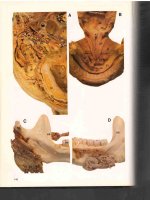
c
D
Figure 30.3 (ContinuedJ (B) Pulsed dye laser treatment to cherry angioma
utilizing diascopy (C) Purpura immediately post pulsed dye laser treat
ment. (D) Complete resolution of cherry angioma after one pulsed dye
laser treatment
1 74
I
Color Atlas of Cosmetic Dermatology
CHAPT E R 3 1
G ra nu l o m a Facia l e
G ra n u loma fac i a l e ( G F ) was fi rst d escri bed by Wigley i n
1 945 w h o la beled t h e d i sease "eos i n o p h i l ic gra n u l o ma . "
P i n kus re n a m ed this d isorder gra n u loma fac i a l e i n 1952.
G F is a n i d i o pathic c h ro n i c c uta neous d isorder that usu
a l ly i nvolves the face, pa rt i c u l a rly the nose . It ca n prese nt
with a si ngle lesion or m u ltiple lesions.
E P I D E M I O LOGY
Incidence: u n c o m m o n
Age: 30 t o 50 yea rs
Race: pri m a ri ly seen in Caucasians
Sex: ma les > fem a l es
Figure 3 1 . 1 Granuloma faciale on the scalp
PATH OG E N ES I S
U n k nown , but may b e mediated b y i m m u ne c o m p lex
d e position .
PHYS I CAL EXAM I NAT I O N
Si ngle i n d u rated facial brown ish-red pa pule o r plaque.
Some lesions may have telangiectasia . M u ltiple lesions may
be present. Extrafacial sites rarely observed . Lesions may
vary in size from m i l l i meters to centimeters ( Fig. 3 1 . 1 ) .
D I FFERENTIAL D I AG N OS ES
Cutaneous l u pus erythematos us, sa rco idosis, lym p h o m a ,
pseudolym phoma , c uta neous T-ce l l
lym p h o m a , fixed
d ru g e r u pti o n , rosacea .
D E R M ATOPATHOLOGY
Dense, polymorphous i nflam matory cell i nfi ltrate i n the
u pper two-t h i rds of the dermis. The i nfi ltrate is com posed
of n u merous eosinoph i ls, neutrophi ls, lym phocytes, a n d
h istiocytes . A pro m i nent grenz zone is c h a racteristica lly
present. Leu kocytoclastic vasc u l itis is freq uently observed .
CO U RS E
The lesions of G F a re usua l ly c h ro n i c a n d o n l y occasion
a l ly resolve s ponta neously.
Sect i o n 6 : Va sc u l a r A l te rat i o n s
I
1 75
MANAG E M ENT
Difficu lt t o treat with a ny modal ity. A n y s uccessfu l treat
ment often leaves sca rring.
• To p i c a l Treat m e n t
•
Corticosteroids: topica l , i ntra lesio n a l
•
Tac ro l i m u s o i ntment (0. 1 % )
• Syste m i c Treat m e n t
•
Da psone
•
Anti m a l a ri a l s
•
Colc h ic i n e
•
Cl ofaz i m i n e
•
G o l d i nj ecti ons
A
S U RG I CAL TREAT M E N T
•
C ryos u rgery:
m u ltiple
reports
i n d icati ng su ccessful
c l ea ra n c e . Resu lts a re u n pred icta ble ( Fig. 3 1 . 2 ) .
•
S u rgical excision .
•
Derm a b rasion .
•
El ectrosu rgery.
• L i g h t Treat m e n t
•
Topica l psora len a n d u l traviolet A ( P UVA) rad iation
thera py
•
Laser thera py: d ifferent lasers have been used in the
treatment of GF with p ro m i s in g resu lts, either as an
a b lative thera py with ca rbon d i oxid e laser o r as a selec
tive thera py ta rget i n g the prom i n ent vasc u latu re in G F
lesions using the Q-switc hed a rgon laser, p u lsed dye,
d i ode laser, and potass i u m tita nyl phosphate ( KT P )
532-nm l a s e r ( F ig. 3 1 .3 ) .
P I T FALLS T O AVO I D
•
G F is often reca lc itra nt to thera py. Patie nts s h o u l d be
cou nseled that successfu l treatment is often el usive.
B I B L I OG RAPHY
A m m i rati CT, H ruza GJ . Treatment o f gra n u l o m a fac i a l e
w i t h the 585- n m p u l sed d y e laser.
Arch Dermatol.
1 999; 135(8) :903-905.
Apfel berg DB, Dru ker D , Maser M R , Las h H, S pence B
J r, Denea u D. G ra n u l o m a fac i a l e . Treatment with the
a rgon laser. Arch Dermatol. 1 983 ; 1 1 9 ( 7 ) : 573-576.
B
Figure 3 1 .2 (A) Multiple lesions of granuloma faciale on the face. (8) No
significant improvement detected after one treatment with cryotherapy on
a 4-month follow-up visit
1 76
I
Color Atlas of Cosmetic Dermatology
Chatrath V, R o h rer TE. G ra n u loma fac i a l e successfu l l y
treated w i t h long-pu lsed t u n a b l e d y e laser. Dermatol
Surg. 2002 ;28( 6 ) : 527-529 .
Elston O M . Treatment of gra n u loma fac i a l e with the
p u l sed dye laser. Cutis. 2000;65(2 ) : 9 7-98.
Khaled A , J ones M, Zerma n i R, et a l . G ra n u loma fac i a l e .
Pathologica. 2007 ;99( 5 ) : 306-308.
M a i l l a rd H, G rogna rd C , Toled a n o C, J a n V, Mac het L,
Va i l la nt L. G ra n u l o m a fac i a l e : Efficacy of c ryosu rgery i n
2 cases. Ann Dermatol Venereal. 2000; 1 2 7 0 ) : 77-79 .
To mson N , Ste rl i ng J C , Sa lva ry I . G ra n u loma fac i a l e
treated successfu l l y w i t h topica l tac ro l i m us . Clin Exp
Dermatol. 2009;34(3) :424-42 5 .
Wheela nd R G , Ash l ey J R , S m ith O A , E l l i s O L, Wheela n d
O N . Ca rbon d ioxid e l a s e r treatment o f gra n u loma fac i a l e .
J Dermatol Surg Oneal. 1 984; 1 0 ( 9 ) : 730-733 .
A
B
Figure 3 1 .3 (A) Indurated brownish-red plaque on the left cheek of a
middle-aged female with granuloma facia/e. (B) Two-year follow-up show
ing resolution of granuloma faciale after m ultiple pulsed dye laser treat
ments
Sect i o n 6: Va sc u l a r A l te rat i o n s
CHAPT E R 3 2
I
1 77
I nfa ntile H e m a ngio m a
I nfa nti le hema ngioma ( I H l , a lso known as strawberry,
ca p i l l a ry,
or
cavernous
hema ngiom a ,
is
a
benign
e n d oth e l i a l prol iferation that re presents the most com
mon tumor i n i nfa ncy. I t ca n be c lassified i nto su perfic i a l
hema ngioma ( S H , 55% o f cases ) , deep hema ngioma
( D H , 30% of cases ) , and m ixed su perfi c i a l and deep
hema ngioma ( M H , 1 5% of cases ) . They occ u r m ost com
m o n ly o n head a n d neck a reas .
EPI D E M I O LOGY
Incidence: 1% to 3 % a re p resent at b i rt h , 10% to 1 2 %
a re p resent b y 1 yea r o f age
Age: majority (80 % ) become a p pa rent between 2 a n d
5 weeks o f age; 2 0 % a re n oted at b i rt h .
Sex: fe ma les a re affected two t o fou r ti mes more t h a n
m a l es
A
Precipitating factors: prematu re i nfa nts a re more com
monly affected
PHYS I CAL EXA M I NAT I O N
The a p pearance depends o n t h e d e pth o f the heman
gioma a n d the phase of evol utio n . S H p resents as bright
red -colored p l a q u e . D H presents as a soft dermal o r s u b
c uta neous nod u l e with a b l u ish- p u r p l e col or. M H shows
featu res of both SH a n d D H . M u lt i p l e truncal heman
giomas
may
be
o bserved .
I nvol uting
hema ngiomas
demonstrate a flatter su rfa ce with a grayis h - p u r p l e h u e
t h a t begi ns ce ntra l l y a n d expa n d s outwa rd . The h e m a n
giomas
m ight
become
u lcerated
and
he morrhag i c .
Resi d u a l fatty tissue, atrop hy, tela ngiecta s i a , s c a r forma
tion , and hypertrophy may be observed .
B
D I F F E R E N T I AL D I AG N OS ES
Congen ita l hema ngiomas ca n be confused with a vasc u
lar
ma lformation
such
as
port-wi n e sta i n
at
b i rt h .
H ema ngiomas a re ge nera l ly present after b i rth versus
vasc u l a r ma lformations, which a re genera l l y present at
b i rth .
LABO RATORY TESTS
• D e r m at o p at h o l ogy
Prol iferations of p l u m p e n d oth e l i a l cel ls that may exte n d
fro m the su perfi c i a l d e r m i s t o the deep su bcuta neous
tiss u e , d e pen d i ng o n the hem a ngioma s u btype.
Figure 32. 1 (A) Left upper eyelid hemangioma in its early growth phase,
a lesion that may threaten the child 's vision. (B) Marked lightening and
flattening of the hemangioma after m ultiple pulsed dye laser treatments
1 78
I
Color Atlas of Cosmetic Dermatology
• A n c i l l a ry Tests
•
A n a bd o m i n a l u ltraso u n d s h o u l d be o bta i ned if m o re
t h a n fo u r tru ncal hema ngiomas a re noted prior to
4 months of age .
•
An electroca rd iogra m ( ECG) a n d a ca rd iac EC H O should
be considered for a n y concern of h igh ca rd iac output.
COU RS E
H ema ngiomas c h a racteristica l l y exh i bit th ree phases of
evol ution : ( a ) prol iferative phase, ( b ) i nvol uting phase,
and (c) i nvo l uted phase. The prol iferati ng phase is c h a r
a cterized by a ra p i d growth p hase that starts at 1 to
2 m o nths of age a n d lasts u nt i l 6 to 9 months of age. This
growth phase is fol l owed by the i nvol uting phase that
usua l l y starts i n the second yea r of l i fe a n d persists for
A
severa l yea rs. M ore than 90% of u ntreated hema ngiomas
i nvol ute, that is, atta i n maxi m a l regression by 9 yea rs of
age. U p to 30% of hema ngiomas leave posti nvol ution
cha nges i n c l u d ing hypopigme ntati o n , sca rring, tela ngiectasi a , and fi b rofatty tiss u e .
COM P L I CAT I O N S
B leed i n g a n d u lceratio n with seco n d a ry i nfection a n d
sca rring, espec ia l ly i n hema ngiomas i nvolvi ng t h e d i a pe r
a rea , a re c o m m o n l y see n . Oth er serious com pl ications
i n c l u d e orbital o bstruction and a m b lyo pia with periorbita l
hema ngiomas, u pper a i rway o bstruction with h e m a n
g i o m a s i n the bea rd d istri bution , s p i n a l a bnorma l ities
with l u m bosacra l hema ngiomas, posterior fossa ma lfor
mation in la rge fac i a l hema ngioma ( P H A C E syn d rome) ,
a n d h igh output c a rd ia c fa i l u re with m u lt i p l e c uta neous
hema ngiomas assoc iated with viscera l i nvolvement.
B
Figure 32.2 (A) Hemangioma on the left fifth toe pad, a location that
in terfered with the child's ability to ambulate. (B) Significant clearing and
near resolution of the hemangioma after multiple pulsed dye laser treat
ments
KEY CO N S U LTAT I V E QU EST I O N S
•
Onset o f lesion
•
N u m ber of lesions noted
•
U l ceration n oted
•
B l eed i ng noted
•
Prior treatm ents a n d res ponse
MANAG E M E N T
T h e treatment o f I H s is controve rsia l . G iven t h e natu ra l
cou rse o f I H with sponta neous reso l ution, m a n y physi
cians c h oose to ca refu l ly o bserve the a rea with no
i ntervention, espec i a l l y i n nonfacia l , sma l l , a n d u ncom
p l icated
hema ngiomas.
Ea rly i ntervention
is recom
m e n d ed for ( a ) all I H s that i nterfere with the function of
vita l
orga ns
(eg,
periorbita l
hema ngiomas,
a i rway
o bstruction with hema ngiomas i n the bea rd d istr i b ution,
Sect i o n 6 : Va sc u l a r A l te rat i o n s
I
1 79
h igh-output cardiac fa i l u re ) ; ( b ) la rge facia l hema ngiomas
that usua l ly i nvo l ute with permanent d i sfiguri ng; (c) u l cer
ated hema ngiomas; and (d) hema ngiomas in the d ia per
a rea that a re very l i kely to u lcerate causing severe pa i n .
•
Medica l treatment
- Steroids i n c l u d i ng topica l steroid a pp l i cation ( c lass 1
corticoste roid a p pl ied twice d a i ly with mon itoring
every 2 wee ks) , i ntra lesiona l steroids (tria m c i nolone
a ceto n i d e 1 0 mg!m L a d m i n istered monthly), and oral
steroids ( 1 . 5-2 mg/kg/d of pred n isone) a re the m a i n
stay o f treatment. Patie nts m ust be mon itored c l osely,
espec ia l ly with oral steroid use given the risk of sys
temic com p l ications i nc l u d i ng growth reta rdation a n d
g l u cose a lterations. Loca l ized side effects i n c l u d e
atrophy a n d yeast infect i o n .
- Other treatment options i nc l u d e to pica l i m i q u i mod
( a p p l ied d a i ly ) , i nterferon-a (3 m i l l ion u n its/m 2/d ,
A
S C ) , a nd v i n c ristine (0.05 mg/kg/d if less than 10 kg,
IV ), espec ia l ly in steroid-resista nt I H . As i nterferon-a
is associated with spastic d i plegi a , patients m u st be
mon itored c l osely.
•
P ro p ra nolol at a d ose of 2 mg/kg/d has been recently
reported to be ve ry effective i n treating severe I H s , even
in steroid-resista nt I H s . T h i s treatment is proposed to
re place ora l or i ntravenous steroids that a re associated
with sign ifica nt side effects. H owever, patients on p ro
pra n olol s h o u l d be c l osely m o n itored for bradyca rd i a ,
hypotension , a n d hypoglycemia espec ia l ly a t the o nset
of the treatment.
•
Laser treatment
- P u lsed dye laser ( P D U treatment i n d u ces sign ifi
ca ntly faster regression of the I H . Fl u e nces lower
than those of PWS a re effective and a re assoc iated
with lowe r risk of laser- i n d u ced sca rri ng ( Figs . 3 2 . 1 ,
3 2 . 2 a n d 3 2 . 3 ) . P D L has been used exte nsively i n
B
the treatment of I H i n th ree c l i n ical scena rios:
Figure 32.3 {A) Segmental hemangioma in volving the hand of a 1 -year
1. U l cerated hema ngiomas res pond effectively to
P D L. PDL ma rked ly dec reases the associated
pa i n a n d i n d uces ra pid hea l i ng of the u l ceration
(75% with i n 2 weeks) ( Fig. 32.4) . Res i d u a l sca r
fo rmation from the u l ce ration is expected .
2. S H s c a n respond wel l to P D L if sta rted either
before
or
early
in
the
prol ife rative
phase.
M u ltiple treatments, every 4 to 6 weeks, a re
req u i red in the prol iferative phase. T h e o n ly
exception is a ra pid ly prol ife rating fa c i a l hema n
gioma . P D L treatment may i n d uce u lceratio n of
these va ria nts so treatm ent s h o u l d be avoided .
I H with deeper components ( M H , D H J res pond
less effectively to PDL beca use of the l i m itation
of penetration of PDL to 1 . 2 mm i n the ski n .
3 . P D L ca n h e l p treat the res i d u a l erythema a n d
tela ngiectasias o n
hemangiomas.
the
s u rface o f i nvol uted
old girl. {B) Complete resolution of the hemangioma after four treatments
with 595-nm pulsed dye laser at low fluences
1 80
I
Color Atlas of Cosmetic Dermatology
- Long-pu lsed N d : YAG lasers a re usefu l for photocoagu
lation of D H s but have a h igher incidence of sca rring.
•
Other
interventions
include
s u rgical
debulking
and
em bol ization . The risks and benefits of each s u rgica l
a pproach should be considered ca refu l ly before i nterven
tion since the sca r from spontaneous regression is usua l ly
better than the surgica l scar. Em bol ization is uti l ized in
hema ngiomas associated with h igh-output ca rd iac fa i l u re.
P I T FALLS TO AVO I D
•
Use of excessive P O L fluences without s k i n coo l i ng ca n
cause sca r.
•
Pa rents a re u nd ersta n d a bly a nxious a bout their c h i l d 's
hema ngioma . A f u l l d iscussion of the natu ra l c o u rse of
A
hema ngiomas is m a ndatory prior to sta rt i n g thera py.
The option of foregoi n g treatm ent a n d c l i n ica l l y m o n i
toring a patient s h o u l d b e reviewed ca refu l ly p r i o r to
sta rt i n g treatment.
•
Pa rents s h o u l d a lso have a rea l i stic idea of the l i m ita
tions of thera py. La rge hema ngiomas res pond less suc
cessfu l ly
to
o ra l ,
s u rgica l ,
and
laser
thera py.
C o m p l icated hema ngiomas that may i n te rfere with the
c h i l d 's health s h o u l d be referred to an a p p ropriate
ped iatric spec i a l i st. P a re nts m ust be awa re that treat
ment wi l l provide an i m provement but may n ot res u lt i n
fu l l resol ution o f t h e h e m a ngioma .
•
Parents n eed to be ed ucated on proper wou n d care,
espec i a l ly for u lcerated hema ngiomas, i n order to
i m prove the c h i l d 's q u a l ity of l ife .
•
F i b rofatty c h a n ges a re ofte n a seq uela of resolved
hema ngiomas.
Such
c h a nges
can
be
B
i m p roved
sign ificantly with n o n a b l ative a n d a blative fract i o n a l
resu rfa c i ng.
B I B L I OG RAPHY
Batta K, G oodyea r H M , M oss C, Wi l l i a m s H C , H i l ler L,
Waters R. R a n d o m ised control led study of early p u lsed
dye laser treatment of u ncompl icated c h i l d hood haeman
giomas: Resu lts of a 1 -yea r a na lysis.
Lancet 2002 ;
360(9332 ) : 5 2 1 -527 .
Lea ute-La breze C, Du mas de Ia Roq ue E, H u biche T,
Bora levi F, Tha m bo J - B , Ta·leb A. Propranolol for severe
hema ngiomas of i n fa n cy. N Eng! J Med. 2008;358: 2649265 1 .
c
L i YC, McCa h a n E , R owe N A , M a rt i n PA, Wilcsek G A ,
Figure 32.4 (A) Ulcerated hemangioma, isolated nodular type, extremely
painful and hemorrhaging, treated twice with pulsed dye laser 6 Jlcm 2 ,
7-mm spot size, 590 nm. (B) At 2 months ' follow-up, significant healing
of the ulceration after a single treatment with pulsed dye laser. (C) Four
months after initial pulsed dye laser treatment and 2 months after
second pulsed dye laser treatment, there is complete healing of the
ulceration
M a rt i n FJ . S uccessfu l treatment o f i nfa nti le h a e m a n
g i o m a s o f the o r b i t w i t h pro p ra n olol . Clin Experiment
Ophthalmol. 2010;38(6): 5 54-559 .
More l l i J G , Ta n OT, Yoh n J J , Weston WL. Treatment of
u l cerated hema ngiomas i nfa n cy. Arch Pediatr Ado/esc
Med. 1 994; 148( 1 0) : 1 1 04- 1 1 0 5 .
Sect i o n 6: Va sc u l a r A l te rat i o n s
CHAPT E R 33
I
1 81
Ke ratosis Pi l a ris At rophica ns
Ke ratosis p i l a ris atro p h ica ns ( K PA) is a gro u p o f i n he rited
d i so rd e rs with th ree su btypes i n c l u d i ng (a) keratosis
p i l a ris atro p h i ca n s fac i e i ( KPAF ) , (b) atrophoderma ver
m ic u latu m (AV ) , a n d (c) ke ratosis fo l l i c u l a ris s p i n u losa
d ecalva n s ( KFS D ) . KPA F a n d AV present m a i n ly on the
face with K FS D often a p pea r i n g o n the eye b row a n d AV
m ost com m o n l y seen on the c heeks, sparing the eye
brows a n d sca l p . KFSD can affect the face, sca l p , a n d
tru n k . I n herita nce pattern can b e a utosom a l d o m i na nt
( KPAF, AV) , recessive (AV ) , or X-l i n ked ( KFS D ) .
EPI D E M I O LOGY
Incidence: very ra re; KPAF is the m ost c o m m o n su btype
Age: KPAF a n d KFSD in i nfa ncy; AV in c h i l d h ood
Sex: ma les a re more seve rely affected in KFSD
Figure 33. 1 Keratosis pilaris: fine, sandpaper-like follicular papules on
PATH OG E N ES I S
the arm of a young man
Abnormal fol l i c u l a r keratin ization of the u pper sectio n of
the h a i r fol l icle that may later res u lt in atro p h i c fo l l i c u l a r
sca rring.
PHYS I CAL EXAM I NAT I O N
Fol l i c u l a r
pl u gging
with
erythema
in
early
stages
( Figu re 33. 1 ) . Atro p h i c fol l i c u l a r sca r fo rmation with
assoc iated a lopecia in later stages .
D I FFERENTIAL D I AG N OS I S
Ke ratos is p i l a ris, keratosis pila ris ru b ra , seborrheic der
matitis ( KPA F ) , atopic d e rmatitis ( KFS D ) , other etiologies
of sca rring a l o pecia ( KFS D ) , acne sca rri ng (AV), Rom bo
syn d rome (AV ) , a n d K I D syn d rome ( K FS D ) .
D E R M ATOPAT H O LOGY
D i lated fo l l ic l es with fo l l i c u l a r hyperkeratosis and i nfla m
m a t i o n i n e a r l y stages . Fol l i c u l a r fi brosis a n d atrophy i n
later stages .
CO U RS E
The cou rse i s c h ro n i c with n o sponta n eous reso l ution .
With t i m e , the e ryt h e m ato u s fo l l i c u l a r hyperkeratotic
pa p u les i nvol u te i nto d e p ressed atro p h i c fo l l i c u l a r sca rs
with a l opec i a .
1 82
I
Color Atlas of Cosmetic Dermatology
MANAG E M ENT
There is n o com pletely effective treatment for KPA.
M u ltiple treatment options have been tried with only va ri
a b le s uccess . Patients should be cou nseled that thera py
may not be effective.
•
Topical thera py may, at best, prod uce modest benefit.
- Lactic acid a n d a-hyd roxy acid lotions ( 1 0 %- 1 2 % )
a p plied twice d a i ly may i m p rove the text u ra l ro ugh
ness. H owever, they may p rod uce i rritatio n .
- R eti n o i d s (taza rote n e , reti n-A) a p p l ied n i ghtly may
i m p rove text u r a l ro ugh ness. T h ey may prod uce i rri
tati o n .
- Corticosteroids a p p l ied s pa ri ngly m a y show i m provement. R i s k of fac i a l atro ph y l i m its their use.
•
A
System i c thera py
- Other o ptions that have p rovided va ria ble su ccess
i n c l u d e o ra l reti noids a n d d a pso n e .
- They a re m ost h e l pfu l fo r the i nfla m m atory stage of
KPA, but provide m i n i m a l i m prove ment in the fol l ic u
l a r hyperkeratos is.
- They req u i re ca refu l mon itoring for potentia l side
effects.
•
Laser thera py
- P u lsed dye laser ( 59 5 n m , 7-m m spot, 7-1 0 J/cm 2 ,
D C D 40/20, p u lse d u ration of 1 . 5-3 ms) c a n be
effective in the treatment of the assoc iated e rythema
of KPAF but will not sign ifica ntly i m prove the text u ra l
rough n ess o f KPA ( Fig. 33 . 2A , B ) .
- Laser-assisted h a i r remova l with long- p u lsed n o n
Q-switc hed ru by l a s e r may be a n effective treatment
i n patients with KFS D .
P I T FALLS T O AVO I D
Pati ent expectations a re ge nera l ly very h i g h . They m ust
be cou nseled as to the c h ro n i c natu re of the cond ition
and m i n i m a l res ponse to ava i la ble thera pies.
B I B L I OG RAPHY
Baden H P, Byers H R . C l i n i c a l fi n d i ngs, c uta neous pathol
ogy, and response to therapy i n 21 patients with keratosis
p i l a ris atro p h ica n s . Arch Dermatol. 1 994; 130(4):469475.
C h u i CT, B e rger TG , P rice VH, Za c h a ry CB. R eca lcitra nt
sca rring fol l ic u l a r d isord e rs treated by laser-assisted h a i r
re mova l : A prel i m i na ry report. Dermatol Surg. 1 999 ;
25( 1 ) : 34-3 7 .
C l a rk S M , M i l l s C M , La n iga n SW. Treatment o f keratosis
p i l a ris atro p h i c a n s with the p u lsed tunable dye laser. J
Cutan Laser Ther. 2000 ; 2 (3 ) : 1 5 1 - 1 56.
B
Figure 33.2 (A) Keratosis pilaris atrophicans. Patient is emotionally both
ered by persistent erythema. (8) Marked lightening of erythema 2 years
following three pulsed dye laser treatments
Sect i o n 6: Va sc u l a r A l te rat i o n s
Ka u n e K M , Haas E, E m m e rt S, Schon M P, Z utt M .
Successfu l treatment of severe keratos is p i l a ris ru bra with
a 595- n m pu lsed dye laser. Dermatol Surg. 2009 ; 3 5 :
1 592- 1 595.
M a rq ue l i ng AL, G i l l ia m AE, P rend ivi l l e J, et al. Keratosis
p i l a ris ru b ra : A c o m m o n but u n d errecogn ized conditi o n .
Arch Dermatol. 2006; 142( 1 2 ) : 1 6 1 1 - 1 6 1 6 .
R i c h a rd
G,
H a rth W . Keratosis fol l ic u l a ris s p i n u losa
d ecalva n s . T he ra py with isotret i n o i n and etreti nate in the
i nfla m matory stage. Hautarzt. 1 993;44(8) : 529-534.
CHAPT E R 34
Po rt-wi n e Stains
Port-wine sta i n s ( PWS) a re low-flow ca p i l lary m a lforma
tions. They represent the m ost common type of vasc u l a r
ma lformations. Any a rea o f t h e body can b e affected .
H owever, the head a n d neck a reas a re m ost co m mo n ly
affected .
EPI D E M I O LOGY
Incidence: 3 per 1 , 000 newborns
Age: prese nt at b i rt h i n the majo rity of patients ; rarely
a p pea r i n adolesce nce o r a d u lthood
Sex: no sex pred i l ection
Race: less common i n Asi a n s a n d African Americans
Associated syndromes: PWS can be a m a n ifestation of
severa l synd romes i n c l u d i n g Stu rge-We ber syn d rome,
K l i ppel-Tre n a u nay synd ro m e , P rote us syn d rome, and
pha komatos is pigmentovasc u la ris
P H YS I CAL EXA M I NAT I O N
PWS prese nts a t b i rth a s l ight p i n k , we l l-dema rcated
m a c u l a r lesions a n d patc hes usua l l y in a segmenta l d is
tri butio n . They ca n tra n sform with age i nto hypertro p h i c
d a r k r e d a n d/or p u r p u ric pla q u es w i t h nod u l a rity. PWS
i nvolves the face m ost c o m m o n l y a l ong the trigem i n a l
n e rve d istri bution : ophtha l m i c b ra n c h V 1 ( u pper eye l i d
a n d forehea d ) , maxi l l a ry b ra n c h V2 ( u pper l i p , cheek,
lower eye l id ) , a n d m a n d i b u l a r b ra n c h V3 .
D I FFERENTIAL D I AG N OS I S
PWS exh i bits c h a racteristic c l i n i cal featu res a n d i s sel
d o m m isd iagnosed . I t can be confused with the mac u l a r
stage o f h e m a ngioma at b i rth .
I
1 83
1 84
I
Color Atlas of Cosmetic Dermatology
D E R M ATOPAT H O LOGY
M u ltiple d i lated t h i n -wa l led vesse ls in the pa p i l l a ry a n d
reti c u l a r d e r m i s .
A N C I LLARY TESTS
•
The pa rents s h o u l d be cou nseled rega rd i n g the possi
b i l ity of Stu rge-We ber synd rome (SWS) i n lesions
l ocated i n a fac i a l Vl o r V2 dermatom a l d istri bution .
SWS is cha racterized by the prese nce of fac i a l PWS
with i psi latera l o c u l a r a n d lepto m e n i ngea l a n o m a l ies.
Ten to fifteen percent of pati ents with PWS i n the V l
d istr i b ution wi l l have SWS . Patients w i t h b i latera l PWS
h ave even a h igher risk of SWS . An ophthal mologic
exa m i nation to ru l e out gla ucoma a nd cata ract forma
tion with conti n ued fo l lowu p is necessa ry for these
patients . A head c o m p uted tomogra phy ( CT) or mag-
A
netic reson a n ce i maging ( M R I ) s h o u l d be o bta i ned to
r u l e out b ra i n i nvolvement that could affect menta l
development a n d res u l t i n sei z u res.
•
PWS overlyi ng the s p i n e ca n be associated with s p i n a l
a n o m a l y s u c h as s p i n a l dysra p h i s m o r tethered s p i n a l
cord . N e u ro l ogic eva l uation a n d a p p ro priate i maging
stu d ies a re recom m e n d ed .
•
Large extremity PWS should ra ise the consideration of
Kl i ppel-Trenau nay syn d rome, cha racterized by capillary
venous ma lformations or ca pil lary-lym phatic-venous mal
formations with hypertrophy of the affected extrem ity. Leg
girth and length should be measu red and followed over
time.
COU RS E
PWS grows proporti o n a l l y with the patient a n d gra d ua l ly
t h i c kens a n d d a rkens i n color from p i n k to d a r k red to
B
deep p u rple. Eleven percent may d eve l o p n od u l a rity a n d
2 4 % may d eve l o p pyoge n i c gra n u lomas. PWS may b e
associated with hypertro phy o f u n derlying soft tissue a n d
bone,
pa rtic u l a rly
in
Stu rge-We ber
syn d rome
and
K l i ppel-Tre n a u nay syn d ro m e .
KEY CO N S U LTAT I V E QU EST I O N S
•
On set o f lesion
•
Assoc iated c l i nical fi n d i ngs
•
Is the c h i l d m eeti ng d eve l o pmenta l m i lestones?
•
Has the c h i l d had an eye exa m i nation?
•
Has the c h i l d had a head M R I or CT?
•
Past treatments a n d response
•
B l eed i ng
•
B l ebs
(B) Significant lightening of the PWS after a single POL treatment.
•
G rowth of PWS
(C) Complete resolution of the PWS after POL treatments
c
Figure 34. 1 (A) PWS on the right inner thigh of an infant girl.
Sect i o n 6: Va sc u l a r A l te rat i o n s
I
1 85
MANAG E M ENT
PWS d e m o nstrates progressive vasc u l a r d i latation a n d
hypertrophy with age, t h u s m a k i ng treatment d u ri ng
ea rly i nfa ncy esse ntial for a bette r res ponse. Treatment
ca n be sta rted as ea rly as 2 weeks of age . Treatment p ro
vides a red uction in the n u m be r of vessels a n d d oes n ot
c o m p l ete ly rem ove the enti re lesio n . T h e refore , the PWS
may exh i bit some d a rke n i n g a n d t h i c ke n i ng over t i m e
despite
i n terventio n .
G e n e ra l
a n esthesia
m ight
be
needed for treati ng la rge PWS i n c h i ld re n .
•
Laser treatm e n t ( F igs . 34. 1-34. 5 ) .
P u lsed dye laser ( P O L) rema i n s the gol d sta n d a rd for
the treatment of PWS . Effective P O L pa ra meters i n c l u d e
wavele ngths o f 5 8 5 t o 600 n m , flue nces o f 6 t o 1 5 J/c m 2 ,
p u l se d u rations of 0.45 or 1 . 5 ms with cryogen spray
A
cool i n g (CSC). Fou r to twe lve laser sessions with 4-to-8week i nterva ls a re u s u a l l y req u i red in order to ach ieve
sign ificant b la n c h i n g of the PWS . Lower fl uen ces a re i n itia l ly uti l i zed for PWS off the face a n d in d a rker s k i n
types . The use o f e s c concom ita ntly d u ri n g P O L treatment sign ificantly dec reases the pa i n associated with the
proced u re a n d the i n c i d ence of bl istering. esc protects
the epidermis a n d a l l ows for d e l ivery of h igher flu ences,
resulting in more effective b l a n c h i ng of the PWS . P O L
treatm ent is fo l l owed b y tem pora ry p u r p u ra that usua l ly
resolves in 7 to 14 days. Complete l ighte n i ng of PWS with
POL treatment is a c h i eved i n l ess than 20% of PWS .
Resista nce to
P O L treatment
is
more freq ue ntly
encou nte red in deeper and hypertro p h i c PWS . H e l pful
m a n e u ve rs to potentiate the efficacy of P O L i n c l u d e
i n c reasi n g t h e fl u e n ces with adeq uate c ryogen cool i n g to
p rotect the epidermis a n d i n c reas i n g the wavelength u p
to 600 n m to ta rget deeper vesse ls. A pi lot study demon
strated that PWS that a re treated with to pica l imiquimod
once d a i ly for 1 month after P O L exposu re m a n ifest
su perior b l a n c h i ng res ponse over time as compared to
P O L a l o n e . Another re port i n vestigated the c o m b i ned use
of POL and a topica l a n giogenesis i n h i bitor, rapamycin,
using the in vivo rodent wi n d ow c ha m ber mode l . There
was no reformation a n d reperfusion of blood vessels after
treatment with P O L fol l owed by topical ra pamyc i n for
14 d ays, i n contrast to P O L a l o n e . With extreme ca ution
to avo i d sca rring and dyspigmentatio n , it is poss i b l e to
treat P O L-resista nt PWS and deeper or hypertro p h i c
a d u lt P W S su ccessfu l ly w i t h longer wavele ngth lasers that
a l low d eeper penetration i nto the skin such as l ongp u l sed a l exa n d rite (755 n m ) laser, long-pu lsed N d :YAG
( 1 , 064 n m ) laser, and d u a l 595- n m P O L a n d 1 ,064- n m
N d :YAG laser cou pled w i t h adeq uate coo l i ng. U s e o f t h e
N d :YAG laser can be treac h e rous as there is a narrow
thera peutic ra nge. R isk of sca r ca n be sign ificant.
•
Light treatment: i ntense pu lsed l ight ( I P L ) may be effec
tive in treatment of PWS , i n c l u d i n g P O L- resista nt PWS .
A green-ye l l ow waveband a n d lowest ava i l a ble p u lse
B
Figure 34.2 (A) Extensive port-wine stain on the right face and forehead
of an infant male. (8) Significant resolution after multiple treatments
with pulsed dye laser
1 86
I
Color Atlas of Cosmetic Dermatology
d u ration s h o u l d be used , with s k i n coo l i ng. A recent
ra ndom ized c l i n ical tria l com pa r i ng P O L a n d I P L side
by side revea led a better efficacy a n d h igher patient
preference after POL treatment. P h otodyna m ic thera py
may a lso prove to be an a lternative efficacious treat
ment for PWS .
•
Other treatment modal ities for PWS that can be effec
tive i n c l u d e tattooing a n d cosmetic m a keu p .
P I T FALLS TO AVO I D
•
Patients s h o u l d be cou nseled that PWS d isplay a va ri
a b le response to treatment. M o re extens ive and th icker
lesions respond less wel l when com pa red to su perfi c i a l
lesions. Fac i a l PWS responds best. P W S treatment effi-
A
cacy decreases as one d escends from face to feet, with
the lower extre m ities d isplaying the least treatment
benefit.
•
M u lt i p l e treatment sessions may be req u i red . B r u i s i n g
is a necessa ry side effect t o o bta i n efficacious thera py.
•
Laser treatment may prod uce "footpri nti ng" or o n ly pa r
tial i m p rovement.
•
Treatme nts should be ceased when the patient is satis
fied with l ighte n i ng, o r when n o fu rther benefit has
been noted , that is, afte r two su bseq uent treatments.
B I B L I OG RAPHY
Alste r TS, Ta nzi EL. C o m b i ned 595- n m a n d 1 , 064- n m
laser i rrad iation o f rec a l c itra nt a n d hypertro p h i c port
wine sta i n s in
c h i l d ren a n d a d u lts.
Dermatol Surg.
2009 ; 3 5 ( 5 ) : 8 1 3-8 1 5 .
C h a n g CJ , Hsiao Y C , M i h m M C J r, N elson J S . P i lot stu d y
B
Figure 34.3 (A) Extensive port-wine stain on the right neck of a young
female. (B) Marked resolution of the port-wine stain after multiple treatments with pulsed dye laser
exa m i n i ng the com b i ned u s e o f p u lsed d y e l a s e r a n d topical l m i q u i mod versus laser a l o n e for treatment of port
wine sta i n b i rt h m a rks. Lasers Surg Med. 2008;40(9 ) :
605-6 1 0 .
C h a pas A M , Eickhorst K, G e ron e m u s R G . Efficacy of
early treatment of fac i a l port w i n e sta i n s in newborns: A
review of 49 cases. Lasers Surg Med. 2007;39 ( 7 ) : 563568 .
C h i u C H , C h a n H H , H o WS , Ye u ng C K , N e lson J S .
P ros pective stu d y o f p u l sed d ye laser i n conj u nction with
c ryogen s p ray coo l i n g fo r treatment of port wine sta i ns i n
C h i n ese patients. Dermatol Surg. 2003;29(9):909-9 1 5 .
Discussion 9 1 5 .
Fa u rsc h o u A , Togsverd- B o K , Zachariae C , Haedersdal
M. P u lsed dye laser vs . i ntense p u lsed l ight for po rt-wine
sta i ns : A ra nd o m ized side-by-side tria l with b l i n ded
res ponse eva l uati o n . Br J Dermatol. 2009 ; 1 60(2) :359-
�.
A
Figure 34.4 (A) Port-wine stain on the lower mucosal and cutaneous lip.
Sect i o n 6: Va sc u l a r A l te rat i o n s
I
1 87
Ho WS, Ying SY, C h a n PC, C h a n H H . Treatment of port
wine sta i n s with i ntense pu lsed l ight: A prospective study.
Dermatol Surg. 2004;30(6):887-890.
H u i keshoven M, Koste r P H , d e B orgie CA, Beek J F, va n
Gernert M J , va n d e r H o rst C M . Reda rken i n g of port-wine
sta i n s 1 0 years after p u l sed-dye-laser treatment. N Eng! J
Med 2007;356( 1 2 ) : 1 235- 1 240.
Li L, Kon o T, G roff WF, C h a n H H , Kitazawa Y, N oza ki M .
Com parison study of a long-pu lse p u lsed dye laser a n d a
long-pu lse p u lsed a lexa nd rite laser in the treatment of
port w i n e sta i ns . J Cosmet Laser Ther. 2008; 1 0( 1 ) :
12-15.
P h u ng T L , O ble D A , J ia W , B enja m i n L E , M i h m M C J r,
N elson J S . Can the wo u n d hea l i ng res ponse of h u ma n
s k i n b e mod u l ated afte r laser treatment a n d t h e effects of
exposu re exte nded? I m pl ications on the c o m b i ned use of
the p u l sed dye laser a n d a topical a ngioge nesis i n h i bitor
B
fo r treatment of port wine sta i n b i rth ma rks . Lasers Surg
Figure 34.4 (Continued) (B) Significant lightening of port-wine stain after
Med. 2008;40( 1 ) : 1-5.
Se l i m M M , Ke l l y K M , N e lson J S, We nd elsc hafe r-Cra b b G ,
Ke n n edy WR , Z e l i c kson B D. Confocal m i c roscopy stu d y
three treatments with a combination of pulsed dye laser to the cutaneous lip
and vermilion and long-pulsed 1 , 064-nm Nd: YAG laser to the inner
mucosa/ lip and vermillion
o f nerves a n d blood vessels i n u ntreated a n d treated
portwine sta i ns : Pre l i m i n a ry o bservati ons. Dermatol Surg.
2004;30:892-897.
Ya ng M , Ya roslavsky A , Fari n e l l i , e t a l .
Long-pu lsed
neodym i u m : Yttri u m -a l u m i n u m -ga rnet laser treatment
for port-wi ne sta i n s . J Am Acad Dermatol. 2005 ; 52(3):
480-490.
Figure 34.5 Hypopigmentation, which can be permanen t, after aggres
sive treatment of a PWS in an A frican-American patient
1 88
I
Color Atlas of Cosmetic Dermatology
CHAPT E R 3 5
Pyoge nic G ra n ulo m a
Pyoge n i c gra n u l o m a ( PG ) c a n be rega rded a s a benign
vasc u l a r tu m o r o r a s a reactive vasc u l a r process a risi ng
at sites of prev i o u s tra u m a or i rritat i o n . PG is a lso k n own
as l o b u l a r ca p i l l a ry h e m a n g i o m a , gra n u l o m a tela ng
iectatic u m , a n d gra n u lo m a gravi d a r u m when p rese nting
o n t h e gi ngiva of preg n a n t wo m e n . I t commonly occ u rs
i n a reas of tra u ma i n c l u d i n g the face a n d finge rs .
EPI D E M I O LOGY
Incidence: c o m m o n
Age: most common i n c h i l d ren a n d yo u ng a d u lts
Precipitating factors: m i nor tra u ma , pregna n cy, laser treat
ment of port-wi ne sta ins, isotretinoin
Figure 35. 1 Classic hemorrhagic pyogenic granuloma
PATHOG E N E S I S
Reactive neovasc u l a rization suggested b y c o m m o n asso
c iation with preexisting tra u m a o r i rritation a n d l i m ited
growth ca pac ity.
PHYS I CAL EXAM I NAT I O N
Red t o violaceous, d o me-sha ped , friable
pa p u l e or
nod u le , 0.5 to 1 . 5 e m i n size, with s m ooth surfa ce that
freq uently ulcerates ( Figs. 35. 1 , 3 5 . 2 and 3 5 . 3 ) .
D I F F E R E N T I A L D I AG N OS ES
N od u l a r a me l a n otic m e l a n o m a , glomus tumor, h e m a n
gioma , sq u a m o us c e l l carci noma ( S C C ) ( F ig. 3 5 . 4 ) ,
nod u la r basa l cel l carc i n o m a , wa rt, bac i l l a ry a ngiomato
sis, Ka posi 's sa rco m a , and m etastatic cancer.
D E R M ATOPAT H O LOGY
Wel l -circ u mscri bed exo phytic l o b u l a r pro l i feration of ca p
i l l a ries with flattened a n d someti mes e roded overlyi n g
epidermis w i t h pe r i p hera l epidermal "colla rettes . "
COU RS E
P G u s u a l l y grows ra p i d ly over the cou rse of weeks o r
months a n d then sta b i l izes. It b l eeds freq u e ntly with
m i nor tra u ma and ca n persist i n d efin itely if n ot treated .
Figure 35.2 Pyogenic granuloma on the palm of a pregnant woman,
bleeding frequently
Sect i o n 6: Va sc u l a r A l te rat i o n s
I
1 89
MANAG E M ENT
•
Laser treatment
- Pu lsed dye laser (585--600 n m , 0.45- 1 . 5 ms, 7-10 m m ,
6-- 1 5 J/cm 2, O C O 20-40/20 with or without d iascopy) is
a safe and effective device for the treatment of small
lesions and for ped iatric patients. Seria l treatments are
usua l ly req uired . Treatment is wel l tolerated without
anesthesia. A recent report suggested shave excision
followed by immed iate pu lse dye laser ( P OLl for larger
lesions. POL has been also reported to be effective i n
gi ngival PG. Nd:YAG laser c a n also be effective.
- Carbon d ioxi d e is effective . Lesional flatte n i ng is the
c l i n ica l end point. l ntra l esional l i doca i n e 1% is neces
sa ry prior to treatment. Postoperative ca re req u i res
twice d a i ly cleansing with soa p a n d water a n d a p p l i
cation o f a nt i b i otic oi ntment over a 2 t o 6 wee ks heal
i n g t i m e . Sca r formation is l i kely. A low rec u rrence
rate is noted .
•
S u rgical treatment: a l l treatments may res u lt in sca r for
Figure 35.3 Pyogenic granuloma overlying a dermal nevus
mati o n .
- Shave exc ision fol l owed b y electrod essication o f t h e
base is t h e proced u re most c o m m o n l y e m p loyed .
Recu rrence is common ( Figs . 3 5 . 5 a n d 3 5 . 6 )
- El l i ptica l exc ision c a n be pe rformed w i t h l o w rec u r
rence but wi l l leave a sca r
- Ligation of the base
- C ryos u rgery
•
Alternative treatment options i n c l ud e
- l m iq u i m od 5 % c rea m h a s been recently reported to
be effective in ped iatric patients a n d in patients with
recu rrent PG
- l ntralesional i njection of a bsol ute etha nol
- Scleroth erapy with monoetha nola m i n e oleate
- To pica l a l itreti n o i n (9- cis-ret i n oic c i d ) ge l , a d rug that
is used for the treatment of Ka pos i 's sa rcoma
P I T FALLS TO AVO I D
•
Patients s h o u l d be awa re that rec u rre nce is common
after treatment.
•
Patie nts s h o u l d be i nformed that all treatments may
result i n sca rring.
•
Amela notic melanoma as wel l as SCC and other skin can
cers can m i mic PG . A biopsy should be performed for
any suspicious lesions in the a ppropriate c l i nical setti ng.
B I B L I OG RAPHY
B o u rguignon
R,
Paq uet
P,
P i e ra rd - F ra n c h i mont
C,
P i e ra rd G E . Treatment o f pyogen ic gra n u lomas with t h e
N d-YAG laser. J Dermatolog Treat. 2006; 1 7(4) : 247-249 .
Figure 35.4 Pyogenic granuloma mimicking a squamous cell carcinoma
on the left lower mucosa/ lip of a patient with multiple nonmelanoma
skin cancers
1 90
I
Color Atlas of Cosmetic Dermatology
Fa l l a h H , Fisc h e r G , Zaga re l l a S. Pyoge n i c gra n u loma i n
c h i ld re n : Treatment with to pical i m i q u i m od . A ustralas J
Dermatol. 2007;48(4) : 2 1 7-220
Kha n d p u r S , Sharma VK. S u ccessfu l treatment of m u lti
p l e gi ngiva l pyoge n i c gra n u lomas with p u lsed-dye laser.
Indian J Dermatol Venereal Lepra/. 2008; 74( 3 ) : 275-27 7 .
M a loney D M , S c h m idt J D , D u v i c M . A l itreti n o i n g e l to
treat pyoge n i c gra n u loma . J Am Acad Dermatol. 2002 ;
47( 6 ) : 969-970.
Mats u m oto K, N a ka n is h i H, Seike T, Koiz u m i Y, M i h a ra K,
Ku bo Y. Treatment of pyogen i c gra n u loma with a scleros
ing agent. Dermatol Surg. 200 1 ;27(6) : 52 1 -523 .
R a u l i n C, G reve B , H a m mes S. The combi ned conti n u
ouswave( pu I sed carbon d ioxide laser for treatment o f pyo
gen i c gra n u lo m a . Arch Dermatol. 2002 ; 138( 1 ) :33-3 7 .
S u d A R , Ta n ST.
Pyoge n i c gra n u loma c o m p l icating
p u lsed -dye laser thera py for c h e rry a ngioma . J Plast
Reconstr Aesthet Surg. 2010;63(8) : 1 364- 1368.
A
B
Figure 35.5 (A) Shaving a hemorrhagic and painful pyogenic granuloma
on the plantar foot with # 1 5 blade. The specimen was sent for histological
confirmation. (B) Electrodessication of the residual pyogenic granuloma
Sect i o n 6: Va sc u l a r A l te rat i o n s
I
1 91
A
B
Figure 3 5 . 6 (A) Biopsy-proven pyogenic granuloma on the right chin of a
young female. (8) Shave excision of pyogenic granuloma with Derma
Blade (Personna Medical, Verona, VA)