Ebook Pediatric critical care medicine (Volume 4: Peri-operative care of the critically ill or injured child - 2nd edition): Part 2
Bạn đang xem bản rút gọn của tài liệu. Xem và tải ngay bản đầy đủ của tài liệu tại đây (13.28 MB, 257 trang )
Part III
Trauma
Richard A. Falcone
Head and Neck Trauma
14
Derek S. Wheeler, Derek Andrew Bruce,
and Charles Schleien
Abstract
While the overall mortality rates have decreased significantly, TBI remains a significant
public health problem. In addition, while cervical spine and spinal cord injuries are less
common in children compared to adults, these injuries are an important source of long-term
morbidity and pose a significant burden on the health care system. The management of
these injuries has continued to evolve over time. Critically injured children with TBI require
the close coordination of management between the PICU team, the trauma surgeon, and the
neurosurgeon.
Keywords
Traumatic brain injury • Depressed skull fracture • Closed head injury • Cervical spine
injury • SCIWORA • Spinal cord injury • Epidural hematoma • Subdural hematoma
• Intracranial hypertension
Introduction
Trauma is the leading cause of pediatric morbidity and
mortality in the United States. The mortality rate due to
trauma has declined significantly in all age groups since
1979, largely as a result of aggressive injury prevention programs. However, accidental injury still accounts for more
than one-third of all childhood deaths [1]. Many of these
D.S. Wheeler, MD, MMM (*)
Division of Critical Care Medicine,
Cincinnati Children’s Hospital Medical Center,
University of Cincinnati College of Medicine,
3333 Burnet Avenue, Cincinnati, OH 45229-3039, USA
e-mail:
D.A. Bruce, MB, ChB
Center for Neuroscience and Behavioral Medicine,
Children’s National Medical Center,
111, Michigan Avenue NW, Washington, DC 20010, USA
e-mail:
C. Schleien, MD, MBA
Department of Pediatrics, Cohen Children’s Medical Center,
Hofstra North Shore-LIJ School of Medicine,
269-01 76 Ave, Suite 111, New Hyde Park, NY 11040, USA
e-mail:
D.S. Wheeler et al. (eds.), Pediatric Critical Care Medicine,
DOI 10.1007/978-1-4471-6359-6_14, © Springer-Verlag London 2014
deaths are due to traumatic brain injury (TBI) [2]. Neck and
cervical spinal cord injuries, although relatively rare in the
pediatric population, often have catastrophic consequences
[3–5]. Clearly, pediatric head and spinal cord trauma creates
a significant burden on society.
Head Trauma
Epidemiology
While the overall mortality rates have decreased significantly, TBI remains a significant public health problem.
Seat-belt and bicycle helmet laws have resulted in a dramatic
decrease in both the number and severity of TBI in children
[6, 7]. Although children generally have better survival rates
than adults [6], the life-long sequelae of even a mild TBI
can be more devastating in children due to their young age
and developmental potential [8, 9]. The usual mechanism of
injury depends on the age of the patient. For example, children under 4 years most often suffer TBI secondary to falls,
motor vehicle accidents, or non-accidental trauma (child
abuse), while TBI in older children usually occurs second199
200
ary to sporting or motor vehicle accidents. In the adolescent
population, motor vehicle accidents and assault or violent
crime are the most common causes of TBI [6, 9]. Males
appear to sustain TBI almost twice as often as females, especially in the adolescent age group. Most large series show an
increased incidence of head trauma in the spring and summer
months when children are more likely to be outdoors [10].
As mentioned above, pediatric TBI is a significant burden to
the health care system, accounting for more than $1 billion in
total hospital charges every year in the United States alone.
These costs do not take into account the costs of future medical care, years of lost work, and the years of lost quality of
life, which are likely to be significantly greater [11].
Physical abuse (inflicted trauma) is the leading cause of serious head injury and death in children under 2 years of age [12].
The mechanism of injury in inflicted or abusive head trauma
is controversial, but likely involves a combination of shaking, asphyxia, and blunt trauma to the head. The distinction
between inflicted and accidental head injury in young children
is important as it greatly affects prognosis. Outcome among
children with non-accidental head trauma is significantly
worse, and the majority of survivors suffer significant disability and neurologic impairment [13]. Inflicted or abusive head
trauma is discussed in greater detail elsewhere in this textbook.
Pathophysiology
The pathophysiology of TBI is specific to either the primary
or secondary insult. Primary injury is the injury that results
directly from the original impact and is best prevented by
aggressive injury prevention programs, including the proper
use of safety devices such as seatbelts, bicycle helmets, and
air bags. Primary head injury can involve damage to the
scalp, cranial bone, dura, blood vessels, and brain tissue as
a result of immediate application of acceleration/deceleration forces with or without impact. Both contact and inertial
forces may be involved in the primary injury. Linear force
vectors occur when the head is struck by a moving object and
are responsible for generating contact force. Inertial forces
are created by acceleration/deceleration or angular-rotational
movement of the head in space. Because the child’s headto-torso ratio is much greater than that of the adult, inertial
forces are magnified in children resulting in more diffuse
brain injury. The relatively higher water content and incomplete myelination of the pediatric brain may also contribute
to the diffuse nature of the injury in the immature brain as
compared to the more focal adult pattern [14].
Impact injuries to a static head have their greatest effects
on the skin and skull and, as a result of absorption of the
force by these tissues, less effect on the brain tissue and brain
blood vessels. For example, children with depressed skull
fractures may have an associated cerebral contusion, though
D.S. Wheeler et al.
the predominant damage occurs to the skull. Acceleration/
deceleration forces, whether associated with impact or not,
result in complex deformations of the brain and its blood vessels that can lead to a variety of pathologies from (i) shearing injury of the white matter, with or without hemorrhage,
(ii) contusion or laceration of the cortex or deep structures,
including the midbrain and medulla, (iii) disruption of arteries or veins with subsequent hemorrhage, and (iv) disruption
of the blood-brain barrier.
Secondary brain injury results from the physiologic and
biochemical events that occur after the initial trauma or primary brain injury. The best recognized of these secondary
injuries are systemic hypotension, hypoxemia, hypercarbia, intracranial hypertension, and cerebrovascular spasm.
Hypotension and hypoxia commonly present on admission
to the emergency department (ED) and thus any secondary
damage may already have been sustained prior to advanced
medical care (i.e., in the ED or PICU). However, intracranial
hypertension tends to progress over several days and is rarely
present during the early stages after initial resuscitation.
Other secondary injuries may be produced by a variety
of molecular events (discussed elsewhere in this textbook in
much greater detail) such as the release of excitotoxic neurotransmitters or free oxygen radicals. Multiple mechanisms
have been implicated in secondary brain injury and include
cerebral ischemia, release of excitatory neurotransmitters,
free radical formation, activation of neuronal apoptosis cascades, and blood-brain barrier disruption leading to cerebral
edema. The role of these mechanisms in human brain trauma
is unclear and no specific therapies are available to correct or
modify these molecular events.
In children, cerebral blood flow (CBF) is reduced shortly
after TBI. Loss of endogenous vasodilators such as nitric
oxide and elaboration of vasoconstrictors such as endothelin1 have been implicated in producing post-traumatic hypoperfusion. Glutamate levels in cerebrospinal fluid have been
shown to increase in humans after brain injury leading to
excitotoxic neuronal death in cell culture. Glutamate exposure leads to elevation of intracellular calcium, oxidative
stress, and production of free radicals. Although a portion
of cell death occurs immediately after the initial insult, some
neurons have been shown to die in a delayed manner by
apoptosis [15]. The immature brain may be more vulnerable
to apoptosis as demonstrated in experimental animal models
where the severity of neurodegeneration after trauma was
highest in the youngest animals [16]. Finally, both osmolar
swelling in contusions and astrocyte swelling as a result of
excitotoxicity contribute to significant cerebral swelling.
This swelling can lead to secondary ischemia and/or herniation with their devastating consequences.
Post-traumatic insults such as hypoxia and systemic
hypotension are common in children and are known to
exacerbate the severity of secondary injury and worsen
14 Head and Neck Trauma
prognosis [17–19]. Since intracranial hypertension, hypoxia,
and systemic hypotension are the leading factors associated
with poor outcome, post-injury interventions which decrease
or ameliorate these events reduce secondary injury to the
injured but still viable brain.
Trauma Systems
The influence of trauma systems and pediatric trauma centers on outcomes of TBI has recently been studied. Children
with severe TBI are more likely to survive if treated in a
pediatric trauma center or an adult trauma center with added
qualifications to treat children [20–24]. Based on the wealth
of experience, pediatric patients in a metropolitan area with
severe TBI should be transported directly to a pediatric
trauma center which will most likely be in close proximity to the accident site [25, 26]. However, for those children
injured in rural areas, stabilization at an outside hospital may
be indicated prior to transfer to the trauma center.
Initial Resuscitation
Immediate attention to airway, breathing, and circulation is
mandatory for all unconscious children. The initial resuscitation of a child with TBI is vitally important since post-injury
hypoxia and systemic hypotension are associated with worse
outcome (as discussed above). It may be possible to minimize the rate of occurrence of these events with proper early
resuscitation measures. Generally all resuscitation interventions are aimed at lowering intracranial pressure (ICP)
and maximizing cerebral perfusion pressure and delivery
of oxygen and substrate to the brain. Providing the injured
brain with adequate substrate to maintain normal function is
dependent on maintaining a stable airway, adequate ventilation, cardiac function, and systemic perfusion (i.e., airway,
breathing, circulation).
Hypotension, defined as systolic blood pressure less than
5th percentile for age, has been associated with a 61 % mortality rate in children with severe TBI and an 85 % mortality rate when combined with hypoxia [27]. Hypotension has
repeatedly been shown to worsen the prognosis for all levels
of severity (as determined by the Glasgow Coma Score, GCS)
of central nervous system (CNS) injury in children as well as
in adults [17–19, 27–32]. Hypotension is present at admission in 20–30 % of severe head injuries and the avoidance of
hypotension, if possible is dependent on timely recognition
and resuscitation prior to arrival to the hospital. Episodes
of hypotension can also occur in the hospital, both in the
ED and in the PICU. The origin of these episodes is unclear
and therefore avoiding them may be difficult. However,
close, intensive multimodality monitoring will identify these
201
episodes early and allow for timely intervention. While
hypotension must always trigger a careful exploration for
possible areas of blood loss, it can occur with isolated head
injury or spinal cord injury [33].
Regardless of the etiology, hypotension must be aggressively treated. TBI can be associated with loss of normal cerebral autoregulation (Fig. 14.1), such that rapid
decreases in mean arterial pressure (MAP) result in profound decreases in cerebral perfusion pressure (CPP) and
cerebral blood flow (CBF). Since children will often maintain their systolic blood pressure despite significant blood
loss until they enter the later stages of hypovolemic shock,
clinical signs of shock (such as tachycardia, diminished central pulses, urine output less than 1 mL/kg/h, cool extremities, and prolonged capillary refill) should be treated as if
hypotension were already present and rapidly corrected
with volume resuscitation. Fluid restriction to avoid exacerbating cerebral edema is contraindicated in the management
of the child with TBI in shock. The use of hypertonic saline
as a resuscitation fluid is gaining popularity because of the
beneficial effects on ICP, though there are no clinical trials
to support this type of fluid over other available agents for
fluid resuscitation.
Transfusion of packed red blood cells is indicated to
replace active blood loss, though the ideal transfusion trigger
for critically injured children with TBI is not known [34–36].
Severe anemia is potentially harmful in patients with TBI.
Furthermore, transfusion can help maintain intravascular
volume and maximize oxygen carrying capacity. However,
observational and retrospective studies have shown that
transfusion does not necessarily improve short- and longterm outcomes [37–39]. Regardless, once the volume deficit
has been corrected, a vasopressor (e.g., dopamine, epinephrine) should be administered to patients with persistent hypotension. Resuscitation fluid should be isotonic to avoid the
risk of worsening cerebral edema. It is recommended that
intravenous glucose be avoided in the first 48 h after injury
as hyperglycemia has been associated with worse outcome
[40]. However blood glucose should be monitored frequently, especially in younger children who are most at risk
for hypoglycemia.
The deleterious effects of hypoxia are less well established (compared to hypotension), though there is evidence to suggest that post-injury hypoxemia, defined as
PaO2 <60–65 mmHg or oxygen saturation <90 %, portends
a worse neurologic outcome in both pediatric and adult
TBI patients [27, 28] and that it is a common occurrence in
children with severe head injury, present in up to 45 % of
patients [41]. Hypoxemia must be avoided and corrected
with the use of 100 % supplemental oxygen. Although
there is no evidence to support that tracheal intubation
provides an advantage over bag-valve-mask ventilation
in children with TBI, the current recommendation is that
202
D.S. Wheeler et al.
a
b
Blood flow =
Resistance
Perfusion pressure
Resistance
Maximal
vasodilation
No autoregulation
Blood flow
Autoregulation
Blood flow
Autoregulation
Autoregulation
Maximal
vasoconstricition
No autoregulation
Pressure
Autoregulatory
pressure range
Time
Perfusion pressure
Fig. 14.1 Cerebral Blood Flow (CBF) autoregulation. (a)
Autoregulation is the intrinsic ability of an organ, independent of neural
and humoral influences, to maintain a constant blood flow despite
changes in perfusion pressure. To maintain constancy in organ blood
flow, as perfusion pressure is altered there must be a responsive reciprocal change in vascular resistance, mediated by a change in arterial
diameter. For example, a decrease in organ blood flow resulting from a
decrease in the perfusion pressure triggers a reflex autoregulatory vasodilation and reduction in vascular resistance, reconciling a return of
arterial blood flow to steady state. (b) Maintenance of organ blood flow
at a constant rate is limited by the ability of the vasculature to vasodilate
and vasoconstrict. As organ perfusion pressure decreases there is a
compensatory vasodilation to maintain constancy of organ blood flow.
As the point of maximal vasodilation is reached, further decreases in
organ perfusion pressure result in an uncompensated decrease in organ
blood flow. Similarly, as organ perfusion pressure increases, there is a
compensatory vasoconstriction to maintain constancy of organ blood
flow. As the point of maximal vasoconstriction is reached, further
increases in organ perfusion pressure result in an uncompensated
increase in organ blood flow
Table 14.1 Indications for tracheal intubation in children with TBI
are present. Chronic hyperventilation can lead to reactive
vasoconstriction resulting in decreased cerebral blood flow,
cerebral hypoperfusion, decreased oxygen delivery, and possibly, ischemia. Conversely, hypercarbia may lead to cerebral
vasodilation which can acutely raise ICP. Most unconscious
head injured children do not have intracranial hypertension
upon initial presentation. Therefore, usually there is no reason to routinely administer hyperosmolar agents such as
mannitol or hypertonic saline as part of the initial resuscitation. Indeed between 30 and 50 % of children with severe
head injuries (depending on the study population) will not
develop significant intracranial hypertension at any time during their hospital course [42–45].
As soon as the child is stable and has had a complete
physical examination (including neurologic examination),
the next step in management is to obtain an imaging study.
Initial plain radiographs should include a lateral cervical
spine, anteroposterior (AP) chest, and AP pelvis radiograph (so-called trauma X-ray panel). The imaging study
of choice for the CNS in most centers is still a noncontrast
CT scan of the head with bone windows. In addition, as
most major trauma centers now have spiral CT scanners, it
is relatively easy to obtain images of the spinal column, as
the clinical history and exam dictate, without unnecessary
delay. There is no question that MRI offers superior definition of the extent of tissue injury compared to CT, though
GCS ≤8
Decrease in GCS of >3 (independent of the initial GCS)
Anisocoria >1 mm
Apnea, bradypnea, irregular respirations
Loss of gag/cough reflex
Cervical spine injury with respiratory compromise
Inadequate oxygenation or ventilation
children with a GCS ≤8 should have their airway secured
by tracheal intubation to avoid hypoxemia, hypercarbia,
and aspiration (Table 14.1). Ideally, this should be performed by an individual with specialized training in the
pediatric airway and with the use of capnometry/capnography to verify proper placement of the airway in the trachea (please see the chapters on Airway Management for
a more in-depth discussion of this topic). These specifications are made for children because success rates of prehospital tracheal intubation in children have been shown to
be lower than in adults. The cervical spine must be stabilized in the midline during tracheal intubation in any child
with suspected cervical spine injury.
Children with TBI should be ventilated with the goal of
maintaining PaCO2 in the normal range. Aggressive hyperventilation to acutely reduce PaCO2 should be reserved for
the acute situation when signs of impending brain herniation
14 Head and Neck Trauma
CT is still better at defining the extent of bony injury.
However, until faster MRI scanners become available and
more MRI compatible equipment is developed, CT remains
the initial imaging study of choice [46]. The results of this
initial CT dictate the next steps in management. If there is
a significant mass lesion (e.g., epidural hematoma), surgery
is usually required. However, if there is no mass lesion, the
CT scan is further examined for evidence of diffuse axonal
injury, ischemic injury, or signs of brain swelling (either
focal or generalized). Imaging studies of other organ systems may dictate the need for surgery as well, e.g. intestinal
rupture. If surgery is necessary on other organ systems in
a child with a GCS ≤8, insertion of an ICP monitor at the
commencement of the operative procedure is indicated, as
ICP monitoring allows the anesthesiologist to monitor ICP
and to control it until surgery on these other organ systems
is completed.
ICP Monitoring
No randomized controlled trials evaluating the effect on outcome of severe TBI with or without ICP monitoring have been
conducted in any age group. However, ICP-focused intensive
management protocols have almost certainly improved outcomes [26, 47, 48]. Given the paucity of pediatric data, the
current recommendations for pediatric ICP monitoring are
largely based upon anecdotal experience and adult studies.
Indeed, the current pediatric guidelines [26] do not make
any firm recommendations and suggest that ICP monitoring
may be considered for critically injured children with severe
TBI (generally defined as GCS ≤8). This recommendation
applies to infants as well since the presence of open fontanels and/or sutures does not negate the risk of developing
intracranial hypertension nor does it alter the utility of ICP
monitoring. Although ICP monitoring is not routinely recommended for infants and children with less severe injury
(GCS ≥8), it may be considered in certain conscious patients
with traumatic mass lesions or for patients whose neurologic
status may be difficult to assess serially because of sedation
or neuromuscular blockade, especially when going to the
operating room for general anesthesia.
Once the decision is made to invasively monitor a patient’s
ICP, there are several types of monitoring devices that can be
used. These are discussed elsewhere in this textbook. The
ventricular catheter has been shown to be an accurate way
of monitoring ICP and has the added advantage of enabling
cerebrospinal fluid (CSF) drainage, making it the preferred
method. Intraparenchymal monitoring devices are used
commonly but have the potential for measurement drift and
do not allow for CSF drainage. Morbidities related to all of
these catheters, including infection, hemorrhage, and seizure
are unusual.
203
Intracranial Hypertension
Intracranial pressure, or the pressure within the intracranial
vault, is determined by the interactions between the brain
parenchyma, the cerebrospinal fluid (CSF), and the cerebral
blood volume. The fundamental principles of intracranial
hypertension were proposed by the two Scottish physicians
Monro and Kellie in 1783 [49] and 1824 [50], respectively,
who stated that (i) the brain is enclosed in a non-expandable,
relatively rigid space; (ii) the brain parenchyma is essentially
non-compressible; (iii) the volume of blood within the skull
is nearly constant; and (iv) a continuous outflow of venous
blood is required to match the continuous inflow of arterial
blood. However, as originally proposed, the Monro-Kellie
doctrine did not take into account the volume of the CSF. As
we now know, reciprocal volume changes of the CSF compartment is an important compensatory mechanism that will
allow reciprocal changes in the volumes of the other cranial
compartments (i.e., blood, brain) [51]. The combined volume
of all of the components of the skull cavity (brain, blood,
CSF) must remain constant because they are encased in a
fixed volume. Therefore, if the volume of one intracranial
element increases, the volume of another (e.g. CSF) must
decrease to compensate and keep ICP in the normal range.
ICP is therefore a reflection of the relative compliance of the
cranial compartments (Fig. 14.2). As shown in Fig. 14.3, ICP
will remain normal in spite of small additions of extra volume, whether edema, tumor, hematoma, etc. However, once
a critical point is reached, at which compensatory mechanisms are maximized, addition of subsequent volume produces a dramatic rise in ICP.
During the initial hours following head injury, there is
a diminished volume of intracranial CSF as a result of displacement of CSF into the spinal subarachnoid space, as
well as increased reabsorption of brain CSF by the choroid
plexus. Intracranial pressure therefore remains in a safe,
normal range. However, as edema worsens or hemorrhage
increases in size, these compensatory mechanisms eventually fail and ICP increases. If cerebral herniation occurs at
the foramen magnum or the tentorium (Fig. 14.4), the normal CSF pathways are blocked and displacement of CSF
cannot occur, resulting in a further decrease in intracranial
compliance and worsening intracranial hypertension.
The perfusion of the brain, like all organs, is determined
by the difference between the upstream and downstream
blood pressures (i.e. perfusion pressure). The driving force
for blood flow to the brain (upstream pressure) is the mean
arterial pressure (MAP) and the downstream pressure, under
normal physiologic circumstances, is the central venous
pressure (CVP). In the case of intracranial hypertension,
when the ICP exceeds the CVP, the cerebral perfusion pressure (CPP) becomes: CPP = MAP – ICP. Therefore, in order
to maximize cerebral blood flow (CBF) after TBI, therapies
204
D.S. Wheeler et al.
Venous
blood
Mass/edema
Mass/edema
Arterial blood
Arterial blood
Arterial blood
Brain
Brain
Brain
Normal
Compensated
Uncompensated
p
c
a
CSF
Venous blood
CSF
Intracranial pressure
Fig. 14.2 The Monro-Kellie
Doctrine. See text for detailed
explanation
b
V
Fig. 14.3 Pressure-volume curve of the craniospinal compartment.
This figure illustrates the principle that in the physiological range, i.e.
near the origin of the x-axis on the graph (point a), intracranial pressure
remains normal in spite of small additions of volume until a point of
decompensation (point b), after which each subsequent increment in
total volume results in an ever larger increment in intracranial pressure
(point c) (Reprinted from Andrews and Citerio [51]. With permission
from Springer Science + Business Media)
must be targeted to optimize MAP and reduce ICP thereby
decreasing the risk of secondary brain injury. It is well known
that intracranial hypertension is associated with poor neurologic outcome and that aggressive treatment of elevated ICP
is associated with the best clinical outcomes.
As stated above, approximately 30–50 % of head injuries
will demonstrate normal to minimally elevated ICP in the
face of adequate CPP and do not require any specific therapy directed to the cranial injury [42–45, 52]. Management
of these patients is therefore directed towards maintaining
cardiorespiratory and hemodynamic stability. The current
pediatric guidelines state that treatment efforts directed
towards intracranial hypertension may be considered when
ICP >20 mmHg. Similarly, the Brain Trauma Foundation
and the European Brain Injury Consortium guidelines also
recommend initiating treatment of intracranial hypertension if ICP ≥20 mmHg to maintain CPP in the range of
50–70 mmHg [53–55]. Two quite different approaches have
been proposed for the treatment of intracranial hypertension to prevent secondary cerebral ischemia. One approach
(presented above) focuses on maintaining the CPP = MAP
– ICP in an acceptable range (i.e. CPP is increased by either
reducing ICP, increasing MAP, or a combination of both)
[56–62], while the other approach focuses on decreasing the
end capillary pressure in the brain and thus reducing brain
edema by slightly lowering arterial pressure and controlling end-capillary pressure and colloid osmotic pressure (the
so-called Lund concept) [63–66]. Most intensive care units
use a combination of these therapies [67, 68]. Both methods
have their (at times passionate) proponents. However, there
is insufficient evidence to support one method over the other
at this time [69–72].
In a single-center observational study comparing ICP and
survival, of the 51 children with severe closed head injury
who underwent ICP monitoring, 94 % of the children in
14 Head and Neck Trauma
205
consensus guidelines [26]. For additional discussion and a
detailed reference list of supportive evidence, the reader is
referred to these guidelines and the chapter on Intracranial
Hypertension in this textbook.
M1
A
B
M2
C
Fig. 14.4 Schematic representation of herniation syndromes.
According to the Monro and Kellie doctrine, increased volume and
pressure in one compartment of the brain may cause shift of brain tissue
to a compartment in which the pressure is lower. M1 is an expanding
supratentorial lesion; M2 is an expanding mass in the posterior fossa. A
Increased pressure on one side of the brain may cause tissue to push
against and slip under the falx cerebri toward the other side of the brain,
B Uncal (lateral transtentorial) herniation. Increased ICP from a lateral
lesion pushes tissue downward, initially compressing third cranial
nerve and, subsequently, ascending reticular activating system, leading
to coma, C Infratentorial herniation. Downward displacement of cerebellar tissue through the foramen magnum producing medullar compression and coma (Reprinted from Citerio and Andrews [205]. With
permission from Springer Science + Business Media)
whom the ICP never exceeded 20 mmHg survived. This is in
sharp contrast to the 59 % survival rate in the children with
maximum ICP’s greater than 20 mmHg [73]. An elevation
of ICP for greater than 1 hour was found to be most deleterious and was associated with worse clinical outcome. This
study, and others of its kind have led to the recommendation that treatment for intracranial hypertension should begin
at an ICP of 20 mmHg or greater. Maintenance of adequate
CPP is important in order to allow for ongoing delivery of
metabolic substrates to the brain. Again, there are insufficient data to establish firm, consensus recommendations
[26], though a minimal CPP of 40–50 mmHg may be considered for children. The pediatric consensus guidelines further
suggested that the appropriate CPP may be age-based – the
lower end of the aforementioned threshold is considered
appropriate for infants while the upper end is considered
appropriate for adolescents. A stepwise approach to the
management of ICP and CPP was proposed in the original
pediatric consensus guideline [25], which is still useful. Here
we will present a very brief overview of the current pediatric
Sedation, Analgesia and Neuromuscular
Blockade
The use of sedatives and analgesics in the setting of raised
ICP remains a difficult challenge. Pain and stress are known
to increase cerebral metabolic demands as well as cause
intracranial hypertension. However, most sedatives cause a
reduction of mean arterial pressure which can decrease CPP.
Additionally, these medications may exacerbate an elevated
ICP by causing cerebral vasodilation, which in turn increases
cerebral blood volume. Long-acting sedatives also may
interfere with the ability to follow serial neurologic exams.
For these reasons, short-acting agents like midazolam are
preferred. Narcotics such as morphine or fentanyl can be
used for pain control. Medications known to raise ICP, for
example ketamine, should be avoided.
Neuromuscular blocking agents may be used to reduce
ICP by preventing shivering, posturing, and breathing against
the ventilator (dysynchrony). Potential harmful effects
include masking of seizure activity and increased infection
risk. Therefore, these agents should be reserved for specific
indications and only with continuous EEG monitoring. In
addition, positioning the patient with the head elevated to 30°
in a midline, neutral position will facilitate adequate venous
drainage through the jugular veins, helping to reduce ICP.
CSF Drainage
CSF drainage can be used as a means of controlling ICP if
a ventriculostomy catheter is in place. A lumbar drain may
be considered as an option for refractory intracranial hypertension if a functioning ventriculostomy is already present.
Since the lateral ventricles are often small in brain injured
patients and up to 30 % of the compliance of the CSF system
is in the spinal axis, lumbar drains have been studied as an
alternate way of diverting CSF and lowering ICP. In a retrospective analysis of 16 pediatric patients, Levy et al. reported
a decrease in ICP in 14 of 16 children and improved survival
after placement of a lumbar drain [74].
Hyperosmolar Therapy
Osmotic diuretics, such as mannitol, have been used extensively in the management of intracranial hypertension.
Mannitol (0.25–1 g/kg IV) is effective in lowering ICP both
by decreasing blood viscosity and thereby decreasing cerebral blood volume, and by gradually drawing water from the
brain parenchyma into the intravascular space. This effect
however requires an intact blood-brain barrier which may not
be present in injured areas of the brain. Mannitol may therefore leak into the injured area and accumulate, exacerbating
206
focal edema. Other risks of mannitol use include acute
tubular necrosis and renal failure, perhaps related to hypovolemia and dehydration. Care should be taken to maintain euvolemia and serum osmolarity below 320 mOsm/L.
Hypertonic, 3 % saline is effective in controlling ICP with
few adverse effects at doses of 0.1–1.0 mL/kg/h. The current consensus pediatric guidelines favor hypertonic saline
over mannitol at doses between 6.5 and 10 mL/kg for acute
increases in ICP [26]. A continuous infusion is an acceptable
alternative. It appears that hypertonic saline can be safely
used up to a serum osmolarity of 360 mOsm/L.
Hyperventilation
Prophylactic hyperventilation is contraindicated in the setting of pediatric TBI. Hypocapnia induces cerebral vasoconstriction and leads to a reduction in cerebral blood volume
and ICP. Chronic hyperventilation depletes the brain’s interstitial bicarbonate buffering capacity and causes a shift in
the hemoglobin-oxygen dissociation curve, impairing oxygen delivery to brain tissue. In a prospective trial of severely
brain injured adults randomized to prophylactic hyperventilation or normocapnic treatment, the patients in the hyperventilation group had a significantly worse outcome [75].
However, based upon the lack of definitive evidence, the
current consensus guidelines recommend against prophylactic severe hypoventilation [26] and further suggest that mild
hyperventilation (PaCO2 30–35 mmHg) may be considered
for intracranial hypertension refractory to sedation, CSF
drainage, and hyperosmolar therapy, only if advanced neuromonitoring methods are used to avoid cerebral ischemia.
Temperature Control
Hyperthermia after TBI has been correlated with worse
injury and functional outcome in both animal models and
clinical studies in adults. The mechanisms of damage
include worsening of the secondary insult by increasing
cerebral metabolic demands, damage by excitotoxicity, and
cell death by stimulation of apoptotic pathways. Therefore
hyperthermia should be aggressively avoided in children
with TBI. The basis for the use of hypothermia (core body
temperature <35 °C) in children is derived from several adult
studies which show a strong correlation between hypothermia and reduced ICP with a trend towards improved outcome at 3 and 6 months after injury in a younger adult group
[76]. The presumed mechanism of neuroprotection involves
a decrease in excitatory amino acid release, preservation of
anti-oxidants, a decrease in the release of free radicals, and
anti-inflammatory effects. Although there are no clinical trials in children that show a positive effect, the current recommendation [26] is that moderate hypothermia (32–33 °C)
should be considered as a treatment for intracranial hypertension in children after severe TBI, beginning within 8 h of
the initial injury. If hypothermia is used, rewarming should
D.S. Wheeler et al.
commence at 48 h and at a rate no greater than 0.5 °C/h [26].
These new recommendations are based upon two randomized, controlled trials in critically ill children which suggested that moderate hypothermia reduced ICP. However,
there was no difference in mortality or long-term outcomes in
these two trials (indeed, there was a trend towards increased
morbidity and mortality in the hypothermia group in the trial
by Hutchison and colleagues) [77, 78].
Barbiturates
High-dose barbiturates decrease ICP by several mechanisms.
They lower both resting cerebral metabolic rate and cerebral blood volume. In addition, they appear to have direct
neuroprotective effects by inhibiting free radical–mediated
lipid peroxidation of membranes [79]. Potential risks include
myocardial depression, risk of hypotension, and the need for
hemodynamic support. Barbiturates may be used for refractory intracranial hypertension in hemodynamically stable
patients [80]. Starting at lower doses and titrating up to burst
suppression on EEG may decrease the risk of coma-induced
complications [26]. Invasive hemodynamic monitoring is
frequently necessary.
Decompressive Craniectomy
Decompressive craniectomy has been shown to be an effective method of lowering ICP in children with severe head
injury in several small studies. Taylor et al. reported lowered
mean ICP after surgery in children with intracranial hypertension refractory to medical management and CSF drainage
[81]. In addition, several case-control studies have suggested
improved outcome in children undergoing early craniectomy
versus a non-surgical control group [82]. The current literature suggests that decompressive craniectomy is most appropriate as a means of lowering ICP and maximizing CPP if
performed within 48 h of injury in patients with diffuse cerebral swelling on CT scan, with an evolving cerebral herniation syndrome or those with secondary clinical deterioration.
There have been reports of exacerbation of cerebral edema
and hemorrhage after surgery and reports of poor outcome
especially after non-accidental trauma [83]. Of interest, a
prospective, randomized, controlled trial of early decompressive craniectomy in critically ill adults with severe traumatic brain injury showed that decompressive craniectomy
decreased ICP and ICU length of stay, but was associated
with more unfavorable outcomes [84]. The current pediatric consensus guidelines suggest that early decompressive
craniectomy can be considered for patients with refractory
intracranial hypertension [26].
Corticosteroids
Corticosteroids are not recommended in the treatment of
raised ICP after pediatric TBI [26]. Multiple prospective,
randomized studies failed to show improvement in ICP
14 Head and Neck Trauma
management or functional outcomes with the use of steroids,
and an increase in infection rate and suppression of endogenous cortisol have been observed.
Anti-convulsants
Children with severe brain injury, especially infants and
toddlers, are at high risk for post-traumatic seizures in the
period immediately following injury. Approximately 20 %
of children will have at least one seizure following moderate
to severe TBI [85–88]. Seizures increase ICP by increasing
cerebral metabolic demand and causing the release of excitatory amino acids. In adults, the benefit of giving 1–2 weeks
of prophylactic phenytoin has been shown to outweigh the
risk. In a retrospective review of 194 children with TBI,
there was a significant reduction in posttraumatic seizures
in children treated with phenytoin [85]. Phenobarbital has
been used for prophylaxis for infants. There is no evidence
to support the use of prophylactic anti-epileptics in children
or adults beyond the first 2 weeks after trauma. The pediatric
consensus guidelines only state that seizure prophylaxis with
phenytoin may be considered [26]. Levetiracetam may be an
acceptable alternative for seizure prophylaxis [88, 89].
Surgical Management
Approximately one-third of severely head injured children
have surgically treatable lesions. It is important to identify
these lesions early since they can be the cause of delayed
deterioration and death. However, for the majority of children with TBI, therapy is directed to maintenance of normal systemic parameters and prevention or treatment of
elevated ICP.
Linear Skull Fractures
Linear fractures, which are generally the most frequently
encountered type of skull fracture in children, do not require
surgery. However, in a small percentage of infants and toddlers under 2 years of age, the fracture is associated with
laceration of the dura and contusion of the underlying brain.
The brain and CSF can insinuate themselves into the fracture
and with the pulsating of the brain produce widening of the
fracture edges (i.e., “growing fracture”) and reabsorbtion of
bone, leading to a growing skull fracture or leptomeningeal
cyst. This requires surgical correction once it is clear that
the fracture line is widening. Soft tissue swelling is usually
palpable over the fracture site [90, 91].
Depressed Skull Fractures
In the unconscious child, closed depressed skull fractures
rarely require emergency surgical correction. Many smaller
lesions, especially those in the parietal region never warrant
surgical elevation. The general rule is that if the fracture
207
is depressed more than the thickness of the skull, surgical
elevation of the depressed fracture should be considered.
The surgical correction can be performed after the child has
recovered from coma. There is no evidence that the surgery
has any beneficial effect on long-term outcome other than
improving appearance [92, 93].
Compound Skull Fractures
Since compound skull fractures are by definition open
wounds, they are associated with a high risk for infection
of the skull or brain. Early operation, within the first 12 h
to debride the brain, close the dura, and reconstruct the
skull is widely accepted. In most cases the broken pieces of
bone can be sterilized and replaced to achieve an immediate
reconstruction of the skull. Removal of all intracerebral bone
debris is important for avoiding delayed abscess formation
[92]. Delayed surgical correction has been proposed in those
children who require intensive management of intracranial
hypertension [94].
Fractures that involve the anterior skull base with brain
extruding into the ethmoid sinus can pose a logistic problem.
These are usually associated with frontal bone fractures and,
in older children, facial fractures [95]. If there is brain swelling it can be difficult to get adequate exposure of the anterior skull base to assure a good reconstruction and therefore
the surgery may have to be delayed until the life-threatening
increases in ICP are controlled. If the child is evaluated early
before significant intracranial swelling has occurred, both
the skull base and facial fractures can be repaired in a single
operation. The advantage is the prevention of increased cerebral herniation through the fracture site during the period of
brain swelling (and the attendant risk of formation of leptomeningeal cysts). Finally, prophylactic antibiotics are not
generally indicated in the management of skull fractures,
except in the case of compound skull fractures. However,
tetanus toxoid and tetanus vaccination booster should be
administered if the vaccination status is not up to date.
Epidural Hematomas
Epidural hematomas occur in 3–8 % of children hospitalized after head injury [96]. Most epidural hematomas are
the result of falls, automobile or bicycle accidents, or skate
boarding accidents, where the head strikes a static object
[97–99]. Despite the relative plasticity of the neonatal and
infantile skull, epidural hematomas also affect children in
this age group with equal frequency to that of older children.
Skull fractures overlying the site of the epidural hematoma
are common and result from the impact injury. Bleeding
occurs in the space between the skull and the dura and arises
from a ruptured meningeal artery, a torn venous sinus, or
from the bone itself. One third of children exhibit the “classic” pattern of immediate unconsciousness followed by
recovery (a lucid interval) and then secondary deterioration.
208
D.S. Wheeler et al.
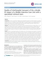
Fig. 14.5 Axial CT scans of an 8-year-old boy who rode his skateboard into a wall 6 h prior to this CT scan. He presented to the ER
unarousable with bradycardia and bradypnea. The CT shows an acute
epidural hematoma with areas of low density in the hematoma and significant midline shift
This pattern is due to traumatic unconsciousness as a result
of the deceleration injury and subsequent recovery from that
event, followed by secondary hemorrhage from the epidural
vessel, artery, or vein, resulting in increased ICP, cerebral
herniation, and loss of consciousness related to brain stem
compression. Another third of children are never unconscious and the final third are in coma from the time of the
injury [100]. Pupillary changes and hemiparesis are initially
found contralateral to the side of the hematoma in only 50 %
of children. Therefore, in contrast to adults, the affected side
of the hematoma is not easily deduced by clinical examination. If a CT scan cannot be obtained because of the rapidity
of the deterioration in the level of consciousness, the best
indicator of the location of the clot is the presence of a skull
fracture. CT scan is especially sensitive for the presence of
epidural hematomas, but if obtained too early the sensitivity
decreases as the hematoma has not yet formed [101]. This
is most likely in cases of venous epidural hematomas. Any
clinical deterioration, including increasing headaches and/
or vomiting, requires a second CT scan. Since the outcome
is closely related to the level of consciousness at the time
of surgical evacuation, early diagnosis is crucial. Low density attenuations found on the CT scan suggest continuing
hemorrhage and if seen is an additional indication for early
evacuation of the clot (Fig. 14.5). Currently about one-third
of epidural hematomas are treated without surgery. Nonsurgical management is more common in awake children
and in epidural hematomas that are frontal in location, less
than 1.5 cm in size, and unassociated with significant midline
shift [102–105]. Many temporal lesions and posterior fossa
lesions require surgery because of the risk of rapid deterioration; conservative management (i.e., non-operative) for these
lesions has also been described [106–108]. Surgery requires
a craniotomy flap and complete evacuation of the lesion with
coagulation of any bleeding points. The skull fracture can
often be used as one limb of the bone flap and repaired at the
time of surgery. Outcome is related to the level of consciousness and the presence of other intracranial lesions. Mortality
rates are low from 0 to 5 % and clinical recovery is usually good [98, 99, 102–104, 109], especially in children. ICP
monitoring is not necessary in the majority of children after
clot evacuation, but if the dura is tight or if there is CT evidence of other intracranial injury, ICP monitoring is generally recommended.
Subdural Hematomas
In the pediatric age group, the majority of subdural hematomas occur in infants and children under 2 years of age who
are the victims of child abuse (see chapter on inflicted head
trauma). Most subdural hematomas are the result of acceleration/deceleration injuries at high speed and therefore occur
after motor vehicle accidents. Both passenger and pedestrian injuries can be associated with subdural hemorrhage.
In contrast to epidural hematomas, the bleeding associated
with subdural hematomas occurs from tearing of the bridging veins from the cortex to the venous sinuses or from direct
14 Head and Neck Trauma
209
Fig. 14.6 Axial CT scans of
a 6 year-old girl who was an
unrestrained passenger in a MVA.
The top right and left views
demonstrate a small subdural
hematoma on the right, lowdensity brain with loss of the
gray/white interface, and midline
shift with trans-tentorial
herniation. Because of the
inability to control the ICP a
decompressive craniectomy was
performed after failure of medical
management. The bottom left
view obtained following
decompression demonstrates an
increase in the low-density area,
resolution of the midline shift,
and herniation of the hemisphere
outside the bony margin. The
bottom right view obtained after
recovery demonstrates evidence
of residual damage to the right
hemisphere
cortical laceration. The location of the bleeding is usually
between the dura and the arachnoid (hence subdural), though
subarachnoid hemorrhage is also common. As a result of the
significant forces required to create subdural bleeding, CT
in affected children will frequently demonstrate evidence of
other brain injury, cerebral contusions, diffuse axonal injury,
intraparenchymal hematoma, or focal or generalized brain
swelling. In many cases the size of the subdural hematoma is
small compared to the degree of brain herniation (Fig. 14.6).
The frequency of surgical drainage of subdural hematomas
varies considerably between neurosurgical centers. If the
hematoma is not large and the main problem is the underlying brain injury and swelling, medical management of intracranial hypertension may be the only therapy that is required.
However, if the hematoma is large and felt to be responsible
for the majority of any brain shift seen on imaging, surgical
evacuation of the hematoma is indicated. Even after surgery
these children frequently require ICP monitoring and aggressive management of intracranial hypertension.
Cerebral Contusions and Intracerebral
Hematomas
Children with cerebral contusions often recover without significant sequelae, and resection of contused brain is therefore
rarely appropriate. The same is also true for the majority of
post-traumatic intracerebral hematomas in children. These
lesions are usually small and located in the deep white matter
or basal ganglia. They are accompanied by diffuse shearing
210
Fig. 14.7 Comparison of CT
(a) and MRI (b) findings on a
6 year-old male who was struck
by an automobile. While both
imaging studies demonstrate
evidence of diffuse axonal injury,
the MRI is noticeably superior,
showing many more areas of
abnormality
D.S. Wheeler et al.
a
injuries in most cases and do not require surgical removal. If
one hematoma is progressively enlarging and the ICP cannot
be easily controlled, evacuation may be necessary, though as
stated rarely necessary.
Serial Imaging
It is now fairly standard practice to repeat a neuro-imaging
study at 24 h following injury in all unconscious children
because of the frequency with which new lesions or most
commonly, progression of lesions are seen [110, 111]. The
value of this approach has recently been questioned, as
studies have shown that findings on repeat head CT rarely
resulted in a change in management [112–115]. In general,
serial imaging may not be necessary for patients with an
improving neurologic examination. Repeat imaging studies
are recommended for any patient with a deteriorating neurologic examination or GCS ≤8 [26, 116]. If an MRI scanner
is available, and the patient is medically stable, an MRI is
preferable to CT for the follow-up study (Fig. 14.7).
Additional Management Considerations
The child with a TBI poses several critical care management
issues, exclusive of ICP and surgical management discussed
above. Secondary brain insults may occur at any point after
the initial injury and are attributed to both intracranial and
systemic factors. Intracranial factors include cerebral edema,
mass lesions, intracranial hypertension, vasospasm (with
b
subsequent ischemia-reperfusion injury), and seizures.
Systemic factors include hypotension, hypoxia, hyperthermia, hyperglycemia, bleeding due to either coagulopathy or
thrombocytopenia [117, 118]. Again, as the extent of primary
brain injury is determined at the time of injury and cannot be
modified, minimizing the degree of secondary brain injury
will ultimately determine outcome.
Electrolyte imbalances are common, especially hyponatremia. As hyponatremia increases the risk of seizures and
potentially worsens cerebral edema (both of which can result
in worsening ICP and secondary brain ischemia), serum
sodium should be monitored closely. Generally, IV fluids
should be isotonic (0.9 % saline) [119] without dextrose,
unless the child is under 2 years of age (5 % dextrose with
0.9 % saline). In the majority of cases, hyponatremia is due
to SIADH (inappropriate secretion of antidiuretic hormone),
though the cerebral salt wasting syndrome is not uncommon.
A fluctuating situation from SIADH to cerebral salt wasting
is not unusual. Hyperglycemia has been shown to worsen
outcome following brain injury and should be avoided [40,
120–124]. However, hypoglycemia should be avoided as
well [125].
Thrombocytopenia and coagulopathy are especially common following severe TBI and appear to be associated with
poor outcome [118, 126–128]. Serial CT scanning suggests
that thrombocytopenia and coagulopathy are significant risk
factors for developing either new or progressive intracranial
hemorrhage following TBI [129–133]. The brain contains
a high concentration of tissue thromboplastin [134–136],
and in fact, the laboratory assay for plasma thromboplastin
time (PTT) at one time was referenced using rabbit brain
14 Head and Neck Trauma
thromboplastin [127, 137]. Therefore, TBI results in release
of tissue thromboplastin from the injured brain, leading to
activation of the extrinsic coagulation pathway. In addition,
diffuse endothelial cell damage leads to platelet activation
and activation of the intrinsic coagulation pathway, leading
to intravascular thrombosis, consumption of platelets and
clotting factors, and eventually, disseminated intravascular
coagulation (DIC). Intravascular thrombosis certainly contributes to secondary ischemic brain injury as well. Platelet
counts, prothrombin time (PT), and plasma thromboplastin
time (PTT) should therefore be monitored closely, and if
abnormal should be corrected with aggressive replacement
of fresh frozen plasma (FFP), cryoprecipitate, or platelets.
Of interest, a retrospective review showed that hemorrhagic
complications were infrequent in critically ill patients with
INR ≤1.6 following ICP monitor placement [138]. While
treatment with recombinant activated factor VII has been
studied, it generally is not necessary for medical management of TBI and is usually reserved for invasive surgical procedures in the face of a severe bleeding diathesis [139–144].
Neurogenic pulmonary edema (NPE) was initially
described in 1908 by Shanahan [145] and colleagues and
is defined as noncardiogenic pulmonary edema that occurs
in patients with acute CNS disease or injury. NPE has been
described in multiple reports and series in both children and
adults after seizures, closed head injury, intracranial hemorrhage, penetrating head trauma, and brain tumors [146]. The
pathophysiology of NPE is currently poorly understood, but
it is thought to be multifactorial in origin. Several theories
have been proposed, but it is likely that NPE results from
a combination of (i) a centrally mediated catecholamine
release (due to acute increases in ICP) leading to increased
peripheral vascular resistance and redistribution of blood
to the pulmonary circulation and (ii) a centrally mediated
increase in capillary permeability [146]. Clinically, the onset
of NPE is relatively acute and can rapidly lead to respiratory compromise. Treatment is largely supportive. Of note,
several studies have demonstrated the safety of mechanical
ventilation with positive end-expiratory pressure (PEEP) in
patients with TBI [147–154].
Spinal Cord Injury
Epidemiology
Spinal column injuries are much less frequent compared to
head injuries, and are relatively uncommon in children compared to adults [3–5, 155–158]. It is estimated that only 5 %
of all spinal cord injuries occur in the pediatric age group
with approximately 1,000 new spinal cord injuries reported
annually in children age 0–16 years [159]. Most likely,
many additional cases go unreported, including immediate
211
fatalities, or those associated with non-accidental trauma or
birth-related injuries. Although spinal cord injuries are less
frequent in children, the mortality rate is significantly higher
as a result of the associated injuries [160]. In addition, detection of spinal injuries in children is more challenging because
children are less likely to report symptoms and many injuries
are radiographically occult. There is no sex-related difference in the incidence of spinal cord injury in younger children, but in the 10–16 years age group, boys are more likely
than girls to sustain a spinal injury [161], probably due to the
higher incidence of sports-related injuries.
The mechanism of injury is related to age and behavioral
differences. In children less than 10 years old, spinal injuries
are usually due to a fall or motor vehicle collision. Abuse
accounts for a significant portion of injuries in children less
than 2 years of age. In children older than 10 years, motor
vehicle accidents and sports-related injuries are the predominant causes of spinal cord injury [162]. While 30–40 % of
children with spinal injuries have multiple trauma, only
1–2 % of multiple trauma patients have spinal injuries [3, 5,
155–158, 163] with 19–50 % of these injuries involving the
spinal cord [164–166].
Anatomic Considerations
Young children exhibit a different pattern of spinal injury
than older children and adults because of anatomic and
bio-mechanical differences. For example, infants and to
some degree young children have a large head-to-body
ratio and poorly developed cervical musculature. In children under 8 years of age, the common levels of injury are
the occiput – C1 and C1- C2, while after 8 years of age
the lower cervical region -C5, C6 and C7 is most commonly affected (Fig. 14.8). In contrast, in adults cervical
injuries constitute only 30–40 % of all vertebral injuries
[3, 5, 155–158, 163–169]. Other anatomic factors in children
include the increased laxity of spinal ligaments, vertebrae
which are not completely ossified, and facets which articulate
at a shallower angle. The net result is less skeletal resistance
to flexion and rotational forces with more force shifted to the
ligaments. This explains why children under 8 years are less
prone to spine fractures and more likely to sustain ligamentous injuries. By 8–10 years of age the child’s spine adopts a
more adult alignment at which time the child’s injury profile
resembles that of the adult. However, recent studies suggest
that injuries to the thoracic spine are the most frequent in children of all ages [169] and that lower cervical injury is more
frequently seen in the younger child than previously assumed
[170]. The highest risk for spinal injury is in association with
severe head injury [171] (Fig. 14.9). Most awake children
with spine injuries have local pain and may have a neurological deficit. Spine injury in this setting is rarely truly occult.
212
a
D.S. Wheeler et al.
b
c
Fig. 14.8 CT scan images (a, b) of C5 compression, flexion, rotation injury with disruption of the pedicle and transverse process of C5. This is
an unstable fracture. (c) Post-surgical stabilization lateral spine X-ray
Clearing the Cervical Spine
Clearing the pediatric cervical spine of injury remains a
challenge to even the most skilled clinician. Assessing bony
tenderness and neurologic deficits in a young child after
trauma is difficult. However, if the child does not have midline cervical tenderness, evidence of intoxication, neurologic
injury, unexplained hypotension, and distracting injury and
has a normal level of consciousness, he/she can be cleared
without any radiologic testing. However, this is unreliable in
children under 3 years of age – these patients can be cleared
clinically if they have GCS >13, absence of no neurologic
deficits, midline cervical spine tenderness, painful distracting injury, unexplained hypotension, and the mechanism of
injury is not a fall from a height >10 ft, motor vehicle collision, or suspected child abuse. Cervical spine radiographs
or high-resolution CT is recommended in the cases not fulfilling these criteria [172, 173]. Anterior-posterior (AP),
14 Head and Neck Trauma
213
structures. In fact, with MRI imaging only 12–15 % of these
children do not exhibit ligamentous injury and/or spinal cord
injury. Several mechanisms have been proposed to explain
the pathophysiology of SCIWORA. One possible mechanism is transient vertebral subluxation followed by spontaneous return to normal alignment undetected on plain films. In
the process, the spinal cord is pinched between the vertebral
body and the adjacent lamina causing injury. A second possible mechanism is that the spinal column is stretched and
deformed elastically exceeding the tolerance of the more
fragile spinal cord. This stretching can lead to vascular injury
of the spinal cord [181]. The probability of recovery of neurologic function is low given that the force needed to disrupt
the spinal axis is great typically producing severe injury to
the spinal cord [182].
Management
Fig. 14.9 Image is a sagittal T2 MRI scan of a 14 years old boy with a
distraction injury at the occiput to C1 (widened occiput to C1 distance),
showing intra spinal cord injury and small anterior subdural hematoma.
This also shows ligamentous disruption between occiput and C1. He
unfortunately had complete tetraplegia without spontaneous ventilation, and initially he was in coma with diffuse head injury. The boy’s
family wished to continue life support despite a very poor prognosis.
This represents an example of a very unstable injury ultimately requiring occiput, C1 and C2 fusion
lateral, and open-mouth cervical spine radiographs are recommended for patients over the age of 9 years who cannot
be cleared clinically. High resolution CT, flexion/extension
radiographs or fluoroscopy, or MRI are adjuncts to these
standard radiographic views [172, 173].
Spinal Cord Injury Without Radiographic
Abnormalities (SCIWORA)
SCIWORA is an entity almost unique to children. It was first
described in 1982 by Pang and Wilberger as traumatic injury
to the spinal cord in children with no fracture or dislocation
evident on radiographic tests [174]. SCIWORA occurs most
frequently in children under 5 years of age with a frequency
of 6–60 % of spinal cord injuries in children [3, 5, 155–158,
163–169, 175–180]. With today’s routine use of MRI, most
cases previously described as SCIWORA actually do have
evidence of injury to the spinal cord itself or ligamentous
The basic principles of acute management of the spinal
cord-injured child are basically the same as with any
trauma patient. Interventions include prompt restoration
of airway, breathing and circulation. There is no evidence
to support an advantage of tracheal intubation over bagvalve-mask ventilation in the pre-hospital setting in the
spinal cord injured child. However, there is data reporting
a lower rate of successful tracheal intubation in infants and
children compared to adults [183] and evidence of further
dislocation of the cervical spine during tracheal intubation
[184]. Therefore, mask ventilation is an acceptable alternative to immediate tracheal intubation if a skilled clinician is not readily available to perform the procedure. The
circulation should be supported with intravenous fluids as
in all trauma patients. Patients with spinal cord injury may
additionally present with neurogenic shock manifested
by loss of sympathetic tone resulting in bradycardia and
hypotension. In these instances, fluid resuscitation alone is
inadequate to restore circulation, and so vasopressors such
as dopamine or norepinephrine should be used early in the
resuscitation.
Complete, neutral immobilization of the spinal axis is
vitally important in any child with suspected spinal injury
to prevent movement and possible exacerbation of the spinal
cord injury. An appropriately sized cervical collar should be
placed and the child should be on a backboard. Care should
be taken to avoid using collars that are too large as they can
distract the neck excessively and worsen injury. Backboards
for young children should have a recess for their disproportionately large occiput to avoid inadvertently placing
the neck in flexion. If such a board is not available, a small
shoulder roll should be placed. In children under 5–6 years
of age the standard backboard tends to result in a flexed neck
and is often not satisfactory. In the unconscious child manual
214
a
D.S. Wheeler et al.
b
c
Fig. 14.10 CT scan images (a, b) of a 10 year-old boy who was a passenger in the rear seat demonstrating evidence of L1 burst fracture with
displacement of bone into the spinal canal. The T2 MRI sagittal image
(c) shows a vertebral fracture, bony displacement into the canal, and
signal change in the conus. The neurological exam showed a complete
paraplegia from T12 down. This was a lap belt injury and the fracture
required surgical fusion of T11- L3
support or sand bags are preferable to a poorly fitting collar
or backboard. In addition many injuries in children have a
traction component and thus further traction is not advisable (Fig. 14.10). This occurs when poorly fitting collars
are used. The neutral position without flexion or extension
and without traction is ideal for transport, but this is hard to
achieve in the child.
Spinal immobilization is maintained until either the child
awakes and an exam can be conducted or the spine is cleared
after MRI. Hypotension in the absence of definable blood
14 Head and Neck Trauma
loss should trigger the suspicion of a spinal cord injury and
may require both volume and vasoactive medication. Acute
bladder distension can occur after fluid resuscitation if the
spinal cord is injured leading to severe hypertension. A bladder catheter should be placed as soon as the absence of urethral injury is established. If the child is on a backboard this
should be removed as soon as possible as skin breakdown
can occur within a few hours after spinal cord injury.
If there is clinical or radiological evidence of spinal cord
injury an early MRI should be performed to establish the
state of the spinal cord and to identify any evidence of spinal cord compression or injury. With the presence of significant cord compression acute surgical decompression may be
necessary with bony stabilization. Unstable spinal fractures,
even in the absence of spinal cord compression may require
early surgical stabilization. In the severely head injured child
with increased ICP, time to surgical intervention is an individual decision. Earlier surgery may shorten the PICU and
acute hospital stay without evidence of improved neurological outcome.
If a spinal cord injury is present, the child should be admitted to the PICU utilizing a bed that is appropriate for a spinal
cord injury allowing for easy and frequent changes of position. An acute illeus may be present and require the placement of a nasogastric tube for decompression. Skull traction
is rarely indicated in children <8 years of age. Maintenance
of normal cardiovascular and respiratory parameters is the
same as for head injury. Deep venous thrombosis is a serious risk especially in younger children. Low dose heparin
is begun as soon as the cranial or other injuries make this
possible. High-dose corticosteroids are the standard of care
in many hospitals for spinal cord injured adults and children
despite on-going controversy regarding their effectiveness.
The current recommendation is to give an initial iv bolus of
30 mg/kg of methylprednisolone followed by an iv infusion
of 5.4 mg/kg/h for 24 h if started within 3 h of the injury
and for 48 h if started 3–8 h after the injury. These recommendations are based on the results of the National Acute
Spinal Cord Injury Study I, II, and III (NASCIS I, II, and III)
and follow-up studies [185–191]. A brief discussion of these
studies is pertinent to the present discussion.
NASCIS I was a multicenter, double-blind randomized
trial comparing high-dose methylprednisolone (1,000 mg
bolus followed by 1,000 mg once daily for 10 days) versus
standard-dose methylprednisolone (100 mg bolus followed
by 100 mg once daily for 10 days) in 330 adults with acute
spinal cord injury. This study failed to demonstrate any significant differences in neurological improvement at 6 weeks
and 6 months after injury between groups, and in fact, there
was a trend towards an increased incidence of wound infection and mortality in the high-dose methylprednisolone
group [185]. These differences persisted at 1 year follow-up
[186]. Unfortunately, the lack of a placebo group precluded
215
any meaningful findings from this study on the efficacy of
corticosteroids in acute spinal cord injury. However, near the
conclusion of the NASCIS I study, preclinical data from animal studies suggested that the dose of methylprednisolone
used in that study was below the therapeutic threshold of
approximately 30 mg/kg body wt [192].
NASCIS II was a multicenter, double-blind, randomized
trial comparing methylprednisolone 30 mg/kg followed by
a continuous infusion of 5.4 mg/kg/h for 23 h, naloxone
(5.4 mg/kg bolus followed by 4 mg/kg/h infusion for 23 h),
and placebo involving a total of 487 adults treated within
12 h of presentation with acute spinal cord injury [187].
Again, there were no significant differences in neurological
recovery between the three groups at 6 weeks, 6 months, and
1 year following injury [187, 188]. However, a post-hoc analysis (unplanned) suggested that patients who were treated
in the methylprednisolone arm within 8 h of injury demonstrated significant improvements in neurological recovery at
6 weeks, 6 months, and 1 year following injury [187, 188].
While the exact numbers were not reported, less than 50 %
of the patients that were enrolled in the study received treatment (methylprednisolone, placebo, or naloxone) within 8 h
of injury. Notably, patients treated with methylprednisolone
after 8 h following injury had worse outcome, suggesting
that corticosteroid treatment could be detrimental in some
patients with acute spinal cord injury.
NASCIS III was a multicenter, double blind, randomized trial comparing methylprednisolone (30 mg/kg bolus
followed by a continuous infusion at 5.4 mg/kg/h) infused
for a total of either 24 or 48 h to tirilazad mesylate in 499
adults with acute spinal cord injury [189]. Again, no significant differences in neurological recovery were demonstrated
between the 24 and 48 h infusions of methlyprednisolone,
although though a post-hoc analysis (unplanned) suggested
that patients who received treatment within 3–8 h of injury
had significantly improved neurological recovery at 6 weeks,
6 months, and 1-year after injury [189, 190].
Numerous methodological concerns exist with the design,
statistical analysis, randomization, and clinical endpoints
used in all three NASCIS studies [193–199]. In addition,
a prospective, randomized clinical trial conducted in 106
adults with spinal cord injury in France failed to replicate
the results of NASCIS studies [200]. Furthermore, there are
no data in children to support or refute the efficacy of corticosteroids in spinal cord injury. Outcomes in patients treated
with this high dose steroid protocol have been disappointing [201], and a publication by the Congress of Neurologic
Surgeons and American Association of Neurologic Surgeons
after reviewing the National Acute Spinal Cord Injury Study
data, stated that the evidence suggesting harmful side effects
of methylprednisolone is more compelling than any suggestion of clinical benefit [202]. However, many physicians continue to prescribe corticosteroids in patients with
216
acute spinal cord injury (despite these data and the position
statement referenced above) due to fears of possible litigation [203, 204], and so it is likely that this controversy will
be debated for years.
Conclusion
While overall mortality rates have decreased significantly,
TBI remains a significant public health problem. In addition, while cervical spine and spinal cord injuries are less
common in children compared to adults, these injuries are
an important cause of long-term morbidity and pose a significant burden on the health care system. The management of these injuries has evolved over time. Critically
injured children with TBI require the close coordination
of management between the PICU team, the trauma
surgeon, and the neurosurgeon.
References
1. Hamilton BE, Hoyert DL, Martin JA, Strobino DM, Guyer
B. Annual summary of vital statistics: 2010–2011. Pediatrics.
2013;131:548–58.
2. Bayreuther J, Wagener S, Woodford M, Edwards A, Lecky F,
Bouamra O, Dykes E. Paediatric trauma: injury pattern and mortality in the UK. Arch Dis Child Educ Pract Ed. 2009;94:37–41.
3. Kokoska ER, Keller S, Rallo MC, Weber TR. Characteristics of
pediatric cervical spine injuries. J Pediatr Surg. 2001;36:100–5.
4. Platzer P, Jaindl M, Thalhammer G, Dittrich S, Kutscha-Lissberg
F, Vecsei V, Gaebler C. Cervical spine injuries in pediatric patients.
J Trauma. 2007;62:389–96.
5. Cirak B, Ziegfield S, Knight VM, Chang D, Avellino AM, Paidas
CN. Spinal injuries in children. J Pediatr Surg. 2004;39:607–12.
6. Coronado VG, Xu L, Basavaraju SV, McGuire LC, Wald MM, Faul
MD, Guzman BR, Hemphill JD. Surveillance for traumatic brain
injury-related deaths – United States, 1997–2007. Morb Mortal
Wkly Rep. 2011;60:1–32.
7. Piatt Jr JH, Neff DA. Hospital care of childhood traumatic brain
injury in the United States, 1997–2009: a neurosurgical perspective. J Neurosurg Pediatr. 2012;10:257–67.
8. Anderson V, Catroppa C, Morse S, Haritou F, Rosenfeld
J. Functional plasticity or vulnerability after early brain injury?
Pediatrics. 2005;116:1374–82.
9. Barlow KM. Traumatic brain injury. Handb Clin Neurol.
2013;112:891–904.
10. Kraus J, Rock A, Hemyari P. Brain injuries among infants, children,
adolescents, and young adults. Am J Dis Child. 1990;144:684–91.
11. Schneier AJ, Shields BJ, Hostetler SG, Xiang H, Smith GA. Incidence
of pediatric traumatic brain injury and associated hospital resource
utilization in the United States. Pediatrics. 2006;118:483–92.
12. Overpeck MD, Brenner RA, Trumble AC, Trifiletti LB, Berendes
HW. Risk factors for infant homicide in the United States. N Engl J
Med. 1998;339:1211–6.
13. Deans KJ, Minneci PC, Lowell W, Groner JI. Increased morbidity
and mortality of traumatic brain injury in victims of nonaccidental
trauma. J Trauma Acute Care Surg. 2013;75:157–60.
14. Mazzola CA, Adelson D. Critical care management of head trauma
in children. Crit Care Med. 2002;30:S393–401.
15. Bayir H, Clark RSB, Kochanek PM. Promising strategies to minimize secondary brain injury after head trauma. Crit Care Med.
2003;31:S112–7.
D.S. Wheeler et al.
16. Bittigau P, Sifringer M, Pohl D, Stadthaus D, Ishimaru M, Shimizu
H, Ikeda M, Lang D, Speer A, Okney JW, Ikonomidou C. Apoptotic
neurodegeneration following trauma is markedly enhanced in the
immature brain. Ann Neurol. 1999;45:724–35.
17. White JR, Farukhi Z, Bull C, Christensen J, Gordon T, Paidas C,
Nichols DG. Predictors of outcome in severely head-injured children. Crit Care Med. 2001;29:534–40.
18. Ducrocq SC, Meyer PG, Orliaquet GA, Blanot S, Laurent-Vannier
A, Renier D, Carli PA. Epidemiology and early predictive factors
of mortality and outcome in children with traumatic severe brain
injury: experience of a French pediatric trauma center. Pediatr Crit
Care Med. 2006;7:461–7.
19. Ramaiah VK, Sharma D, Ma L, Prathep S, Hoffman NG, Vavilala
MS. Admission oxygenation and ventilation parameters associated
with discharge survival in severe pediatric traumatic brain injury.
Childs Nerv Syst. 2013;29:629–34.
20. Johnson DL, Krishnamurthy S. Send severely head-injured children
to a pediatric trauma center. Pediatr Neurosurg. 1996;25:309–14.
21. Potoka DA, Schall LC, Gardner MJ, Stafford PW, Peitzman AB,
Ford HR. Impact of pediatric trauma centers on mortality in a statewide system. J Trauma. 2000;49:237–45.
22. Potoka DA, Schall LC, Ford HR. Improved functional outcome
for severely injured children treated at pediatric trauma centers.
J Trauma. 2001;51:824–34.
23. Sherman HF, Landry VL, Jones LM. Should level I trauma centers
be rated NC-17? J Trauma. 2001;50:784–91.
24. Oyetunji TA, Haider AH, Downing SR, Bolorunduro OB, Efron
DT, Haut ER, Chang DC, Cornwell 3rd EE, Abdullah F, Siram SM.
Treatment outcomes of injured children at adult level 1 trauma centers: are there benefits from added specialized care? Am J Surg.
2011;201:445–9.
25. Adelson PD, Bratton SL, Carney MA, et al. Guidelines for the acute
medical management of severe traumatic brain injury in infants,
children, and adolescents. Crit Care Med. 2003;31:S417–90.
26. Kochanek PM, Carney N, Adelson PD, et al. Guidelines for the
acute medical management of severe traumatic brain injury in
infants, children, and adolescents – second edition. Pediatr Crit
Care Med. 2012;13:S1–82.
27. Pigula FA, Wald SL, Shackford SR, et al. The effect of hypotension
and hypoxia on children with severe head injuries. J Pediatr Surg.
1993;28:310–4.
28. Chesnut RM, Marshall SB, Piek J, Blunt BA, Klauber MR, Marshall
LF. Early and late systemic hypotension as a frequent and fundamental source of cerebral ischemia following severe brain injury in the
Traumatic Coma Data Bank. Acta Neurochir Suppl. 1993;59:121–5.
29. Kokoska ER, Smith GS, Pittman T, Weber TR. Early hypotension
worsens neurological outcome in pediatric patients with moderately severe head trauma. J Pediatr Surg. 1998;33:333–8.
30. Manley G, Knudson MM, Morabito D, Damron S, Erickson V, Pitts
L. Hypotension, hypoxia, and head injury: frequency, duration, and
consequences. Arch Surg. 2001;136:1118–23.
31. Chiaretti A, Piastra M, Pulitano S, et al. Prognostic factors and outcome of children with severe head injury: an 8-year experience.
Child Nerv Syst. 2002;18:129–36.
32. Chiaretti A, De Benedictis R, Della Corte F, et al. The impact of
initial management on the outcome of children with severe head
injury. Child Nerv Syst. 2002;18:54–60.
33. Partrick DA, Bensard DD, Janik JS, Karrer FM. Is hypotension a
reliable indicator of blood loss from traumatic injury in children?
Am J Surg. 2002;184:555–60.
34. Leal-Noval SR, Munoz-Gomex M, Murillo-Cabezas F. Optimal
hemoglobin concentration in patients with subarachnoid hemorrhage, acute ischemic stroke, and traumatic brain injury. Curr Opin
Crit Care. 2008;14:156–62.
35. Kramer AH, Zygun DA. Anemia and red blood cell transfusion in
neurocritical care. Crit Care. 2009;13:R89.
14 Head and Neck Trauma
36. LeRoux P. Haemoglobin management in acute brain injury. Curr
Opin Crit Care. 2013;19:83–91.
37. McIntyre LA, Fergusson DA, Hutchison JS, Pagliarello G, Marshall
JC, Yetisir E, Hare GM, Hebert PC. Effect of a liberal versus restrictive transfusion strategy on mortality in patients with moderate to
severe head injury. Neurocrit Care. 2006;5:4–9.
38. Carlson AP, Schermer CR, Lu SW. Retrospective evaluation
of anemia and transfusion in traumatic brain injury. J Trauma.
2006;61:567–71.
39. Utter GH, Shahlaie K, Zweinberg-Lee M, Muizelaar P. Anemia in
the setting of traumatic brain injury: the arguments for and against
liberal transfusion. J Neurotrauma. 2011;28:155–65.
40. Smith RL, Lin JC, Adelson D, et al. Relationship between hyperglycemia and outcome in children with severe traumatic brain
injury. Pediatr Crit Care Med. 2012;13:85–91.
41. Silverston P. Pulse oximetry at the roadside: a study of pulse oximetry in immediate care. BMJ. 1989;298:711–3.
42. Poca MA, Sahuquillo J, Baguena M, Pedraza S, Gracia RM, Rubio
E. Incidence of intracranial hypertension after severe head injury: a
prospective study using the Traumatic Coma Data Bank classification. Acta Neurochir Suppl. 1998;71:27–30.
43. Guerra SD, Carvalho LF, Affonseca CA, Ferreira AR, Freire HB.
Factors associated with intracranial hypertension in children and
teenagers who suffered severe head injuries. J Pediatr (Rio J).
2010;86:73–9.
44. Farahvar A, Gerber LM, Chiu YL, Carney N, Hartl R, Ghajar
J. Increased mortality in patients with severe traumatic brain injury
treated without intracranial pressure monitoring. J Neurosurg.
2012;117:729–34.
45. Sigurta Z, Zanaboni C, Canavesi K, Citerio G, Beretta L, Stocchetti
N. Intensive care for pediatric traumatic brain injury. Intensive Care
Med. 2013;39:129–36.
46. Bruce DA. Imaging after head trauma: why, when and which.
Childs Nerv Syst. 2000;16:755–9.
47. Alberico AM, Ward JD, Choi SC, Marmarou A, Young HF.
Outcome after severe head injury. Relationship to mass lesions,
diffuse injury, and ICP course in pediatric and adult patients.
J Neurosurg. 1987;67:648–56.
48. Jagannathan J, Okonkwo DO, Yeoh HK, et al. Long-term outcomes
and prognostic factors in pediatric patients with severe traumatic
brain injury and elevated intracranial pressure. J Neurosurg Pediatr.
2008;2:240–9.
49. Monro A. Observations on the structure and function of the nervous
system. Edinburgh: Creech and Johnson; 1823. p. 5.
50. Kellie G. An account of the appearances observed in the dissection
of two of the three individuals presumed to have perished in the
storm of the 3rd, and whose bodies were discovered in the vicinity of Leith on the morning of the 4th of November, 1821, with
some reflections on the pathology of the brain. Trans Med Chir Sci.
1824;1:84–169.
51. Andres PJD, Citerio G. Intracranial pressure: Part one: historical
overview and basic concepts. Intensive Care Med. 2004;30:1730–3.
52. Lazaridis C, Smielweski P, Steiner LA, Brady KM, Hutchinson P,
Pickard JD, Czosnyka M. Optimal cerebral perfusion pressure: are
we ready for it? Neurol Res. 2013;35:138–48.
53. Maas AI, Dearden M, Teasdale GM, et al. EBIC-guidelines for
management of severe head injury in adults. European Brain Injury
Consortium. Acta Neurochir (Wien). 1997;139:286–94.
54. Bratton SL, Chestnut RM, Ghajar J, et al. Guidelines for the management of severe traumatic brain injury. VIII. Intracranial pressure
thresholds. J Neurotrauma. 2007;24:S55–8.
55. Bratton SL, Chestnut RM, Ghajar J, et al. Guidelines for the management of severe traumatic brain injury. IX. Cerebral perfusion
thresholds. J Neurotrauma. 2007;24:S59–64.
56. Rosner MJ, Daughton S. Cerebral perfusion pressure management
in head injury. J Trauma. 1990;30:933–41.
217
57. Lewis S, Wong M, Myburgh J, Reilly P. Determining cerebral
perfusion pressure thresholds in severe head trauma. Acta Neurochir
Suppl. 1998;71:174–6.
58. Murr R, Schurer L. Correlation of jugular venous oxygen saturation to spontaneous fluctuations of cerebral perfusion pressure in
patients with severe head injury. Neurol Res. 1995;17:329–33.
59. Pillai S, Praharaj SS, Rao GS, Kolluri VR. Cerebral perfusion pressure management of severe diffuse head injury: effect on brain compliance and intracranial pressure. Neurol India. 2004;52:67–71.
60. Cremer OL, van Dijk GW, Amelink GJ, de Smet AM, Moons
KG, Kalkman CJ. Cerebral hemodynamic responses to blood
pressure manipulation in severely head-injured patients in the
presence or absence of intracranial hypertension. Anesth Analg.
2004;99:1211–7.
61. Coles JP, Steiner LA, Johnston AJ, et al. Does induced hypertension reduce cerebral ischaemia within the traumatized human
brain? Brain. 2004;127:2479–90.
62. Downard C, Hulka F, Mullins RJ, et al. Relationship of cerebral
perfusion pressure and survival in pediatric brain-injured patients.
J Trauma. 2000;49:654–9.
63. Asgeirsson B, Grände PO, Nordström CH. A new therapy of posttrauma brain oedema based on haemodynamic principles for brain
volume regulation. Intensive Care Med. 1994;20:260–7.
64. Asgeirsson B, Grande PO, Nordstrom CH, Berntman L, Messeter
K, Ryding E. Effects of hypotensive treatment with alpha 2-agonist
and beta 1-antagonist on cerebral haemodynamics in severely head
injured patients. Acta Anaesth Scand. 1995;39:347–51.
65. Grande PO. Pathophysiology of brain insult. Therapeutic implications with the Lund concept. Schweiz Med Wochenschr.
2000;130:1538–43.
66. Wahlström MR, Olivecrona M, Koskinen L-WD, Rydenhag B,
Naredi S. Severe traumatic brain injury in pediatric patients: treatment and outcome using an intracranial pressure targeted therapythe Lund concept. Intensive Care Med. 2005;31:832–9.
67. Meyer P, Legros C, Orliaguet G. Critical care management of neurotrauma in children: new trends and perspectives. Childs Nerv
Syst. 1999;15:732–9.
68. Robertson CS, Cormio M. Cerebral metabolic management. New
Horiz. 1995;3:410–22.
69. Grande PO. The “Lund Concept” for the treatment of severe head
trauma – physiological principles and clinical application. Intensive
Care Med. 2006;32:1475–84.
70. Sharma D, Vavilala MS. Lund concept for the management of traumatic brain injury: a physiological principle awaiting stronger evidence. J Neurosurg Anesthesiol. 2011;23:363–7.
71. Grande PO. The Lund concept for the treatment of patients
with severe traumatic brain injury. J Neurosurg Anesthesiol.
2011;23:358–62.
72. Dizdarevic K, Hamdan A, Omerhodzic I, Kominlija-Smajic E.
Modified Lund concept versus cerebral perfusion pressure-targeted
therapy: a randomized controlled study in patients with secondary
brain ischemia. Clin Neurol Neurosurg. 2012;11:142–8.
73. Michaud LJ, Rivara FP, Grady MS, et al. Predictors of survival
and severity of disability after severe brain injury in children.
Neurosurgery. 1992;31:254–64.
74. Levy DI, Rekate HL, Cherny WB, et al. Controlled lumbar drainage
in pediatric head injury. J Neurosurg. 1995;83:452–60.
75. Muizelaar JP, Marmarou A, Ward JD, et al. Adverse effects of prolonged hyperventilation in patients with severe head injury: a randomized clinical trial. J Neurosurg. 1991;75:731–9.
76. Marion DW, Obrist WD, Carlier PM, et al. The use of moderate
therapeutic hypothermia for patients with severe head injuries: a
preliminary report. J Neurosurg. 1993;79:354–62.
77. Adelson PD, Ragheb J, Kanev P, et al. Phase II clinical trial of moderate hypothermia after severe traumatic brain injury in children.
Neurosurgery. 2005;56:740–54.
218
78. Hutchison JS, Ward RE, Lacroix J, et al. Hypothermia
therapy after traumatic brain injury in children. N Engl J Med.
2008;358:2447–56.
79. Piatt JH, Schiff SJ. High dose barbiturate therapy in neurosurgery
and intensive care. Neurosurgery. 1984;15:427–44.
80. Mellion SA, Bennet KS, Ellsworth GL, Moore K, Riva-Cambrin
J, Metzger RR, Bratton SL. High-dose barbiturates for refractory
intracranial hypertension in children with severe traumatic brain
injury. Pediatr Crit Care Med. 2013;14:239–47.
81. Taylor A, Warwick B, Rosenfeld J, et al. A randomized trial of
very early decompressive craniectomy in children with traumatic
brain injury and sustained intracranial hypertension. Child Nerv
Syst. 2001;17:154–62.
82. Polin RS, Shaffrey ME, Bogaev CA, et al. Decompressive bifrontal craniectomy in the treatment of severe refractory posttraumatic
cerebral edema. Neurosurgery. 1997;41:84–94.
83. Oluigbo CO, Wilkinson CC, Stence NV, et al. Comparison of outcomes following decompressive craniectomy in children with
accidental and nonaccidental blunt cranial trauma. J Neurosurg
Pediatr. 2012;9:125–32.
84. Cooper DJ, Rosenfeld JV, Murray L, et al. Decompressive craniectomy in diffuse traumatic brain injury. N Engl J Med.
2011;364:1493–502.
85. Lewis RJ, Yee L, Inkelis SH, et al. Clinical predictors of posttraumatic seizures in children with head trauma. Ann Emerg Med.
1993;22:1114–8.
86. Liesemer K, Bratton SL, Zebrack CM, Brockmeyer D, Statler KD.
Early post-traumatic seizures in moderate to severe pediatric traumatic brain injury: rates, risk factors, and clinical features.
J Neurotrauma. 2011;28:755–62.
87. Arango JI, Deibert CP, Brown D, Bell M, Dvorchik I, Adelson PD.
Posttraumatic seizures in children with severe traumatic brain
injury. Childs Nerv Syst. 2012;28:1925–9.
88. Pearl PL, McCarter R, McGavin CL, Yu Y, Sandoval F, Trzcinski
S, Atabaki SM, Tsuchida T, van den Anker J, He J, Klein P.
Results of phase II levetiracetam trial following acute head injury
in children at risk for posttraumatic epilepsy. Epilepsia.
2013;54:e135–7.
89. Torbic H, Forni AA, Anger KE, Degrado JR, Greenwood BC. Use
of antiepileptics for seizure prophylaxis after traumatic brain
injury. Am J Health Syst Pharm. 2013;70:759–66.
90. Muhonen MG, Piper JG, Menezes AH. Pathogenesis and treatment of growing skull fractures. Surg Neurol. 1995;43:362–73.
91. Johnson DL, Helman T. Enlarging skull fractures in children.
Childs Nerv Syst. 1995;11:265–8.
92. Bullock MR, Chestnut R, Ghajar J, et al. Surgical management of
depressed cranial fractures. Neurosurgery. 2006;58:S56–60.
93. Ersahin Y, Mutluer S, Mirzai H, Palali I. Pediatric depressed skull
fractures: analysis of 530 cases. Childs Nerv Syst. 1996;12:323–31.
94. Curry DJ, Frim DM. Delayed repair of open depressed skull fracture. Pediatr Neurosurg. 1999;31:294–7.
95. Bruce DA. Craniofacial trauma in children. J Cranio-Maxillofacial
Trauma. 1996;1:1–9.
96. Maggi G, Aliberti F, Petrone G, Ruggiero C. Extradural hematomas in children. J Neurosurg Sci. 1998;42:95–9.
97. Rocchi G, Caroli E, Raco A, Salvati M, Delfini R. Traumatic epidural hematoma in children. J Child Neurol. 2005;20:569–72.
98. Gerlach R, Dittrich S, Schneider W, Ackermann H, Seifert V,
Kieslich M. Traumatic epidural hematomas in children and adolescents: outcome analysis in 39 consecutive unselected cases.
Pediatr Emerg Care. 2009;25:164–9.
99. Zhong W, Sima X, Huang S, Chen H, Cai B, Sun H, Hu Y, Liu Y,
You C. Traumatic extradural hematoma in childhood. Childs Nerv
Syst. 2013;29:635–41.
100. Schutzman SA, Barnes PD, Mantello M, Scott RM. Epidural
hematomas in children. Ann Emerg Med. 1993;22:535–41.
D.S. Wheeler et al.
101. Summers LE, Mascott CR. Delayed epidural hematoma: presentation in a pediatric patient. J La State Med Soc. 2001;153:81–4.
102. Hamilton M, Wallace C. Nonoperative management of acute epidural hematoma diagnosed by CT: the neuroradiologist’s role. Am
J Neuroradiol. 1992;13:853–62.
103. Ceylan S, Kuzeyli K, Ilbay K, Akturk F. Non operative management of acute extradural hematomas in children. J Neurosurg Sci.
1992;36:85–8.
104. Balmer B, Boltshauser E, Altermatt S, Gobet R. Conservative
management of significant epidural hematomas in children. Childs
Nerv Syst. 2006;22:363–7.
105. Jamous MA, Abdel Aziz H, Al Kaisy F, Elogayli H, Azab M,
Al-Jarrah M. Conservative management of acute epidural hematoma in a pediatric age group. Pediatr Neurosurg. 2009;45:181–4.
106. Otsuka S, Nakatsu S, Matsumoto S, Sato S, Motozaki T, Ban S,
et al. Study on cases with posterior fossa epidural hematoma–clinical features and indications for operation. Neurol Med Chir
(Tokyo). 1990;30:24–8.
107. Malik NK, Makhdoomi R, Indira B, Shankar S, Sastry K. Posterior
fossa extradural hematoma: our experience and review of the literature. Surg Neurol. 2007;68:155–8.
108. Sencer A, Aras Y, Akcakaya MO, Goker B, Kiris T, Canbolat AT.
Posterior fossa epidural hematomas in children: clinical experience with 40 cases. J Neurosurg Pediatr. 2012;9:139–43.
109. Lee EJ, Hung YC, Wang LC, Chung KC, Chen HH. Factors influencing the functional outcome of patients with acute epidural
hematomas: analysis of 200 patients undergoing surgery.
J Trauma. 1998;45:946–52.
110. Stein SC, Spettell CM. Delayed and progressive brain injury in
children and adolescents with head trauma. Pediatr Neurosurg.
1995;23:299–304.
111. Lobato RD, Gomez PA, Alday R, et al. Sequential computerized
tomography changes and related final outcome in severe head
injury patients. Acta Neurochir (Wien). 1997;139:385–91.
112. Kaups KL, Davis JW, Parks SN. Routinely repeated computed
tomography after blunt head trauma: does it benefit patients?
J Trauma. 2004;56:475–81.
113. Figg RE, Stouffer CW, Vander Kolk WE, Connors RH. Clinical
efficacy of serial computed tomographic scanning in pediatric
severe traumatic brain injury. Pediatr Surg Int. 2006;22:215–8.
114. da Silva PS, Reis ME, Aguiar VE. Value of repeat cranial computed tomography in pediatric patients sustaining moderate to
severe traumatic brain injury. J Trauma. 2008;65:1293–7.
115. Schnellinger MG, Reid S, Louie J. Are serial brain imaging scans
required for children who have suffered acute intracranial injury
secondary to blunt trauma? Clin Pediatr. 2010;49:569–73.
116. Brown CV, Zada G, Salim A, Inaba K, Kasotakis G, Hadjizacharia
P, Demetriades D, Rhee P. Indications for routine repeat head
computed tomography (CT) stratified by severity of traumatic
brain injury. J Trauma. 2007;62:1339–45.
117. Chestnut RM. Secondary brain insults after head injury: clinical
perspectives. New Horiz. 1995;3:366–75.
118. Carrick MM, Tyroch AH, Youens CA, Handley T. Subsequent
development of thrombocytopenia and coagulopathy in moderate
and severe head injury: support for serial laboratory examination.
J Trauma. 2005;58:725–30.
119. Talmor D, Shapira Y, Artru AA, Gurevich B, Merkind V, Katchko
L, Reichenthal E. 0.45 % saline and 5 % dextrose in water, but not
0.9 % saline or 5 % dextrose in 0.9 % saline, worsen brain edema
after closed head trauma in rats. Anesth Analg. 1998;86:1225–9.
120. Chiaretti A, De Benedictis R, Langer A, Di Rocco C, Bizzari C,
Iannelli A, Polidori G. Prognostic implications of hyperglycaemia
in paediatric head injury. Childs Nerv Syst. 1998;14:455–9.
121. Rovlias A, Kotsou S. The influence of hyperglycemia on neurological outcome in patients with severe head injury. Neurosurgery.
2000;46:335–3343.
14 Head and Neck Trauma
122. Walia S, Sutcliffe AJ. The relationship between blood glucose,
mean arterial pressure, and outcome after severe head injury: an
observational study. Injury. 2002;33:339–44.
123. Cochran A, Scaife ER, Hansen KW, Downey EC. Hyperglycemia
and outcomes from pediatric traumatic brain injury. J Trauma.
2003;55:1035–8.
124. Jeremitsky E, Omert LA, Dunham CM, Wilberger J, Rodriguez A.
The impact of hyperglycemia on patients with severe brain injury.
J Trauma. 2005;58:47–50.
125. Kramer AH, Roberts DJ, Zygun DA. Optimal glycemic control in
neurocritical care patients: a systematic review and meta-analysis.
Crit Care. 2012;16:R203.
126. Kaufman HH, Mattson JC. Coagulopathy in head injury. In:
Becker DP, Povlishock JT, editors. Central nervous system trauma
status report. Bethesda: National Institute of Neurological and
Communicative Disorders and Stroke; 1985. p. 187–206.
127. Stein SC, Smith DH. Coagulopathy of traumatic brain injury.
Neurocrit Care. 2004;1:479–88.
128. Talving P, Lustenberger T, Lam L, Inaba K, Mohseni S, Plurad D,
Green DJ, Demetriades D. Coagulopathy after isolated severe
traumatic brain injury in children. J Trauma. 2011;71:1205–10.
129. Stein SC, Spettell C, Young G, Ross SE. Delayed and progressive
brain injury in closed-head trauma: radiologic demonstration.
Neurosurgery. 1993;32:25–31.
130. Miner ME, Kaufman HH, Graham SH, Haar FH, Gildenberg PL.
Disseminated intravascular coagulation fibrinolytic syndrome following head injury in children: frequency and prognostic implications. J Pediatr. 1982;100:687–91.
131. Bullock R, Hannemann CO, Murray L, Teasdale GM. Recurrent
hematomas following craniotomy for traumatic intracranial mass.
J Neurosurg. 1990;72:9–14.
132. Oertel M, Kelly DF, McArthur D, et al. Progressive hemorrhage
after head trauma: predictors and consequences of the evolving
injury. J Neurosurg. 2002;96:109–16.
133. Stein SC, Chen XH, Sinson GP, Smith DH. Intravascular coagulation: a major secondary insult in nonfatal traumatic brain injury.
J Neurosurg. 2002;97:1373–7.
134. Astrup T. Assay and content of tissue thromboplastin in different
organs. Thromb Diath Haemorrh. 1965;14:401–16.
135. Keimowitz RM, Annis BL. Disseminated intravascular coagulation associated with massive brain injury. J Neurosurg.
1973;39:178–80.
136. Goodnight SH, Kenoyer G, Rapaport SI, Patch MJ, Lee JA, Kurze
T. Defibrination after brain-tissue destruction: a serious complication of head injury. N Engl J Med. 1974;290:1043–7.
137. Quick AJ. Determination of prothrombin. Proc Soc Exp Biol Med.
1939;42:788–9.
138. Davis JW, Davis IC, Bennink LD, Hysell SE, Curtis BV, Kaups
KL, Bilello JF. Placement of intracranial pressure monitors: are
“normal” coagulation parameters necessary? J Trauma. 2004;
57:1173–7.
139. Morenski JD, Tobias JD, Jimenez DF. Recombinant activated factor VII for cerebral injury-induced coagulopathy in pediatric
patients. Report of three cases and review of the literature.
J Neurosurg. 2003;98:611–6.
140. Park P, Fewel ME, Thompson BG, Hoff JT. Recombinant activated factor VII for the rapid correction of coagulopathy in nonhemophilic neurosurgical patients. Neurosurgery. 2003;53:34–9.
141. Aiyagari V, Menendez JA, Diringer MN. Treatment of severe
coagulopathy after gunshot injury to the head using recombinant
activated factor VII. J Crit Care. 2005;20:176–9.
142. Bartal C, Freedman J, Bowman K, Cusimano M. Coagulopathic
patients with traumatic intracranial bleeding: defining the role of
recombinant factor VIIa. J Trauma. 2007;63:725–32.
143. Narayan RK, Maas AI, Marshall LF, Servadei F, Skolnick BE,
Tillinger MN. Recombinant factor VIIa in traumatic intracerebral
219
144.
145.
146.
147.
148.
149.
150.
151.
152.
153.
154.
155.
156.
157.
158.
159.
160.
161.
162.
163.
164.
165.
166.
hemorrhage: results of a dose-escalation trial. Neurosurgery.
2008;62:776–8.
Stein DM, Dutton RP, Kramer ME, Scalea TM. Reversal of coagulopathy in critically ill patients with traumatic brain injury:
recombinant factor VIIa is more cost-effective than plasma.
J Trauma. 2009;66:63–75.
Shanahan WT. Acute pulmonary edema as a complication of epileptic seizures. N Y Med J. 1908;37:54–6.
Rubin DM, McMillan CO, Helfaer MA, Christian CW. Pulmonary
edema associated with child abuse: case reports and review of the
literature. Pediatrics. 2001;108:769–75.
Huynh T, Messer M, Sing RF, Miles W, Jacobs DG, Thomason
MH. Positive end-expiratory pressure alters intracranial and cerebral perfusion pressure in severe traumatic brain injury. J Trauma.
2002;53:488–93.
Videtta W, Villarejo F, Cohen M, et al. Effects of positive endexpiratory pressure on intracranial pressure and cerebral perfusion
pressure. Acta Neurochir Suppl. 2002;81:93–7.
Wolf S, Schurer L, Trost HA, Lumenta CB. The safety of the open
lung approach in neurosurgical patients. Acta Neurochir Suppl.
2002;81:99–101.
Mascia L, Grasso S, Fiore T, Bruno F, Berardino M, Ducati A.
Cerebro-pulmonary interactions during the application of low levels of positive end-expiratory pressure. Intensive Care Med.
2005;31:373–9.
Caricato A, Conti G, Della Corte F, et al. Effects of PEEP on the
intracranial system of patients with head injury and subarachnoid
hemorrhage: the role of respiratory system compliance. J Trauma.
2005;58:571–6.
Berning EL, Anderson MR. Respiratory support of the headinjured child. Respir Clin North Am. 2001;7:39–57.
Zhang XY, Yang ZJ, Wang QX, Fan HR. Impact of positive endexpiratory pressure on cerebral injury patients with hypoxemia.
Am J Emerg Med. 2011;29:699–703.
Lou M, Xue F, Chen L, Xue Y, Wang K. Is high PEEP ventilation
strategy safe for acute respiratory distress syndrome after severe
traumatic brain injury? Brain Inj. 2012;25:887–90.
DeVivo MJ, Vogel LC. Epidemiology of spinal cord injury in children and adolescents. J Spinal Cord Med. 2004;27:S4–10.
de Amoreira Gepp R, Nadal LG. Spinal cord trauma in children
under 10 years of age: clinical characteristics and prevention.
Childs Nerv Syst. 2012;28:1919–24.
Schottler J, Vogel LC, Sturm P. Spinal cord injuries in young children: a review of children injured at 5 years of age and younger.
Dev Med Child Neurol. 2012;54:1138–43.
Galvin J, Scheinberg A, New PW. A retrospective case series of
pediatric spinal cord injury and disease in Victoria, Australia.
Spine. 2013;38:E878–82.
Hamilton MG, Myles ST. Pediatric spinal injury: review of 174
hospital admissions. J Neurosurg. 1988;68:18–24.
Nitecki S, Moir CR. Predictive factors of the outcome of traumatic
cervical spine fracture in children. J Pediatr Surg. 1994;29:1409–11.
Hadley MN, Zabramski JM, Browner CM, et al. Pediatric spinal
trauma: review of 122 cases of spinal cord and vertebral column
injuries. J Neurosurg. 1988;68:18–24.
Proctor MR. Spinal cord injury. Crit Care Med. 2002;30:S489–99.
Bayless P, Ray VG. Incidence of cervical spine injuries in association with blunt head trauma. Am J Emerg Med. 1989;7:139–42.
Finch GD, Barnes MJ. Major cervical spine injuries in children
and adolescents. J Pediatr Orthop. 1998;18:811–4.
Carreon LY, Glassman SD, Campbell MJ. Pediatric spine fractures: a review of 137 hospital admissions. J Spinal Disord Tech.
2004;17:477–82.
Viccellio P, Simon H, Pressman BD, Shah MN, Mower WR,
Hoffman JR. A prospective multicenter study of cervical spine
injury in children. Pediatrics. 2001;108:e20.
220
167. Zuckerbraun BS, Morrison K, Gaines B, Ford HR, Hackam DJ.
Effect of age on cervical spine injuries in children after motor
vehicle collisions: effectiveness of restraint devices. J Pediatr
Surg. 2004;39:483–6.
168. Turgut M, Akpinar G, Akalan N, Ozcan OE. Spinal injuries in the
pediatric age group: a review of 82 cases of spinal cord and vertebral column injuries. Eur Spine J. 1996;5:148–52.
169. Reddy SP, Junewick JJ, Backstrom JW. Distribution of spinal fractures in children: does age, mechanism of injury, or gender play a
significant role? Pediatr Radiol. 2003;33:776–81.
170. Givens TG, Polley KA, Smith GF, Hardin WDJ. Pediatric cervical
spine injury: a three-year experience. J Trauma. 1996;41:310–4.
171. Laham JL, Cotcamp DH, Gibbons PA, Kahana MD, Crone KR.
Isolated head injuries versus multiple trauma in pediatric patients:
do the same indications for cervical spine evaluation apply?
Pediatr Neurosurg. 1994;21:221–6.
172. Rozell CJ, Aarabi B, Dhall SS, Gelb DE, Hurlbert RJ, Ryken TC,
Theodore N, Walters BC, Hadley MN. Management of pediatric
cervical spine and spinal cord injuries. Neurosurgery.
2013;72:205–26.
173. Walters BC, Hadley MN, Hurlbert RJ, Aarabi B, Dhall SS, Gelb
DE, Harrigan MR, Roxelle CJ, Ryken TC, Theodore N. Guidelines
for the management of acute cervical spine and spinal cord injuries: 2013 update. Neurosurgery. 2013;60:S82–91.
174. Pang D, Wilberger Jr JE. Spinal cord injury without radiographic
abnormalities in children. J Neurosurg. 1982;57:114–29.
175. Baker C, Kadish H, Schunk JE. Evaluation of pediatric cervical
spine injuries. Am J Emerg Med. 1999;17:230–4.
176. Brown RL, Brunn MA, Garcia VF. Cervical spine injuries in children: a review of 103 patients treated consecutively at a level 1
pediatric trauma center. J Pediatr Surg. 2001;36:1107–14.
177. Dickman CA, Rekate HL, Sonntag VK, Zabramski JM. Pediatric
spinal trauma: vertebral column and spinal cord injuries in children. Pediatr Neurosci. 1989;15:237–56.
178. Martin BW, Dykes E, Lecky FE. Patterns and risks in spinal
trauma. Arch Dis Child. 2004;89:860–5.
179. Pang D. Spinal cord injury without radiographic abnormality in
children, 2 decades later. Neurosurgery. 2004;55:1325–43.
180. Pang D, Pollack IF. Spinal injury without radiographic abnormality in children – the SCIWORA syndrome. J Trauma. 1989;29:
654–64.
181. Bosch PP, Vogt MT, Ward T. Pediatric spinal cord injury without
radiographic abnormality (SCIWORA). Spine. 2002;27:
2788–800.
182. Dickman CA, Zabramski JM, Hadley MN, et al. Pediatric spinal
cord injury without radiographic abnormalities: report of 26 cases
and review of the literature. J Spinal Disord. 1991;4:296–305.
183. Nakayama DK, Gardner MJ, Rowe MI. Emergency endotracheal
intubation in pediatric trauma. Ann Surg. 1990;211:218–23.
184. Lennarson PJ, Smith DW, Sawin PD, et al. Cervical spine motion
during intubation: efficacy of stabilization maneuvers in the setting of complete segmental instability. J Neurosurg. 2001;94:
265–70.
185. Bracken MB, Collins WF, Freeman DF, et al. Efficacy of methlyprednisolone in acute spinal cord injury. JAMA. 1984;251:45–52.
186. Bracken MB, Shepard MJ, Hellenbrand KG, et al.
Methylprednisolone and neurological function 1 year after spinal
cord injury. J Neurosurg. 1985;63:704–13.
187. Bracken MB, Shepard MJ, Collins WF, et al. A randomized, controlled trial of methylprednisolone or naloxone in the treatment of
acute spinal-cord injury. N Engl J Med. 1990;322:1405–11.
D.S. Wheeler et al.
188. Bracken MB, Shepard MJ, Collins WF, et al. Methylprednisolone
or naloxone treatment after acute spinal cord injury: 1-year follow-up data. Results of the second National Acute Spinal Cord
Injury Study. J Neurosurg. 1992;76:23–31.
189. Bracken MB, Shepard MJ, Holford TR, et al. Administration of
methylprednisolone for 24 or 48 hours or tirilazad mesylate for 48
hours in the treatment of acute spinal cord injury. Results of the
third National Acute Spinal Cord Injury randomized, controlled
trial. JAMA. 1997;277:1597–604.
190. Bracken MB, Shepard MJ, Holford TR, et al. Methylprednisolone
or tirilazad mesylate administration after acute spinal cord injury:
1 year follow up. J Neurosurg. 1998;89:699–706.
191. Bracken MB, Holford TR. Neurological and functional status 1
year after acute spinal cord injury: estimates of functional recovery in National Acute Spinal Cord Injury Study II from results
modeled in National Acute Spinal Cord Injury Study III.
J Neurosurg Spine. 2002;96:259–66.
192. Braughler JM, Hall ED. Lactate and pyruvate metabolism in
injured cat spinal cord before and after a single large intravenous
dose of methylprednisolone. J Neurosurg. 1983;59:256–61.
193. Nesathurai S. Steroids and spinal cord injury: revisiting the
NASCIS 2 and NASCIS 3 trials. J Trauma. 1998;45:1088–93.
194. Short DJ, El Masry WS, Jones PW. High dose methylprednisolone in the management of acute spinal cord injury – a systematic
review from a clinical perspective. Spinal Cord. 2000;38:
273–86.
195. Hurlbert RJ. Methylprednisolone for acute spinal cord injury: an
inappropriate standard of care. J Neurosurg Spine. 2000;
93:1–7.
196. Coleman WP, Benzel E, Cahill DW, et al. A critical appraisal of
the reporting of the National Acute Spinal Cord Injury Studies (II
and III) of methylprednisolone in acute spinal cord injury. J Spinal
Disord. 2000;13:185–99.
197. Hurlbert RJ. The role of steroids in acute spinal cord injury: an
evidence-based analysis. Spine. 2001;26:S39–46.
198. Hugenholtz H, Cass DE, Dvorak MF, et al. High-dose methylprednisolone for acute closed spinal cord injury – only a treatment
option. Can J Neurol Sci. 2002;29:227–35.
199. Hugenholtz H. Methylprednisolone for acute spinal cord injury:
not a standard of care. Can Med Assoc J. 2003;168:1145–6.
200. Pointillart V, Petijean ME, Wiart L, et al. Pharmacological therapy
of spinal cord injury during the acute phase. Spinal Cord.
2000;38:71–6.
201. Bydon M, Lin J, Macki M, Gokaslan ZL, Bydon A. The current
role of steroids in acute spinal cord injury. World Neurosurg.
2013; 20 Feb 2013 [Epub ahead of print].
202. Guidelines for the management of acute cervical spine and spinal
cord injuries: Guidelines Committee of the Section on Disorders
of the Spine and Peripheral Nerves of the American Association
of Neurological Surgeons/Congress of Neurological Surgeons.
Neurosurgery. 2002;50:S1–199.
203. Hurlbert RJ, Moulton R. Why do you prescribe methylprednisolone for acute spinal cord injury? A Canadian perspective and a
position statement. Can J Neurol Sci. 2002;29:236–9.
204. Vellman WP, Hawkes AP, Lammerse DP. Administration of corticosteroids for acute spinal cord injury: the current practice of
trauma medical directors and emergency medical system physician advisors. Spine. 2003;28:941–7.
205. Citerio G, Andrews PJD. Intracranial pressure: part two: clinical
applications and technology. Intensive Care Med. 2004;30(10):
1882–5.
Craniofacial Trauma
15
Brian S. Pan, Haithem E. Babiker, and David A. Billmire
Abstract
Management of the pediatric facial trauma patient presents a unique challenge to the clinician given the differences in anatomy, physiology and psychological development compared to the adult patient. These differences account for the overall low incidence of these
injuries in the United States. Although many of these craniofacial injuries are treated in a
conservative fashion, a high index of suspicion, and a thorough clinical exam should guide
treatment even in the setting of normal radiographic studies. Often a multidisciplinary surgical team is needed to treat the various components of these complex injuries, especially
considering the long-term effects that treatment may have on growth and development. This
chapter discusses the treatment of frontal, orbital, nasal, mid-face and mandibular fractures,
in addition to the management of soft tissue injuries as they pertain to the pediatric critical
care specialist.
Keywords
Pediatric facial trauma • Pediatric facial fractures • Treatment and management of facial
trauma
Introduction
The management of the pediatric facial trauma patient presents a unique challenge to the clinician given the differences
in anatomy, physiology and psychological development
compared to the adult patient. While the principles of management are in essence the same, the techniques utilized to
evaluate and treat the injury must be modified based upon the
injured child’s age. Thus, the clinician must consider the
potential impact on long-term craniofacial growth and devel-
B.S. Pan, MD, FAAP (*) • D.A. Billmire, MD, FAAP, FACS
H. E. Babiker, MD
Division of Pediatric Plastic Surgery,
Cincinnati Children’s Hospital Medical Center,
3333 Burnet Avenue, Cincinnati, OH 45229-3030, USA
e-mail: ; ;
D.S. Wheeler et al. (eds.), Pediatric Critical Care Medicine,
DOI 10.1007/978-1-4471-6359-6_15, © Springer-Verlag London 2014
opment as treatment plans are formulated. The goal of this
chapter is to present a concise review of pediatric facial injuries, including the evaluation and management as it pertains
to the pediatric critical care specialist.
Epidemiology
In comparison to the adult population, pediatric facial fractures in the United States are uncommon, comprising only
15 % of all facial fractures and decreasing in incidence
proportionately with age [1, 2]. These statistics are directly
attributable to the inherent differences in anatomy, physiology and social factors that exist between adults and especially pre-adolescent children. Statistics from the 2010
National Trauma Data Bank pediatric report demonstrate an
increased incidence of craniofacial trauma in males, most
commonly due to motor vehicle accidents and falls. Other
etiologies include violence and sports-related injuries,
221
222
B.S. Pan et al.
especially in adolescents, as well as child abuse. Similar to
general pediatric trauma, the frequency of craniofacial injuries increases during the winter and summer months when
children are not attending school [3].
Anatomic and Physiologic Considerations
in Pediatric Craniofacial Injuries
The concept that is repeatedly emphasized in this chapter
is the anatomic and physiologic differences of the craniofacial skeleton in the pediatric population compared to adults.
The key characteristics that separate the two populations
are surface area, structure and skeletal elasticity. In regards
to surface area, children have small faces in comparison
to the size of their heads. The ratio of cranium size to face
decreases throughout childhood and stabilizes during adolescence to a ratio of 2.5:1 [4]. Structurally, facial projection
increases throughout childhood although the composition
of the craniofacial skeleton also changes over the course
of development [5]. Dense facial fat pads, unerupted teeth
in both the mandible and maxilla, in addition to unpneumatized sinuses pad and protect the face from fractures.
Finally, there is increased elasticity of the skeleton compared to adults, leading to a higher incidence of greenstick
and non-displaced fractures [6]. Although these properties
contribute to the decreased incidence of facial fractures
in children, the clinician must not discount the possibility
of injuries to the underlying brain in both the presence or
absence of a fracture. It follows then that the severity of the
fracture positively correlates with an increased incidence
of concomitant injuries [6, 7]. In addition, fractures located
in facial growth centers (i.e. nasal septum and mandibular condyle) can have a significant impact on future facial
growth (Fig. 15.1).
Clinical Examination
The management principles of Advanced Trauma Life
Support are broadly applicable to both children and
adults. When considering the ABCD’s of trauma, the airway is of particular importance in the setting of facial
trauma as it can be compromised by fractures, swelling
and bleeding. Children specifically possess a high surface
area-to-volume ratio, which in cases of severe intravascular volume depletion, can lead to rapid decompensation.
However, children and adolescents possess a greater
physiologic reserve than adults and their compensatory
mechanisms may mask early signs and symptoms of
Fig. 15.1 Mandibular asymmetry secondary to a history of a left condylar fracture
volume depletion. Thus, aggressive volume resuscitation
is important in initial management. Once the patient has
been stabilized, a comprehensive physical examination
can be performed.
A detailed examination of the head and neck of an
injured child can pose multiple challenges secondary to
patient anxiety and often an inability of the patient to verbalize an exact complaint. If the patient cannot provide a
history, an account from the caregiver or a witness describing the mechanism of injury can help focus the physical
exam. In some cases, sedation and restraints may be
required to obtain an adequate exam; however, the airway
must be stable or secured prior to examination. The examination should be performed in a systematic fashion beginning cephalically and proceeding caudally. Starting with
superficial inspection, the clinician should catalogue any
superficial lacerations or gross deformation. This is followed by gentle palpation of the face, noting any step-offs,
asymmetries and crepitus, although significant edema can
mask underlying deformities.
Special attention should be given to the ophthalmic,
nasal, dental and cranial nerve examinations. Any gross
disturbances or the inability to assess the visual acuity,
pupillary response to light or extraocular muscle function
warrants consultation with ophthalmology. In addition to
evaluating for fractures, a thorough examination of the nose
includes an evaluation of the nasal airway to rule out septal
hematoma and cerebrospinal fluid leakage. These findings
will be addressed in subsequent sections of this chapter.