Ebook ABC of ear, nose and throat (5/E): Part 2
Bạn đang xem bản rút gọn của tài liệu. Xem và tải ngay bản đầy đủ của tài liệu tại đây (5.2 MB, 62 trang )
CHAPTER 11
Sore Throats
William McKerrow, Patrick J Bradley
OVERVIEW
• A sore throat as a presenting symptom to a general practitioner
is very common. The majority of such symptoms are due to viral
infections with symptoms that last for a few days, and most will
respond to simple analgesics.
• A bacterial infection generally presents with soreness, otalgia and
dysphagia with systemic upset and pyrexia, and requires analgesia
as well as antibiotics for 7+ days.
• Indication for tonsillectomy currently remains controversial, but,
when performed, great symptomatic relief is reported by the
majority of patients.
• Complications, such as peritonsillar and parapharyngeal abscess,
must be considered when symptoms are not resolving quickly,
and specialist referral is to be encouraged.
• Sore throat with airway distress must be considered very serious
and, when encountered, patients of whatever age should be
referred to hospital, to ensure that an airway is maintained, and
appropriate treatment with fluid replacement, antibiotics and
possibly surgery is available with urgency should the scenario
deteriorate suddenly.
• Chronic sore throat is seldom due to bacteria, and rarely if ever responds to courses of antibiotics. Other causes need to be excluded,
such as cancers and specific infections which may require examination under anaesthetic. Referral should be considered if such
symptoms persist for several weeks without a specific diagnosis.
Throat symptoms
Symptoms affecting the throat are very common, especially in children. The most common symptom is one of soreness or pain, which
can vary in severity and periodicity. Pain is usually related to activity
of and/or infections in the lymphoid tissue surrounding the upper
airway (Waldeyer’s ring), consisting of paired lingual and pharyngeal tonsils, as well as the adenoids that are placed behind the soft
palate. There is overlap between pharyngitis and tonsillitis and between bacterial and viral infections, which cannot be reliably differentiated clinically. Bacteria are the primary pathogens identified
in less than a third of cases.
Presentation
Relevant factors in the history include duration, severity (including
50
presence of systemic upset and neck lymphadenopathy), history of
previous episodes and response to antibiotics. Symptoms localized
to one side may indicate peritonsillar abscess (quinsy; see below).
Referred pain to the ear is common, and sore throat radiating to the
ear raises a possibility of neoplasm, particularly in older patients.
Other causes of sore throat include infectious mononucleosis and
rare conditions such as vasculitis, agranulocytosis and neoplasms,
as well as complications of tonsillitis including deep neck space abscess. Sore throat, which may be associated with a potential airway
obstruction, can present and affect all patient age groups, as the
cause and source of the obstruction may be located at the epiglottis
and the supraglottis.
Patients presenting with difficulty breathing, with severe systemic
symptoms or with external swelling in the neck should be referred
urgently.
Diagnosis and treatment
In most cases, the diagnosis of acute sore throat is straightforward,
and symptomatic management with analgesia and gargles is all that
is required. Antibiotic use in sore throat is controversial, as is the
place for tonsillectomy.
There are no clinical or laboratory tests that reliably differentiate
bacterial from viral sore throat quickly enough to help the general
practitioner. Throat swabs may grow pathogenic bacteria, including
beta haemolytic streptococci, even if the infection is primarily viral,
and rapid antigen testing has variable specificity and sensitivity. The
more reliable anti-streptolysin O titre is not usually available in time
to inform treatment.
Antibiotics may reduce the incidence of the septic complications of tonsillitis, such as otitis media, sinusitis and peritonsillar
abscess, and shorten the duration of the illness somewhat. This
modest benefit needs to be balanced against the adverse effects of
diarrhoea, skin rashes and (rarely) severe allergic reaction. There is
also the danger of encouraging antibiotic-resistant organisms.
It is wise to manage most sore throats without antibiotics, apart
from those with severe systemic upset or worsening symptoms.
Penicillin V remains the drug of choice with erythromycin for those
who are penicillin allergic and cephalosporins as an alternative.
Amoxycillin and its derivatives should be avoided because of the
risk of severe skin rashes in unsuspected infectious mononucleosis.
Sore Throats
51
usual. Traditional management by incision in the unanaesthetized
patient (topical anaesthesia being potentially dangerous because of
the risk of aspiration of pus) has been replaced by aspiration with a
large-bore hypodermic needle. Antibiotics, usually penicillin V, with
or without metronidazole are administered.
Figure 11.1 Acute tonsillitis.
Indications for tonsillectomy
Indications for tonsillectomy have become more stringent recently,
with a reduction in numbers as risk/benefit analysis has developed
(Fig. 11.1). There is also a better understanding of the natural history, at least in children with relatively mild symptoms who are likely
to improve over 3 years. Tonsillectomy is reasonable if the infection
is due to true tonsillitis, is severe enough to preclude normal activity
and has been recurring on and off for at least a year, with at least five
episodes. The benefits must be balanced against a small but significant risk of complications, particularly haemorrhage, which had an
incidence of 2–8% in one national audit.
The incidence of surgery diminishes with age, particularly over
30. There is a distinct group of adults with low-grade continuing
sore throat symptoms, punctuated by occasional acute episodes due
to chronic tonsil sepsis, who may be helped by tonsillectomy.
Sore throats with cervical adenopathy
Severe sore throat with marked neck lymphadenopathy in young
people, particularly with no history of recurrent sore throat, may be
due to infectious mononucleosis, and the monospot test should be
checked in these cases. Management is supportive, but severe cases
may need admission for intravenous rehydration. Antibiotics, and
sometimes steroids, are usually given in hospital despite the viral
cause, as secondary bacterial infection, sometimes with anaerobes,
is common. Epstein–Barr virus is the most common cause, although
cytomegalovirus, toxoplasmosis, rubella and human immunodeficiency virus (HIV) are also implicated. During recovery, patients
should be warned to avoid contact sports for at least 6 weeks because
of a risk of damage to an enlarged liver and spleen, and abnormal
liver function should be monitored until recovery is complete.
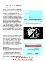
Parapharyngeal abscess
Deep neck space infection secondary to tonsil, or sometimes dental,
sepsis is less common, but needs management in hospital, sometimes
with airway protection by intubation or tracheostomy before surgical incision and drainage and infusion of intravenous antibiotics (see
Fig. 11.2). The commonest variety is parapharyngeal abscess presenting in a severely ill patient with marked unilateral, tender, often red,
neck swelling.
Acute retropharyngeal abscess
This is uncommon and usually secondary to tonsil or adenoid sepsis,
sometimes in the immune compromised (Fig. 11.3). Airway protection before incision and drainage, which may be peroral, is essential. Tuberculosis is a rare cause for chronic retropharyngeal abscess
nowadays in Western countries, but needs to be excluded.
Sore throat with acute airway distress
Acute upper airway distress or obstruction is most commonly inflammatory (Fig. 11.4), usually due to bacterial infection with Haemophilus influenzae. Vaccination programmes have reduced the incidence of this once common paediatric illness, usually manifesting as
acute epiglottitis, but it is still seen quite commonly in adults where
the infection tends to affect the whole supraglottic region. Mild
cases in cooperative adults may be investigated by nasopharyngeal
endoscopy, but there is a risk, particularly in children, of precipitating complete airway obstruction, and interference of any kind
should be avoided – even the use of a tongue depressor or attempts
at imaging. Large doses of intravenous broad-spectrum antibiotics,
usually a third generation cephalosporin, with intravenous steroids
are the management of choice and will usually result in improvement if not complete resolution within 48–72 hours.
The life-threatening nature of this disease cannot be over-emphasized. Rapid deterioration, even in apparently stable patients, may
occur to precipitate critical airway obstruction.
Sore throat with subacute airway
obstruction
In adults, subacute stridor may be caused by neoplasia, most commonly in the subglottic region (see Chapter 19), or rarely due to
bilateral vocal cord palsy from neurological disease, or may be a
complication of thyroid neoplasia or surgery.
Complications of throat sepsis
Quinsy
Peritonsillar abscess (quinsy) presents as a unilateral erythematous
swelling lateral to the tonsil, and in a patient with systemic upset is
the commonest septic complication. Restricted mouth opening is
Sore throat with a chronic upper airway
obstruction
Low-grade upper airway obstruction in adults may be due to neoplasms or rarer conditions such as amyloidosis, sarcoid or vascu-
52
ABC of Ear, Nose and Throat
(a)
(a)
(b)
Figure 11.3 (a) Lateral soft tissue of neck showing widening of
retropharyngeal space and (b) CT scan of the same patient showing abscess
cavity in retropharyngeal space.
Pharyngitis
(b)
Figure 11.2 (a) Tonsillar and parapharyngeal abscess and (b) large neck
abscess secondary to tonsillitis.
litis, notably Wegener’s granuloma. Patients present with noisy
breathing or hoarseness and may be distressed or cyanosed. They
should be referred early for specialist care. Examination with the
flexible nasolaryngoscope is the first step, followed up by endoscopy and biopsy under general anaesthesia when appropriate (Fig.
11.5).
Pharyngitis may be acute and is most often caused by viruses, rhinoviruses, influenza A and B, herpes simplex and zoster and other infections involving pharyngeal lymphoid tissue. The symptoms of pharyngitis may not correlate with the clinical picture on inspection.
Chronic pharyngitis may be specific or non-specific. Non-specific pharyngitis is more difficult to define and diagnose. Patients
usually complain of long-standing discomfort in the throat, pain or
catching on swallowing, and sometimes earache. The observation of
red patches on the posterior pharyngeal wall is not a reliable indication for a firm diagnosis, nor are there any helpful laboratory tests.
There are a number of sources of infection of the lymphoid tissue of
Sore Throats
Figure 11.4 Rare case of diphtheria oropharyngitis.
Figure 11.5 Lingual tonsillar tissue which may present with chronic upper
airway obstruction.
the posterior pharyngeal wall – chronic sinusitis with postnasal pus
irritation from above, chronic bronchitis and bronchiectasis from
the respiratory tract below, as well as laryngo-pharyngeal reflux
from the upper gastrointestinal system. Local irritants from tobacco,
alcohol or industrial fumes are possible causes. Referral is indicated
to exclude patients with primary pharyngeal carcinoma. Chronic
pharyngitis is expressed by the patient as ‘soreness in the throat’,
sometimes associated with catarrh. It is rarely associated with pyrexia or systemic upset. Rarely, if ever, will this symptom respond to
antibiotics, and they should not be given as they are ineffective and
may result in the development of resistance or side effects. Chronic
specific pharyngitis may be associated with specific bacterial organisms: syphilis, tuberculosis, toxoplasmosis, leprosy, and scleroma.
These are recognized by a localized physical abnormality, followed
by biopsy and culture of the tissue. Specific treatment usually resolves the infection, but patients may remain symptomatic from
scarring of local tissues.
Chronic inflammation of the oral mucosa follows ingestion of
irritants (spices, spirits or heavy tobacco use), and may eventually
progress to neoplasia. Inflammation also arises from irritation by
rough, irregular teeth or ill-fitting dentures.
Candidal or fungal infection is commonly seen in the oral cavity of the elderly, and may manifest as redness and soreness of the
mouth, sometimes with angular chelitis. Frequent recurrent fungal infections can occur, and are most commonly seen in denture
wearers as prosthetic teeth can harbour the candidal organisms.
Other patients who may present with fungal infections are those
on steroid inhalers for chronic respiratory airway diseases, and
also patients who are immunocompromised. Severe cases exhibit
white plaques on the oral mucosa, commonly on the soft palate,
but frequently generalized redness is the only evidence. Culture is
diagnostic but the swab must be taken by vigorous rubbing of the
tongue if it is to yield a positive result. Prolonged treatment with
nystatin or clotrimazole, and occasionally with a systemic antifungal agent, is required for eradication. Dentures should be sterilized
daily to prevent reinfection.
Ulceration
Ulceration commonly occurs in the form of aphthous ulcers (Fig.
11.6), which are painful and self-limiting in the course of about 10
days. They have a well-demarcated edge with a white sloughy base
and are usually a few millimetres in diameter. Management is with
topical pain-relieving medication such as ‘Bonjela’ (cetalkonium &
choline salicylate) or ‘Difflam’ (benzydamine hydrochloride). Referral is advisable for further patient management. Topical steroid
preparations are available and may help to provide symptomatic relief. Occasionally, ulcers are larger and more painful and persistent,
in the condition known as major aphthi. The cause is unknown.
Oral ulceration is occasionally seen in serious systemic disease
such as agranulocytosis and vasculitic disorders, and oral mucosal
changes may occur as the manifestation of HIV in the form of ‘hairy
leucoplakia’. The typical ‘punched out’ ulcer of primary syphilis of
the oral cavity is now relatively rare in the developed world, but
Oral mucosal lesions
Mucosal abnormalities generally take the form of altered colour
– generalized redness, or white or red patches. Ulcerative defects,
swellings and occasionally papillomatous lesions also occur. Some
common lesions may be identified from appearance alone, but many
require biopsy for diagnosis, which may usually be achieved under
local anaesthesia.
Inflammation
Acute inflammation of the oral cavity mucosa may arise from contact with irritants, or allergic responses to foodstuffs. Peanut allergy
in children and young adults is increasingly common and manifests
as irritation and swelling of the lips and oral mucosa. Progression to
airway obstruction can develop. Prompt hospital treatment with antihistamine and intravenous steroids is essential to abort this potential
danger.
53
Figure 11.6 Aphthous ulcer.
54
ABC of Ear, Nose and Throat
should be considered as part of the differential diagnosis. The benign incidental finding of ‘geographical tongue’ is often mistaken
for oral ulceration, but is in fact a normal variant. Many of these
conditions and lesions may affect the posterior tongue and, unless
diagnosed early, patients may try all remedies that are available ‘over
the counter’ (and ultimately may become a clinical nuisance).
Neoplasia
Field change of the oral mucosa ranging from chronic inflammation through mild to severe dysplasia is common in association with
heavy tobacco and alcohol usage, particularly in combination, and
may progress via carcinoma in situ to frank squamous carcinoma. Any
persistent white or red patches in the oral cavity, particularly in areas
directly exposed to high concentrations of smoke and particularly
dark spirits, should be regarded as suspicious and submitted to biopsy.
Leucoplakia is easily confused with lichen planus, which is typified by
a lacy white pattern on the mucosa, particularly in the buccal region.
Carcinomatous change is discussed in Chapter 19.
Further reading
Del Mar CB, Glasziou PP, Spinks AB. (2004) Antibiotics for sore throat. The
Cochrane Database of Systematic Reviews, 2, CD000023; DOI: 10.1002/
14651858.CD000023.pub2
Katori H, Tsukuda M. (2005) Acute epiglottis: Analysis of factors associated with Airway Intervention. Journal of Laryngology and Otology; 119:
967–72.
Mckerrow WS. Tonsillitis. BMJ Clinical Evidence, www.clinicalevidence.com
van Staaij BK, van den Akker EH, Rovers MM et al. (2004) Effectiveness of
adenotonsillectomy in children with mild symptoms of throat infections
or adenotonsillar hypertrophy: open, randomised controlled trial. British
Medical Journal; 329: 651–4.
Ridder GJ, Technau-Ihling K, Sander A, Boedeker CC. (2005) Spectrum and
Management of Deep Neck Space Infections: An 8 year Experience of 234
cases. Otolaryngol Head Neck Surg; 133: 709–14.
Scully C, Felix DH. (2005) Oral Medicine: Aphthous and other common ulcers. British Dental Journal; 199: 259 –64.
Scully C, Felix DH. (2005) Oral Medicine: Mouth Ulcers of more serious connotation. British Dental Journal; 199: 339 – 343.
CHAPTER 12
Breathing Disorders
Vinidh Paleri, Patrick J Bradley
OVERVIEW
• Stridor is a symptom NOT a diagnosis, and always requires
examination and investigation. It denotes a harsh, vibratory noise
from turbulent flow through a partially obstructed segment of
respiratory tract.
• All patients presenting with stridor, acute or chronic, must be
investigated urgently.
• The prime aim of managing a patient with stridor is to establish a
secure and stable airway by intubation or tracheostomy.
Box 12.2 Historical information
•
•
•
•
•
•
•
•
•
•
•
Age of onset
Duration/phase of stridor
Worsening/improvement of stridor since onset
Precipitating causes
Failure to gain weight
Breath-holding spells
Fever
Feeding/swallowing problems
Hoarse/muffled voice
Intubation in the past
Cough/chest infections
Pathophysiology
Breathing is involuntarily controlled by the respiratory centre in the
brain stem. The vocal cords abduct during inspiration and, with the
negative pressure caused by diaphragmatic contraction and expansion of the chest, air is drawn into the lungs. The recurrent laryngeal
branches of the vagus nerves control vocal cord movement, with intrinsic laryngeal muscles providing fine control. The cricoid cartilage is the only complete ring in the respiratory tract, surrounding
the subglottic region. Any airway oedema there reduces its lumen.
One millimetre of mucosal oedema reduces the cross-sectional area
by more than 40%.
Stridor is a harsh, vibratory noise from turbulent flow through a
partially obstructed segment of the respiratory tract. This is differentiated from stertor, where noise is caused by vibration of pharyngeal
structures, leading to a lower pitched sound. Stridor can be present
during the inspiratory or the expiratory phase or be biphasic (Box
12.1).
Stridor in children
Evaluation
History provides pointers to the diagnosis (Box 12.2). A previously
Box 12.1 Types of stridor
• Inspiratory: supraglottic and glottic obstruction.
• Expiratory: low tracheal obstruction.
• Biphasic: glottic and subglottic obstruction.
well child presenting with acute onset stridor arouses suspicion of
foreign body aspiration. Preceding upper respiratory tract infection (URTI) indicates croup or bacterial tracheitis. Epiglottitis (supraglottitis) typically presents as rapid onset fever, dysphagia and
drooling in children from 2 to 7 years old.
A child with acute stridor must be assessed where instrumentation and experienced personnel are available for emergency intervention to protect the airway. Clinical assessment is shown in Box 12.3.
Respiratory rate and level of consciousness are the most important
indicators of severity of obstruction. Intensity of the sound does not
indicate severity, as severe obstruction so reduces airflow that stridor
is inaudible. The child must not be upset in case of precipitating
acute obstruction.
Box 12.3 Clinical evaluation
•
•
•
•
•
•
•
•
•
•
•
Respiratory rate
Cyanosis
Apnoeic spells
Use of accessory muscles
Intercostal/sternal retraction
Nasal flaring
Timing/severity of stridor
Hoarseness
Temperature/toxicity
Level of consciousness
ENT examination in controlled setting
55
56
ABC of Ear, Nose and Throat
Noisy breathing
Stertor
Stridor
Adenotonsillar hypertrophy
Macroglossia
Micrognathia
Choanal atresia
Inspiratory or biphasic
Hoarse voice
Afebrile
Vocal cord paralysis
Respiratory papillomatosis
Laryngeal cyst/web
Laryngeal cleft
Expiratory
Normal voice
Febrile
Croup
Supraglottitis
Afebrile
Laryngomalacia
Subglottic stenosis
Subglottic haemangioma
Afebrile
Febrile
Foreign body
Bronchial asthma
Febrile
Bronchiolitis
Retropharyngeal abscess
Figure 12.1 Differential diagnosis of stridor in children.
The probable cause is usually surmised before direct examination
(Fig. 12.1). Most conditions are evolving when first seen, and observation needs intensive care or a high dependency setting.
Congenital structural lesions rarely present acutely. Chronic stridor usually needs diagnostic laryngotracheoscopy unless mild and
easily diagnosed on clinical examination alone. In a cooperative
child with no evidence of hypoxia, flexible laryngoscopy in the clinic
can be very informative.
Acute stridor
Epiglottitis (supraglottitis)
Haemophilus influenzae type B is the usual infective agent. Incidence has decreased with HiB vaccination. Children between 2 and
7 years are affected, with peak incidence at 3. The disease presents
with rapid onset of high fever, toxicity, agitation, stridor, dyspnoea,
muffled voice and painful swallowing. The child sits leaning forward
with mouth open and drooling. If epiglottitis is suspected, no further examination should be performed outside a controlled setting.
The risk of complete obstruction is high. Endotracheal intubation is
preferred as the supraglottic swelling usually subsides in a few days.
A swollen, cherry red epiglottis is seen on direct laryngoscopy. Intravenous antibiotics are essential.
Laryngotracheobronchitis
The most common cause of acute stridor in childhood is laryngotracheobronchitis or ‘croup’. Parainfluenza virus is the commonest cause, with influenza virus types A or B, respiratory syncytial virus and rhinoviruses sometimes being implicated. Children
between 6 months and 3 years are affected, with peak incidence at
2. Symptoms include low-grade fever, barking cough, inspiratory
stridor and hoarseness, worse at night and aggravated by crying.
No endoscopy is needed. Nebulized epinephrine with intravenous
steroids is recommended. Rarely, intubation and ventilation are
necessary.
Acute retropharyngeal and peritonsillar abscesses
Drooling, painful swallowing and systemic upset are usually seen at
presentation, usually with a preceding URTI. Retropharyngeal abscesses in the lower pharynx may cause stridor, neck stiffness and torticollis.
A soft tissue lateral X-ray of the neck shows diagnostic widening of
the space between the vertebral column and the airway. Peritonsillar
abscesses cause trismus and stertor. Urgent drainage is required.
Chronic stridor
Gastro-oesophageal reflux is a problem in children with chronic
stridor – in up to 80% of cases. This is caused by the strong thoracoabdominal pressure gradient of airway obstruction.
Laryngomalacia
This accounts for 75% of all causes of stridor in infants. Weakness
of the supraglottic structures leads to prolapse of the supraglottis
during inspiration (Figs 12.2 and 12.3). It presents as inspiratory or
variable stridor between the fourth and sixth weeks of life. Stridor is
worsened by crying and feeding and is relieved in the prone position.
It is a self-limiting condition.
Subglottic stenosis
This may be congenital or iatrogenic (secondary to prolonged intubation and ventilation) (Fig. 12.4). Symptoms include inspiratory or
biphasic stridor, usually in the first year of life. Iatrogenic stenosis
Breathing Disorders
57
Figure 12.2 Laryngomalacia showing open airway during expiration
(Courtesy Dr H. Kubba).
Figure 12.4 Subglottic stenosis (Courtesy Dr H. Kubba).
Figure 12.3 Laryngomalacia showing epiglottic collapse during inspiration
(Courtesy Dr H. Kubba).
is suspected if stridor presents after extubation. Mild stenoses can be
observed during laryngeal growth. Surgical reconstruction may be
needed.
Vocal cord paralysis
This is usually met within the first month of life with stridor, cyanosis,
apnoea and feeding problems. Concomitant neurological disease,
such as hydrocephalus and Arnold-Chiari malformation, is present in
most patients. Diagnosis is established by rigid endoscopy and assessment of vocal cord mobility. Management depends upon severity and
progression. Spontaneous recovery may take up to 3 years. Tracheostomy may be needed.
Subglottic haemangioma
A capillary haemangioma in the subglottis presents between 6
weeks and 6 months of life (Fig. 12.5). Cutaneous haemangiomas
offer a hint to the diagnosis. Intermittent stridor and a tendency
to recurrent episodes of ‘croup’ are typical. Haemangiomas may
grow for a year, followed by spontaneous regression, so they can be
observed. A tracheostomy may be needed until regression. Other
Figure 12.5 Subglottic haemangioma causing airway compromise (Courtesy
Dr H. Kubba).
treatment options include laser vaporization, excision and systemic
steroids.
Respiratory papillomatosis
This is caused by the human papilloma virus. Transmission can
occur from the mother to the child during labour. Hoarse voice is
the usual presenting symptom, and the airway may be compromised.
Stridor may need urgent debulking of the papillomatous lesions
(Fig. 12.6). Tracheostomy should be avoided as this may provoke
spread of papillomas into the lower airways. Resolution usually
occurs during adolescence. Regular surveillance is needed with debulking or vaporization by a laser as necessary. Addition of topical
cidofovir (an antiviral agent) reduces recurrences.
58
ABC of Ear, Nose and Throat
Figure 12.6 Papilloma on the right true vocal cord.
Evaluation of stridor in adults
Without definite precipitating cause or relevant history, acute and
chronic stridor in adults should be considered neoplastic unless
proven otherwise. A careful history may indicate causes such as
previous thyroid surgery (bilateral recurrent laryngeal nerve injury)
and intubation trauma. Assessment of the extent of hypoxia and the
work of breathing is described in Boxes 12.2 and 12.3. It is possible
to assess the larynx with a flexible nasolaryngoscope and to achieve
a diagnosis in the outpatient setting in most adults.
Bilateral vocal cord palsy
The commonest cause of this condition used to be thyroid surgery,
but now most causes are idiopathic. Voice is preserved, with stridor
most evident on exertion. Flexible laryngoscopy reveals limitation
of abduction of the cords on inspiration. Management includes observation only, a choice of intralaryngeal procedures to increase the
airway at the glottic level, or tracheostomy.
Malignancy
Malignant lesions of the larynx and hypopharynx can present with
stridor due to tumour obstruction of the airway or by causing vocal
cord palsy and oedema. Stridor can also occur after radiation for
laryngeal cancers. It is not always possible to secure the airway before
tracheostomy. Debulking the tumour to improve the airway while
awaiting definitive management is an option. For factors that determine treatment, see Chapter 19. Tumours presenting with stridor
are usually well advanced locally and may need total laryngectomy
for clearance (Fig. 12.7).
Intubation trauma
Intubation for any length of time causes laryngeal inflammation.
Extensive inflammation and ulceration lead to fibrosis and scarring. This usually affects the subglottis. Neonates tolerate intubation for weeks with little long-term harm, but it is reasonable to
consider conversion to tracheostomy after a week to 10 days of intubation in adults if no extubation is planned. Reconstruction of the
stenotic segment is needed in established stenosis.
Figure 12.7 Laryngeal cancer causing complete obstruction of the glottis
with superficial bleeding caused by intubation.
Laryngeal trauma
Blunt and penetrating trauma cause airway obstruction. Other findings include hoarseness, subcutaneous emphysema and haemoptysis. Intubation causes further disruption to the larynx and the airway
is best secured by an urgent tracheostomy.
Angioedema
Angioedema is explained by abnormal vascular permeability beneath the dermis. The causes are shown in Box 12.4. The onset of
oedema can occur within a few hours and can lead to rapid airway
obstruction. Management is primarily medical with epinephrine,
steroids and antihistamines.
Surgical management of the acutely
obstructed airway
Children should be transferred to a centre with medical and nursing
expertise in managing paediatric airway problems, where the airway
is secured in conjunction with a direct laryngoscopy. If endotracheal
intubation is difficult, a laryngeal mask airway or a rigid bronchoscope is used to maintain the airway and ventilate the patient while
tracheostomy is performed. Tracheostomy in children, especially
neonates, is associated with a high risk of complications. If rapid deterioration occurs and there is not sufficient time for a tracheostomy,
a cricothyrotomy can provide emergency oxygenation. In adults,
endotracheal intubation is usually possible. Adult patients with supBox 12.4 Causes of angioedema
The following are possible causes:
• IgE mediated – atopy, allergens, physical stimuli;
• complement mediated – hereditary (production of low or dysfunctional C1 INH*);
• non-immunologic – drug induced (e.g. angiotensin-converting
inhibitors, beta lactam antibiotics);
• idiopathic.
* C1 INH: C1-esterase inhibitor.
Breathing Disorders
59
raglottitis may be observed in a high dependency setting. Obstructive
lesions may need tracheostomy or debulking.
Tracheostomy
Tracheostomy can be used for three reasons: to bypass the upper
airway in airway obstruction, to provide pulmonary toilet and for
access during head and neck surgery. This is performed under general anaesthesia if possible.
Ideally, a horizontal incision is made 2 cm above the suprasternal notch. Dissection proceeds in the midline to separate the strap
muscles and expose the thyroid isthmus, which is ligated and cut.
The tracheal rings are exposed and ‘stay’ sutures inserted, especially
in children. These help with finding the track should the tube become displaced after operation. A vertical slit tracheostomy is made
through the third and fourth rings, and the chosen tracheostomy
tube is inserted (Fig. 12.8). The integrity of the tube and the cuff
must be checked in advance. The tube is secured in place with sutures and tape as necessary. A tube change is performed after 4 to 7
days, allowing time for the track to mature. An uncuffed tube can be
used at this time if there is little concern about significant aspiration.
The cricoid cartilage must not be damaged to avoid stenosis.
Tracheostomy tubes
There are many types of tracheostomy tube, made of PVC, silicone
or silver. A cuffed tube is usually used in the early days after operation, especially in a ventilated patient. This is changed to an uncuffed
tube prior to discharge, unless there are significant problems with
aspiration. This is often seen in patients with neurological disabili-
Figure 12.9 Tracheostomy tubes.
ties. A fenestrated tube with holes on the shoulder allows phonation
when the tube is occluded. Most tracheostomy tubes used in hospital
and community practice have an inner tube protruding just beyond
the outer tube at its distal tip. The longer end of the inner tube picks
up the dried mucus and can be removed for cleaning, while the outer
tube is left in place (Fig. 12.9).
Care of a tracheostomy in the community
Patients who have a tracheostomy for chronic airway obstruction or
pulmonary toilet may be managed at home. Care in the community
needs skilled nursing. A good network of communication needs to
be set up before discharge to ensure that the home is equipped with
suction apparatus, a humidification system, if required, and a supply
of spare tracheostomy tubes. The patient’s family should be taught
about tracheostomy care: how to perform competent suction and
to replace the tube in the event of a blockage. A community physiotherapist and speech and language therapist may also be needed.
Some problems faced in the community, such as narrowing of the
tract and persistent granulations with bleeding around the stoma,
may need specialist ENT advice.
Further reading
Figure 12.8 Total laryngectomy showing end tracheostoma with speaking
valve in place.
Leung AK, Cho H. (1999) Diagnosis of stridor in children. American Family
Physician; 60(8): 2289–96.
Lewarski JS. (2005) Long-term car of the patient with a tracheosotomy. Respiratory Care; 50; (4): 534–7.
Mount J, Uner A, Kaku S. (2004) Pediatric wheezing and stridor. Emergency
Medical Services; 33(7): 55–56, 58–60.
Oberwaldner B, Eber E. (2006) Tracheostomy care in the home. Paediatric
Respiratory Review 7; (3): 185–90.
Yellon RF, Goldberg H. (2001) Update on gastroesophageal reflux disease in
pediatric airway disorders. American Journal of Medicine; 111(Suppl 8A):
78S–84S.
CHAPTER 13
Swallowing Problems
Vinidh Paleri, Patrick J Bradley
OVERVIEW
• Dysphagia is the symptom of swallowing impairment.
T
• Swallowing can be divided into four stages: oral preparatory, oral,
pharyngeal and oesophageal (Box 13.1).
• The majority of the swallowing mechanism is located above the
clavicle.
P
• Weight loss is associated with significant disease or condition,
difficult to reverse, and is usually a late sign.
• Aspiration is defined as liquid or solids penetrating below the
level of the vocal cord and frequently may not precipitate any
symptoms, coughing as a symptom is not reliable.
D
E
G
• Evaluation of the swallowing mechanism should be undertaken
by a multidisciplinary team, not only for diagnosis but for treatment and rehabilitation.
Swallowing and its mechanisms
The pharyngeal stage is controlled at brain stem level (Fig. 13.1). The
soft palate closes against the nasopharynx to prevent nasal regurgitation and laryngeal closure occurs to prevent aspiration. The epiglottis
may play a greater role in directing the bolus into the piriform sinuses
than in protecting the airway. Following laryngeal closure by cord
adduction, the pharyngeal constrictor muscles sequentially contract
to propel the bolus. The suprahyoid muscles raise the larynx, clearing
the bolus down as the cricopharyngeal sphincter opens, admitting it
to the upper oesophagus. The cricopharyngeus relaxes with laryngeal
elevation, which mechanically pulls open the sphincter under bolus
pressure. The oesophageal stage then follows for 8–20 seconds.
Symptoms
Swallowing problems present as difficulty in initiating the swallow,
choking or coughing upon swallowing (aspiration) or a sensation
(a)
(b)
Figure 13.1 (a) Pharyngeal phase of swallowing – early phase. (b) Pharyngeal
phase of swallowing – late phase. D, depression; E, elevation; G, gravity; T,
tongue; +, positive pressure; – negative pressure.
of obstruction in the neck or behind the sternum. Dysphagia may
involve liquids, solids or both. Progressive difficulty with weight loss
suggests malignant disease. Slow progression over years occurs in
achalasia of the cardia and pharyngeal pouches (Zenker’s diverticulum), associated with regurgitation of undigested food. Dry mouth
(xerostomia), caused by autoimmune diseases (Sjogren’s syndrome)
and radiation therapy, also causes dysphagia (Box 13.2).
Box 13.2 Investigations for dysphagia
Primary care:
Box 13.1 Stages of swallowing
The four stages are:
• oral preparatory;
• oral;
• pharyngeal;
• oesophageal.
60
• full blood count;
• contrast swallow.
Secondary care:
• flexible and rigid endoscopy;
• videofluoroscopy;
• flexible endoscopic evaluation of swallowing;
• oesophageal manometry.
Swallowing Problems
Weakness of the oral and lingual musculature leads to drooling
and poor mastication. Altered sensation of the pharynx arising centrally in neurological disease, or peripherally after radiotherapy, delays
initiation of the pharyngeal stage. This may cause aspiration, with
coughing and choking, or a ‘wet’ voice quality. Aspiration also arises
from laryngeal protective mechanism impairment in neurological or
neoplastic processes. Aspiration can also be silent if laryngeal sensation is impaired, with risks of chest infection.
Clinical evaluation
There are good pointers towards the diagnosis in 80% of histories.
Acute dysphagia is most usually caused by the presence of a foreign
body or of a candidal infection. Dysphagia lasting longer than three
weeks needs specialist referral. Otolaryngological examination must
include flexible fibreoptic assessment of the pharynx (Fig. 13.2) and
larynx. Lesions in the apex of the piriform sinus and the postcricoid
region are not always apparent on flexible endoscopy and rigid endoscopy must be used when suspicion is high. Contrast swallows
are useful to identify the presence and size of pharyngeal pouches,
and oesophageal lesions. Videofluoroscopy provides dynamic assessment of the anatomy and coordination of the oral, pharyngeal and
oesophageal stages of swallowing. The flow chart in Fig. 13.3 shows
the role of investigations.
Common otolaryngological conditions causing chronic dysphagia
are shown in Fig. 13.4.
Presbyphagia
Physiological changes occur in the swallowing reflex with ageing.
There is reduction in muscle mass and strength. Presentation is
that of chronic dysphagia with malnutrition and aspiration. Treatment involves modifying the consistency of food, and swallowing
therapy with correction of concurrent contributory factors.
Globus pharyngeus
This is a sensation of a lump or tightness and irritation in the throat,
where no organic cause is identified. It may be an atypical manifestation of gastro-oesophageal reflux or oesophageal dysmotility, or of
psychogenic origin. Presentation is usually in middle age. Diagnosis
is based on the history and lack of findings with no weight loss. Intermittent symptoms are typically between meals, accompanied by
61
a continual urge to swallow. Unless presentation is atypical, further
investigation is unwarranted. Treatment involves reassurance and an
explanation, with anti-reflux therapy. Symptoms can last for up to
2 years and are often recurrent.
Pharyngeal pouch (Zenker’s diverticulum)
There is natural weakness in the posterior aspect of the hypopharynx (Fig. 13.5), between the fibres of the thyropharyngeus and the
cricopharyngeus muscles (upper oesophageal sphincter) of the inferior pharyngeal constrictor. Pulsion diverticula can form at this
site. Various hypotheses of aetiology include poor relaxation of the
cricopharyngeal muscle during swallowing, increased resting tone
of the muscle and myopathy of the cricopharyngeus.
The condition usually arises in the elderly, and presents with progressive dysphagia and weight loss. Symptoms include regurgitation
of undigested food many hours after eating, gurgling sounds in the
neck during swallowing, halitosis, coughing episodes and aspiration.
Endoscopic examination may reveal some pooling of residue in the
hypopharynx, and reflux. Contrast swallow establishes the diagnosis
(Fig. 13.6). Patients are prone to oesophageal perforation during endoscopic examination if a pouch is not recognized pre-operatively.
Treatment depends on the size of the pouch and its symptoms. Small
pouches discovered incidentally need no treatment. The management of larger, symptomatic pouches includes endoscopic stapling
of the party wall between the pouch and the oesophagus to prevent
food from stagnating in the pouch (Fig. 13.7). There is a very small
incidence of malignancy in these pouches and careful telescopic inspection is essential prior to stapling. Difficult exposure due to a
short neck and an anteriorly placed larynx may preclude stapling. An
external excision of the pouch is then needed.
Postcricoid web
This condition is usually found in women in their forties and fifties, in
association with iron deficiency anaemia and weight loss. The onset is
gradual and patients have altered dietary habits to compensate for the
dysphagia. Examination findings may include angular cheilitis and
atrophy of the dorsum of the tongue from iron deficiency (Fig. 13.8).
Contrast X-ray shows a thin filling defect, usually arising on the anterior wall. Early webs may be reversed by iron supplementation, but
the majority need rigid endoscopic dilatation of the web for relief. A
postcricoid carcinoma, unlike other hypopharyngeal cancer, is more
commonly found in younger women, and up to two-thirds have experienced the presence of previous or persistent symptoms of a web.
Neurological diseases
Figure 13.2 Regions of the hypopharynx (purple – piriform sinus, blue
– postcricoid space, green – posterior pharyngeal wall).
Myasthenia gravis, multiple sclerosis, motor neurone disease, muscular dystrophies and other degenerative disorders may affect swallowing. The problem is with initiation of swallow for both solids and
liquids, and symptoms are usually progressive. Swallowing therapy
may be helpful. These patients are prone to aspiration due to diminished sensation of the pharynx and impaired lingual and pharyngeal
constrictor activity. If aspiration is intractable, and the patient is
having difficulty maintaining daily calorific intake, tube feeding (by
gastrostomy) may be considered.
62
ABC of Ear, Nose and Throat
Preliminary examination
Prelimonary
examination
History and preliminary examination
Provisional diagnosis of dysphagia
Diagnosis
No known diagnosis
Oesophageal
Oro-pharyngeal
Oropharyngeal
Investigations
Refer to gastroenterology
Structural
Suspect, for example, neurological problem
Functional
Refer to ear, nose, and throat
Refer to appropriate consultant
Refer to speech and language therapist
Videoendoscopic swallow study
Clinical swallow examination
Further investigation
Barium swallow
Examination under anaesthesia
Videoendoscopic swallow study
Oesophageal manometry
Videofl
uoroscopic swallow
swallow study
study
Videoendoscopic
Flexible endoscopy
Management
Surgery or radiotherapy
Management plan
Reduce risk
Non-oral feed
Modify consistency
Modify posture
Optimise
z hydration and nutrition
Supplements and dietary advice
Figure 13.3 Flow chart showing the evaluation of dysphagia. Reproduced with permission from Leslie et al. (2003) British Medical Journal; 326: 433–6.
Acute onset palsy of the vagus presents with dysphagia, aspiration
(Fig. 13.9) and a breathy voice. The aetiology is often idiopathic. Possible causes are viral neuritis or damage to the neural microvasculature
by underlying systemic causes such as diabetes mellitus. Structural
lesions must be excluded by imaging the course of the recurrent laryngeal nerve from the skull base to the diaphragm for left cord palsy,
and to the superior mediastinum for right cord palsy. Improvement
in symptoms may take place by compensation from the contralateral
cord over a few months. A head turn to the affected side on swallowing can help to reduce aspiration. If aspiration continues with poor
speech, the affected cord can be moved surgically to meet its fellow,
reducing aspiration and improving the voice.
Percutaneous gastrostomy
High-dose radiation therapy to a primary site of cancer-related dysphagia, especially when combined with concurrent chemotherapy,
leads to severe mucositis and restriction in oral intake. Supplementation of feeds may be needed by nasogastric tube or preferably a
gastrostomy. A gastrostomy tube is inserted before treatment under
endoscopic, ultrasound or fluoroscopic guidance without the need
for laparotomy (Fig. 13.10). It is easily performed with minimal morbidity. It is also widely used in neurological practice, when long-term
dysphagia is expected, being more comfortable than a nasogastric
tube and offering the patient greater mobility.
Swallowing Problems
63
Dysphagia
Acute
Painful
Pharyngitis
Tonsillitis
Neck space infection*
Eg: peritonsillar abscess, parapharyngeal abscess
Foreign body
Radiation reaction
h
Infective oesophagitis eg: Herpes
simplex
Chronic
Painless
body
Foreignbody
Neuromuscular*
Vocal cord palsy
Progressive
Oro/hypopharyngeal malignancy*
Oesophageal malignancy
Pharyngeal pouch*
Achalasia cardia*
Benign stricture
Neuromuscular*
Stable/intermittent
Globus phenomenon
Presbyphagia
Postcricoid web
* Can be associated with aspiration
Figure 13.4 Flow chart showing the differential diagnostic options for dysphagia of otolaryngological origin.
Muscle
(a)
Muscle
Mucosa
Mucosa
Muscle
Figure 13.5 Anatomy of pharyngeal pouch.
(b)
(c)
Figure 13.6 Radiological image of a ‘large pharyngeal pouch’.
Figure 13.7 Illustration of how a pouch is stapled (endoscopic
cricopharyngeal myotomy): (a) a prominent cricopharyngeal ‘bar’ muscle, the
oesophagus opening anteriorly and the pouch posteriorly; (b) the method of
stapling, with two parallel rows of three sets of staples, between which the
muscle and the mucosa are divided; (c) the bar has been divided – increasing
the opening into the oesophagus.
64
ABC of Ear, Nose and Throat
Figure 13.8 Angular chelitis and atrophic glossitis.
Figure 13.10 A percutaneous gastrostomy.
gies to improve nutritional status and prevent chest infections or
pneumonia. Common interventions may include head or body postures to improve control and direction of bolus flow, manoeuvres
to improve airway closure and protection or the efficiency of bolus
clearance, exercises to increase the range or strength of movements
of the swallowing musculature and/or dietary modifications to suit
the patient’s abilities.
Further reading
Figure 13.9 Residual coloured contrast remaining in the hypopharynx.
Swallowing therapy
Speech and language therapists may be involved in the management of patients with oropharyngeal dysphagia. Following clinical
assessment, and videofluoroscopy or endoscopic assessment when
indicated, they may recommend management or rehabilitation strate-
Amin MR, Postma GN. (2004) Office evaluation of swallowing. Ear, Nose &
Throat Journal; 83(7,Suppl 2): 13–16.
Hiss, SG, Postma, GN. (2003) Fiberoptic endoscopic evaluation of swallowing.
Laryngoscope; 113(8): 1386–93.
Leslie P, Carding PN, Wilson JA. (2003) Investigation and management of
chronic dysphagia. British Medical Journal; 326: 433–6.
Spieker MR. (2000) Evaluating dysphagia. American Family Physician; 61:
3639–48.
CHAPTER 14
Snoring and Obstructive Sleep Apnoea
Anshul Sama
OVERVIEW
• Snoring, noisy breathing during sleep, is caused by vibration of
one or more areas of the upper airway.
• Such noisy breathing occurs in 45% of the population from time
to time and an estimated 25% are habitual snorers.
• Although snoring is the cardinal symptom of obstructive sleep
apnoea, the prevalence of obstructive sleep apnoea is notably
lower at 0.5–4%.
Spectrum of the condition
Snoring and obstructive sleep apnoea form the opposite ends of a
spectrum of disorders under the umbrella of obstructive sleep-related breathing disorders (SRBDs) (Fig. 14.1). Depending on the degree
of obstruction and associated symptoms, individuals are categorized
into one of the following categories.
Simple snoring is disruptive snoring without any impact on the
patient’s sleep pattern or increased daytime sleepiness. As the obstruction increases, greater respiratory effort leads to increased sleep
disruption and daytime sleepiness. Upper airways resistance syndrome (UARS) is categorized by the presence of these symptoms
without evidence of obstructive apnoea or oxygen desaturation.
There is increased respiratory effort recognized by oesophageal pressure analysis. Further progression of airway obstruction leads to
near total or total obstruction of airflow. Obstructive sleep apnoea
hypopnoea syndrome (OSAHS) comprises excessive daytime sleepiness with interrupted and repeated collapse of the upper airway during sleep, causing oxygen desaturation. Collapse may be complete
with cessation of airflow (apnoea), or partial with significant hypoventilation (hypopnoea).
The frequency of apnoea and hypopnoea is used to grade the
severity of OSAHS as the apnoea/hypopnoea index (AHI), or the
respiratory disturbance index (RDI). OSAHS is mild (5–14 events
per hour), moderate (15–30 events per hour) or severe (more than
30 events per hour). Clinically significant OSAHS is only likely to be
present when the AHI is greater than 15 events per hour, in association with unexplained daytime sleepiness or a minimum of two of
the other features of the condition identified in Box 14.1.
Aetiology
Snoring and obstructive apnoea only occur during sleep. In humans,
the airway between the posterior end of the nose and the larynx is unprotected by cartilaginous or bony structures and is reliant on muscle
tone for its patency. With the onset of sleep, pharyngeal muscle tone
falls progressively as sleep deepens. This phenomenon is present in all
30–50%
0.5–4%
Prevalence
Simple snoring
Diagnosis
Upper airways
resistance
syndrome
Mild obstructive
sleep apnoea
syndrome
Moderate
obstructive
sleep apnoea
syndrome
Severe obstructive
sleep apnoea
syndrome
Site of obstruction
Local/single site
Diffuse/multilevel
Recommended treatment
Surgery
Continuous positive airway pressure
Figure 14.1 Sleep-related breathing disorders – a spectrum of conditions.
65
66
ABC of Ear, Nose and Throat
Box 14.1 Symptoms associated with OSAHS
•
•
•
•
•
•
•
•
•
•
Excessive daytime sleepiness
Impaired concentration
Snoring
Unrefreshed sleep
Choking episodes during sleep
Witnessed apnoeas
Restless sleep
Irritability/personality change
Nocturia
Decreased libido
humans and yet not all snore or have OSAHS. Factors that have been
found to increase the risk of SRBDs are as follows.
• Age. There is a progressive increase in the prevalence of snoring
and obstructive sleep apnoea up to the sixth and seventh decades
of life independent of the body mass index (BMI).
• Sex. Men have between a two- and fivefold increased risk of OSAHS
compared with age- and weight-matched women. The reasons for
the sex difference are unknown. The prevalence of snoring and obstructive sleep apnoea in women increases post-menopause. Oestrogen plus progesterone replacement therapy in post-menopausal
women has been shown to reduce occurrences.
• Obesity. The most important risk factor. The prevalence of respiratory events (RDI) has been shown to directly correlate with
BMI. Seventy per cent of individuals with BMI of 40 or greater suffer with OSAHS. Conversely, up to 50% of patients with OSAHS
may have a BMI below 30. Central obesity indicators such as neck
circumference index and waist to hip ratio are better predictors of
OSAHS than obesity or BMI in general.
• Obstructive upper airway anatomy. Craniofacial abnormalities
are associated with a higher prevalence of SRBDs. These include
abnormalities such as retro- or micrognathia, midfacial or mandibular hypoplasia and macroglossia. Adenotonsillar hypertrophy
is common in children as a cause of snoring and OSAHS. Obstruction of the nasal airway due to turbinate hypertrophy, septal deviations or nasal polyposis increases upper airway resistance. These
contribute to snoring and UARS, but are unlikely to be the sole
cause for OSAHS.
• Social habits. Smoking and alcohol consumption increase the risk
of SRBDs.
• Other risk factors. The relative risk of OSAHS may be two- to
fourfold greater in first degree relatives even after adjustment for
BMI and craniofacial variations. Certain medical conditions such
as hypothyroidism and acromegaly are associated with OSAHS.
Neuromuscular diseases also predispose to OSAHS, although central apnoeas are more likely. Drugs associated with central depression such as hypnotics and opioids increase the risk of SRBDs.
Chronic lung disease does not pose a direct risk for SRBDs. However, in both obstructive and restricted lung disease, OSAHS tends
to be more severe with deeper events of oxygen desaturation resulting from hypoventilation and the lower lung reserve.
Consequences of sleep-related breathing
disorders
Although simple snoring has a significant social impact, it has no
detrimental impact on an individual’s health. However, OSAHS is
known to have important cardiovascular and other consequences.
Neurocognitive effects
Excessive daytime sleepiness (EDS) is the commonest complaint of
patients with obstructive sleep apnoea. Cognitive performance is
notably impaired with deterioration in memory, intellectual capacity and motor co-ordination. There is an increase in accident rates
amongst patients with OSAHS. Sleepiness at the wheel is estimated
to cause 20% of road traffic accidents on major highways.
Cardiovascular consequences
There is compelling evidence that OSAHS is associated and contributes to systemic hypertension. This association is independent
of confounding factors such as obesity, age, gender and alcohol consumption. Furthermore, treatment with continuous positive airway
pressure (CPAP) reduces blood pressure by up to 5 mmHg over
24 hours. There are some data suggesting an association between
OSAHS and coronary artery disease and cerebrovascular events.
Other issues
Patients with OSAHS have been known to have potential problems
with impotence and increased likelihood of gastro-oesophageal reflux.
Assessment
The aims are to:
1 identify if the patient has OSAHS;
2 identify the potential causes and predisposing factors;
3 localize the level(s) of obstruction in the upper airway.
The Epworth sleepiness scale (ESS) is a validated method of identifying EDS. However, the correlation between ESS and OSAHS is
relatively weak and it cannot be used as a screening tool for OSAHS.
Physical examination of the upper airway is essential and is usually
performed by an ENT surgeon. However, there is poor correlation
between the clinical findings and predictability of OSAHS. A formal
assessment of nasal airway and pharyngeal anatomy needs to be undertaken, preferably with an endoscope (flexible or rigid). An assessment should be made of the oropharyngeal inlet including tonsil,
tongue and mandibular size. The possibility of hyperthyroidism,
acromegaly and Marfan’s syndrome should always be considered in
patients presenting with snoring or OSAHS.
Sleep studies
Sleep studies are indicated in all patients presenting with snoring or
suspected sleep apnoea. OSAHS should always be excluded in patients before considering surgery for snoring. OSAHS can be present
in over 30% of snorers presenting without symptoms of overt sleepiness. Patients with chronic obstructive pulmonary disease (COPD)
Snoring and Obstructive Sleep Apnoea
and snoring should have an urgent sleep study as the combination is
potentially dangerous. All patients who drive long distances and/or
heavy goods vehicles, or handle hazardous machinery as part of their
profession, must have a sleep study as part of their assessment. There
are many levels of sleep study depending on the local circumstances.
Polysomnography
Polysomnography (PSG) is the gold standard for diagnosis of OSAHS.
The technique entails an inpatient study involving overnight assessment of a number of measures, including: EEG, electromyogram, electro-oculogram, respiratory airflow, thoraco-abdominal
movement, ECG, oximetry, body position, snoring sound and video.
Clearly, it is a relatively intrusive and costly study whose interpretation can be complex (Fig. 14.2).
67
site specific. Therefore, the efficacy of the treatment is dependent
on accurate localization of the obstruction. Clinical and radiological examinations are poor for localizing the level of obstruction.
Fibreoptic upper airway endoscopy, with or without sedation, is of
limited use, as it is not performed during natural sleep and is unipositional. Upper airway pressure recordings and acoustic reflectometry are promising techniques, although they are not widely
available or practised in the UK.
Behavioural changes
For simple snoring, simple measures, such as allowing the partner to
fall asleep first, using ear plugs, or sleeping on one side rather than
the back, can often suffice.
Weight loss
Treatment options
The choice of treatment is dictated by the following.
• Diagnosis. In moderate to severe OSAHS, the aim is to eliminate
the episodes of apnoeas/hypopnoeas, desaturations and associated
daytime sleepiness. The ideal treatment is CPAP. In the simple
snorer, the aim is a reduction of the duration and intensity of snoring to socially acceptable levels. Lifestyle changes, oral devices and
limited surgery are appropriate. In UARS and mild OSAHS, the
aim is reduction in snoring but also the upper airway resistance
with sleep fragmentation. Most modalities of treatment are appropriate depending on patient choice and the predominant symptom
– snoring noise reduction or sleep disturbance.
• Accurate localization of the level of airway obstruction. Other
than lifestyle changes and CPAP, other modalities of treatment are
Figure 14.2 Polysomnography trace.
Obesity is the single most important factor in increasing upper airway
resistance. Weight reduction has been shown to reduce snoring and
the number of apnoeas and hypopnoeas, and improve sleep efficiency
and oxygenation. The most dramatic results have been reported with
surgical weight loss. It should be recognized that substantial weight
loss by non-surgical means is both difficult to achieve and hard to
maintain.
Lifestyle changes
Patients should be encouraged to stop smoking. Although there is evidence linking smoking with OSAHS, there is no evidence that stopping smoking improves apnoeic events. Alcohol, especially close to
bedtime, exaggerates loss of pharyngeal muscle tone during sleep and
episodes of airway collapse. For similar reasons, sleeping tablets, sedative antihistamines and tranquillizers should be avoided at bedtime.
68
ABC of Ear, Nose and Throat
Continuous positive airway pressure
CPAP applied via a nasal mask has been shown to be the most effective treatment for OSAHS. It may eliminate apnoeas/hypopnoeas
and improve daytime alertness, neurocognitive functions, mood
and cardiovascular sequelae. Unfortunately, it suffers from compliance limitations. One-third of patients offered CPAP are unwilling to use it and nocturnal use averages only 4–5 hours per night.
Compliance can be improved by initial habituation to the mask for
several days before CPAP usage, eliminating oral leakage with chin
straps and heated humidification to reduce nasal dryness, Bi-level
positive airway pressure (BIPAP) or Auto-CPAP to reduce exhalation pressure. The most important factor is supportive and accessible medical staff.
Intra-oral appliance
Several intra-oral devices have been designed to enlarge the pharyngeal airway by moving and fixing the mandible in an anterior position. These are effective in improving snoring and mild OSAHS. Side
effects relating to excessive salivation, jaw discomfort, teeth/gum
discomfort and temporomandibular joint dysfunction affect the majority of patients.
Pharmacological treatment
Drugs used in the treatment of OSAHS are either respiratory stimulants for increasing upper airway muscle tone, or drugs for treating
excessive daytime hypersomnolence. Protryptyline, acetazolamide
and progesterone are respiratory stimulants and suppress rapid eye
movement sleep (when airway collapse is most likely). These drugs
are not curative in the treatment of OSAHS. There is some evidence
to show that the addition of alerting drugs, such as modafinil, may
be beneficial in reducing daytime sleepiness in those who remain
sleepy despite CPAP usage.
Upper airway surgery
The success of upper airway surgery depends on accurate identification of the level/s of obstruction, and effective surgical treatment.
Identification of the level of obstruction has traditionally been based
on clinical assessment and/or investigations under sedation.
Tracheostomy was the first surgical procedure used in the treatment of OSAHS. It is rarely performed today. Current surgical approaches are designed to widen the upper airway: nasal, oropharyngeal or retrolingual. These procedures are usually single site and
non-invasive for simple snoring, and multiple level and invasive for
moderate to severe OSAHS.
Nasal surgery
Nasal disease increases the upper airway resistance, with increased
negative pressure in the pharynx during inspiration. Surgical correction of a deviated septum, removal of nasal polyps and turbinate
reduction can reduce upper airway resistance. However, the reported
impact on snoring is variable (39–87%) and there is relapse after several years. In patients with OSAHS, nasal procedures can improve
compliance with nasal CPAP but they do not improve OSAHS per
se.
Figure 14.3 Traditional uvulopalatopharyngoplasty (UPPP).
Uvulopalatopharyngoplasty
Uvulopalatopharyngoplasty (UPPP) has a long track record for
widening the oropharyngeal airway. The procedure involves tonsillectomy, uvulectomy and excision of a variable segment of the soft
palate (Fig. 14.3). In the appropriately selected group, i.e. suspected
obstruction solely at the level of the soft palate/oropharynx, success rates for simple snoring vary between 75 and 85% and around
50% for OSAHS. It is notable that the success rate decreases with
increasing follow-up periods.
Laser-assisted uvulopalatoplasty
Laser-assisted uvulopalatoplasty (LAUP), although originally developed as a modification of the traditional UPPP, has evolved notably in the last decade. There are many techniques in the literature.
Radiofrequency
Radiofrequency (RF) procedures of the tonsil, palate and tongue
base are based on the principle of submucosal application of low-frequency radiowaves to create thermic lesions and subsequent volume
reduction and scarring (Figs 14.4 and 14.5).
Maxillofacial and multilevel surgery
Maxillofacial and multilevel surgery is usually performed for patients with moderate to severe OSAHS. It includes a range of procedures to improve the retrolingual airway and the retropalatal airway.
These are extremely invasive and their use is limited to patients who
fail to use CPAP.
Snoring and obstructive sleep apnoea in
children
Although OSAHS in children has many similarities with the adult
form, there are some notable differences (Table 14.1). Unlike adults,
the incidence is equal in both sexes and does not increase with age.
The peak occurrence is between the ages of 2 and 5 years, when the
adenoids and tonsils are largest in relation to the oropharyngeal size.
Children with OSAHS frequently show signs of failure to thrive
rather than obesity. Symptoms in children are similar to those in
adults, with the exception of sleepiness. Paradoxically, children often
demonstrate restlessness and hyperactivity. Other potential consequences in children include secondary enuresis.
Snoring and Obstructive Sleep Apnoea
69
Figure 14.4 Minimally invasive radiofrequency
techniques (cautery-assisted uvulopalatoplasty;
CAUP).
(a)
(b)
Figure 14.5 Outcome with (cautery-assisted uvulopalatoplasty; CAUP) palatal surgery. (a) Before. (b) After.
Table 14.1 Differences in characteristics of obstructive sleep apnoea between children and adults
Age
Gender
Weight
Daytime somnolence
Neurobehaviour
Children
Adults
Peak: 2–5 years
Male = female
Usually undernourished
Uncommon
Hyperactive, developmental delay
Increases with age
Male > female
Usually obese
Primary symptom
Cognitive impairment, impaired vigilance
Although OSAHS in children is associated with adenotonsillar
hypertrophy (Fig. 14.6), it is unlikely to be the only cause. Other
anatomical factors that are predisposing to OSAHS include choanal
stenosis/atresia, macroglossia, micrognathia, midface hypoplasia
(e.g. Down’s, Crouzon’s and Apert’s syndromes, achondroplasia) and
mandibular hypoplasia (e.g. Pierre-Robin and Cornelle De Lange
syndrome).
Medical sequelae, such as pulmonary hypertension, systemic
hypertension, cor pulmonale and congestive heart failure, are rare.
Neurobehavioural and developmental consequences are more com-
70
ABC of Ear, Nose and Throat
• those describing daytime sleepiness and those who work with
dangerous machinery or drive for their profession;
• those being considered for surgery for snoring.
• Surgical procedures for SRBD should be guided by the diagnosis
and level/s of obstruction.
• There are notable differences between the adult and childhood
forms of OSAHS.
Further reading
Figure 14.6 Obstructive tonsillar hypertrophy in a child.
mon, including poor school performance, poor learning skills, attention deficit hyperactivity disorder and behavioural problems.
Key points
• SRBDs include a spectrum of conditions from simple snoring to
severe obstructive sleep apnoea.
• OSAHS is associated with systemic hypertension and notable neurocognitive sequelae.
• Patients who must be considered for sleep studies are:
• those with COPD as the combination is potentially dangerous;
Faber CE, Grymer L. (2003) Available techniques for objective assessment of
upper airway narrowing in snoring and sleep apnoea. Sleep and Breathing;
7: 77–87.
Friedman M, Tanyeri H, Lim JW et al. (2000) Effect of improved nasal breathing on obstructive sleep apnea. Otolaryngolology – Head and Neck Surgery;
122: 71–4.
Gozal D. (1998) Sleep disordered breathing and school performance in children. Pediatrics; 102: 616–20.
Hicklin LA, Tostevin P, Dasan S. (2000) Retrospective survey of long-term results and patient satisfaction with uvulopalatopharyngoplasty for snoring.
Journal of Laryngology and Otolaryngology; 114: 675–81.
Marcus CL. (2001) Sleep disordered breathing in children. American Journal of
Respiratory Critical Care Medicine; 164: 16–30.
Miljeteig H, Mateika S, Haight JS, Cole P, Hoffstein V. (1994) Subjective and
objective assessment of uvulopalatopharyngoplasty for treatment of snoring and obstructive sleep apnea. American Journal of Respiratory Critical
Care Medicine; 150: 1286–90.
Ross SD, Allen IE, Harrison KJ et al. (1999) Systematic review of the literature
regarding the diagnosis of sleep apnea. Agency for Health Care Policy and
Research, Rockville, MD. AHCPR publication No. 99-E002, www.ncbi.nlm.
nih.gov/books/bv.fcgi?rid=hstat1.chapter.2 [accessed on 28 April 2003].
Worsnop CJ, Naughton MT, Barter CE et al. (1998) The prevalence of obstructive sleep apnoea in hypertensives. American Journal of Respiratory Critical
Care Medicine; 157: 111–15.
CHAPTER 15
Hoarseness and Voice Problems
Julian McGlashan, Declan Costello, Patrick J Bradley
OVERVIEW
• Hoarseness or dysphonia is a symptom of altered laryngeal function and merits investigation and treatment.
• The causes of hoarseness include; structural and neoplastic,
inflammatory, neuromuscular and muscle tension imbalance, and
most patients have a combination of causes.
• Patients who smoke and are hoarse only, inspection of the larynx
will always identify the cause, a chest x-ray is indicated when
patients have additional symptoms – dyspnoea, chest pain or
haemoptysis.
• Voice disorders should be investigated and managed by a multidisciplinary team and may require many tests including stroboscopy and voice analysis.
The need to use the voice (Box 15.1) for prolonged periods, especially at raised intensity levels, increases the risk of dysphonia. The
commonest voice complaints are outlined in Box 15.2.
Box 15.2 Commonest voice complaints
• Change in voice quality (hoarseness, roughness and breathiness)
• A deeper or higher pitched voice that is not appropriate for the
age and gender
• Problems controlling the voice, described as pitch breaks, squeaky
voice or the voice cutting out
• Difficulty making oneself heard in a noisy environment or in raising
the voice
• Efforts in producing voice
• Reduced stamina of voice, which tires with use
• Difficulties or restrictions in the use of voice at different times of the
day or related to specific daily, social or occupationally-related tasks
• Reduced ability to communicate effectively
• Difficulty in singing
• Throat-related symptoms (soreness, discomfort, aching, dryness,
mucus) particularly related to voice use
• Emotional and psychological aspects of the above
Aetiology of voice problems
Anatomy, physiology and pathophysiology
The vocal cords are attached anteriorly to the thyroid cartilage and
posteriorly to a pair of arytenoid cartilages, perched on the superior
rim of the cricoid cartilage. The intrinsic laryngeal muscles abduct
the cords for respiration and adduct the cords for lower airway protection, coughing and phonation (Fig. 15.1). The vocal cords are
folds of mucosa and are better called vocal folds. They consist of a
superficial epithelial layer separated from the underlying ligament
and muscle by the so-called Reinke’s space. This allows the epithelial
layer to slide and oscillate passively over the ligament (Fig. 15.2). The
larynx is divided into three regions: supraglottis, glottis and subglottis (Fig. 15.3).
Box 15.1 Definition of a normal voice
A pragmatic definition of a normal voice is one that is:
• audible in a wide range of acoustic settings;
• appropriate for the gender and age of speaker;
• capable of fulfilling its linguistic and paralinguistic functions;
• not easily fatigued;
• not associated with phonatory discomfort or pain.
Voice problems are classified as: structural/neoplastic, inflammatory,
neuromuscular and muscle tension imbalance. Many patients have
evidence of more than one. A vocal fold polyp (Fig. 15.4), for instance
(structural/neoplastic cause), may arise as a result of primary muscle tension from voice abuse, such as shouting, during viral upper
respiratory tract infection (inflammation). This polyp can cause secondary trauma to the vocal folds (inflammation) and muscle tension
imbalance. An important part of the assessment of a patient is the
determination of which of these conditions are present, which are
primary and which are secondary, and which cause the complaint.
Guidelines and referral
Examination of the larynx is rarely possible by the general practitioner. As laryngeal visualization is the key examination, most patients need to be referred. Patients with persistent hoarseness or
change in voice for 3 weeks, especially smokers and heavy drinkers
aged 40 years or older, need urgent chest X-rays to exclude recurrent laryngeal nerve palsy from lung cancer, and referral to an ENT
surgeon. Positive X-ray findings indicate urgent referral to a lung
cancer specialist team. Negative X-ray findings with upper aerodi71
72
ABC of Ear, Nose and Throat
gestive tract symptoms (Table 15.1) demand referral to a head and
neck cancer specialist team. Others should be seen in a specialist
voice disorders clinic.
Assessment
Comprehensive assessment of a voice problem includes the items
listed in Box 15.3.
The first aim is to identify the presence of the main aetiological factors (Fig. 15.5). History details are outlined in Table 15.2. It
is essential to decide whether the voice is rough, breathy, weak or
strained, its appropriateness for the age and gender of the patient,
whether the voice is too loud or too soft and whether vocal character
is variable.
A flexible endoscope allows inspection of the vocal tract from
nasal cavity to larynx. It is the preferred technique in neuromuscular
and muscle tension imbalance disorders. A rigid endoscope placed
on the tongue provides higher quality, with larger images, and is better
for examining the vocal fold structure and function. The use of stroboscopic light improves the accuracy of diagnosis. Recording images
digitally means that they can be stored, and recalled for slow motion
playback (Fig. 15.6). Neck masses must be sought, and abnormalities
of shape and movement of the laryngeal cartilages must be identi-
(a)
Regions of larynx
Supraglottic
(b)
Figure 15.1 Vocal folds in abduction for respiration (a) and adduction for
phonation (b).
Glottic
Subglottic
Epithelium
Reinke’s space
Figure 15.3 Sagittal and coronal views of the larynx showing regions.
Vocal ligament
Inflammation
Neoplastic/structural
Muscle tension imbalance
Neuromuscular
Vocalis muscle
Mucus gland
e
Conus Elasticus
Figure 15.2 Cross-section of the vocal cord (fold) showing layered structure.
Figure 15.4 Example of structural abnormality: vocal polyp.
Hoarseness and Voice Problems
Table 15.1 Symptoms and signs of laryngeal and lung cancer
Laryngeal cancer (UK incidence of Lung cancer (UK incidence of
3.7/100 000; Cancer Research UK) 63.9/100 000; Cancer Research UK)
• Hoarseness:
• Haemoptysis or any of the
• Rough voice due to vocal cord
following unexplained persistent
lesion
(>3 weeks) symptoms and signs:
• Hoarseness
• (A subtle change in voice may be
only symptom)
• Weak breathy voice due to vocal
• Unexplained lump in neck recently
cord palsy
appeared or changed over 3–6
• Cervical and/or supraclavicular
lymphadenopathy
weeks
• Unexplained pain in head and neck • Dyspnoea
region
• Chest and/or shoulder pain
• Referred otalgia (normal otoscopy)
• Weight loss
• Unexplained persistent sore or
• Finger clubbing
painful throat
• Cough with or without any of
• Other:
the above
• Inspiratory stridor
• Features suggestive of
• Haemoptysis
metastases from lung cancer
• Dysphagia
73
Box 15.3 Requirements for comprehensive assessment of a
voice problem
Requirements are:
• detailed clinical history;
• perceptual evaluation of the voice quality;
• flexible nasoendoscopic examination of vocal tract for (a) structure
and (b) function;
• videolaryngostroboscopic assessment of vocal fold vibration;
• neck inspection and palpation for:
• lymphadenopathy and other masses;
• increased muscle tension and imbalance;
• larynx position in neck and relationship of the thyroid and
cricoid cartilages;
• neck, back and overall body posture;
• breathing patterns during speech;
• neurological assessment;
• psychological assessment.
Further investigations
• diagnostic microlaryngoscopy;
• quality of life measures using validated questionnaires;
• objective measurements of vocal function, e.g. acoustic, electrolaryngographic and aerodynamic measures, electromyography;
• 24-hour dual-site pH monitoring with impedance;
• oesophagogastroduodenoscopy.
fied. Tenderness of the extralaryngeal muscles may indicate increased
muscle tension.
Electromyography is helpful in detecting paresis and in distinguishing cricoarytenoid joint fixation from vocal cord palsy. Microlaryngoscopy is helpful in cases of persistent chronic laryngitis or
when outpatient laryngoscopy is not possible. Laryngopharyngeal
reflux (Fig. 15.7) can be investigated by 24-hour pH measurements
with probes coupled to oesophageal impedance measurements.
leucoplakia (white mucosal lesions) and many cases of papillomas,
polyps and vocal cord palsy (Fig. 15.8). Other structural lesions may
require surgery. Post-operative voice therapy may also help.
Treatment
Structural/neoplastic conditions
Treatment options (Table 15.3) depend on the diagnosis, reasons
for seeking referral, vocal requirements and risk/benefit of intervention. If the diagnosis is not initially clear, a trial of simple vocal
hygiene measures, medical treatment or voice therapy can be tried.
Primary surgery is performed for suspected neoplastic lesions,
The voice depends on the effects of the lesion on vocal fold vibration
and glottic closure. If the vocal fold vibration is disorganized, there
will be a rough voice, whereas, if glottic closure is incomplete, the
voice may be breathy. Superficial mucosal lesions may be congenital
(sulcus vergeture), secondary to inflammation (cysts, sulcus vocalis,
Malignant
Infective
Non-infective
Hyperfunction
Structural/
neoplastic
Inflammation
Primary
Neuromuscular
Muscle tension
imbalance
Mixed
Secondary
Hypofunction
Control
Figure 15.5 Aetiological categories of
voice disorders and their interactions (With
acknowledgement to Professor J. Koufman).
Reassurance
and
education
VHA/dietary
and
lifestyle
advice
Benign
Treatment options
Medical
treatment or
medication
review
Voice
therapy
Specialist
therapy
Surgery
Other
medical
specialist
74
ABC of Ear, Nose and Throat
Table 15.2 Important points in patient history
• Voice problem, e.g. abnormal quality, • Past and current medical history including conditions
affecting the respiratory, gastro-oesophageal, neurological,
pitch, loudness, loss of voice function
musculoskeletal and endocrine systems
• Onset and duration
• Psychological and psychiatric conditions
• Constant/intermittent
• Effects of drugs*:
• Relieving and exacerbating factors
• Reduced laryngeal secretions and mucosal drying, e.g. anti• Voice requirement in terms of fine
cholinergics, diuretics
precision and control, voice projection
• Irritant, e.g. bronchial inhalers
and continuous periods of use
• Androgenic , e.g. danazol
• Social history, hobbies and lifestyle
• Predisposition to infection, e.g. candidosis from steroid inhalers
factors
• Predisposition to gastro-oesophageal reflux
• Smoking and alcohol consumption
• Central nervous system, e.g. Parkinsonism secondary to anti• Dietary habits
psychotics
• Caffeine and fluid intake
*Frequently prescribed medications and effects on voice and speech can be found at www.ncvs.org/ncvs/info/
vocol/rx.html
Figure 15.6 A series of images obtained using laryngostroboscopy taken at eight phases of the vibratory cycle and demonstrating the opening and closing of
the glottis during phonation due to the mucosal wave motion.
Figure 15.7 Marked laryngeal inflammation secondary to laryngopharyngeal
reflux.
mucosal bridges, granulomas), due to surgical trauma (intubation,
granulomas, scarring), or due to physical damage by voice abuse with
inflammation (nodules, ectasia, polyps, pseudocysts, Reinke’s oedema) (Fig. 15.9). Androgens can cause the female larynx to enlarge,
and hypothyroidism can cause vocal fold thickening. The vocal fold
and false cords may be distorted by neoplasms, deformities of the
laryngeal skeleton or external trauma (Table 15.4).
The diagnosis usually follows videolaryngostroboscopy or microlaryngoscopy. In some cases, adequate improvement in vocal function is achieved with vocal hygiene, voice therapy and treatment of
inflammation. Surgical resection is often required and microlaryngoscopy and biopsy should always be performed where malignancy
is suspected.