Ebook Oxford textbook of critical care (2/E): Part 2
Bạn đang xem bản rút gọn của tài liệu. Xem và tải ngay bản đầy đủ của tài liệu tại đây (27.66 MB, 956 trang )
SECTION 7
Nutrition
Part 7.1Physiology 950
Part 7.2 Nutritional failure 960
PART 7.1
Physiology
201 Normal physiology of nutrition 951
Annika Reintam Blaser and Adam M. Deane
202 The metabolic and nutritional
response to critical illness 956
Linda-Jayne Mottram and Gavin G. Lavery
CHAPTER 201
Normal physiology of nutrition
Annika Reintam Blaser and Adam M. Deane
Key points
◆ Ingested
carbohydrate, glycogenolysis, and gluconeogenesis
are essential for function of brain and anaerobic tissues that
depend on glucose as their main energy source.
◆ Fat
is the most energy-rich nutrient, but most of ingested
lipids will be stored in adipose tissue because the oxidative
capacity for lipids is low.
◆ During
periods of inadequate energy delivery, ingested or
endogenous proteins are diverted into glucose metabolism,
and this provides a rationale to deliver more protein during
these periods.
◆
Basic metabolic rate (BMR) is the largest component of total
daily energy requirements, even in the case of very high physical activity or acute illness.
◆ Daily
energy requirements range from 1800 to 2800 kcal/
day or 25 to 30 kcal/kg body weight (BW)/day roughly—
carbohydrates should provide 55–60%, lipids 25–30%, and
proteins 10–15%.
Body composition
Water (approximately 60% in adult males and 50% in females [1]),
protein, minerals, and fat are the main components of human
body. Essential fat is contained in bone marrow, the heart, lungs,
liver, spleen, kidneys, intestines, muscles, and central nervous
system. Fat located in adipose tissue is called storage fat. The
two-component model distinguishes between fat and fat-free mass
(FFM), while the three-component model further divides FFM
into body cell mass and extracellular mass [2]. Lean body mass
is an indirect estimation of the weight of bones, muscles, ligaments, and internal organs, which can be calculated using various
equations [3].
Direct methods of assessing body composition, such as skinfolds,
bioelectrical impedance analysis, and hydrostatic weighing are not
routinely used in the critically ill.
While being underweight is associated with poorer outcomes in the
critically ill, obesity does not appear to be harmful (and may be
beneficial) [4].
Estimation of the ideal body weight [5]is often inaccurate, but
the range may be useful and can be calculated (Broca’s index):
(Height (cm) − 100) ± 15% for women or 10% for men [eqn 1]
Clinical examination and laboratory tests
General clinical examination of skin, hair, eyes, gums, tongue,
bones, muscles, and thyroid gland tends only to reveal signs in
cases of marked malnutrition or vitamin/mineral deficiencies.
Laboratory tests such as blood haemoglobin, total lymphocyte
count, glucose, serum albumin, prealbumin, transferrin, total
protein measurements, and calculation of nitrogen balance have
limitations, but have been used. Nitrogen balance is considered the
most dynamic nutritional indicator.
Essential nutrients: substrate
and energy metabolism
Essential nutrients are substances that are not synthesized (or are
synthesized in too small amounts) within the body and must,
therefore, be ingested or administered. They include essential fatty
acids, essential amino acids, vitamins, and dietary minerals.
Energy
Energy is derived from three major categories of macronutrient—
protein, carbohydrate, and lipids—and is released by breaking
down carbon–carbon bonds created in plants via photosynthesis.
Energy requirements to maintain stable weight can be estimated,
using calculations or measured using calorimetry.
Oxidative (burning for energy) and non-oxidative (storage,
synthesis) substrate metabolism occur to a different extent according to the type of macronutrient and state of energy stores [2].
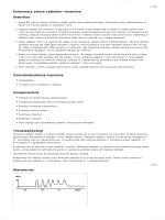
Respiratory quotient (RQ) is used to describe oxidative substrate
metabolism.
Estimation of nutritional status
Carbohydrates
Body mass index (BMI) = weight (kg)/height (m) and provides a
rough estimation of nutritional status [2]. A limitation of BMI is
that the calculation does not distinguish between muscle and fat
mass. A BMI of 18.5–24.9 kg/m2 is considered ‘normal’ regardless
of age or population [1]. BMI has a U-shape relation to morbidity and mortality [2]. In persons >60 years old being slightly heavier (BMI 26–27) is associated with the longer life expectancy [2].
Carbohydrates are compounds comprised of carbon, hydrogen, and
oxygen. Depending on the composition of these molecules, carbohydrates are divided into mono-, di-, oligo-, and polysaccharides.
While carbohydrates are non-essential nutrients, they comprise a
substantial proportion of calories (4.1 kcal/g) in a normal diet [1].
In plants, carbohydrate is stored as starch, whereas in animals it is
stored as glycogen.
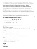
Table 201.1 Nutritional requirements in adults
Nutrient
Role
Daily needs
Deficiency
Abnormalities in ICU
Macronutrients
Energy, structure, all functions
No absolute daily
requirements exist
Starvation
Carbohydrates
Major energy source, energy
storage and transport, structure
55–60% of calories,
max. 4 g/kg BW at rest
Hypoglycaemia, ketoacidosis
Hyperglycaemia
(insulin resistance,
counter-regulatory
hormones)
Lipids
Energy storage, cell membrane
structure, signalling molecules
25–30% of calories
Malabsorption of fat-soluble
vitamins
Hyperlipidaemia with
excessive parenteral
nutrition and propofol
Proteins
Enzymes, structure, signalling,
immune response
10–15% of calories
0.8 g/kg BW
Kwashiorkor, cachexia
Protein energy wasting
occurs frequently
Vitamin
Role
RDA
Deficiency
In ICU
A (retinol)
Retinal pigment
Male 1000 µg female 800 µg
Night blindness, follicular
hyperkeratosis
B1 (thiamine)
Coenzyme in decarboxylation of
pyruvate and alpha-keto acids
Male 1.5 mg, female 1.1 mg
Beriberi, Wernicke’s
encephalopathy
Threshold for thiamine
administration should
be low
B2 (riboflavin)
Coenzyme for oxidative enzymes
Male 1.7 mg, female 1.3 mg
Mouth ulcers, normocytic
anaemia
B3 (niacin) also vitamin
PP or nicotinic acid
Coenzyme, precursor for NAD
and NADP
Male 19 mg, female 15 mg
Pellagra, neurological symptoms
B6 (pyridoxine)
Coenzyme in synthesis of amino
acids, haeme, neurotransmitters
Male 2.0 mg, female 1.6 mg
Muscle weakness, depression,
anaemia
B12 (cobalamin)
coenzyme (deoxyribonucleotids),
formation of erythrocytes, myelin
2 µg
Neurological symptoms
With pernicious anaemia
(lack of intrinsic factor)
C (ascorbic acid)
Cofactor in collagen synthesis
60 mg
Scurvy
Antioxidant effect, cave
oxalosis
D (1,25-cholecalciferol)
Ca2+ absorption and metabolism
5–10 µg
Rickets
E (alpha-tocopherol)
Antioxidant
Male 10 mg, female 8 mg
Peripheral neuropathy
Antioxidant effect
K
Clotting, synthesis of
prothrombin, factors VII,IX,X
Male 70–80 µg
female 60–65 µg
Coagulopathy
Biotin (also vitamin B7
or H, or coenzyme R)
Cofactor for several carboxylase
enzymes; cell growth
30–100 µg
Neurological symptoms, alopecia,
conjunctivitis, dermatitis
Folate (vitamin B9)
Necessary for synthesis of DNA,
haemopoesis
Male 200 µg female 180 µg
(pregnancy 400)
Megaloblastic anaemia,
peripheral neuropathy, neural
tube defects
Panthothenic acid
(vitamin B5)
Synthesis of CoA. Carbohydrate,
and fat metabolism
4–7 mg
Gastrointestinal and neurological
symptoms
Mineral
Role
RDA
Deficiency
In ICU
Calcium
Bone, intracellular signalling
800–1200 mg
Osteoporosis, arrhythmia
hypertension
Chromium
Cofactor
50–200 µg
Impaired glucose tolerance,
peripheral neuropathy
Deficiency reported
during long-term
parenteral nutrition
Copper
Cofactor (oxidative
phosphorylation,
neurotransmitter synthesis etc.)
1.5–3 mg
Myelodysplasia, anaemia,
leucopenia, neurological
symptoms
Deficiency after
gastric bypass. Reduce
replacement in liver failure
Iron
Haemoglobin and cytochromes
Male 10 mg, female 15 mg
Microcytic anaemia, mucosal
atrophy
(continued)
Chapter 201
normal physiology of nutrition
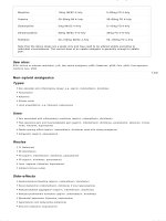
Table 201.1 Continued
Nutrient
Role
Daily needs
Deficiency
Abnormalities in ICU
Iodine
Thyroid hormones
150 µg
Goitre, hypothyroidism
Magnesium
Complex with ATP
Male 350 mg, female 280 mg
Muscle weakness, GI, and cardiac
symptoms
Deficiency common and
associated with major
adverse outcomes
Manganese
Antioxidant
2–5 mg
Reduces replacement in
liver failure
Molybdenum
Cofactor
75–250 µg
Liver dysfunction
Phosphorus
Major component of the
skeleton, nucleic acids, and ATP
800–1000 mg
Potentially catastrophic
reduction during refeeding
syndrome
Potassium
Membrane potential
At least 3510 mg (conditional
recommendation by WHO)
Arrhythmias
Often life-threatening
hyper- or hypo-K, narrow
therapeutic/normal range
Selenium
Antioxidant
Male 70 µg, female 55 µg
Cardiomyopathy
Antioxidant effect
Zinc
Antioxidant, cofactor
Male 15 mg, female 12 mg
Skin lesions, loss of appetite
Antioxidant effect
RDA, Recommended daily allowance.
Adapted from a table published in Medical Physiology, Second Edition, Boron WF and Boulpaep EL, Copyright Elsevier 2012.
As an energy source, glucose is oxidized to CO2 and water:
C 6H12O6 + 6O2 + 32ADP + 32P − > 6 CO2 + 6H2O
(
+ 32ATP + heat RQ = 6CO2 / 6O2 = 1.0
)
[eqn 2]
At rest, the maximum oxidative capacity is approximately 4 g glucose/
kg BW/day [2]. If the glucose intake is greater non-oxidative metabolism occurs, resulting in glycogenesis (glycogen store is limited to
200–500 g; storing consumes about 6% of energy stored in glucose)
and, after reaching the limit, in lipogenesis (storing costs 23% of
energy) [6]. As an isolated energy source blood glucose covers energy
needs for about 30 minutes, whereas glycogen would last for approximately one day [2]. Glycogen is stored in hydrated form, making it less
energy-efficient, but easily available. Hepatic or muscle glycogenolysis
occurs rapidly in response to hypoglycaemia or anaerobic demands.
Gluconeogenesis is the synthesis of glucose from non-hexose precursors (lactate, pyruvate, intermediates of the citric acid cycle, 18 of
20 amino acids and glycerol) [1]. Leucin and lysine together with fatty
acids are not gluconeogenic, but ketogenic. Their breakdown-product
is acetyl coenzyme A (CoA), which cannot generate pyruvate or
oxalo-acetate. Gluconeogenesis is essential for the brain and anaerobic tissues (blood cells, bone marrow, renal medulla) that depend
on glucose as their main energy source [1], but is energy-expensive,
consuming 24% of energy contained in amino acids (AA) [6].
Lipids
Lipids are hydrophobic compounds that are soluble in organic solvents such as acetone.
Lipids contain 9.4 kcal/g of energy and can be ingested as triglycerides, sterol esters or phospholipids [1]. To generate energy, fatty
acids are oxidized to CO2 and water.
C15H31COOH ( palmitic acid ) + 23O2
+ 106ADP + 106P −> 16CO2 + 16H2O
(
+ 106ATP + heat RQ = 16CO2 /23O2 = 0.70
)
[eqn 3]
In resting humans the oxidative capacity for lipids is 0.7 g/kg BW/
day [2]. Greater amounts of ingested lipid will be stored as triglycerides in fat tissue. Fat constitutes approximately 20% of body
weight, and the standard triglyceride store has the capacity to cover
the body’s energy requirements for about 2 months.
Ketone bodies are produced when accelerated oxidation of fatty
acids leads to incomplete breakdown, producing acetyl CoA faster
than the citric acid cycle can utilize it [1]. Ketone bodies (acetoacetate, β-hydroxybutyrate, and acetone) may serve as an alternative
energy resource, e.g. during starvation up to 50% of brain energy
demands might be met via ketone bodies [2].
Protein
Nitrogen differentiates protein from carbohydrates and fats. The
major source of endogenous protein is muscle, which is converted
into energy via complex metabolic pathways. When AA, either
endogenous proteins or ingested, are metabolized to CO2 and water,
4.3 kcal/g of energy can be released [1]. Protein metabolism in cells
additionally results in the production of energy-containing metabolites (urea, ureic acid, and creatinine). The RQ for protein oxidation
is 0.80–0.85 [1]. Protein stores are about 14% of body weight, but
only half of it is available as an energy source, lasting for 10 days
approximately. In health, protein catabolism contributes less than 5%
of energy requirements, but this increases to 15% during starvation.
The body constantly breaks down proteins to AAs and synthesizes
other proteins according to the current needs of the body (protein
turnover). Nine out of 20 AAs are essential—the body cannot synthesize them at sufficient rates for long-term survival and they must
be ingested to replace the proteins oxidized during daily turnover.
Excess protein is converted to glycogen or triacylglycerols [1].
To maintain nitrogen balance in an average adult individual,
ingestion of 0.6–0.8 g/kg BW/day of protein is needed [2]. However,
during periods of inadequate energy delivery greater amounts
of protein are diverted into glucose metabolism and, in catabolic
states, there is a marked increase in endogenous protein breakdown. Accordingly, more protein either ingested or administered
may be beneficial during these periods [7].
953
954
Section 7
nutrition: physiology
Nitrogen (N) balance is the sum of protein degradation and protein synthesis, reflecting the changes in protein stores where:
N Balance = N int ake − N losses,
N intake = protein intake ( g/day ) /6.25, and
N losses = urinary urea N ( UUN ) , g/day, determined
from a 24-hour urine collection) + 4 g miscellaneous
other N losses from skin, mucosa and with faeces.
[eqn 4]
N balance is used to estimate current protein requirements.
Positive or negative N balances indicate anabolic or catabolic states
respectively.
As in patients with renal replacement therapy (RRT) measurement of UUN is not applicable; the total nitrogen appearance
(TNA) is used to express nitrogen losses [8]:
urea nitrogen loss +
TNA in a patient with RRT =
oss during RRT ( g )
AA nitrogen lo
change in interdialytic blood urea
+
×
nitrogen
BUN
g/L
total
body
water
L
(
)( )
( )
[eqn 5]
Protein energy wasting [9]due to inadequate protein intake and
high catabolism is thought to occur frequently in the critically ill.
While preventing or limiting protein deficiency in this group is
often proposed as beneficial, it is not yet established that such an
approach improves outcomes.
Other
Other essential nutrients include inorganic elements like calcium,
potassium, iodine, iron, trace elements (dietary minerals that are
needed in very small quantities) and vitamins, which are necessary
for normal functioning of the body. The role, recommended daily
allowances and signs of deficiency of these essential vitamins and
minerals are presented in Table 201.1.
Vitamins
Vitamins are divided into water-soluble (B,C) and fat-soluble (A,
D, E and K) groups. Bonds between fat-soluble vitamins with
proteins are broken by the acidity of gastric juice and proteolysis.
Assimilation of fat-soluble vitamins relies on lipid absorption and
their deficiency occurs in various fat malabsorption states.
Thiamine (B1) with its phosphorylated derivatives plays a fundamental role in energy metabolism and is used in the biosynthesis
of the neurotransmitters. Its best-characterized derivate thiamine
pyrophosphate is a coenzyme in the catabolism of sugars and amino
acids. Thiamine derivatives and thiamine-dependent enzymes are
present in all cells of the body, but the nervous system and the heart
are particularly sensitive to its deficiency. Thiamine deficiency
may occur because of concomitant chronic disease or alcoholism
and can lead to severe neurological impairment and contribute to
increased mortality [10].
Vitamin K is pivotal for synthesis of coagulation factors VII, IX,
X, protein C and protein S in liver, acting as a co-factor for carboxylation. Its deficiency occurs with fat malabsorption, but also during
severe bleeding (disseminated intravascular coagulation). Previous
treatment with vitamin K antagonists, blocking carboxylation of
prothrombin (factor II), factors VII, IX and X, and making their
complexes with Ca2+ and therefore usage for coagulation impossible, is common in hospitalized patients.
Supplementation of vitamins E and C has been proposed as having beneficial antioxidant effects in the critically ill [11]. This has
yet to be established and particularly in patients with renal failure
excessive vitamin C may lead to oxalosis.
Minerals
Calcium is necessary for the structure (calcium phosphate in
bones), signalling, and enzymatic processes (co-enzyme for clotting factors, pre-synaptic release of acetylcholine) in the body.
Magnesium is essential for energy in every cell type in organism, as ATP, the main source of energy in cells, must be bound to a
magnesium ion in order to be biologically active.
Supplementation of minerals such as selenium and zinc has been
described in the critically ill and warrants further study [12].
Next to the essential nutrients food includes fibres and other ballast substances, carotinoides, bioflavonoids etc. considered important for health, but their functions are clarified incompletely.
Energy consumption
Estimation of energy consumption
Basal metabolic rate (BMR) is an estimation of metabolism,
measured under standardized conditions in the absence of stimulation. Resting metabolic rate (RMR) is measured during less
strict conditions and is therefore higher than BMR [1]. BMR
and RMR are measured by gas analysis through either direct
(the body is positioned in a chamber to measure the body’s
heat production) or indirect (CO2 production is measured)
calorimetry [13].
BMR can be estimated by a number of calculations of Basal
Energy Expenditure (BEE) with Harris-Benedict equation being
the most frequently used in critical care:
Adult males:
(
)
)
[eqn 6]
) (
)
BEE ( kcal/day ) = 13.8 × weight in kg
(
+ 5 × height in cm − (6.8 x age ) + 66.5.
Adult females:
(
BEE ( kcal/day ) = 9.6 × weight in kg + 1.8 × height in cm
− 4.7 × age + 655.
[eqn 7]
(
)
As body weight is the major factor that determines BEE, a simplified estimate of 25 kcal/kg BW/day is often used and our experience is that the latter approach is adequate for clinical purposes.
The BMR is the largest component of total daily energy requirements, even in case of very high physical activity, as well as in the
most hypermetabolic patients. The various estimations of stress/
activity factors available to calculate the total energy expenditure
(TEnE = BEE × stress/activity factor) tend to overestimate the
TEnE in ICU patients, as measured TEnE is often close to calculated BEE [6].
Chapter 201
References
1. Boron WF and Boulpaep EL. (2012). Medical Physiology, 2nd edn.
Philadelphia: Saunders.
2. Speckmann EJ, Hescheler J, Köhling R. (2008). Physiologie, 5th edn.
Munich: Elsevier.
3. R. Hume. (1966). Prediction of lean body mass from height and weight.
Journal of Clinical Pathology, 19, 389–91.
4. Heyland DK, Dhaliwal R, Jiang X, and Day AG. (2011). Identifying
critically ill patients who benefit the most from nutrition therapy: the
development and initial validation of a novel risk assessment tool.
Critical Care, 15, R268.
5. Pai MP and Paloucek FP. (2000). The origin of the ‘ideal’ body weight
equations. Annals of Pharmacotherapy, 34, 1066–9.
6. Fontaine E and Müller MJ. (2011). Adaptive alterations in metabolism: practical consequences on energy requirements in the severely ill patient.
Current Opinion in Clinical Nutrition and Metabolic Care, 14, 171–5.
7. Shils ME, Shike M, Ross AC, Caballer B, and Cousins RJ. (2006).
Modern Nutrition in Health and Disease, 10th edn. London: Lippincott
Williams & Wilkins.
normal physiology of nutrition
8. Chua HR, Baldwin I, Fealy N, Naka T, and Bellomo R. (2012). Amino
acid balance with extended daily diafiltration in acute kidney injury.
Blood Purification, 33, 292–9.
9. Kopple JD. (1999). Pathophysiology of protein-energy wasting in
chronic renal failure. Journal of Nutrition, 129(1 Suppl.), 247S–51S.
10. Berger MM, Shenkin A, Revelly JP, et al. (2004). Copper, selenium,
zinc, and thiamine balances during continuous venovenous hemodiafiltration in critically ill patients. American Journal of Clinical
Nutrition, 80, 410–16.
11. Casaer MP, Mesotten D, and Schetz MRC. (2008). Bench-to-bedside
review: Metabolism and nutrition. Critical Care, 12, 222.
12. Andrews PJ, Avenell A, Noble DW, et al. (2011). Randomised trial
of glutamine, selenium, or both, to supplement parenteral nutrition for critically ill patients; Scottish Intensive care Glutamine or
selenium Evaluative Trial Trials Group. British Medical Journal,
342, d1542.
13. Singer P, Anbar R, Cohen J, et al. (2011). The tight calorie control
study (TICACOS): a prospective, randomized, controlled pilot study
of nutritional support in critically ill patients. Intensive Care Medicine,
37, 601–9.
955
CHAPTER 202
The metabolic and nutritional
response to critical illness
Linda-Jayne Mottram and Gavin G. Lavery
Key points
◆
The metabolic response to critical illness is biphasic, the acute
stage being accompanied by increased hypothalamic pituitary
function and peripheral resistance to effector hormones.
◆
The acute phase has been considered adaptive, increasing the
availability of glucose, free fatty acids, and amino acids as substrates for vital organs.
◆ Prolonged
critical illness results in damped hypothalamic
responses that are implicated in the critical illness wasting
syndrome.
◆Cytokines
can stimulate the hypothalamic pituitary axis
directly as part of the stress response in critical illness.
◆
Gastrointestinal failure may in part be a neuroendocrine phenomenon, with disordered hormonal and enteric nervous system responses.
Introduction
The metabolic response to critical illness is complex and affects
every body system. The response to acute critical illness differs
from the response to more prolonged states. These differences or
the dynamic complexity of the neuroendocrine changes themselves, may explain the failure of pharmacological manipulation to
date. The gut response to critical illness is also an example of neuro
endocrine derangement. The interaction between body systems
becomes apparent when gastrointestinal failure and inadequate
nutrition combine to exacerbate the catabolic state. Ultimately, the
consequence is to lengthen the illness, prolong intensive care stay,
and hamper the recovery process.
The somatotrophic axis
Normal physiology
Human growth hormone (GH) is produced in the somatotrophic
cells of the anterior pituitary in response to hypoglycaemia, exercise, sleep, high protein intake, and acute stress. This process is
regulated by the stimulatory effect of growth hormone releasing hormone (GHRH) from the hypothalamus and also by the
hunger-stimulating hormone, ghrelin. Inhibitory effects on GH
release occur via somatostatin secretion from the hypothalamus.
GH acts directly on the tissues causing lipolysis, anti-insulin effects,
sodium and water retention, and immunomodulation. It also acts
indirectly via hepatic production of insulin-like growth factor
1 (IGF-1) to bring about protein synthesis and thus protect lean
body mass.
Acute critical illness
Serum GH levels are elevated overall and demonstrate increased
pulsatility. However, IGF-1 levels are lower and GH receptor
expression is reduced, which together produce a state of peripheral
GH resistance. Energy-consuming anabolic processes are halted,
permitting the release of amino acids for use as an energy substrate.
The direct effects of lipid breakdown and antagonism of insulin are
permitted, which again favourably releases energy reserves in the
acute phase of critical illness [1].
Prolonged critical illness
Levels of GH are reduced with a more erratic and less pulsatile
pattern of secretion, a process that is compounded by low ghrelin levels. Despite less peripheral resistance to GH, a state of relative deficiency persists and contributes to critical illness wasting
[2]. The return of peripheral responsiveness to GH was thought
to provide a therapeutic target for exogenous GH administration,
but actually results in higher morbidity and mortality. These findings may be a function of timing of GH administration and remain
under investigation. Greater abnormalities are seen in the male GH
axis, which has been theorized to account for some gender differences in ICU outcome.
The thyrotropic axis
Normal physiology
In health, thyrotropin-releasing hormone (TRH) is released from
the hypothalamus and in turn the anterior pituitary secretes
thyroid-stimulating hormone (TSH), with negative feedback via
the thyroid hormones triiodothyronine (T3) and thyroxine (T4).
Acute critical illness
The adaptive response of the thyroid to critical illness is an energy
conservation strategy, reducing expenditure on metabolic processes. It is often called ‘non-thyroidal illness syndrome’, but may
also be known as ‘low T3 syndrome’ or ‘sick euthyroid syndrome’.
Laboratory parameters include low serum T3 levels, increased
reverse T, while TSH and free T4 remain largely normal [3].
Low T3 levels are partly due to reduced peripheral conversion
from T4. The enzyme 5’-monodeiodinase catalyses this peripheral
Chapter 202
conversion and accounts for 80% of free T3 in the circulation. This
enzyme is inhibited during the stress response and in particular by
glucocorticoids. It contains the novel amino acid selenocysteine
and so may be affected by selenium deficiency.
Prolonged critical illness
As the illness progresses, free T4 decreases and is a reflection of illness severity. Those with the lowest T3 and T4 levels in critical care
have the highest mortality [4]. There is dampening of the normal
negative feedback loop. TSH fails to increase and loses its pulsatile
secretion pattern, only doing so as the patient starts to recover.
Non-thyroidal illness syndrome is associated with prolongation
of mechanical ventilation in the ICU population [5]. Despite the
biological rationale for treating such a state of continued relative
hypothyroidism, there is little convincing proof of efficacy. Others
[6] have argued for treatment with hypothalamic releasing peptides, rather than thyroid hormone per se, but again definitive evidence to support this strategy is lacking.
The adrenocortictrophic axis
In health, corticotrophin-releasing hormone (CRH) from the
paraventricular nucleus is carried in the hypophyseal-portal tract
and stimulates release of adrenocorticotrophic hormone (ATCH).
Cortisol is produced in the zona fasiculata of the adrenal cortex and
a negative feedback loop exists to regulate secretion and synthesis.
Acute critical illness
Plasma ACTH and cortisol levels increase with loss of the normal
circadian rhythm. The hypothalamus is stimulated by a direct effect
of cytokines. The typical effects of glucocorticoids are manifest
in order to maintain homeostasis after the stressful insult. These
include use of alternative energy strategies, such as mobilization
of amino acids from extrahepatic tissues, lipolysis, and subsequent
utilization of glycerol, and gluconeogenesis in the liver. They have
a regulatory role in the acute inflammatory response, by blocking
cytokine gene expression and up-regulating specific anti-inflammatory processes. The cardiovascular effects of glucocorticoids
include the maintenance of vascular responsiveness to catecholamines, endothelial integrity, and intravascular volume via their
mineralocorticoid actions [7]. These anti-inflammatory and vascular effects explain the biological rationale for the use of low-dose
corticosteroids in septic shock [8].
Prolonged critical illness
When critical illness is protracted, plasma cortisol levels remain
high, but ACTH decreases. It is likely that this effect is mediated
via peripheral mechanisms, such as substance P, atrial natriuretic
peptide, endothelin, and cytokines. The adverse effects of sustained hypercortisolism, such as muscle wasting, hyperglycaemia,
hypokalaemia, poor wound healing, and psychiatric sequelae
become apparent and can be seen as a maladaptive response [9,10].
Sex hormones and prolactin
In health gonadotrophin-releasing hormone (GNRH) is secreted
in a pulsatile pattern and stimulates the anterior pituitary to release
luteinizing hormone (LH) and follicle-stimulating hormone (FSH)
from the gonadotroph cells. In males, LH drives testosterone production in the Leydig cells of the testes
metabolic and nutritional response
In the acute phase of critical illness, serum testosterone levels are
low (in spite of elevated LH levels) and prolactin is high. Low testosterone switches off the anabolic processes that maintain skeletal
muscle mass. High oestradiol levels are found—an adaptation that
was originally thought to be beneficial as oestrogens inhibit proinflammatory cytokine production. Recent findings appear to contradict this and there is an association with increased mortality [11].
In prolonged critical illness there is a state of hypogonadal hypogonadism and prolactin deficiency. T- and B-lymphocytes possess prolactin receptors, requiring it for their function. Hypoprolactinaemia
may play a role in the immune paralysis seen in illnesses of longer
duration. The use of exogenous dopamine could theoretically suppress prolactin secretion and negatively impact immune function.
Despite these concerns and a higher incidence of adverse events in
shocked patients treated with dopamine, the evidence falls short of
it having an adverse impact on mortality [12].
The role of the autonomic nervous system
The classical ‘fight or flight’ response is mediated via adrenaline
and noradrenaline. A variety of physiological insults, such as pain,
hypotension, hypoxia, acidosis, and hypercarbia can stimulate the
sympathetic nervous system. Pre-ganglionic sympathetic fibres terminate in the adrenal medulla and cathecholamines are released
rapidly from synaptic vesicles. The leukocyte itself can be an additional source of cathecholamines.
Cardiovascular responses occur via B1 receptors and include
positive inotropy and chronotropy. Stimulation of the reninangiotensoin system at the juxta-glomerular cells acts to maintain
intravascular volume and tone. B2 receptor activation results in
gluconeogensesis and glycongenolysis. B2 stimulation dampens
the pro-inflammatory cytokine response and in sepsis it alters the
balance of T helper cells from THC1 to THC2. Some regulation
also occurs via α-receptors. Alpha-1-mediated vasconstriction acts
to maintain blood pressure, but reduced gut perfusion and motility
are adverse consequences discussed in ‘Loss of Barrier Function’.
The parasympathetic response to traumatic and infectious insults
is largely anti-inflammatory and occurs through the activation
of α7 nicotinic acetylcholine receptors. This acetylcholine-mediated reduction in cytokine production occurs, not only from a
direct effect on macrophages, but also indirectly via vagal splenic
innervation.
Vitamin D metabolism
Vitamin D deficiency is common in critical illness for two reasons:
◆
Vitamin D is lost through lack of serum binding proteins in acute
illness.
◆
Many chronic conditions predisposing to critical illness will reduce
sunlight exposure and thus synthesis of Vitamin D in the skin.
The clinical consequences of Vitamin D deficiency are bone resorption, hypercalcaemic immune dysfunction, namely reduced innate
responses and heightened adaptive responses, such as prolonged
hypercytokinaemia [13].
The role of cytokines
Cytokines are intercellular messenger proteins that act on various
cell types to bring about pro- and anti-inflammatory responses
957
958
Section 7
nutrition: physiology
during critical illness. They can have local (autocrine or paracrine)
or widespread (endocrine) effects.
Cytokines are produced via stimulation of Toll-like receptors
(TLRs), which may be a future pharmacological target. At the cellular level, TLRs are activated not only by the presence of microbial proteins as part of the innate immune response, but also by
non-infectious insults, such as tissue injury. Here, endogenous intracellular proteins released from dying cells are the trigger, and are
known as ‘alarmins’. The cell surface TLRs initiate the nuclear factor
kappa-beta (NF-κβ) transcription pathway, which ultimately generates cytokine proteins. Note that some cytokines can be released
more readily in response to catecholamines with no requirement for
gene transcription. Tumour necrosis factor α (TNFα) has a positive
effect on NF-κβ and is responsible for triggering further cytokine
release, in what is described clinically as the ‘cytokine storm’.
There are several cytokine families (Table 202.1) including the
interleukins, interferons, tumour necrosis factors, chemokines,
and colony-stimulating factors. Burns, tissue trauma, or infection
results in a cascade of pro-inflammatory cytokines, of which the
key players are TNFα, IL-1, IL-6, and IL-8. Levels of these cytokines
correlate with illness severity and outcome. Cytokine gene polymorphisms and aberrant responses to TLR ligands are partly
accountable for the individual response to sepsis and other insults.
However, despite the wealth of research in this area, modulation of
interleukins and TNFα with recombinant pharmacological agents
has not been widely successful.
Pathophysiology of the gastrointestinal
tract in critical illness
The normal functions of the gastrointestinal (GI) tract extend
beyond digestion, absorption, and elimination. Important immune
and metabolic functions are performed by the gut, and crucially it
forms a barrier between bacteria in the intestinal lumen and the
sterile internal milieu.
The GI dysfunction associated with critical illness has been
poorly defined and lacked universal terminology until recently
[14]. A number of clinical manifestations of GI dysfunction are
recognized, including stress ulceration, gastro-oesophageal reflux,
intolerance of enteral nutrition, ileus, acalculous cholecystitis,
abdominal compartment syndrome, intestinal ischaemia, and gastrointestinal hypermotility.
The pathophysiology of these well recognized clinical phenomena can be explained by the complex interplay between the epithelium, commensal bacteria, and the mucosal immune system [15].
The gut has been described as the ‘motor’ of multi-organ dysfunction syndrome and a number of key factors in its response to critical illness reinforce that status as a driver of systemic inflammation.
Loss of barrier function
Table 202.1 Effects of cytokines in the inflammatory process
Cytokine
Effects
TNFα
◆
Rises early in response to sepsis and trauma
Activates HPA axis
◆ Induces fever and increases insulin resistance
◆ Major trigger for other cytokine release (IL-1 and IL-6)
◆ Promotes phagocytosis and neutrophil chemotaxis
◆
IL-1
◆Fever
◆
◆
IL-6
◆
◆
T cell activation and B cell proliferation
Activates HPA axis and suppresses anabolic activity
Major activator of acute phase protein synthesis
B and T cell differentiation
IL-8
Neutrophil chemotaxis and activation
HMGB1
◆
Macrophage
migration inhibitory
factor (MIF)
◆
Multiple effects including acting as an alarmin and
cytokine
◆ Can be induced via NF-κβ and cell death
◆ Therefore, an initiator and effector of the
inflammatory response
◆ Role in vascular endothelium and enterocyte
permeability
Key link between immune and endocrine system
Expressed by leucocytes and stored intracellularly
unlike other cytokines
◆ Secreted by HPA axis in response to stress or infection
◆ Antagonizes the immunosuppressive actions of
endogenous steroids
◆
Although perfusion of the gut is autoregulated, the gastrointestinal epithelium is predisposed to ischaemia for anatomical reasons. Macroscopically, endogenous cathecholamines acting on
alpha-receptors constrict the splanchic circulation. Arginine
vasopressin and angiotensin also contribute to this non-occlusive
ischaemia. The small bowel is particularly prone to this.
Microscopically, the mucosa at the tips of the villi are most at
risk of hypoxia. A countercurrent blood supply to the metabolically
active villus via a central arteriole and network of venules renders
it extremely supply dependent. The damaged enterocytes slough off
and permit translocation of endotoxins and bacteria. In addition,
ischaemia-reperfusion injury and oxidant stress are likely to further exacerbate mucosal injury.
Even in the absence of epithelial cell death, the barrier function
of the intestine can be lost through disruption of cellular tight junctions. This paracellular route is another way in which endotoxin
and bacteria may enter the circulation or lymphactics, resulting in
sepsis or the systemic inflammatory response syndrome. Cytokines
are likely to be responsible, with IL-4, interferon-gamma and
HMGB-1 being implicated.
Alteration of gut microflora in critical illness can also compromise intestinal barrier function [16]. This shift from commensal
bacteria to pathogenic strains can occur as a result of antibiotic
use, acid suppression or the illness itself. It is likely that commensal Gram-negative anaerobes provide protection to the mucosa
through promotion of mucosal repair, increased mucus production
and the induction of selective bactericidal proteins, which preferentially target Gram-positive pathogens.
In the stomach, stress ulceration may be regarded as loss of barrier
function and classically affects the gastric fundus. Reduced mucosal
prostaglandin synthesis and lower secretion of bicarbonate-rich
Chapter 202
mucus by goblet cells is implicated. In fact, gastric acid secretion
may not be increased at all in critical illness [17].
Motility disturbances
Up to 50% of critically-ill patients suffer from gastrointestinal motility disorders, the adverse consequences of which include inadequate
nutrition and aspiration of gastric contents. Common factors contribute to this problem, including electrolyte imbalance, gut oedema
and drugs used in intensive care such as opioids, synthetic cathecholamines and alpha-2 agonists [18]. Although, the contribution of
deranged physiology is significant.
Gastrointestinal motility in health is regulated via neural and
hormonal mechanisms. Cholecystokinin (CCK), a peptide hormone that normally inhibits gastric emptying, is found at higher
levels in critical illness. Peptide YY may also have a role in slowing gastric emptying and small intestine transit in these patients.
Neither of these hormones has been exploited pharmacologically in
clinical settings, although a CCK anatagonist does exist.
In contrast, motilin and ghrelin act to accelerate gastric emptying, but ghrelin levels are reduced in early critical illness by up to
50%. Erythromycin is a drug with agonist activity at the motilin
receptor, hence the rationale for using it to treat feed intolerance.
Ghrelin agonists have potential use in the treatment of gastro
paresis and appetite stimulation. They may also have a wider role
as ghrelin is an endogenous ligand of the GH secretagogue receptor and theoretically could reverse the catabolic state and negative
nitrogen balance described previously. In summary, the gut hormone response can be considered like any other endocrine organ
dysfunction in the critically ill [19].
The enteric nervous system of the gut contains the largest
amount of neuronal cells outside the central nervous system. The
myenteric plexus regulates motility while the submucous plexus
controls secretory functions and blood flow. The migrating motility complex (MMC) is the collective term for the three phases of
motility seen in the small bowel between meals, also known as the
‘interdigestive’ pattern. It has a cleansing effect, sweeping gastrointestinal debris into the colon, but is rendered defective during acute
illness and contributes to ileus. The usual ‘digestive’ motility pattern
occurs after a meal producing segmentation of the bowel and peristalsis. In critical illness it can be abnormally increased and promotes diarrhoea. Local and systemic factors essentially produce an
imbalance between sympathetic and parasympathetic motor inputs
as the single common pathway for these clinical manifestations.
References
1. Elijah I, Branski L, Finnerty C, and Herndon D. (2011). The GH/
IGF-1 system in critical illness. Best Practice & Research Clinical
Endocrinology & Metabolism, 25,759–67.
metabolic and nutritional response
2. Van den Berghe G. (2002). Dynamic neuroendocrine responses to
critical illness. Frontiers in Neuroendocrinology, 23, 370–91.
3. Economidou F, Douka E, Tzanela M, et al. (2011). Thyroid function
during critical illness. Hormones, 10, 117–24.
4. Mebis L and Van den Berghe G. (2011). Thyroid axis function and
dysfunction in critical illness. Best Practice & Research Clinical
Endocrinology & Metabolism, 25, 745–57.
5. Bello G, Pennisi M, Montini L, et al. (2009). Nonthyroidal illness
syndrome and prolonged mechanical ventilation in patients admitted
to the ICU. CHEST Journal, 135, 1448–54.
6. Mebis L and Van den Berghe G. (2009). The
hypothalamus-pituitary-thyroid axis in critical illness. Netherlands
Journal of Medicine, 67, 332–40.
7. Venkatesh B and Cohen J. (2011). Adrenocortical (dys) function in
septic shock-A sick euadrenal state. Best Practice & Research Clinical
Endocrinology & Metabolism, 25, 719–33.
8. Annane D. (2011). Corticosteroids for severe sepsis: an evidence-based
guide for physicians. Annals of Intensive Care, 1, 1–7.
9. Vanhorebeek I and Van den Berghe G. (2006). The neuroendocrine response to critical illness is a dynamic process. Critical Care
Clinics, 22, 1.
10. Gibson S, Hartman D, and Schenck J. (2005). The endocrine response
to critical illness: update and implications for emergency medicine.
Emergency Medicine Clinics of North America, 23, 909–30.
11. Kauffmann R, Norris P, Jenkins J, et al. (2011). Trends in estradiol
during critical illness are associated with mortality independent of
admission estradiol. Journal of the American College of Surgeons, 212,
703–12.
12. De Backer D, Biston P, Devriendt J, et al. (2010). Comparison of
dopamine and norepinephrine in the treatment of shock. New England
Journal of Medicine, 362, 779–89.
13. Lee P. (2011). Vitamin D metabolism and deficiency in critical illness.
Best Practice & Research Clinical Endocrinology & Metabolism, 25,
769–81.
14. Reintam Blaser A, Malbrain MN, Starkopf J, et al. (2012).
Gastrointestinal function in intensive care patients: terminology, definitions and management. Recommendations of the
ESICM Working Group on Abdominal Problems. Intensive Care
Medicine, 1–11.
15. Clark J and Coopersmith C. (2007). Intestinal crosstalk-a new paradigm for understanding the gut as the ‘motor’ of critical illness. Shock,
28, 384.
16. Balzan S, De Almeida Quadros C, De Cleva R, Zilberstein B, and
Cecconello I. (2007). Bacterial translocation: overview of mechanisms
and clinical impact. Journal of Gastroenterology and Hepatology, 22,
464–71.
17. Stannard V, Hutchinson A, Morris D, and Byrne A. (1988). Gastric
exocrine ‘failure’ in critically ill patients: incidence and associated
features. British Medical Journal, 296, 155.
18. Fruhwald S, Holzer P, and Metzler H. (2007). Intestinal motility disturbances in intensive care patients pathogenesis and clinical impact.
Intensive Care Medicine, 33, 36–44.
19. Deane A, Chapman M, Fraser R, and Horowitz M. (2010).
Bench-to-bedside review: The gut as an endocrine organ in the critically ill. Critical Care, 14, 228.
959
PART 7.2
Nutritional failure
203 Pathophysiology of nutritional
failure in the critically ill 961
Jan Wernerman
204 Assessing nutritional status in the ICU 964
Pierre-Yves Egreteau and Jean-Michel Boles
205 Indirect calorimetry in the ICU 969
Joseph L. Nates and Sharla K. Tajchman
206 Enteral nutrition in the ICU 973
Shaul Lev and Pierre Singer
207 Parenteral nutrition in the ICU 977
Jonathan Cohen and Shaul Lev
CHAPTER 203
Pathophysiology of nutritional
failure in the critically ill
Jan Wernerman
Key points
◆
There is no evidence supporting nutritional supply of calories
in excess of energy expenditure in critical illness.
◆ Early
enteral nutrition in critical illness is associated with
more favourable outcomes.
◆
In the acute phase of critical illness parenteral nutritional supplementation is not evidence based.
◆
The exact time-point when full nutrition should be provided
in critical illness is based on individual factors, and not well
defined.
◆
The optimal protein nutrition in critical illness remains to be
established.
Background
Nutritional failure in critical illness is poorly defined. The term
nutritional failure implies there is a definition of correct nutrition.
This is not the case. At best, we know the energy expenditure of the
patient together with whole body balance of a number of substances
and nutrients. Nevertheless, optimal nutrition should be a part of
optimal medical care of the critically-ill patient. There is considerable evidence that nutritional care and metabolic care makes a difference [1]. This is particularly true for overweight and underweight
patients, while normally-fed patients have a larger safety margin [2].
There is a dogma that critically-ill patients should be in a positive energy balance. In current guidelines this results in recommendations of 20–25–30 kcal/kg/day [3–5]. The background is not
survival advantage demonstrated by randomized controlled trials,
but rather studies of nitrogen balances, where whole-body nitrogen
economy is more favourable when patients are in a positive energy
balance [6]. This concept has historically led to massive overfeeding, which has repeatedly been demonstrated to be harmful for
critically-ill patients [1,7,8].
Overall, two extrapolations that are not validated to be true, are
commonly used in guidelines for critically-ill patients:
◆Findings
from post-operative patients have been thought to be
valid for all critically-ill patients.
◆ Measurements
and observations made at times not related to the
admission to the ICU.
This is particularly troublesome as most post-operative patients
have quite different characteristics compared with patients with
septic shock, with multi-organ failure, or with mechanical ventilation. Similarly, the time course for an individual patient may
change rather dramatically in terms of energy expenditure during a
prolonged period of critical illness.
Optimal energy supply
Measurement of energy expenditure by the use of indirect calorimetry has been used for many years. The technique is not easy to use
and the availability of indirect calorimetry for critically-ill patients
is often limited. Still the most important question is if actual energy
expenditure should be the nutritional target calorie-wise? There
is limited literature indicating that feeding in excess of energy
expenditure is not a very good idea during critical illness. A pilot
study with daily measurements of energy expenditure gave a signal
of better outcomes compared with protocolized energy intake [9].
In the classic study by Krishnan et al., 33–67% of an arbitrary energy
target of 27 kcal/kg/day (9–18 kcal/kg/day) was associated with a
better outcome than 67–100% [8]. Another study demonstrated an
advantage in hospital mortality when permissive hypocaloric feeding was employed and 58% of an energy target of 20–25 kcal/kg/
day was compared with 71% of the energy target among the controls [10]. None of these studies properly characterized the temporal relation to ICU admission. The EPaNIC study suggests delayed
parenteral nutritional supplementation shortens ICU stay and
prevents infections [1]. In another classic study, Sandström et al.
demonstrated full parenteral nutrition following elective surgery is
a disadvantage, while parenteral nutrition may be an advantage for
patients developing post-operative complications [11]. Again, in
this study, the temporal relationship of extraparenteral nutrition to
the course of critical illness was not well defined.
Underfeeding
In epidemiology, malnutrition is strongly associated with an unfavourable outcome. This is true also in critical illness, where the
highest mortality is seen in the cohort of patients with a BMI < 20
[2,12]. The possible benefit of nutritional support in this high risk
group of patients is not very strong. Observational data indicate an
advantage, but again the relation between admission and treatment
has been poorly characterized. Within the EPaNIC study patients
with BMI < 17 were excluded, although patients with BMI > 17,
but with a high nutritional risk score [13], did not benefit from
early parenteral nutrition supplementation [1]. This is clearly an
962
Section 7
nutrition: nutritional failure
area where more evidence is badly needed, as depleted underweight patients with limited physiological reserve are very vulnerable. Optimal nutrition is therefore particularly important for these
patients.
Overfeeding
A caloric surplus above energy expenditure leads to fat accumulation and is well characterized in healthy individuals, as well as in
critical illness [7]. The crucial question is if a marginal surplus of
calories is a disadvantage as compared with hypocaloric feeding?
It is probably important to differentiate between the acute phase
of critical illness and the chronic phase. Indirect evidence suggests
marginal overfeeding is harmful, particularly in the early phase of
critical illness [1,8,10]. The positive results obtained when employing early enteral nutrition [14] may be interpreted as a beneficial
effect, directly related to nutrition in the gut at an early time-point.
An alternative interpretation of the results is that tolerance of early
enteral nutrition selects patients with sufficient reserve to tolerate feeding in the early phase of critical illness. As success rate of
enteral feeding will always have a large scatter, these questions of
interpretation will always remain.
A mechanistic hypothesis concerning the harmful effects of full
feeding in the early phase of critical illness is the inhibited autophagy
as a result of feeding. Autophagy represents the necessary turnover
of cellular structures and body proteins. Insufficient autophagy is
frequently seen in muscle and liver tissue of critically-ill patients
and proteins that are normally eliminated by autophagy are accumulated [15]. Early feeding and insulin therapy are potent inhibitors of autophagy [16], while blood sugar control offers a possibility
to eliminate the inhibition. More research to clarify the mechanisms behind the negative effects of marginal overfeeding during
the early phase of critical illness is needed.
Optimal protein supply
Available evidence concerning the protein or amino acid requirements of ICU patients is sparse and not very recent [6,17]. In summary, an amino acid supply of more than 0.2 g nitrogen/kg/day
does not improve nitrogen balance if the energy provided is on the
level of energy expenditure. Techniques to estimate whole-body
protein content have insufficient precision and proxy measures,
such as nitrogen balance or protein turnover, are not always easy
to interpret [18]. The obvious increased losses associated with continuous renal replacement therapy have attained special interest
[19]. This group of patients in the ICU are at particular risk to be
under-fed in terms of proteins and/or amino acids.
Several authors who have reviewed this area recently recommend not less than 1.5 g of protein/kg/day for critically-ill patients
[20], which is more than what is usually given today. However, the
shortage of solid evidence and the poorly-understood underlying mechanisms regulating protein economy in critical illness are
underlined.
Conclusion
Nutritional failure implies there may be a concept of correct or optimal nutrition. Today sufficient knowledge is not at hand to define
such optimal nutrition in critical illness. Over time in longstanding
critical illness malnutrition develops, which may be attenuated or
delayed by nutrition therapy. On the other hand, overfeeding in
the very early phase of critical illness may be detrimental for the
patient. Knowledge is particularly sparse concerning the optimal
protein intake during critical illness.
References
1. Casaer MP, Mesotten D, Hermans G, et al. (2011). Early versus late
parenteral nutrition in critically ill adults. New England Journal of
Medicine, 365(6), 506–17.
2. Alberda C, Gramlich L, Jones N, et al. (2009). The relationship between
nutritional intake and clinical outcomes in critically ill patients: results
of an international multicenter observational study. Intensive Care
Medicine, 35(10), 1728–37.
3. Kreymann KG, Berger MM, Deutz NE, et al. (2006). ESPEN Guidelines
on Enteral Nutrition: Intensive care. Clinical Nutrition, 25(2), 210–23.
4. McClave SA, Martindale RG, Vanek VW, et al. (2009). Guidelines
for the Provision and Assessment of Nutrition Support Therapy in
the Adult Critically Ill Patient: Society of Critical Care Medicine
(SCCM) and American Society for Parenteral and Enteral Nutrition
(A.S.P.E.N.). Journal of Parenteral and Enteral Nutrition, 33(3),
277–316.
5. Singer P, Berger MM, Van den Berghe G, et al. (2009). ESPEN
Guidelines on Parenteral Nutrition: intensive care. Clinical Nutrition,
28(4), 387–400.
6. Larsson J, Lennmarken C, Martensson J, Sandstedt S, and Vinnars
E. (1990). Nitrogen requirements in severely injured patients. British
Journal of Surgery, 77(4), 413–16.
7. (1991). Perioperative total parenteral nutrition in surgical patients. The
Veterans Affairs Total Parenteral Nutrition Cooperative Study Group.
New England Journal of Medicine, 325(8), 525–32.
8. Krishnan JA, Parce PB, Martinez A, Diette GB, and Brower RG. (2003).
Caloric intake in medical ICU patients: consistency of care with guidelines and relationship to clinical outcomes. Chest, 124(1), 297–305.
9. Singer P, Anbar R, Cohen J, et al. (2011). The tight calorie control
study (TICACOS): a prospective, randomized, controlled pilot study
of nutritional support in critically ill patients. Intensive Care Medicine,
37(4), 601–9.
10. Arabi YM, Tamim HM, Dhar GS, et al. (2011). Permissive underfeeding and intensive insulin therapy in critically ill patients: a randomized
controlled trial. American Journal of Clinical Nutrition, 93(3), 569–77.
11. Sandstrom R, Drott C, Hyltander A, et al. (1993). The effect of postoperative intravenous feeding (TPN) on outcome following major surgery
evaluated in a randomized study. Annals of Surgery, 217(2), 185–95.
12. Gupta R, Knobel D, Gunabushanam V, et al. (2011). The effect of low
body mass index on outcome in critically ill surgical patients. Nutrition
in Clinical Practice, 26(5), 593–97.
13. Kondrup J, Allison SP, Elia M, Vellas B, and Plauth M. (2003). ESPEN
guidelines for nutrition screening 2002. Clinical Nutrition, 22(4),
415–21.
14. Doig GS, Heighes PT, Simpson F, Sweetman EA, and Davies AR.
(2009). Early enteral nutrition, provided within 24 h of injury or intensive care unit admission, significantly reduces mortality in critically ill
patients: a meta-analysis of randomised controlled trials. Intensive Care
Medicine, 35(12), 2018–27.
15. Vanhorebeek I, Gunst J, Derde S, et al. (2011). Insufficient activation of autophagy allows cellular damage to accumulate in critically
ill patients. Journal of Clinical Endocrinology & Metabolism, 96(4),
E633–45.
16. Klionsky DJ. (2007). Autophagy: from phenomenology to molecular
understanding in less than a decade. Nature Reviews Molecular Cell
Biology, 8(11), 931–7.
17. Pitkanen O, Takala J, Poyhonen M, and Kari A. (1991). Nitrogen and
energy balance in septic and injured intensive care patients: response to
parenteral nutrition. Clinical Nutrition, 10(5), 258–65.
Chapter 203
18. Ishibashi N, Plank LD, Sando K, and Hill GL. (1998). Optimal protein
requirements during the first 2 weeks after the onset of critical illness.
Critical Care Medicine, 26(9), 1529–35.
19. Bellomo R, Seacombe J, Daskalakis M, et al. (1997). A prospective comparative study of moderate versus high protein intake for
pathophysiology of nutritional failure
critically ill patients with acute renal failure. Renal Failure, 19(1),
111–20.
20. Sauerwein HP and Serlie MJ. (2010). Optimal nutrition and its potential effect on survival in critically ill patients. Netherlands Journal of
Medicine, 68(3), 119–22.
963
CHAPTER 204
Assessing nutritional
status in the ICU
Pierre-Yves Egreteau and Jean-Michel Boles
Key points
◆
All the traditional markers of malnutrition lose their specificity in the sick adult as each may be affected by a number of
non-nutritional factors.
◆
Nutritional assessment is required for patients presenting with
clinical evidence of malnutrition, patients with chronic diseases, patients with acute conditions accompanied by a high
catabolic rate, and elderly patients.
◆The
initial nutritional status and the extent of the
disease-related catabolism are the main risk factors for nutrition related complications.
◆
◆
Muscle function evaluated by hand-grip strength and serum
albumin provide an objective risk assessment. Calculating a
nutritional index is helpful in subsets of patients to determine
complication risk and the need for nutritional support.
A strong suspicion remains the best way of uncovering potentially harmful nutritional deficiencies.
Introduction
Normal nutritional status is a key element in the ability to overcome critical illness. Normal body composition and function are
maintained in adults by a daily diet providing nutrients meeting the
needs of the individual.
Why assess nutritional status?
Nutrition and disease interact in several ways. Decreased nutrient
intake, increased body requirements, and/or altered nutrient utilization are frequently combined in critically-ill patients. The frequency of malnutrition in hospital in-patients has been estimated
to be between 30 and 50% of both medical and surgical patients.
There is an established relationship between initial nutritional
status and in-hospital morbidity and mortality [1]. Many complications are related to protein energy malnutrition (PEM): increased
nosocomial infection rates due to diminished immune competence,
delayed wound healing due to decreased ability to repair tissue,
delayed weaning from mechanical ventilation due to altered vital
functions, and frequent depression and psychological disturbances.
Assessing nutritional status pursues several goals—determination
of nutritional deficiencies and evaluation of risk factors of
nutrition-related complications that could affect patient outcome,
evaluation of the need and potential value of nutritional support,
and monitoring the efficacy of and therapeutic response to nutritional support, including tolerance.
An international committee proposed a nomenclature
based on recognition of acute systemic inflammatory response
[2]
. The aetiology-based malnutrition definitions include
‘starvation-associated malnutrition’:
◆ When
there is chronic starvation without inflammation.
◆ ‘Chronic
disease-associated malnutrition’, when inflammation is
chronic and of mild to moderate degree.
◆‘Acute
disease or injury-related malnutrition’, when inflammation is acute and of severe degree [3,4].
Which patients should be assessed?
Obviously, patients with apparently normal physical build, normal diet intake, and no reason for significant increased nutrient
requirements need no further investigation. Several subsets of
patients require a more precise assessment:
◆ Patients
presenting with clinical evidence of malnutrition (marasmus or the hypoalbuminaemic form of protein energy malnutrition or a mixed form).
◆Patients
with chronic disease, such as malignancy, alcoholism,
organ dysfunction, particularly those undergoing treatment,
which impairs nutrient absorption and/or utilization.
◆ Patients
with acute conditions accompanied by a high catabolic
rate, such as severe sepsis, trauma, or burns, and emergency
surgery.
◆ Elderly
patients: ageing is associated with a physiological anorexia, and poor dentition, economic problems, and chronic illness affect nutritional status.
How can nutritional status be assessed?
Nutritional assessment should include assessment of body composition, the presence and duration of inadequate nutrient intake,
and the degree and duration of metabolic stress. The main markers of nutritional assessment in healthy adults are shown in Table
204.1. All the current criteria for objective evidence of malnutrition
are non-specifically affected by many diseases and are subject to
wide errors; also, disease and inactivity alone can result in the same
effects as malnutrition.
Chapter 204
assessing nutritional status in the icu
Table 204.1 Markers of nutritional assessment
Anthropometric
measurements
Body mass (usual, actual, ideal)
BMI = BM/H2 (kg/m2)
Mid-arm circumference (mid-AC) (cm)
Triceps skinfold thickness (TSF) (mm)
Mid-arm muscle circumference (MAMC)
MAMC = mid-AC – (0.314 × TSF)
Female
16.5
28.5
23.2
19–25
Male
12.5
29.3
25.3
Biological tests
Plasma proteins
Albumin (g/L)
Transferrin (g/L)
Prealbumin (TTr) (mg/L)
Retinol-binding protein (mg/L)
IGF1
Urinary index
Creatinine height index (mg/kg ideal body weight)
Urinary 3 methyl histidine/urinary creatinine
Normal values
40 ± 5
2.8 ± 0.3
307 ± 36
62 ± 7
Female 18
23 ± 7.10–3
Half-life (days)
21
10
2
0.5
0.08–0.16
Male 23
Muscle function testing
◆
◆
History
Hand-grip strength
Force-frequency curve and relaxation rate of the adductor pollicis muscle
◆
Usual nutritional intake
Impossibility of oral intake
◆ Physical and mental capacities
◆
Body composition
◆
◆
Bioelectrical impedance analysis
Ultrasound, CT, MRI, X-Ray absorptiometry, isotopic evaluation
In current practice, a comprehensive assessment of nutritional
status relies on a step-by-step clinically based approach and cautious interpretation of measurements and results.
Clinical assessment
Recording the patient’s history and physical examination is the first
stage of nutritional assessment.
History
The history includes dietary habits, nutrient intake, and interference between nutrition and the disease process itself. The latter may
be responsible for either inadequate intake or excessive losses.
Physical examination
Signs of nutritional deficiency, such as muscle wasting, loss of subcutaneous fat, skin rashes, hair thinning, oedema, ascites, fingernail abnormalities, such as koilonychia, glossitis, and other mucosal
lesions, should be sought. Particular signs of specific nutrient deficiencies may also be observed.
Estimation of weight loss
A loss of 10% of the usual body weight over a 6-month period or 5%
over a 1-month period are indicative of a compromised nutritional
status. Weight and weight variations do not reflect nutritional status or nutritional support efficacy when oedema or dehydration are
or have been present.
Other anthropometric measurements
Anthropometric measurements must be interpreted with care
as they may be affected by non-nutritional factors. Bed-ridden
patients will lose muscle mass without malnutrition.
Measurements include weight, height, and body mass index
(BMI) and mid-arm circumference (mid-AC) and triceps skinfold
thickness (TSF) of the non-dominant side measured with a skin
caliper. Mid-arm muscle circumference (MAMC), which is calculated from the preceding two measures, reflects skeletal muscle.
TSF reflects fat stores. Mid-AC < 15th percentile defines serious
malnutrition and predicts a high mortality and complication rate in
critical patients [5]. High coefficients of variation between observers suggest that measurements should always be recorded by the
same observer. These measurements are of no value in cases of subcutaneous emphysema or generalized oedema. Because of slow variations, they cannot be used to evaluate nutritional support efficacy.
Functional tests
Functional changes, such as a reduction in muscle power due to
reduced nutrient intake, occur long before demonstrable anthropometric changes and are better predictors of complications than
other anthropometric measurements (6,7). Muscle function can be
considered as a specific measure of the effect of nutrient inadequate
intake and refeeding. Two methods can be used in critically-ill
patients.
◆ Assessment
of hand-grip strength (of the non-dominant side)
with a hand-grip dynamometer is reserved for co-operative
patients: it has been shown to correlate with MAMC and to be the
most sensitive test for predicting postoperative complications [6].
◆ Measurement
of the contraction of the adductor pollicis muscle in response to an electrical ulnar nerve stimulation at the wrist
can be performed in unconscious patients. The combination of an
abnormal force–frequency curve and a slow relaxation rate is the
965
966
Section 7
nutrition: nutritional failure
most specific and sensitive predictor of nutritionally-associated
complications in surgical patients [7].
In a critically-ill population, SGA is a reliable, easy to handle and
reproducible method of nutrition assessment [9].
Plasma proteins
Nutritional indices
Plasma proteins reflect the visceral protein mass. They include albumin, transferrin, thyroxin-binding pre-albumin, and in patients
with normal kidney function, retinol-binding protein.
Serum albumin level is the most widely used measure of plasma
proteins in nutritional assessment. A fall in albumin level reflects
more the severity and duration of the metabolic stress than the
nutritional status itself. Sensitivity to predicting complications is
better when measurements of serum albumin and transferrin are
combined. Although dependent on the iron status, transferrin has
a better response than albumin to nutritional repletion.
Transthyretin (TTr, called prealbumin or thyroxin-binding
pre-albumin), retinol-binding protein or insulin-like growth factor
1 (IGF1), are particularly useful for following the efficacy of nutritional support [8].
Several nutritional indices have been developed using mathematical and statistical methods to identify patients at risk of
nutritionally-mediated complications. These indices were designed
and generally validated in specific groups of patients, usually cancer
or surgery.
The most widely studied is the Prognostic Nutritional Index
(PNI) calculated from albumin, TSF, transferrin, and evaluation
of delayed hypersensitivity reactivity. This equation correctly predicts the percentage risk of post-operative complications. Adequate
nutritional support in patients with a high PNI has been shown to
improve post-operative outcome [10].
The Nutritional Risk Index (NRI), using serum albumin and
weight variation [11], allows identification of patients who can
profit from nutrition therapy.
Creatinine height index
The daily urinary creatinine excretion is correlated with the lean
body mass. Averaged over three consecutive days, it is matched
with normal controls for sex and height. Creatinine Height Index
(CHI) is a reliable index of muscle mass in patients without renal
failure or rhabdomyolysis.
Urinary 3 methyl-histidine also reflects muscular catabolism. Repeated measurements allow an evaluation of therapeutic
response.
Immune competence
Cellular immunity is the most sensitive component of malnutrition, but reduced immune competence is not specific of malnutrition, thus making it a poor predictor of such a state in sick patients.
Subjective global assessment
Subjective global assessment (SGA) is based on history and physical examination of the patient.
◆ Weight change: loss in past 6 months, and change in past 2 weeks
(in the case of recent weight gain, previous loss is not considered).
◆ Dietary
intake: no change or suboptimal intake, liquid diet, or
hypocaloric fluids or starvation.
◆ Gastrointestinal
symptoms for more than 2 weeks (none, anorexia and nausea, vomiting, diarrhoea).
◆ Functional
bedridden.
capacity: normal, suboptimal work, ambulatory, or
◆ Stress: none,
minimal, or high.
◆ Physical
signs: loss of subcutaneous fat, muscle wasting, fluid
retention, or mucosal lesions suggestive of deficiency.
The patient is classified into one of three classes.
◆ Well
nourished: no or minimal restriction of food intake and/or
absorption with minimal change in function and body weight.
◆ Moderate
malnutrition: clear evidence of food restriction with
functional changes but little evidence of any changes in body mass.
◆ Severe malnutrition: changes in both food intake and body mass
with poor function.
NRI = [1.519 × albumin(g / L)]
+ (0.417 × % usual body weight )
[eqn 1]
The Pronostic Inflammatory Nutritional Index (PINI) reflects
inflammation influence on plasma nutritional protein levels in
critically-ill patients and discriminates risk of complications [12].
PINI = [CRP (mg / L) × orosomucoid (mg / L)]
/ [albumin (g / L) × TTr (mg / L)].
[eqn 2]
where CRP is C-reactive protein. Two scores associate clinical
assessment and severity of disease: the Malnutrition Universal
Screening Tool (MUST) [13] and the Nutritional Risk Screening
tool 2002 (NRS-2002) [14]. In a study comparing NRS-2002,
MUST, and the NRI to SGA, NRS-2002 was the most reliable [15].
The NUTRIC scores age, severity of disease (APACHE II, SOFA),
comorbidities, days from hospital to ICU admission and serum
interleukin-6. As the score increases, so does the mortality and the
duration of mechanical ventilation [16].
Assessment methods of human body composition
Bioelectric impedence provides a reliable estimate of total body
water, fat-free mass, and body fat in healthy individuals and in
critically-injured patients. Disturbance of water distribution is frequent in critically-ill patients, making this technique irrelevant in
the ICU setting [17].
Sophisticated methods measuring body composition have been
developed, such as multiple isotope dilution methods, dual-photon
absorption, and g-neutron activation. Because of their technical complexity, scientific limitations, and high cost, none of these
methods is of clinical utility in routine critical care [17].
Computed tomography and MRI also allow for estimation of adipose tissue, skeletal muscle.
Guidelines for the assessment of nutritional status
Before initiation of nutrition, assessment of nutritional status
should include evaluation of weight loss and nutrient intake before
admission, level of disease severity, comorbid conditions, and function of the gastrointestinal tract [4,18,19].
Chapter 204
assessing nutritional status in the icu
BMI ≤ 18.5
Level 1
Day 1
and/or Weight loss 2% 1 week
5% 1 month
10% 6 months
and/or TTr < 110 mg/l
and/or CRP > 50 mg/l
Stop
TTr twice a week
No
Yes
Level 2
Day 2
N.R.I.
97.5
No malnutrition
83.5–97.5
Moderately malnourished
< 83.5
Severely malnourished
TTr
mg/l
Level 3
50–110
Artificial nutrition
± Pharmaco nutrients
< 50
Yes
Sepsis
Trauma
Intake < 35 kcal/kg/day
Impossibility of oral intake
No
Surveillance
Dietary
± Artificial nutrition
Fig. 204.1 Algorithm to screen for malnutrition.
Obese patients should be assessed similarly. Guidelines require
body weight (usual, actual, and ideal) and BMI as ‘vital signs’.
Biomarkers of the metabolic syndrome (serum levels of triglyceride, cholesterol, and glucose) and the degree of systemic inflammatory reaction should also be assessed [20].
An algorithm to screen for malnutrition using BMI, weight loss,
TTr, CRP, NRI, and critical illness severity should be performed
upon admission and during the ICU stay (Fig. 204.1).
7.
8.
9.
References
1. Hiesmayr M. (2012). Nutrition risk assessment in the ICU. Current
Opinion in Clinical Nutrition and Metabolic Care, 15(2), 174–80.
2. Jensen GL and Wheeler D. (2012). A new approach to defining and
diagnosing malnutrition in adult critical illness. Current Opinion in
Critical Care, 18(2), 206–11.
3. Jensen GL, Mirtallo J, Compher C, et al. (2010). Adult starvation and
disease-related malnutrition: a proposal for etiology-based diagnosis in the clinical practice setting from the International Consensus
Guideline Committee. Clinical Nutrition, 29(2), 151–3.
4. White JV, Guenter P, Jensen G, Malone A, and Schofield M. (2012).
Consensus statement: Academy of Nutrition and Dietetics and
American Society for Parenteral and Enteral Nutrition: characteristics
recommended for the identification and documentation of adult malnutrition (undernutrition). Journal of Parenteral and Enteral Nutrition,
36(3), 275–83.
5. Ravasco P, Camilo ME, Gouveia-Oliveira A, Adam S, and Brum G.
(2002). A critical approach to nutritional assessment in critically ill
patients. Clinical Nutrition, 21(1), 73–7.
6. Klidjian AM, Foster KJ, Kammerling RM, Cooper A, and Karran
SJ. (1980). Relation of anthropometric and dynamometric variables
10.
11.
12.
13.
14.
15.
to serious postoperative complications. British Medical Journal,
281(6245), 899–901.
Zeiderman MR and McMahon MJ. (1989). The role of objective
measurement of skeletal muscle function in the pre-operative patient.
Clinical Nutrition, 8(3), 161–6.
Raguso CA, Dupertuis YM, and Pichard C. (2003).The role of visceral
proteins in the nutritional assessment of intensive care unit patients.
Current Opinion in Clinical Nutrition and Metabolic Care, 6(2), 211–16.
Sheean PM, Peterson SJ, Gurka DP, and Braunschweig CA. (2010).
Nutrition assessment: the reproducibility of subjective global assessment in patients requiring mechanical ventilation. European Journal of
Clinical Nutrition, 64(11), 1358–64.
Buzby GP, Mullen JL, Matthews DC, Hobbs CL, and Rosato EF. (1980).
Prognostic nutritional index in gastrointestinal surgery. American
Journal of Surgery, 139(1), 160–7.
Buzby GP, Knox LS, Crosby LO, et al. (1988). Study protocol: a randomized clinical trial of total parenteral nutrition in malnourished surgical patients. American Journal of Clinical Nutrition, 47(2 Suppl.), 366–81.
Ingenbleek Y and Carpentier YA. (1985). A prognostic inflammatory
and nutritional index scoring critically ill patients. International Journal
for Vitamin and Nutrition Research, 55(1), 91–101.
Malnutition Advisory Group (2000). In: Elia M (ed.) Guidelines for
the Detection and Management of Malnutrition. A report by the
Malnutrition Advisory Group, a standing committee of the British
Association for Parenteral and Enteral Nutrition, Proceedings of a
Consensus Conference, organized by BAPEN.
Kondrup J, Allison SP, Elia M, Vellas B, and Plauth M. (2003). ESPEN
guidelines for nutrition screening 2002. Clinical Nutrition, 22(4),
415–21.
Kyle UG, Kossovsky MP, Karsegard VL, and Pichard C. (2006).
Comparison of tools for nutritional assessment and screening at hospital admission: a population study. Clinical Nutrition, 25(3), 409–17.
967
968
Section 7
nutrition: nutritional failure
16. Heyland DK, Dhaliwal R, Jiang X, and Day AG. (2011). Identifying
critically ill patients who benefit the most from nutrition therapy: the
development and initial validation of a novel risk assessment tool.
Critical Care, 15(6), R268.
17. Lee SY and Gallagher D. (2008). Assessment methods in human body
composition. Current Opinion in Clinical Nutrition and Metabolic Care,
11(5), 566–72.
18. Kreymann KG, Berger MM, Deutz NE, et al. (2006). ESPEN Guidelines
on Enteral Nutrition: Intensive care. Clinical Nutrition, 25(2), 210–23.
19. Martindale RG, McClave SA, Vanek VW, et al. (2009). Guidelines for
the provision and assessment of nutrition support therapy in the adult
critically ill patient: Society of Critical Care Medicine and American
Society for Parenteral and Enteral Nutrition: Executive Summary.
Critical Care Medicine, 37(5), 1757–61.
20. McClave SA, Kushner R, Van Way CW, et al. (2011). Nutrition therapy
of the severely obese, critically ill patient: summation of conclusions
and recommendations. Journal of Parenteral and Enteral Nutrition,
35(5 Suppl.), 88S–96S.
CHAPTER 205
Indirect calorimetry in the ICU
Joseph L. Nates and Sharla K. Tajchman
Key points
◆ Oxygen
consumption can be used to determine a patient’s
energy expenditure.
◆ Indirect
calorimetry is the gold standard for determining
nutrition requirements in critically-ill patients.
◆
Interpretation of indirect calorimetry results should be performed in conjunction with the patient’s clinical condition
and should take into consideration any factors that may
potentially alter energy expenditure.
◆
Despite the potential benefits of indirect calorimetry to prevent adverse effects associated with over- and underfeeding,
widespread utilization is limited due to its cost and the need
for trained personnel to perform gas exchange measurements.
◆
Aside from nutrition purposes, indirect calorimetry can assist
clinicians in weaning mechanical ventilation in patients with
limited respiratory reserve and increased work of breathing.
Introduction
Despite the advancement in nutrition and medicine since Antoine
Lavoisier conducted the first indirect calorimetry study over
200 years ago, the assessment of energy expenditure (EE) in
critically-ill patients remains a clinical challenge. The metabolic
stress response, acuity of illness, and underlying comorbidities commonly present in ICU patients yield a wide variation of unpredictable metabolic derangements that are difficult, if not impossible, to
quantify. Unmet metabolic demand has deleterious consequences
in critically-ill patients and accurately assessing energy expenditure
throughout a patient’s ICU stay is vital to optimizing care and preventing adverse outcomes.
O2 + substrate → CO2 + H2O + heat [eqn 1]
Circulatory ICal (CICal) is based on a thermodilution technique
that requires the insertion of a pulmonary artery catheter to measure cardiac output and mixed venous O2 saturation. EE is calculated using measurements taken from an arterial blood gas via the
Fick method (eqn 2). Catheter and arterial cannula measurements
are both instantaneous and do not allow for continuous assessment
of values. While CICal can provide useful results, it is an invasive
technique that requires placement of a pulmonary artery catheter
that may contribute to complications. This method of calorimetry
is reserved for patients who already have a catheter inserted and
who are not eligible for respiratory ICal due to major air leaks or
other contraindications.
Fick method for determining EE
(
)
EE ( kcal/day ) = CO × Hb SaO2 × SvO2 × 95.18 [eqn 2]
where CO is cardiac output in L/min, Hb is haemoglobin concentration in mg/L, SaO2 is the oxygen saturation of arterial blood, and
SvO2 is the oxygen saturation of mixed venous blood.
Respiratory ICal is what most clinicians refer to as ‘indirect
calorimetry’. By measuring VO2 and VCO2 via pulmonary gas
exchange, EE can be calculated using the Weir equation (eqn 3).
The urinary nitrogen component (uN2) is often omitted from EE
calculations (eqn 4) as it accounts for <4% of true EE in critically-ill
patients and results in <2% error in the final EE calculation. The
VO2 accounts for 70–80% and the VCO2 for 20–30% of the equation [1,2]. The Weir equation can also facilitate the identification
of the substrate that is predominantly being metabolized for fuel,
although it is not commonly used in this capacity.
Indirect calorimetry methodology
Weir equation
Calorimetry is a direct measurement of heat production and usually requires 24-hour patient isolation in a hermetically-sealed
room to assess temperature change. Indirect calorimetry (ICal)
quantifies the amount of heat generated by the body (or resting
energy expenditure (REE)) in relation to the amount of substrate
used and by-product generation. Fuel substrates (carbohydrates,
protein, and lipids) are oxidized to CO2, water, and heat in the
presence of O2. By measuring the amount of O2 consumed (VO2)
and CO2 produced (VCO2), ICal can be used to calculate EE. ICal
was validated using direct calorimetry and is considered to be the
gold standard for determining EE in the intensive care unit (ICU).
VO2 × 3.941 + VCO2 × 1.11
× 1440 [eqn 3]
EE ( kcal/day ) =
+ uN2 × 2.17
(
(
) (
)
)
Simplified Weir equation
EE ( kcal/day ) = VO2 × 3.941 + VCO2 × 1.11 × 1440 [eqn 4]
(
) (
)
where VO2 and VCO2 are both measured in L/min, uN2 is the urinary nitrogen component measured in g/day and 1440 accounts for
the number of minutes in a day.
970
Section 7
nutrition: nutritional failure
Box 205.1 Common contraindications for performing ICal
◆ Mechanical ventilation with FIO2 ≥ 0.6.
◆ Mechanical ventilation with positive end expiratory
pressure >12 cmH2O.
◆ Hyper- or hypoventilation.
◆ Leak in the sampling system.
◆ Moisture in the system, which can affect the oxygen analyser.
◆ Continuous flow through the system >0 L/min during exhalation.
◆ Inability to collect all expiratory flow.
◆ Unstable inspiratory FiO2 (>± 0.01).
◆ Chest tube with air leak.
Table 205.1 Factors affecting energy expenditure
Non-modifiable
Modifiable
Age
Body composition
(e.g. obesity, ascites, oedema)
Disease processes
(e.g. malignancy)
Gender
Genetics
Hormonal status
Limb amputation
Post-operative organ
transplantation
Acute or chronic respiratory distress
syndrome
Burn
Diet
Fever/Infection
Large or multiple open wounds
Nutrition status
Medications (e.g. sedatives, paralytics)
Multisystem organ failure
Sepsis
Systemic inflammatory response syndrome
Trauma
Use of paralytic agents or sedation
◆ Bronchopleural fistula.
◆ Supplemental oxygen in spontaneously breathing patients.
◆ Haemodialysis in progress.
◆ Indirect calorimeter calibration error.
Data from Branson RD and Johannigman JA, ‘The measurement of energy
expenditure’, Nutrition in clinical practice: official publication of the American
Society for Parenteral and Enteral Nutrition, 2004, 19, 6, pp. 622–636. Epub
2005/10/11.
ICal can be performed on mechanically-ventilated or spontaneously breathing patients. Canopies, face masks, mouthpieces,
or nose pieces used to trap all gas exchange can be utilized to
perform ICal in spontaneously breathing patients. Mechanical
ventilators may be equipped with ICal modules to measure VO2
and VCO2 continuously in ventilated patients. In order to achieve
the most accurate results, all patients should be screened for ICal
study eligibility and only trained personnel should perform the
test. Technical factors that can affect the accuracy of ICal results
and are thus considered exclusion criteria for ICal are listed in
Box 205.1 [3]. Many of the exclusion criteria listed can be linked
to the addition or elimination of O2 or CO2 from the ICal study
circuit, which will alter VO2 and VCO2 measurements. The duration of an ICal study will depend on the achievement of steady
state conditions, defined as stable VO2 and VCO2 that vary by
<10% for 5 consecutive minutes or the coefficient of variation for
the two values is <5% for 5 minutes [4]. Steady state represents a
period of metabolic equilibrium and ensures the reliability of the
measurements obtained from the ICal study. When performed
under appropriate conditions, respiratory ICal is non-invasive,
reliably reproducible, and accurate. Although the use of ICal has
expanded significantly over the past 25 years, its use remains limited by availability, cost, and the need for trained personnel for its
correct use.
Determining energy expenditure
A patient’s total daily energy expenditure is the summation of basal
energy expenditure (BEE) or basal metabolic rate, diet-induced
thermogenesis, and activity-related thermogenesis. The BEE is
the energy required to maintain the body’s basic cellular metabolic activity and organ function. Many factors may affect or alter
Data from Brandi LS et al., ‘Indirect calorimetry in critically ill patients: clinical applications
and practical advice’, Nutrition, 1997, 13, 4, pp. 349–358. Epub 1997/04/01; McClave SA
and Snider HL, ‘Use of indirect calorimetry in clinical nutrition’, Nutrition in clinical practice:
official publication of the American Society for Parenteral and Enteral Nutrition, 1992, 7, 5,
pp. 207–221. Epub 1992/10/01.
the body’s BEE and are listed in Table 205.1 [5,6]. BEE can only
be measured when a person is in a deep sleep. ICal measures resting EE (REE), which has traditionally been described as the energy
expended when a patient is lying in bed, awake, and aware of his
or her surroundings. To measure REE, ICal should be performed
under strict testing conditions including a minimum of 5 hours
of fasting, at least 30 minutes to an hour of resting with no physical activity, and in the absence of any stimulants or depressants.
Critically-ill patients rarely meet the previously mentioned conditions, thus the term measured energy expenditure (MEE) is more
commonly used to describe EE in critically-ill patients.
Clinical measurement of REE
At least four different organizations have published guidelines for
the provision of nutrition support in critically-ill patients, although
none of them provide specific recommendations on the use of ICal
in the ICU [7–10]. Ideally, all critically-ill patients should receive
ICal if their ICU length of stay is estimated to be >72 hours, especially if they are mechanically-ventilated (Fig. 205.1) However,
due to cost considerations, and the availability of equipment and
trained personnel, obtaining ICal may not be possible for all
patients or at every institution. All factors considered, it is strongly
recommended that ICU patients with any of the following conditions have an ICal study performed:
◆Any
clinical condition that significantly alters EE (e.g. acute or
chronic respiratory distress syndrome, acute pancreatitis, burns,
multiple trauma, multisystem organ failure, sepsis, systemic
inflammatory response syndrome).
◆Failure
to respond to presumed adequate nutritional support
(wound dehiscence, loss of lean body mass).
◆Long-term
ICU patients with multiple insults for the provision
of individualized nutrition support (baseline and serial ICal
measurements).
Chapter 205
Box 205.2 Recommendations to improve the accuracy of ICal
Mechanical
ventilation
for > 24 h?
Resting conditions
No
Yes
Reassess after
24 h of
mechanical
ventilation
Expected
extubation within
24–48 h?
indirect calorimetry in the icu
Yes
No
Reassess in 24 h
Is the patient
eligible for ICal?
(see Table 205.1)
◆ Supine position at least 30 minutes prior to the study.
◆ Quiet, thermoneutral environment.
◆ Normal voluntary muscle movement is present during the study.
◆ No/minimum involuntary muscle movement is present during
the study.
◆ Adequate pain control.
◆ No/minimal agitation.
◆ All sedatives and/or analgesics should be administered at least
30 minutes prior to the study when clinically feasible.
Nutrition considerations
Yes
No
Perform ICal
study
Reassess ICal
eligibility in 24 h
Fig. 205.1 Algorithm for performing indirect calorimetry.
Interpretation of ICal results
It is important to note the ICal study results are a MEE and may
not accurately represent the BEE or REE depending on patient
conditions during the study. Ideally, ICal should be measured
under resting conditions. Many patient, environmental, and
equipment-related factors can affect the accuracy of ICal measurements (Box 205.2) [3,6,11,12]. Keeping this in mind, ICal results
give an accurate measure of the EE for the patient under the specific testing conditions. If patient conditions drastically change
(discontinuation of paralytic agents, sedatives, initiation of nutrition, etc.) a follow-up ICal should be considered to evaluate the
change in EE.
The MEE should serve as the daily caloric target for the provision of nutrition support in critically-ill patients. The addition
of stress or activity factors to the MEE is not necessary and can
increase the risk of overfeeding as the MEE has been shown
to closely approximate 24-hour EE [6]. Also, since most ICU
patients are receiving continuous feeding, diet-induced thermogenesis is accounted for in the MEE. However, if nutrition
is intermittent, MEE should be increased by 5% to account for
thermogenesis.
Another value derived from ICal is the respiratory quotient
(RQ), which is defined as the ratio between VO2 and VCO2. The
RQ value is a reflection of substrate utilization. Complete oxidation
of glucose in a closed system yields an RQ value of 1. The use of
protein and lipids as substrates for fuel yield different values within
the physiological range of RQ values (see Fig. 205.2). It is important to remember the RQ value is a summation of whole body substrate utilization. Also, since many factors can influence the RQ,
and result in false or inaccurate RQ values, the RQ value is mainly
used as a measure of ICal study quality. An RQ value around 0.7
suggests underfeeding and a shift toward lipolysis for fuel substrate;
alternatively, an RQ > 1 suggests overfeeding due to lipogenesis as
substrate is stored as fat.
◆ Intermittent nutrition:
• If thermogenesis is to be included in the REE—perform
study 1 hour after feeding.
• If thermogenesis is not to be included in the REE—perform
study 4 hours after feeding.
◆
Continuous nutrition: no changes to the rate and/or composition of continuous nutrition for at least 12 hours prior to the study.
Oxygen considerations
◆Non-mechanically ventilated patients—no supplemental oxygen provided during the study.
◆ Mechanically-ventilated patients:
• FiO2 must remain constant during the study.
• No changes in ventilatory settings for at least 90 minutes.
• No leaks are present in the ventilation or sampling system.
Procedural considerations
◆ Haemodialysis:
• Intermittent—must wait 3–4 hours after completion of
haemodialysis.
• Continuous—cannot perform study until continuous renal
replacement therapy is discontinued.
◆ No general anaesthesia within 6–8 hours prior to the study.
◆ Painful procedures: wait at least 1 hour and ensure pain is
adequately controlled prior to study.
◆ Avoid routine nursing care or procedures during the study.
Measurement considerations
◆ Data used to calculate EE and RQ are taken from steady state
conditions.
Data from Branson RD and Johannigman JA, ‘The measurement of energy
expenditure’, Nutrition in clinical practice: official publication of the American
Society for Parenteral and Enteral Nutrition, 2004, 19, 6, pp. 622–636. Epub
2005/10/11; McClave SA and Snider HL, ‘Use of indirect calorimetry in
clinical nutrition’, Nutrition in clinical practice: official publication of the
American Society for Parenteral and Enteral Nutrition, 1992, 7, 5, pp. 207–221.
Epub 1992/10/01; Matamis D et al., ‘Influence of continuous haemofiltrationrelated hypothermia on haemodynamic variables and gas exchange in septic
patients’, Intensive care medicine, 1994, 20, 6, pp. 431–436. Epub 1994/07/01;
Matarese LE, ‘Indirect calorimetry: technical aspects’, Journal of the American
Dietetic Association, 1997, 97, 10, Suppl 2, pp. S154–160. Epub 1997/10/23.
971
972
Section 7
nutrition: nutritional failure
References
Acidosis
Hyperventilation
Overfeeding
Alkalosis
Hyperventilation
ketosis
Underfeeding
P
H
Y
S
I
O
L
O
G
I
C
A
L
R
A
N
G
E
RQ = VCO2/VO2
1.3
1.0
Carbohydrate
0.8
Protein
0.7
Fat
0.67
Fig. 205.2 Interpretation of the RQ value.
Clinical benefits of ICal
Malnutrition occurs in approximately 43–88% of ICU patients and
accurate determination of energy requirements is essential to avoid
feeding-associated adverse effects [13]. Underfeeding may result in
the development of malnutrition and associated adverse outcomes,
such as decreased wound healing, increased infectious complications, increased duration of mechanical ventilation, and increased
ICU length of stay [14,15]. Recent literature has suggested that a
negative caloric balance in the ICU may lead to injurious consequences, including an increased ICU length of stay, duration of
mechanical ventilation, overall rate of complications (pressure
ulcers, acute kidney insufficiency, acute respiratory distress syndrome, and sepsis), and death [15–17]. Conversely, overfeeding
critically-ill patients can lead to hyperglycaemia, hepatic dysfunction, prolonged mechanical ventilation, fluid overload including pulmonary oedema, and congestive heart failure [18]. More
recently, the concept of tight caloric control has been advocated
that critically-ill patients should avoid the deleterious effects of
under- and overfeeding, although the studies have had conflicting
results [19,20].
Weaning from mechanical ventilation
Determination of accurate daily caloric needs with ICal can also
assist in the facilitation of weaning from mechanical ventilation.
Overfeeding results in excessive production of CO2 and results in
increased work of breathing, which can be detrimental to weaning
efforts, especially in patients with limited respiratory reserves such
as those with chronic obstructive pulmonary disease and acute
respiratory distress syndrome. Performing ICal allows clinicians
to determine whether overfeeding is contributing to unsuccessful ventilator weaning attempts. Subsequently, decreasing caloric
intake can help reduce excessive CO2 production and decrease
respiratory efforts required to successfully wean a patient from
mechanical ventilation. Due to the dynamic metabolic profile of
critically-ill patients, it is difficult to estimate daily caloric needs
accurately.
1. Bursztein S, Saphar P, Singer P, and Elwyn DH. (1989). A mathematical
analysis of indirect calorimetry measurements in acutely ill patients.
American Journal of Clinical Nutrition, 50(2), 227–30.
2. Ferrannini E. (1988). The theoretical bases of indirect calorimetry: a
review. Metabolism, 37(3), 287–301.
3. Branson RD and Johannigman JA. (2004). The measurement of energy
expenditure. Nutrition in Clinical Practice, 19(6), 622–36.
4. McClave SA, Spain DA, Skolnick JL, et al. (2003). Achievement of
steady state optimizes results when performing indirect calorimetry.
Journal of Parenteral and Enteral Nutrition, 27(1), 16–20.
5. Brandi LS, Bertolini R, and Calafa M. (1997). Indirect calorimetry
in critically ill patients: clinical applications and practical advice.
Nutrition, 13(4), 349–58.
6. McClave SA and Snider HL. (1992). Use of indirect calorimetry in
clinical nutrition. Nutrition in Clinical Practice, 7(5), 207–21.
7. Heyland DK, Dhaliwal R, Drover JW, Gramlich L, and Dodek P. (2003).
Canadian clinical practice guidelines for nutrition support in mechanically ventilated, critically ill adult patients. Journal of Parenteral and
Enteral Nutrition, 27(5), 355–73.
8. Kattelmann KK, Hise M, Russell M, Charney P, Stokes M, and
Compher C. (2006). Preliminary evidence for a medical nutrition
therapy protocol: enteral feedings for critically ill patients. Journal of
the American Dietetic Association, 106(8), 1226–41.
9. Kreymann KG, Berger MM, Deutz NE, et al. (2006). ESPEN Guidelines
on enteral nutrition: intensive care. Clinical Nutrition, 25(2), 210–23.
10. McClave SA, Martindale RG, Vanek VW, et al. (2009). Guidelines for
the Provision and Assessment of Nutrition Support Therapy in the
Adult Critically Ill Patient: Society of Critical Care Medicine (SCCM)
and American Society for Parenteral and Enteral Nutrition (A.S.P.E.N.).
Journal of Parenteral and Enteral Nutrition, 33(3), 277–316.
11. Matamis D, Tsagourias M, Koletsos K, et al. (1994). Influence of
continuous haemofiltration-related hypothermia on haemodynamic
variables and gas exchange in septic patients. Intensive Care Medicine,
20(6), 431–6.
12. Matarese LE. (1997). Indirect calorimetry: technical aspects. Journal of
the American Dietetic Association, 97(10 Suppl. 2), S154–60.
13. Giner M, Laviano A, Meguid MM, and Gleason JR. (1996). In 1995
a correlation between malnutrition and poor outcome in critically ill
patients still exists. Nutrition, 12(1), 23–9.
14. Singer P, Pichard C, Heidegger CP, and Wernerman J. (2010).
Considering energy deficit in the intensive care unit. Current Opinion
in Clinical Nutrition and Metabolic Care, 13(2), 170–6.
15. Villet S, Chiolero RL, Bollmann MD, et al. (2005). Negative impact of
hypocaloric feeding and energy balance on clinical outcome in ICU
patients. Clinical Nutrition, 24(4), 502–9.
16. Dvir D, Cohen J, and Singer P. (2006). Computerized energy balance
and complications in critically ill patients: an observational study.
Clinical Nutrition, 25(1), 37–44.
17. Faisy C, Lerolle N, Dachraoui F, et al. (2009). Impact of energy deficit
calculated by a predictive method on outcome in medical patients
requiring prolonged acute mechanical ventilation. British Journal of
Nutrition, 101(7), 1079–87.
18. Port AM and Apovian C. (2010). Metabolic support of the obese intensive care unit patient: a current perspective. Current Opinion in Clinical
Nutrition and Metabolic Care, 13(2), 184–91.
19. Singer P, Anbar R, Cohen J, et al. (2011). The tight calorie control
study (TICACOS): a prospective, randomized, controlled pilot study
of nutritional support in critically ill patients. Intensive Care Medicine,
37(4), 601–9.
20. Strack van Schijndel RJ, Weijs PJ, et al. (2009). Optimal nutrition during the period of mechanical ventilation decreases mortality in critically ill, long-term acute female patients: a prospective observational
cohort study. Critical Care, 13(4), R132.
CHAPTER 206
Enteral nutrition in the ICU
Shaul Lev and Pierre Singer
Key points
Indications
◆
Enteral feeding is an integral part of patient care and should
be started early as soon as the patient is stabilized.
◆
Nasogastric or nasojejunal tubes are the main routes of enteral
nutrition (EN) administration.
◆
Monitor gastric residual volume and follow protocols to start
enteral feeding.
The European Society for Clinical Nutrition and Metabolism
(ESPEN) recommends ‘all patients who are not expected to be on
a full oral diet within 3 days should receive enteral nutrition’ [3].
The European [3], American [4], and Canadian [1] guidelines recommend that ventilated, haemodynamically stable patients, with
a functioning digestive system, will begin enteral nutrition 24–48
hours after admission to the ICU. Bowel sounds are a poor indicator of small bowel activity, particularly in patients subject to tracheal intubation and mechanical ventilation. Their absence should
not delay a trial of enteral nutrition.
◆Choice
of feed composition should depend on the main
disease—acute lung injury, diabetes, trauma, or others.
◆
The main complications are aspiration and diarrhoea.
Introduction
Artificial nutritional support is considered an integral part of critical care. Artificial feeding can be in the form of enteral nutrition
(EN), parenteral nutrition (PN) or as a combination. The primary
goal of nutrition support in the critically ill is to supply patients with
macro- and micronutrients that are needed for new protein synthesis, energy production, and to sustain enzymatic function. A secondary goal is to modulate immune function in order to improve
infection rates, wound healing, and to avoid non-adaptive proteolysis of vital proteins and hyper-inflammatory reactions. This field of
nutrition is called immunonutrition. The intensive care unit (ICU)
population is very heterogenic and the appropriate nutritional
intervention should be chosen with care. First the calorie–protein
targets should be defined followed by the timing for commencing
EN and choice of route of feeding. EN is currently viewed as the first
line of feeding for critically-ill patients who cannot be fed by mouth
and has many benefits in maintaining the functionality of the intestine. The timing for starting feeding is a matter of controversy, but
it is usually started in the early stages of ICU hospitalization, during
the first 2 days, in order to avoid major caloric and protein deficits.
The concept of commencing early nutrition for critically-ill patients
is based on observation that feeding started within short time frame
is associated with less gut permeability, diminished activation, and
release of inflammatory cytokines and reduced systemic endotoxaemia. A meta-analysis by Heyland et al. [1]showed a trend toward
reduced mortality and infectious morbidity. A systematic review by
Marik and Zaloga [2] showed significant reduction in infectious
morbidity and hospital stay with early EN compared with delayed
feedings. While most experts agree that patients who can tolerate
feeding should be nourished as soon as they are stabilized, controversy exists regarding the best management of patients who cannot
tolerate EN matched to their estimated needs.
Methods of administration
Enteral feeds are usually given continuously by gravity feed or
pump-assisted infusion. Intermittent bolus feeds may also be given
every 6 hours and may have a more positive effect on protein synthesis than the same quantity of continuous feeds. The administration set must be sterile and have connectors incompatible with
intravenous infusions to minimize the risk of confusion with fluids
intended for intravenous use. Enteral feeds should not be left hanging at the bedside for more than 24 hours at room temperature,
since bacterial colonization of enteral feeds have been found in up
to 24% of enteral feed reservoirs at 24 hours.
Routes of feeding: stomach
versus small bowel
Routes of feeding include nasogastric (NG), nasoduodenal, nasojejunal (NJ), gastrostomy, and jejunostomy. Nasal tube feeding should
be performed via a soft, fine-bore tube in order to avoid ulceration of the nose or oesophagus. Patients tolerate nasal tubes better
than oral tubes, but the nasal route is associated with increased frequency of bleeding during insertion and with sinusitis. Nasal intubation is relatively contraindicated in patients with a fractured base
of skull. Nasogastric feeding usually starts using a 12–14-French
tube to allow aspiration of gastric contents to check feed absorption, and administration of viscous elixirs or crushed tablets.
The placement of nasoduodenal tubes should be considered
if gastric residues are large (250–500 mL). This kind of feeding,
directly to the small bowel, bypasses the stomach, enables nutritional goals to be reached faster and eliminates the need for parenteral feeding. Direct feeding to the small bowel does not cause
special complications. The main disadvantage relates to the difficulty in tube placement. Fewer than 50% of fine-bore tubes pass
through the pylorus spontaneously within 24 hours of insertion.