Ebook ABC of asthma (6/E): Part 2
Bạn đang xem bản rút gọn của tài liệu. Xem và tải ngay bản đầy đủ của tài liệu tại đây (4.3 MB, 50 trang )
CHAPTER 9
Treatment of Acute Asthma
John Rees
Sherman Education Centre, Guy’s Hospital, London, UK
OVERVIEW
•
Most problems in acute severe asthma result from
under-treatment and failure to appreciate severity
•
Forty to sixty percent oxygen should be given with a reservoir
mask to achieve oxygen saturations above 94%
•
A spacer device can deliver bronchodilators as effectively as a
nebuliser in most cases of acute asthma
•
Corticosteroids should be used early in acute attacks of asthma
•
Discharge too early after an acute attack is associated with
increased readmission and mortality
Introduction
The initial assessment of a patient with increased symptoms of
asthma is very important. Most problems result from undertreatment and failure to appreciate severity. Monitor the peak flow
rate and other signs before and after the first nebuliser treatment
and then as appropriate (Figure 9.1). In hospital, peak flow should
be monitored at least four times daily for the duration of the stay.
A flow chart for the management of asthma at home is shown in
Chapter 8 and a flow chart for management in hospital is shown
later in this chapter. The various aspects of treatment are considered
individually in this chapter.
or 28% oxygen by Venturi mask until the results of blood gas
measurements are available.
Details of oxygen delivery and target saturation should be written
clearly on the prescription sheet. Nasal cannulae, simple facemasks
or reservoir masks should be prescribed to obtain a target saturation
of 94–98%
β-agonists
Adrenaline has been used in the treatment of asthma since just after
the First World War. The specific short-acting β2 -agonists such as
salbutamol and terbutaline have replaced the earlier non-selective
preparations for acute use. There are no great differences in practice
between the commonly used agents. If long-acting bronchodilators
are used they can be continued during the attack.
Use and availability of nebulisers
In acute asthma, metered dose inhalers often lose their effectiveness.
This is largely due to difficulties in the delivery of the drugs to the
airways because of coordination problems and narrowing and
occlusion of the airways.
An alternative method of giving β-agonist is necessary – usually
by nebuliser or intravenously. A spacer device (e.g. Aerochamber,
Acute severe asthma is always associated with hypoxia, although
cyanosis develops late and is a grave sign. Death in asthma is caused
by severe hypoxia; oxygen should be given as soon as possible. It
is very unusual to provoke carbon dioxide retention with oxygen
treatment in asthma, so oxygen should be given freely aiming for
saturations above 93% during transfer to hospital where blood gas
measurement can be made. Masks can provide 40–60% oxygen.
Nebulisers should be driven by oxygen whenever possible. In
older subjects with an exacerbation of chronic obstructive pulmonary disease (COPD) there is a potential danger of carbon
dioxide retention. In these cases, treatment should begin with 24%
Peak expiratory flow (l/min)
Oxygen
700
Height (cm)
Men
Women
175
190
160
175
152
160
650
600
550
500
450
400
350
20
30
40
50
60
70
80
Age (yr)
ABC of Asthma, 6th edition. By J. Rees, D. Kanabar and S. Pattani.
Published 2010 by Blackwell Publishing.
44
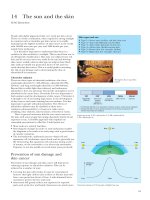
Figure 9.1 Predicted values for peak expiratory flow (adapted from Nunn
AJ, Gregg I. British Medical Journal 1989; 298: 1068–1070).
Asthma in Adults: Treatment of Acute Asthma
45
retention. The driving gas, flow rate, drug diluent and volume of
fill should be clearly written on the prescription chart. Dilutions
should always be done with saline to avoid bronchoconstriction
from nebulisation of hypotonic solutions. There is no real advantage
of nebulisation with a machine capable of producing intermittent
positive pressure.
For adults the initial dose should be 5 mg salbutamol or its
equivalent. This should be halved if the patient has ischaemic heart
disease. It is essential to continue the intensive treatment after the
first response; many of the problems in acute asthma arise because
of complacency after the initial response to the first treatment. In
severe attacks, the nebulisation may need to be repeated every 15
to 30 minutes and can be given continuously at 5–10 mg per hour
with the same effect.
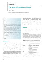
Figure 9.2 Attaching a spacer to a metered dose inhaler avoids the need for
coordination between firing and inhalation.
Parenteral delivery
If nebulised drugs are not effective then parenteral treatment should
be considered. A reasonable plan is to give a β2-agonist the first time,
combine with an anticholinergic drug for the second nebulisation
or initially in life-threatening asthma and move to intravenous
bronchodilators if there is no improvement. If life-threatening
features such as a raised carbon dioxide tension, an arterial oxygen
tension less than 8 kPa on oxygen or a low pH are present, the
intravenous agent should be considered from the start.
The bronchodilator given parenterally in an acute attack can
be β2 -agonist or aminophylline; there is little to choose between
them. If the patient has been on theophylline and a level is not
immediately available it is safer to use the β2 -agonist. Salbutamol
or terbutaline can be given intravenously over 10 minutes, or as an
infusion, usually at 5 to 15 µg per minute. The adverse effects of
tachycardia and tremor are much more common after intravenous
injection than after nebulisation.
Figure 9.3 In acute asthma β-stimulants should be given by oxygen-driven
nebuliser.
Anticholinergic agents
Nebuhaler or Volumatic) can be as effective as a nebuliser in
most cases (Figure 9.2). Like the nebuliser, it has the advantage of
removing the need to coordinate inhaler actuation and breathing.
There is little or no difference in the effectiveness of drugs that
are nebulised or given intravenously in acute severe asthma, so
nebulisation is generally preferable.
It is helpful for general practitioners (GPs) to have nebulisers
available for acute asthmatic attacks (Figure 9.3). β2 -agonists are
best given by nebulisers driven by oxygen in acute asthma, as they
may even worsen hypoxia slightly through an effect on the pulmonary vasculature. In general practice the use of oxygen as the
driving gas is not usually practical. Domiciliary oxygen sets do not
produce a flow rate adequate to drive most nebulisers. If available
they can be used with nasal cannulae at the same time as an air
driven nebuliser for a patient having an acute attack. Many ambulance services are able to give nebulised drugs and oxygen during
transfer to hospital.
In hospital, nebulisers used to treat asthmatic patients should be
driven by oxygen unless the patient has COPD with carbon dioxide
Ipratropium bromide is the only anticholinergic agent available
in nebulised form in the United Kingdom (Figure 9.4). Nebulised
ipratropium seems to be as effective as a nebulised β-agonist in
acute asthma. The dose of ipratropium is 500 mcg and there are no
problems with increased viscosity of secretions or mucociliary clearance at such doses. Ipratropium starts working more slowly than
salbutamol; the peak response may not occur for 30 to 60 minutes.
Adverse reactions such as paradoxical bronchoconstriction have
been reported occasionally. These were related mainly to the osmolality of the solution or to the preservatives and they have been
corrected in the current preparations.
Although the combination of β-stimulant and anticholinergic
agents produces a greater effect than use of a single agent, the
difference is small and β2 -agonists are sufficient for most patients.
It is reasonable to start with a β2 -agonist alone in moderate
exacerbations and add ipratropium if the response to the first
nebulisation is not considered adequate. If the initial assessment
indicates that it is a severe or life-threatening attack then the
combination should be used from the start. After stabilisation the
ipratropium can be stopped.
46
ABC of Asthma
Table 9.1 Drug interactions with theophylline.
Drug
Effect
Increase in theophylline concentration
Alcohol
Allopurinol
Cimetidine
Ciprofloxacin
Interferon alfa
Macrolides
(erythromycin)
Oestrogen
Ticlopidine
Zafirlukast
Decreases theophylline clearance
Decreased clearance
Inhibits cytochrome P450, reducing clearance
As cimetidine
Marked decrease in clearance
Decreased clearance
Decreased clearance
Decreased clearance, concentrations may rise by 60%
Decreased clearance
Decrease in theophylline concentration
Carbamazepine
Cigarette smoking
Phenytoin
Rifampicin
50% increase in clearance
Increased clearance around 30%
Up to 70% increased clearance
Increases cytochrome P450, increasing theophylline
clearance up to 80%
Effect on other drugs
Benzodiazepines
Lithium
Pancuronium
Larger doses of benzodiazepine may be required,
effects may increase if theophylline is discontinued
Lithium clearance increased
Antagonised by theophylline, larger doses may be
necessary
blood concentrations should be measured and the rate adjusted as
necessary.
Corticosteroids
Figure 9.4 Atropa belladonna (deadly nightshade) contains several
anticholinergic substances.
Methylxanthines
Aminophylline is an effective bronchodilator in acute asthma but
most studies have shown that it is no more effective than a
β2 -agonist given by mobilisation or intravenously. There are more
problems with its use than with nebulised drugs and it should be
reserved for patients with life-threatening features or who have
failed to respond to nebulised drugs. Toxic effects are common
and can occur with drug concentrations in or just above the
therapeutic range. Concentrations are difficult to predict from the
dose given because of individual differences in metabolic rate and
interactions with drugs such as nicotine, cimetidine, erythromycin
and ciprofloxacin (Table 9.1).
The position is further complicated if patients are already taking oral theophyllines. The usual starting dose for intravenous
aminophylline is 5 mg/kg given over 20 to 30 minutes. If the
patient has taken oral theophylline or aminophylline in the previous 24 hours and a blood concentration is not available then
the initial dose should be omitted or halved. A continuous infusion is then given at a rate of 0.5–0.7 mg/kg/hr though this dose
should be reduced if the patient also has kidney or liver disease.
If intravenous treatment is necessary for more than 24 hours then
Corticosteroids are effective in preventing the development of acute
asthma.
Oral delivery
Oral prednisolone should be given if control of asthma is deteriorating despite usual regular treatment (Box 9.1). A single oral dose
of prednisolone, 40 to 50 mg according to body weight, should be
given each day for at least 5 days until recovery according to the
speed of the response. If this opportunity is missed and an acute
attack of asthma does develop, corticosteroids are still an important element in treatment. Fatal attacks of asthma are associated
with failure to prescribe any or adequate doses of corticosteroids.
No noticeable response occurs for 4 to 6 hours, so corticosteroids
should be started as early as possible and intensive bronchodilator
treatment used while waiting for them to take effect.
Box 9.1 Adverse effects of short course of oral corticosteroids
•
•
•
•
•
•
•
Fluid retention
Hyperglycaemia
Indigestion
Sleep disturbance
Steroid-induced psychosis
Susceptibility to severe herpes zoster
Weight gain
Asthma in Adults: Treatment of Acute Asthma
47
Intravenous delivery
Antibiotics
In most cases oral corticosteroids are adequate, but when there are
life-threatening features or difficulties with swallowing or absorption intravenous hydrocortisone should be used in an initial dose
of 100 mg followed by 100 mg six hourly for 24 hours. Prednisolone
should be started at a dose of 40 to 50 mg daily whether or not
hydrocortisone is used (50 mg prednisolone is equivalent to 200 mg
hydrocortisone). If the patient is first seen at home and transferred
to hospital, the first dose of corticosteroid should be given together
with initial bronchodilator treatment before leaving home.
Upper respiratory tract infections are the most common trigger
factors for acute asthma and most of these are viral. In only a few
cases are exacerbations of asthma precipitated by bacterial infection.
There is no evidence of benefit from the routine use of antibiotics.
They should be reserved for patients in whom there is presumptive
evidence of infection – such as fever, neutrophils in the blood or
sputum or radiological changes, although all these features may
occur in acute attacks without bacterial infection.
Controlled ventilation
Length of steroid course
When intensive initial treatment has been required prednisolone
should be maintained at a dose of 40 mg per day for at least
5 days. One to three weeks of treatment may be needed to obtain
the maximal response with deflation to normal lung volumes and
loss of excessive diurnal variations of peak flow. There are few
side effects of such short courses of corticosteroids. Increased
appetite, fluid retention, gastrointestinal upset and psychological
disturbance are the most common. Exposure to herpes zoster
may produce severe infections in susceptible individuals. Steroids
can be stopped abruptly after courses lasting up to 3 weeks.
Tapering off the dose is not needed for adrenal suppression or
does not help prevent relapse although many patients are used
to such regimes. Inhaled steroids should be continued or started
during inpatient treatment in accordance with the plans for routine
management.
Magnesium
Intravenous magnesium sulphate has been shown to be effective
and safe in acute asthma. Magnesium sulphate is given as an
infusion, at a dose of 1.2–2 g over 20 minutes. It provides a possible
additional therapy in acute severe asthma in hospital when the
initial response to nebulised bronchodilators is inadequate or when
the initial assessment indicates life-threatening or near fatal asthma.
Doses can be repeated for episodes of deterioration in hospital.
Patients with acute severe asthma who need hospital admission
should be treated in an area equipped to deal with acute medical emergencies, with adequate nursing and medical supervision.
If hypoxia is worsening, hypercapnia is present or patients are
exhausted or drowsy, then they should be nursed in an intensive
care unit.
Occasionally, mechanical ventilation may be necessary for a
short time while the treatment takes effect. It is usually needed
because the patient becomes exhausted; experience and careful
observation are necessary to judge the right time to begin ventilatory
support. Non-invasive ventilation may be tried in expert hands in
an intensive care unit.
High inflation pressures and long expiratory times may make
ventilation difficult in asthmatic patients, but most experienced
units have good results, provided that the decision to ventilate the
patient is made electively and is not precipitated by respiratory
arrest. When patients being mechanically ventilated fail to improve
on adequate treatment, bronchial lavage may occasionally be considered to reopen airways that have become plugged by mucus. In
very severe unresponsive cases other treatments such as inhalational
anaesthetics may be helpful, or a mixture of helium and oxygen
may improve airflow while the other treatment takes effect.
Other factors
Patients with acute asthma tend to be dehydrated because they
are often too breathless to drink and because fluid loss from the
respiratory tract is increased. Dehydration increases the viscosity of
mucus, making plugging of the airways more likely, so intravenous
fluid replacement is often necessary. Three litres should be given
during the first 24 hours if little oral fluid is being taken.
Most patients with acute severe asthma improve with these measures
(Figure 9.5). Occasionally physiotherapy may be useful to help
patients cough up thick plugs of sputum, but mucolytic agents to
change the nature of the secretions do not help.
An episode of asthma is frightening. The dangerous use of
sedatives such as morphine was common before effective treatment
became available. Unfortunately, this practice still continues, with
occasional fatal consequences. Treatment of agitation should be
aimed at reversing the asthma precipitating it, not at producing
respiratory depression.
Potassium supplements
Discharge from hospital
Increased alveolar ventilation, sympathomimetic drugs and corticosteroids all tend to lower the serum potassium concentration.
This is the most common disturbance of electrolytes in acute
asthma; the serum potassium concentration should be monitored
and supplements given as necessary.
Discharge too early is associated with increased readmission and
with mortality. Patients should have stopped nebuliser treatment
and be using their own inhalers, with the proper technique checked,
for at least 24 hours before discharge (Box 9.2). Ideally, peak flow
should be above 75% of the patient’s predicted or best-known
Fluid and electrolytes
48
ABC of Asthma
Immediate management
Oxygen 40–60%
Salbutamol 5 mg or terbutaline and
ipratropium 0.5 mg by oxygen driven
nebuliser
Prednisolone 40–50 mg orally or
hydrocortisone 100 mg intravenously
No sedation
Consider need for chest radiograp
Life–threatening features
• Peak flow <33% Predicted or best
• Silent chest, feeble respiratory effort
• Cyanosis, SaO2 <92%
• Bradycardia, hypotension, dysrhythmia
• Exhausion, confusion, coma
• PCO2 ≥ 4.6 kPa, PO2 ≤ 8 kPa, acidosis
If life–threatening features are present
• Discuss with ICU team
• IV magnesium sulphate 1.2–2 g iv
over 20 min
• Frequent or continuous β2-agoinst
nebulisation
Improving
Continue
• Oxygen
• Prednisolone 40−50 mg daily
• β-agonist and ipratropium 4−6 hourly
Not improving after 15–30 min
Continue
Oxygen and steroids
β-agonist up to every 15 min or
continuously
Ipratropium bromide 0.5 mg 4–6 hourly
Monitor
• Peak flow before and after nebulisations
• Oximetry (keep saturation >92%)
• Blood gas tensions if initial PaO2
<8 kPa and saturation <93%
or PaCO2 normal or high
or patient deteriorates
If still not improving
Aminophylline infusion 0.5 mg/kg/hr
(monitor concentrations if longer than
24 hr)
or
salbutamol or terbutaline infusion 5 to
15 µg/min
Discuss with ICU team
reading. Diurnal variability should be below 25%. A few patients
may never lose their morning dips and may have to be discharged
with them still present (Figure 9.6).
Box 9.2 Discharge after acute severe asthma admission
Patients discharged should have the following:
•
•
•
•
•
•
•
•
•
•
•
•
Planned discharge medication for 24 hours before discharge
Inhaler technique checked
Peak expiratory flow (PEF) >75% best or predicted
PEF diurnal variation <25%
Oral and inhaled steroids
Bronchodilators
PEF meter
Written asthma management plan
Discharge summary for GP
GP follow-up within 2 working days
Chest clinic follow-up within 4 weeks
Circumstances of acute exacerbation and patient response
explored
Peak flow (I/min)
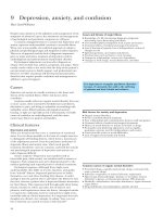
Figure 9.5 Treatment of acute severe asthma in hospital (adapted from guidelines from the British Thoracic Society
and Scottish Intercollegiate Guidelines Network).
400
300
Discharge
200
100
0
0
1
2
3
4
5
6
7
8
9
10
11
Time (days)
Figure 9.6 Peak flow during recovery from acute attack.
For every patient the reason for the acute episode should be
sought and appropriate changes made in their routine treatment
and in their response to any deterioration in an attempt to avoid
similar attacks in the future. Patients with an acute attack of asthma
should be looked after or at least seen by a physician with an interest
Asthma in Adults: Treatment of Acute Asthma
in respiratory disease during their inpatient stay. Follow-up should
be arranged and a respiratory specialist nurse will be helpful in
education, management and support.
49
Hospital follow-up
The patient should return to the chest clinic within a month. Good
communication between hospital and the GP is vital around this
vulnerable period – telephone, fax and electronic links may help.
Subsequent management
At the time of their discharge, patients should be stable on the
treatment that they will take at home. They should leave with a
plan of further management. This should include advice on asthma,
symptoms and peak flow measurement and a plan to respond to
deterioration in the control of their asthma. The GP should be
informed of the admission and the subsequent plans and should
see the patient within two working days.
Further reading
British Thoracic Society. Emergency Oxygen Guideline Group Guideline for
emergency oxygen use in adult patients. Thorax 2008; 63 (Suppl VI).
Silverman RA, Osborn H, Runge J et al.; Acute Asthma/Magnesium Study
Group. IV magnesium sulfate in the treatment of acute severe asthma: a
multicenter randomized controlled trial. Chest 2002; 122: 489–497.
C H A P T E R 10
Methods of Delivering Drugs
John Rees
Sherman Education Centre, Guy’s Hospital, London, UK
OVERVIEW
•
With the combinations of drug and inhaler available it is possible
for nearly all patients to take drugs by inhalation
•
Even when a metered dose inhaler (MDI) is used properly, only
about 10% of the drug reaches the airways below the larynx
•
Inhaler technique should be checked regularly since errors can
develop and interfere with treatment
•
Chlorofluorocarbon (CFC)-free beclometasone MDIs need to be
prescribed by brand because of differences in lung deposition
•
Spacer devices help coordination problems with MDIs and
reduce pharyngeal deposition
Various inhaler devices and formulations have been developed to
deliver drugs efficiently, minimise side effects and simplify use. With
over 100 combinations of drug and inhaler available, it is possible
for nearly all patients to take drugs by inhalation, but there is scope
for confusion for patients and prescribers. All the available devices
used appropriately can provide adequate drug to the airways,
but inhalers should only be prescribed with confidence that the
patient can use the device satisfactorily. This should be rechecked
on subsequent visits since errors can develop and interfere with
treatment. Even after training, at least one-third of patients continue
to make errors in their inhalation technique in most studies. The
scores used in assessing technique may not all relate similarly to
clinical effectiveness, but some result in no drug delivery and poor
technique is related to poor asthma control. Some drugs such as
leukotriene receptor antagonists and theophylline cannot be given
by inhalation.
first-pass metabolism in the liver. Absorption directly from the lung
bypasses liver metabolism.
An MDI should be shaken and then fired into the mouth
shortly after the start of a slow full inspiration. At full inflation the breath should be held for 10 seconds. The technique
should be checked periodically. At least a quarter of patients
have difficulty using an MDI and the problems increase with
Inhaler
50–60% recoverable
from the mouth and
pharynx by washing
<10% reaches
the lungs
>90%
swallowed
Figure 10.1 Inhalers deliver the drug direct to the airways.
Metering chamber
Metering valve
Metered dose inhalers
Inhalers deliver the drug directly to the airways. Even when an MDI
is used properly only about 10% of the drug reaches the airways
below the larynx (Figures 10.1 and 10.2). Nearly all the rest of the
drug gets no further than the oropharynx and is swallowed. This
swallowed portion may be absorbed from the gastrointestinal tract
but drugs such as inhaled corticosteroids are largely removed by
ABC of Asthma, 6th edition. By J. Rees, D. Kanabar and S. Pattani.
Published 2010 by Blackwell Publishing.
50
Actuator orifice
Opening for emptying
of metering chamber
Figure 10.2 The mechanisms inside a metered dose inhaler.
Asthma in Adults: Methods of Delivering Drugs
Clear plastic
One way valve
51
Spray output
Metered dose inhaler
Figure 10.4 An extension tube (spacer) used with a metered dose inhaler.
Some large volume spacers are being replaced by smaller volume devices.
Figure 10.3 The autoinhaler is triggered by inspiratory airflow.
Breath-actuated metered dose inhalers are available for β-agonists,
anticholinergics, cromoglicate and corticosteroids.
age. The common problems are coordination of firing with inspiration. The ‘cold Freon effect’, stopping inspiration when the
inhaler activates, is much less common with replacement of
CFC-containing inhalers. Arthritic patients can find it hard to
activate MDIs and may be helped by a Haleraid device, which
responds to squeezing, or be given a breath-actuated or dry powder
system.
Breath-actuated aerosol inhalers
Breath-actuated MDIs are available for most classes of drug
(Figure 10.3). The pressurised canister is actuated via a spring
triggered by inspiratory airflow. The devices respond to a low flow
rate and are useful for those who have difficulty coordinating actuation and breathing. Errors are less frequent than with MDIs. They
require a propellant similar to that caused in a standard inhaler.
Metered dose inhaler propellants
Most current MDIs have now moved from CFC propellants. The
production, import and use of CFCs have been stopped in most
developed countries because of the effect on the ozone layer. There
is a temporary exemption for medical use under the Montreal
Protocol, but CFC inhalers are being removed now that adequate
non-CFC products are available.
The challenge has been to develop safe alternatives that are
as convenient, effective and clinically equivalent. The process of
development of alternative propellants was more of a problem than
first appreciated, particularly for inhaled steroids. Adaptations to
the method of adding the drug to the propellant and to the valve
and jet mechanisms have been necessary. Hydrofluoroalkanes 134
and 227 are used in the new devices.
Short- and long-acting β-agonists, inhaled steroids and combinations are now available in HFA-containing MDIs. Each new
device has to be tested carefully since total and regional delivery
to the lung will differ with the new devices. The beclometasone
product QVar is prescribed at half the dose of a conventional
MDI because of its smaller particle size, resulting in better lung
deposition. Other preparations are substituted in the ratio of 1:1.
Patients will notice differences in the speed of the aerosol cloud and
taste.
The switch to CFC-free MDIs should be taken as an opportunity
to review patient understanding, inhaler technique and general
asthma management.
Spacer devices
The coordination of firing and inspiration becomes slightly less
important when a short extension tube is used. This may help if
problems are minor but a larger reservoir removes the need for
coordination of breathing and actuation (Figure 10.4). The inhaler
is fixed into the chamber and breath is taken from a one-way valve
at the other end of the chamber. Inhalation should be as soon
as possible after each actuation, certainly within 30 seconds; tidal
breathing is as effective as deep breaths. In young children they can
be used with a facemask.
Pharyngeal deposition is greatly reduced as the faster particles
strike the walls of the chamber, not the mouth. Evaporation of propellant from the larger and slower particles produces a small-sized
aerosol that penetrates further out into the lungs and deposits a
greater proportion of drug beyond the larynx. This reduces the
risk of oral candidiasis and dysphonia with inhaled corticosteroids
and reduces potential problems with systemic absorption from the
gastrointestinal tract. Spacers should be used routinely when doses
of inhaled steroid of more than 800 µg daily are given by MDI.
Most devices are cumbersome, but this is not a great disadvantage
for twice daily treatment such as corticosteroids. Chambers can be
used as effectively as nebulisers in mild to moderate exacerbations
of asthma. Output characteristics of MDIs vary and inhalers and
extension tubes need to be matched appropriately. It cannot be
assumed that results transfer to different combinations.
Electrostatic charge can reduce drug delivery. Chambers should
be washed in detergent and left to air dry rather than be wiped
dry, just once a month and changed every 6–12 months. Metal
chambers without static charge can also be used (Box 10.1).
52
ABC of Asthma
Nebulisers
Box 10.1 Use of spacer devices
•
•
•
•
•
•
•
Match the MDI and spacer
Inhale as soon as possible after each single actuation
Empty the chamber by single large breaths or tidal breathing
Clean chamber monthly
Wash chamber in detergent and water and leave to dry
Wipe any detergent from mouthpiece
Replace spacer every 6–12 months
Dry powder inhalers
Dry powder inhalers (DPIs) of various types are available for
β-agonists, sodium cromoglicate, corticosteroids, anticholinergic
agents and combinations (Figure 10.5). Because inspiratory airflow
releases the fine powder, many problems of coordination are
avoided and there are none of the environmental worries of MDIs.
The dry powder makes some patients cough. The Turbohaler
contains drug with no carrier and patients may feel that nothing is
coming from the device. It has good lung deposition but requires a
flow rate of >60 l/min, achieved easily by most patients.
The problems of reloading for each dose have been eased by the
development of multiple dose units with up to 200 doses, and most
DPIs have a dose counter that helps the patient to know when the
inhaler needs renewing and provides a compliance monitor.
Soft mist inhalers
Soft mist inhalers (SMIs) contain liquid but no propellants and
produce a slow-moving aerosol cloud (the soft mist). They are fired
by the patient with inspiration but coordination is easier because
of the slow velocity and the long duration.
(a)
Nebulisers can be driven by compressed gas (jet nebuliser) or an
ultrasonically vibrating crystal (ultrasonic nebuliser). They provide
a way of giving inhaled drugs to those unable to use any other
device – for example, the very young – or in acute attacks when
inspiratory flow is limited.
Nebulisers also offer a convenient way of delivering a higher
dose to the airways (Figure 10.6). Generally, about 12% of the drug
leaving the chamber enters the lungs but most of the dose stays in
the apparatus or is wasted in expiration. Delivery depends on the
type of nebuliser chamber, the flow rate at which it is driven and
the volume in the chamber. In most cases, flow rates of less than
6 l/min in a jet nebuliser give too large a particle and nebulise too
slowly. Some chambers have a reservoir and valve system to reduce
loss to the surrounding room during expiration.
In many situations, equivalent effects can be obtained with MDI
and a spacer but patients often feel confidence in their nebuliser.
Tablets and syrups
Tablets and syrups are available for oral use. This route is necessary
for theophyllines and leukotriene antagonists, which cannot be
inhaled effectively. Very young children who are unable to inhale
drugs can take the sugar-free liquid preparations. Slow-release
tablets are used when a prolonged action is needed, particularly for
nocturnal asthma in which theophyllines can be helpful. Various
slow-release mechanisms or long-acting drugs have been developed
to maintain even blood concentrations (Figure 10.7).
Bambuterol is a pro-drug of terbutaline which can be given once
daily at night in those unable to use the inhaled route. Tablets avoid
the need to learn the coordination needed for inhalers and might
(b)
Figure 10.5 Dry powder inhalers are used for delivery of inhaled drugs. Two commonly used devices are the (a) accuhaler and the (b) turbohaler.
Asthma in Adults: Methods of Delivering Drugs
53
Serum theophylline concentration (mg/l)
Figure 10.6 The use of nebulisers must be associated with careful
instructions on use and hygiene as well as arrangements for maintenance
and support.
20
15
10
5
0
20
24
4
8
12
16
20
Figure 10.8 In severe cases β2 -agonists can be delivered by subcutaneous
infusion.
Clock time (24 hr)
Figure 10.7 Steady theophylline concentrations in the therapeutic range can
be obtained with 12-hourly slow-release preparations (reproduced with
permission from Ferrari M et al. Effect of once daily and twice daily sustained
release theophylline formulations on day-time variation of bronchial
hyper-responsiveness in asthmatic patients. Thorax 1997: 52; 969–974).
allow delivery to lung tissue beyond blocked airways but at the
expense of potential side effects from body distribution.
Injections and infusions
Injections are used for the treatment of acute attacks. Subcutaneous injections may be useful in emergencies when nebulisers are
unavailable. Occasional patients with severe chronic asthma seem
to benefit from the high levels of β-stimulant obtained with subcutaneous infusion through a portable pump (Figure 10.8). Rates
may need to be adjusted, depending on severity. The infusion site
is changed by the patient every 1 to 3 days.
Further reading
D’Alonzo GE, Smolensky MH, Feldman S et al. Twenty-four hour lung
function in adult patients with asthma. Chronoptimized theophylline
therapy once-daily dosing in the evening versus conventional twice-daily
dosing. The American Review of Respiratory Disease 1990; 142: 84–90.
Giraud V, Roche N. Misuse of corticosteroid metered-dose inhalers is associated with decreased asthma stability. European Respiratory Journal 2002;
19: 246–251.
Pitcairn G, Reader S, Pavia D, Newman S. Deposition of corticosteroid aerosol
in the human lung by Respimat Soft Mist inhaler compared to deposition
by metered dose inhaler or by Turbohaler dry powder inhaler. Journal of
Aerosol Medicine 2005; 18: 264–272.
Virchow JC, Crompton GK, Dal Negro R et al. Importance of inhaler devices in
the management of airway disease. Respiratory Medicine 2008; 102: 10–19.
C H A P T E R 11
Definition, Prevalence and Prevention
Dipak Kanabar
Evelina Children’s Hospital, Guy’s and St Thomas’ Hospitals, London, UK
OVERVIEW
•
Childhood asthma is most likely a spectrum of disorders
•
A good clinical history is important in diagnosing childhood
asthma
•
Asthma affects one in six children at some point in their lives
•
Atopy is probably the single strongest risk factor for
asthma – exposure to relevant allergens in infancy or childhood
may predispose a person to continued allergic responses later
•
The hygiene hypothesis is an attractive hypothesis to explain
rising prevalence of childhood asthma
exercise-induced wheeze. Wheezy episodes in children are a common phenomenon and up to 30% of children under the age of
5 may wheeze at some time point.
Labelling a child as asthmatic can still cause anxiety within the
family and controversy among paediatricians (Figure 11.1). Most
children under 5 presenting with asthmatic symptoms (see International Consensus Report) are either transient early wheezers
or non-atopic wheezers, without a family or personal history of
Defining asthma in children
Western Europe has seen a dramatic increase in children suffering
from asthma. Not only has the prevalence increased but also the
severity of the illness. It is likely that events in early life lead to
changes in the lung and immune systems which predispose the
child to chronic asthmatic symptoms. It is becoming increasingly
apparent that asthma is a spectrum disorder and probably has many
definitions, however a working definition is given in Box 11.1.
Box 11.1 ICS report
The International Consensus Report on the Diagnosis and Management of Asthma gives the following definition: ‘Asthma is a chronic
inflammatory disorder of the airway in which many cells play a role,
in particular mast cells, eosinophils, and T lymphocytes. In susceptible
individuals this inflammation causes recurrent episodes of wheezing, breathlessness, chest tightness, and cough particularly at night
and or in the early morning. These symptoms are usually associated
with widespread but variable airflow limitation that is at least partly
reversible either spontaneously or with treatment. The inflammation
also causes an associated increase in airway responsiveness to a
variety of stimuli.’
Childhood asthma is most likely a spectrum of disorders characterised by episodes of cough, wheeze, shortness of breath and
ABC of Asthma, 6th edition. By J. Rees, D. Kanabar and S. Pattani.
Published 2010 by Blackwell Publishing.
54
Figure 11.1 A definition of asthma.
Asthma in Children: Definition, Prevalence and Prevention
Infant
0.4 mm
Adult
0.7 mm
Figure 11.2 Comparative diameter of bronchioles.
atopy and tend to outgrow their wheezy symptoms at an early
age (<7 years).
Atopic (immunoglobulin E (IgE)-associated) wheezers have
raised IgE concentrations, positive radioallergosorbent (RAST) and
skin prick tests and raised exhaled nitric oxide (FENO ) concentrations.
Presenting symptoms
For example, respiratory syncitial virus (RSV) bronchiolitis itself
causes wheezing and up to half of affected children will go on
to develop recurrent episodic wheeze. Many children have mild
wheezing during viral infections (virus-associated wheeze), but
their prognosis is better than that of children who show bronchial
hyper-reactivity to methacholine (non-atopic wheezers). In addition, the airways of preschool children are small relative to lung
size (Figure 11.2). The airways and chest walls are also less rigid, so
during expiration, they are more likely than those of older children
to collapse, or become obstructed by desquamated airway epithelial
cells and secretions or mucosal changes that are not the result of an
inflammatory process like asthma.
Older children can describe symptoms of cough, wheeze, dyspnoea and chest tightness, and confirm whether there is an
improvement with bronchodilator and steroid therapy. In addition, peak flow measurements, forced expiratory volume in 1 second
(FEV1) by spirometery, exercise testing and recordings of diurnal
variations will assist diagnosis.
Thus, in practice, in the absence of an easily recognised or readily
available diagnostic marker, a clinical diagnosis of asthma usually
relies on a combination of history of characteristic symptoms and
evidence of airway lability and a reduction in symptoms after
treatment with a short-acting β2 -agonist showing reversible airflow
obstruction.
Prevalence of asthma
Asthma is the most common chronic disease of childhood. About
one in six (17%) or more children aged between 2 and 15 years
in the United Kingdom have symptoms of asthma at some time in
their lives which requires treatment.
55
Is prevalence increasing or reaching a plateau?
While several epidemiological studies show that the prevalence of
asthma and other atopic disorders such as eczema and hayfever is
increasing in many countries throughout the world more recent
studies indicate that, perhaps in the Western world at least, prevalence rates are reaching a plateau (Figure 11.3).
The observation that all forms of allergic disease have increased
simultaneously suggests an increase in host susceptibility, rather
than a rise in allergic sensitisation. Associations between the prevalence of asthma and small family size, environmental exposure to
cigarette smoke, affluence, reduced cross infection and BCG status
(decreased asthma with BCG vaccine) are all recognised and, coupled with our understanding of the immunology of asthma, hint at
the possibility of factors either in utero or in early life, which might
modify an individual’s atopic tendency.
Based on self-reported data, international comparison studies
(International Study of Asthma and Allergies in Childhood [ISAAC]
phases I and III) have placed the United Kingdom near the top
of the world league of asthma and allergy prevalence (Figure 11.4)
and while there is some objective data to support large differences
between the United Kingdom and countries such as Albania, it
is not clear why other westernised nations with low levels of air
pollution (e.g. New Zealand) also appear near the top of the table.
Phase III of ISACC confirms that English-speaking countries and
Western Europe have recently seen a decrease in asthma prevalence,
whereas regions where prevalence was previously low (Africa, Latin
America and parts of Asia) have seen an increase (Figure 11.3).
Public health issues
In terms of burden of disease, childhood asthma presents a serious
public health problem. More than half of all cases of asthma
present before the age of 10, and over 30% of children experience a
wheezing illness during the first few years of life. More absence from
school is caused by asthma than any other chronic condition; 30%
of asthmatic children miss more than 3 weeks of schooling each
year. Asthma influences educational attainment even in children of
above average intelligence, the extent of this adverse effect being
related to severity of the disease.
Reasons for the increasing global
prevalence
It is unlikely that there is a single cause and effect association to
account for the rising global burden of asthma and atopic disorders.
Recent immunological studies, however, have indicated that the first
3 years of life (including life before birth) are probably the most
critical in terms of environmental influences on the development of
the asthma phenotype. For example, there are strong links between
cigarette smoking in pregnancy and narrow airways in the offspring,
and the risk of a child developing asthma is more closely associated
with allergy in the mother than in the father.
Further data from ISAAC phase III suggests that use of paracetamol in the first year of life and in later childhood, is associated with
an increased risk of symptoms of asthma and other atopic disorders.
Country (prevalence %)
ABC of Asthma
Country (prevalence %)
56
Costa Rica (27.4)
Australia (27.0)
New Zealand (26.9)
United Kingdom (24.8)
Japan (20.6)
Panama (19.8)
Barbados (18.8)
Singapore (17.0)
Canada (15.1)
Taiwan (13.5)
Chile (11.6)
Malaysia (11.3)
Malta (11.2)
Sultanate of Oman (10.5)
Portugal (9.8)
South Korea (9.1)
Spain (8.8)
Italy (8.6)
Sweden (8.2)
Hong Kong (7.8)
Thailand (7.6)
Brazil (6.2)
Ukraine (6.2)
Indonesia (5.7)
India (5.3)
Belgium (5.1)
Germany (4.3)
Mexico (4.3)
Poland (4.2)
Georgia (3.9)
Austria (3.8)
Iran (3.7)
Nigeria (3.4)
Albania (2.8)
Estonia (2.8)
Russia (2.1)
Lithuania (1.8)
−2
−1
0
1
2
Peru (30.5)
New Zealand (27.7)
Singapore (23.7)
Isle of Man (23.7)
Channel Islands (23.1)
United Kingdom (22.9)
Barbados (20.9)
Costa Rica (20.8)
Sultanate of Oman (20.3)
Japan (19.4)
Philippines (19.3)
Panama (18.7)
Republic of Ireland (18.4)
USA (18.3)
Uruguay (16.7)
Kuwait (15.8)
Tunisia (15.6)
Nigeria (15.0)
Morocco (14.7)
Brazil (14.6)
South Africa (13.7)
Portugal (13.2)
Taiwan (13.1)
Kenya (12.9)
Chile (12.8)
Malta (12.6)
Paraguay (12.5)
Thailand (12.3)
Spain (11.8)
Malaysia (11.5)
Sweden (11.1)
Hong Kong (10.7)
Italy (10.5)
Argentina (9.6)
Belgium (8.3)
Germany (7.5)
Pakistan (7.4)
Indonesia (7.0)
Romania (6.3)
Austria (6.2)
Finland (6.1)
Algeria (5.9)
Ukraine (5.8)
India (5.7)
Mexico (5.6)
Latvia (5.6)
China (5.4)
Poland (4.1)
Estonia (4.0)
South Korea (3.9)
Russia (3.6)
Georgia (3.5)
Iran (3.1)
Lithuania (2.7)
Ethiopia (2.6)
Albania (2.6)
−2
−1
0
Mean change per year
(a)
1
2
Mean change per year
(b)
Figure 11.3 Ranking plot showing the change per year in the lifetime prevalence of asthma (‘asthma ever’) in children aged (a) 6–7 years and (b) 13–14 years
for each centre by country, with countries ordered by their mean prevalence (for all centres combined) across phase I and phase III. The plot also shows the
confidence interval about zero change for a given level of prevalence (i.e. the mean prevalence across phases I and III) given a sample size of 3000 and no cluster
sampling effect. Reproduced with permission from Pearce N et al. Thorax 2007; 62: 758–766.
Changes such as those in housing that allow proliferation of
house dust mite, the effects of outdoor and indoor pollutants
such as cigarette smoke, dietary changes, low birth weight and
prematurity may all account for some of the increased prevalence.
To account for the increase in disease prevalence from 10% to 15%
(such as has occurred in the United Kingdom over the last 30 years),
however, the proportion of the population exposed to these hazards
would need to have increased from 10% to nearly 70%, suggesting
that other, as yet unidentified, risk factors may be operating.
The relevance of atopy
Figure 11.4 Electron micrograph of pollen grains.
Atopy, defined as the predisposition to raise specific IgE to common
allergens (such as house dust mite, wheat and cat dander), is
probably the single strongest risk factor for asthma, carrying up to a
20-fold increased risk of asthma in atopic individuals compared with
non-atopic individuals. The strongest association is with maternal
atopy – a maternal history of asthma or rhinitis, or both – and is a
significant risk factor for late childhood onset asthma and recurrent
wheezing (Figures 11.4 and 11.5).
Percentage
Asthma in Children: Definition, Prevalence and Prevention
57
Observations such as these make the ‘hygiene hypothesis’ an attractive model when explaining the general rise in atopic disorders.
The ‘hygiene hypothesis’ argues that the increase in atopic asthma
is due to a decrease in exposure to infection in early life. Frequent
infections in childhood generate Th1 cytokines such as the interleukins IL-12, IL-18 and IFNγ, and these in turn inhibit the growth
of Th2 cells, thus preventing development of the atopic asthma
phenotype.
40
Atopic
Non-atopic
30
20
10
Prospects for prevention
0
5–7
13–15
Age (years)
Figure 11.5 Atopy in children with bronchial hyper-reactivity.
A routine enquiry should be made about other atopic disorders
such as atopic dermatitis (eczema), food allergies and rhinitis as
they may be coexisting morbidities in a child with allergy-associated
asthma.
Lymphocytes
T lymphocytes – in particular T-helper type 2 (Th2)
lymphocytes – are also believed to be important in the pathogenesis of asthma. The fetal immune system is primarily polarised
towards a Th2 response as a result of interleukin 4 and 10 (IL-4 and
IL-10) production by the placenta. Furthermore, T lymphocytes
isolated from cord blood of newborn babies of atopic mothers are
able to respond to aeroallergens, suggesting that they may have
been exposed to antigens ingested by the mother and transferred
across the placenta in the last trimester of pregnancy.
During early childhood, environmental allergens – in particular
intestinal microflora – are thought to influence the immune deviation of T-helper cells towards the Th1 type in non-atopic children
and towards the Th2 type in atopic children. In atopic children with
recurrent wheezing illness, bronchoalveolar lavage studies indicate
increased mast cell and eosinophil concentrations in children as
young as 3. Up to the age of 10, the peripheral blood mononuclear
cell response to specific stimulation in children who develop atopic
disease is deficient in its capacity to generate interferon gamma
(IFNγ), thereby causing upregulation of Th2 responses and an
allergic phenotype.
Early exposure to infections
Children growing up in rural and farming communities are much
less likely to develop atopy and bronchial hyper-responsiveness
than children raised in inner city areas. There is an inverse association between socio-economic status and asthma and allergy,
and firstborn children have a higher prevalence of asthma than
their siblings, with the assumption that children from higher social
classes and firstborns are exposed to fewer infections in early life.
Allergen avoidance studies such as the Isle of Wight study, where
infants born to mothers with a strong family history of atopy
were randomised to receive prophylaxis, with the mother eating
a hypoallergenic diet and breastfeeding or giving a soya milk
preparation to their babies, showed a significant decrease in the
prevalence of eczema and a positive skin prick test to aeroallergen
and dietary factors, but no sustained benefit in relation to reduction
in asthma.
Other environmental avoidance studies have shown a reduction
in respiratory symptoms in the first year of life, but subsequent
results showed a paradoxical effect of increased allergy but better
lung function.
Dietary manipulation (e.g. introduction of fish in the diet or fish
oil supplementation) has shown some positive results in reducing
the risk of eczema. Breastfeeding is still advised in all children not
only for its other health benefits, but also for a preventative effect in
development of asthma it may have in those children born to atopic
families or in babies identified by high cord blood IgE, although the
evidence is not conclusive (Figure 11.6).
These results seem to indicate that the development of asthma
is a combination of genetic susceptibility and exposure in early
life to allergic stimuli and pollutants that augment a Th2 immune
response. Once the asthma is established, cycles of acute and chronic
inflammation triggered by allergens, viruses, pollutants, diet and
stress are responsible for exacerbations.
Recent studies indicate that the rise in childhood obesity may
also be linked with the rise in childhood asthma. Children with
high body mass indices were more likely to have symptoms of
asthma, suggesting that increased weight might lead to a risk of
inflammation in the respiratory tract or might hinder respiratory
flow (Figure 11.7).
Primary preventative measures to reduce risk might therefore
include allergen avoidance, cessation of smoking and attenuation
of a Th2 response by vaccination. Once asthma is established,
however, T cells and eosinophil responses may have enhanced
capacity to generate the leukotrienes IL-3, IL-4 and IL-5 and it
may be more difficult to reverse an established Th2 response. In
this situation, secondary prevention measures to reduce exposure
to trigger factors are appropriate.
Trigger factors in asthma
During the preschool years viral infections, exercise, and emotional
upset are common triggers of asthma. Young children contract six
58
ABC of Asthma
Box 11.2 Trigger factors in asthma
•
•
•
•
•
•
Viral infections
Dusts and pollutants including cigarette smoke and diesel
particulates
Allergens – house dust mite, pollens, moulds, spores, animal
dander and feathers, certain foods and Alternaria in dry arid
conditions
Exercise
Changes in weather patterns and cold air
Psychological factors such as stress and emotion
The domestic environment
If asthmatic children are sensitised to house dust mite, parents
can reduce exposure by removing carpets or vacuum cleaning
regularly and dusting surfaces with damp cloths, as well as encasing
mattresses and pillows in plastic sheets, washing covers, blankets,
duvets, and furry toys regularly, and applying acaricides to soft
furnishings (Figure 11.8).
A recent Cochrane review (issue 4 2004), however, suggests that
chemical and physical measures to reduce house dust mite cannot
be recommended on the basis of present evidence.
Figure 11.6 Breastfeeding is advocated for children from atopic families.
Figure 11.7 Childhood obesity may be linked to an increase in childhood
asthma.
to eight viral upper respiratory tract infections each year; so it is
not surprising that these infections are more common precipitants
of asthma in children than in adults. Asthmatic children tend to
have more symptoms during the winter than the summer, probably
because viral respiratory infections are more common in winter and
because exercise-induced asthma is more likely to develop outdoors
in cold weather (Box 11.2).
Figure 11.8 Vacuuming.
Asthma in Children: Definition, Prevalence and Prevention
It must be borne in mind that intensive cleaning measures may
also reduce the child’s exposure to endotoxin and other bacterial
components. Some studies indicate that early life exposure to cats
and dogs may reduce the subsequent prevalence of asthma and
allergy, giving further credence to the ‘hygiene hypothesis.’
Antigen
Macrophage
Peptide
Antigen
B
Cell
Smoking
Tobacco smoke has consistently been found to trigger exacerbation
of asthma in children, and families should be encouraged to stop
smoking or smoke in areas away from children outside the house.
In addition, in families with a strong family history of asthma,
and in children exposed to maternal smoking during pregnancy,
there is a fourfold risk of developing wheezing illnesses in young
children.
Studies have also demonstrated a decrease in asthma severity in
children whose parents have ceased smoking (Figures 11.9–11.11).
59
IgE
IL-4
IgE
• Tryptase
• Leukotrienes
Mast
Cell
IL-5
Th2
IL-3
IL-5
GM-CSF
Eosinophil
Other cytokines
Initiation and amplification
of inflammation
• Major basic protein
• Eosinophil cationic protein
• Leukotrienes
Recruitment
activation
Airways
hyper-responsiveness
Figure 11.11 Mechanisms of mast cell and eosinophil-dependent airway
hyper-responsiveness. Adapted from Drazen JM et al. Journal of Expiratory
Medicine 1996; 183: 1–5.
Air pollution
Percentage
Epidemiological studies have suggested that certain types of outdoor air pollution (sulphur dioxide and high diesel particulate
5
Mother non-smoker
4
Mother smoker
(more than 20 per day)
3
2
1
0
Prevalence
of asthma
Current
asthma
medication
Onset of
asthma
first year
Figure 11.9 Maternal smoking and asthma in 4331 children aged 0–5,
based on National Health Service (NHS) interview survey.
Figure 11.12 Diesel particles.
environment) may provoke emergency admissions for asthma or
aggravate existing chronic asthma (Figure 11.12).
Indoor air pollution from gas stoves, for example, may prove to
be a bigger culprit, and further research is required in this area.
Intervention
Tertiary prevention includes the provision of up to date guidelines
to improve bronchodilation, reduce inflammation and improve
quality of life. In addition, airway remodelling may occur early
in the course of disease and may then lead to irreversible loss of
pulmonary function. The early administration of topical steroids
may modify this development, particularly in those with an allergic
phenotype.
Airway inflammation
and hyper-responsiveness
Figure 11.10 Smoking mother next to child.
Airway hyper-responsiveness in young children can be assessed
by a methacholine challenge test and a good clinical history and
60
ABC of Asthma
examination is probably a better diagnostic tool. However, a negative methacholine test in children has a high negative predictive
value, that is, children are unlikely to have asthma with a negative
challenge.
Indirect evidence of an inflammatory process in the airways
of young children has come from measurement of markers of
inflammation (e.g. eosinophils) in the blood and bronchoalveolar
lavage, and measurement of exhaled nitric oxide concentrations
(FENO ). Higher sputum eosinophil counts are associated with
atopy, airways obstruction and reversibility and a greater asthma
severity. A higher FENO is more indicative of an atopic child with
other atopic disorders (allergic rhinitis and eczema) than with
asthma.
No component of the inflammatory process can be used as a
diagnostic test for childhood asthma or as a reliable way to assess
response to treatment. Diagnosis and the choice of treatment still
depend on clinical judgement based on the nature, frequency and
severity of symptoms combined with physiological assessment of
airway function.
Further reading
Alm B, Aberg N, Erdes L et al. Early introduction of fish decreases the risk of
eczema in infants. Archives of Disease in Childhood 2009; 94: 11–15.
Asher IM, Montefort S, Bjorksten B et al. Worldwide time trends in the prevalence of symptoms of asthma, allergic rhinoconjunctivitis, and eczema in
childhood: ISAAC phases one and three repeat multicountry cross-sectional
surveys. Lancet 2006; 368: 733–743.
Illi S, von Mutius E, Lau S et al. For the Multicentre Allergy Study (MAS)
Group. Perennial allergen sensitisation early in life and chronic asthma in
children: A birth cohort study. Lancet 2006; 368: 763–770 (with correction
on page 1154).
Malmberg LP, Pelkonen AS, Haahtela T, Turpeinen M. Exhaled nitric oxide
rather than lung function distinguishes preschool children with probable
asthma. Thorax 2003; 58: 494–499.
Prasad A, Langford B, Stradling JR, Ho LP. Exhaled nitric oxide as a screening
tool for asthma in school children. Respiratory Medicine 2006; 100 (10):
67–73.
Priftanji A, Strachan D, Burr M et al. Asthma and allergy in Albania and the
UK. Lancet 2001; 358: 1426–1427.
Woodcock A, Lowe LA, Murray CS et al. Early life environmental control
effect on symptoms, sensitization and lung function at age 3 years. American
Journal of Respiratory and Critical Care Medicine 2004; 170 (4): 433–
439.
C H A P T E R 12
Patterns of Illness and Diagnosis
Dipak Kanabar
Evelina Children’s Hospital, Guy’s and St Thomas’ Hospitals, London, UK
•
It is important to distinguish between wheeze and upper airway
noises
•
The spectrum of childhood asthma distinguishes between
transient wheezers, persistent wheezers and
methacholine-responsive wheezers
•
The goals of treatment for teenagers with asthma are
psychological well-being, full physical activity and minimal
effects on the underlying developmental progression from
childhood to adulthood
•
With appropriate explanation and reassurance about the
condition, parental anxiety is more likely to be reduced and
compliance with therapy increased
Wheezing in infancy
Wheezing is a high-pitched musical sound arising from the lower
airways of the lung. It is important to distinguish this respiratory
noise from stridor and stertor, which are upper airways noises.
As discussed earlier, young children up to the age of 5 are
particularly prone to wheezing illnesses caused by rhinoviruses and
respiratory syncytial virus. Researchers have differentiated early
transient wheezers from persistent wheezers by analysis of risk
factors and lung function tests. Transient wheezers had smaller
airways and their mothers smoked, whereas the persistent wheezers
had a more classical atopic history with a positive family history
of maternal asthma, raised serum immunoglobulin E (IgE) levels
and positive results to skin prick tests. A third group of children
with transient symptoms which can sometimes persist into school
age fall into the category of non-atopic wheezers. This latter group
also show bronchial hyper-reactivity to methacholine (Figure 12.1)
(Box 12.1).
826 children (51%) had never wheezed. Three patterns were identified in the others: 20% of children who had wheezed early on
with respiratory tract infections had no wheezing by the age of 6
(early transient group), 15% had no wheezing at the age of 3
but had wheezing at the age of 6 (late onset group) and 14%
had wheezing before the age of 3 and at the age of 6 (persistent
wheezers).
Respiratory tract infections
Many young children have repeated episodes of wheezing associated
with viral respiratory tract infections, and in particular, those who
are suffering from or have had RSV bronchiolitis. These infections
cause obstruction of the airways with desquamated airway epithelial
cells, polymorphonuclear cells and lymphocytes. Recurrent cough
and wheezing commonly follow, but in most cases stop before
school age.
The mechanism by which this happens is still not fully understood, but genetic constitution and environmental influences in
early life may predispose to wheeze by causing changes in airway
calibre or lung function. For example, wheezy lower respiratory
illnesses are more common among boys, among infants of parents
who smoke and among babies born prematurely who have needed
prolonged positive-pressure ventilation. Thus, pre-existing factors
other than asthma that cause narrowing of the airways account for
more than half of the wheezing developed by infants.
About 40% of babies with atopic eczema also develop recurrent
wheezing and there is a strong association between a family history
Odds ratio
OVERVIEW
6.1
5.1
4.1
Box 12.1 Results of a prospective study by Martinez
et al. (1995)
A prospective study by Martinez and his colleagues in 1995 looked
at over 1200 children born in Tucson, Arizona. By the age of 6,
ABC of Asthma, 6th edition. By J. Rees, D. Kanabar and S. Pattani.
Published 2010 by Blackwell Publishing.
3.1
2.1
Even
Neither parent
has asthma
One parent
has asthma
Both parents
have asthma
Figure 12.1 Odds ratios for asthma in children (adapted from Weitzman M
et al., Pediatrics 1990; 85: 505–511).
61
62
ABC of Asthma
of atopic disease and wheezing in early childhood. According to
Martinez’s data, 14% of children had persistent wheezing from
infancy to the age of 6 years (persistent wheezers), and this group
also had the highest proportion of viral respiratory disease in
the first year of life, suggesting that some viral infections may
facilitate the development of asthma, whereas others (as discussed
in Chapter 11) may help to modify the immune response in such a
way as to protect against asthma.
Progression of asthma from childhood
to adolescence
The outcome of early onset wheeze is still controversial. Children
seen in referral centres have poorer outcomes than those followed
up in longitudinal studies of general populations, probably because
those with more severe asthma are referred to hospital.
Predictability
The data from Martinez and colleagues would suggest that early
onset asthma is associated with poor outcome in terms of lung
function and persistent bronchial hyper-responsiveness. Another
study in infants aged 1 month showed that those who were more
responsive to histamine challenge were more likely to have asthma
diagnosed at the age of 6, and other studies have shown a clear
relationship between degree of airway hyper-responsiveness to
histamine challenge and persistence of asthma.
In a review of patients aged 29–32 who had previously been
studied at the age of 7 by questionnaire and spirometery, however,
Jenkins and colleagues found that of those who had reported asthma
at age 7, only 26% had symptoms as adults. Other childhood risk
factors which predict asthma in adult life include later onset of
disease (aged over 2), female sex, a family history of asthma and
more severe asthma at a young age.
A population study in New Zealand reported that as children grow older bronchial hyper-reactivity decreases. Judged by
the response to inhaled histamine, the number of children with
hyper-responsive airways halved between the ages of 6 and 12.
In contrast, the total number of children with atopy doubled. Of
those between the ages of 5 and 7 who had evidence of bronchial
reactivity, about 50% were atopic; of the children aged 13 with
bronchial hyper-responsiveness over 90% were atopic.
Teenagers with asthma
Asthmatic teenagers are coping with a period of intense emotional
and psychological change, and this can have a considerable impact
on quality of life. They also have concerns about body image, peer
acceptance, physical capabilities in terms of exercise and activity
and physiological delay of puberty caused by their asthma, all of
which can complicate their asthma treatment goals.
In addition, because of a need to emphasise their own identity, they may become isolated and may experience anxiety and
depression, especially if they are excluded from participation in
the decision-making process regarding their condition. They may
also participate in risky behaviour such as cigarette smoking and
non-compliance with treatment, which may account for their
increased morbidity and mortality (Figure 12.2) (Box 12.2).
Box 12.2 The Goals of treatment for teenagers with asthma
The goals of treatment for teenagers with asthma are psychological
well-being, full physical activity and minimal effects of the underlying
developmental progression from childhood to adulthood.
The weekly incidence of acute asthma attacks diagnosed by
a general practitioner increased markedly during the 1970s and
1980s, peaked in the early 1990s, and by 2000 declined quite
substantially for the age groups of <5 and 5–14. Between 1990
and 2000, hospital admission rates had decreased by 52% among
children under 5 years and by 45% among children aged 5 to
14 years.
These are all very encouraging statistics and suggest that perhaps
greater awareness of the problem and better management guidelines
have helped reduce the burden of disease for the population of UK
teenagers and reduce the need for urgent consultation in general
practice or admission to hospital.
Sympathetic consultation
Paediatricians need to recognise the needs of these vulnerable
teenagers by spending more time listening to their needs, helping
them make choices of treatment and negotiating a plan of action
Results of studies
These results support the clinical observations that non-specific
factors – notably viral infections and exercise – are important triggers of asthma during pre-school years and allergic triggers assume
greater importance as children grow older. Other similar longitudinal studies suggest that children with mild disease usually outgrow
their asthma as a result of the increase in airway size with growth
and the apparent spontaneous decline in airway responsiveness
with age. However, females and those with more severe disease,
greater airway hyper-responsiveness and an atopic history have
persistent disease.
Figure 12.2 Asthma is often diagnosed in teenagers.
Asthma in Children: Patterns of Illness and Diagnosis
63
that allows for compromise on both sides. Holding separate clinics
for young people and being prepared to discuss wider issues other
than asthma may go some way to improve understanding and
compliance.
Diagnosis of asthma
The diagnosis of asthma is made after an appropriate clinical
history and examination, testing for reversibility of bronchoconstriction and assessing a response to therapy. Demonstrating airway
reversibility or a short-term trial with anti-asthma therapy may be
useful diagnostic markers, especially in those children with episodic
symptoms (see Chapter 3, p. 11).
Figure 12.3 A peak flow metre can be used by some children (over 4 years)
to test lung function.
Presentation
In school-age children, there is little difficulty in recognising asthma,
especially when one asks specifically about cough, wheeze, shortness
of breath and exercise-induced symptoms. Pre-school children
sometimes present with cough alone. The other characteristics
that suggest asthma are episodic cough or wheeze, and symptoms
worse at night, after exercise or exposure to allergens and with
viral respiratory tract infections. Asthmatic babies sometimes have
attacks of breathlessness without obvious wheezing.
Hypersecretory asthma
Some asthmatic children produce large amounts of bronchial secretions. This is called hypersecretory asthma. Increased production of
mucus is associated with a productive cough, airway plugging
and areas of collapse on the chest radiograph. These children
may be misdiagnosed as having recurrent lower respiratory tract
infection.
Most wheezing in infancy is due to accumulation of secretions
in the airway in response to bronchial inflammation. However,
certain features suggest that the cough or wheezing may be caused
by conditions other than asthma. These factors include onset after
birth, chronic diarrhoea or failure to thrive, recurrent infections, a
persistent wet cough, stridor, choking or difficulty with swallowing,
mediastinal or focal abnormalities on the chest radiograph and the
presence of cardiovascular abnormalities (see Table 12.1).
Lung function and other tests
When possible, the diagnosis should be confirmed by lung function
testing. This can be done at any age, but in infants and very young
children the facilities are available only in specialised centres. From
Table 12.1 Other causes of noisy breathing in children.
• Bronchiolitis
• Inhalation – such as foreign
body, milk
• Gastro-oesophageal reflux
• Cystic fibrosis
• Ciliary dyskinesia
•
•
•
•
•
•
Laryngeal problem
Tuberculosis
Bronchomalacia
Tracheal/bronchial stenosis
Vascular rings
Mediastinal masses
the age of 4 years some children can use a peak flow meter, and the
peak flow reading can be compared with a range of values related to
the child’s height. A normal peak flow reading at one examination
does not exclude asthma, and several recordings made at home
may be more valuable. If the result of spirometry is normal, then
reversibility testing is of little use. Occasionally, an exercise test or
therapeutic trial is necessary to confirm the diagnosis. Measurement
of total IgE concentration will ascertain only whether the child is
atopic. A chest radiograph is more useful to look for other causes
of wheezing than to diagnose asthma (Figure 12.3).
Labelling
Making a diagnosis of asthma carries with it a certain stigma, for
no parent likes to be told that their child may have a chronic
illness with the possibility of recurrent exacerbations. However,
with appropriate explanation and reassurance, parental anxiety is
more likely to be reduced and compliance with therapy increased.
Assessment of severity
Ideally, the management of asthma should include serial measurement of markers of disease activity, but as yet, there are none which
can be applied to the clinical care of asthmatic children. Evaluation
of severity and response to treatment, therefore, has to be made by
clinical assessment, complemented when possible by measurements
of peak flow and lung function. A sound approach is to classify the
asthma as mild, moderate or severe; to base the initial treatment
regimen on this assessment; and then decide at regular reviews
whether there is scope to modify medication.
Mild asthma
For asthma to be categorised as mild, symptomatic episodes should
occur less frequently than once a month. Symptoms do not interfere with day-time activity or sleep. There is a good response to
bronchodilator treatment, and lung function returns to normal
between attacks.
64
ABC of Asthma
Moderate asthma
Reference
Children with moderate asthma have some symptoms several days
a week and have attacks of asthma more than once a month but
less than once a week. There is no chest deformity and growth is
unaffected. Attacks may be triggered by viral infection, allergens,
exercise, cigarette smoke, climatic changes and emotional upset.
Martinez FD, Wright AL, Taussig LM, Holberg CJ, Halonen M, Margan
WJ. Asthma and wheezing in the first six years of life. The Group Health
Medical Associates. The New England Journal of Medicine 1995; 332:
133–138.
Further reading
Severe asthma
The third category, severe asthma, is the least common. Children
have troublesome symptoms on most days, wake frequently with
asthma at night, miss school and are unable to participate fully in
school or outdoor activities. They may be growth retarded and have
chest deformities.
Some children do not fit into any of these categories. Seasonal
asthma caused by allergy to grass pollen generally affects older
children. A few children have sudden very severe attacks of asthma,
which result in admission to hospital and may be life threatening,
separated by long periods without symptoms during which their
lung function returns to normal. This latter group are very difficult
to treat.
Asher IM, Montefort S, Bjorksten B et al. Worldwide time trends in the prevalence of symptoms of asthma, allergic rhinoconjunctivitis, and eczema in
childhood: ISAAC phases one and three repeat multicountry cross-sectional
surveys. Lancet 2006; 368: 733–743.
Custovic A, Simpson BM, Simpson A et al. Effect of environmental manipulation in pregnancy and early life on respiratory symptoms and atopy
during the first year of life: a randomised trial. Lancet 2001; 358 (9277):
188–193.
Liu AH. Endotoxin exposure in allergy and asthma; reconciling a paradox.
Journal of Allergy and Clinical Immunology 2002; 109: 379–392.
Sears MR, Green JM, Willan AR et al. Long term relation between breastfeeding
and development of atopy and asthma in children and young adults.
A longitudinal study. Lancet 2002; 360 (9337); 901–907.
C H A P T E R 13
Treatment
Dipak Kanabar
Evelina Children’s Hospital, Guy’s and St Thomas’ Hospitals, London, UK
OVERVIEW
•
Asthma treatment should have clearly defined goals of
therapy
•
A stepwise approach to treatment is best for the
patient
•
A partnership arrangement should be encouraged
•
Non-pharmacological therapies may have some benefit
There are several non-pharmacological therapies for the management of paediatric asthma, some of which have been discussed
in earlier chapters. These include allergen avoidance measures
and reduction of cigarette smoke exposure. Cochrane reviews
(The Cochrane library) of other therapies, including complementary therapies, have shown some beneficial effect in the general
well-being of the patient but no direct benefit in terms of asthma
symptoms.
Pharmacological management
Box 13.2 Outcomes of successful self-management
1. Absence of or minimal cough, shortness of breath and
wheeze, including nocturnal symptoms
2. Minimal or infrequent exacerbations
3. Minimal need for bronchodilator therapy
4. No limitation of activity, especially exercise and games
5. Restoration of normal lung function and reduction of
variations in peak flow
6. Minimal or no adverse effects of the medications
Partnership in management
Self-management plans allow a partnership to be established
between the doctor, the child and his or her family. The aim
of the plan is to allow families to become more confident about the
day-to-day management of asthma, to cope with exacerbations and
to prevent hospital admission with early intervention and thereby
ultimately reduce health costs. The goals of the partnership are
listed in Box 13.3.
The aims of treatment are shown in Box 13.1.
Box 13.3 Goals of partnership
Box 13.1 Aims of treatment
1. To control symptoms and allow children to lead a full and
active life at home and at school
2. To restore normal lung function and reduce variations in
peak flow
3. To minimise the requirement for bronchodilator therapy and
prevent exacerbations
4. To enable normal growth and development and avoid
adverse effects of medication
They can be achieved by prompt diagnosis, identification of
trigger factors, evaluation of severity, establishment of a partnership
of management with the asthmatic child and the family and regular
review Box 13.2.
ABC of Asthma, 6th edition. By J. Rees, D. Kanabar and S. Pattani.
Published 2010 by Blackwell Publishing.
1.
2.
3.
4.
An understanding of asthma and goals of treatment
Monitoring of symptoms
Use of a peak flow meter when appropriate
An agreed plan of action of what to do when the
child’s asthma improves, gets worse or there is an
acute attack
5. Clear written instructions
In young children, plans are based on the child’s symptoms and
less so on objective assessments such as peak flow measurements.
In older children, peak flow assessments are useful, especially for
those who are poor perceivers of symptoms.
Respiratory nurses working in asthma clinics, schools and general
practice play a pivotal role in establishing this partnership. They also
keep regular personal contact and reassure and encourage children
and their families. In addition, there is a wealth of information
available from organisations such as Asthma UK.
65
66
ABC of Asthma
Changing the environment
As mentioned earlier, the avoidance of cigarette smoking is important, especially during pregnancy. Families with asthmatic children
should be discouraged from acquiring pets. With a pet already
present, pet allergy has to be established with a good history of
exacerbation following contact, as well as skin prick tests or specific
immunoglobulin E (IgE) levels, before removal is advised. It may
take several months before the animal dander completely disappears, and factors such as the emotional well-being of the child also
have to be considered. There is some evidence, however, that maintaining a high cat-allergen exposure in the domestic environment
might induce tolerance of the immune system.
However, only considerable environmental changes to reduce house
dust mite have been shown to be effective in improving asthma
Box 13.4.
Box 13.4 Who are Asthma UK?
Asthma UK is a charity dedicated to improving the health and
well-being of people in the United Kingdom whose lives are affected
by asthma.
Asthma UK produce useful leaflets for parents of newly diagnosed children and those who are living with asthma (available from
website).
Website: www.asthma.org.uk
Advice line: 08457 01 02 03
House dust mite
House dust mite sensitivity is the most common allergy in asthmatic
children. At high altitudes where concentrations of house dust
mite and other inhaled antigens are low, symptoms, bronchial
reactivity and the need for medication are considerably reduced.
Further reading
Platts-Mills T, Vaughan J, Squillance S et al. Sensitisation, asthma, and a
modified TH2 response in children exposed to cat allergen: a population
based cross-sectional study. Lancet 2001; 357: 752–756.
C H A P T E R 14
Pharmacological Therapies for Asthma
Dipak Kanabar
Evelina Children’s Hospital, Guy’s and St Thomas’ Hospitals, London, UK
OVERVIEW
•
Most clinicians follow the BTS guidelines on management of
childhood asthma
•
It is important to monitor lung function at regular intervals
•
It is important to monitor a child’s growth during treatment with
long-term steroids
•
Refer to a specialist when there is uncertainty about the
diagnosis or poor symptom control despite adequate therapy
The British Guideline on the Management of Asthma (2008)
proposes a stepwise and algorithmic approach to drug management
in paediatric asthma (Box 14.1).
Box 14.1 Important points to remember when following
the guidelines
•
•
•
•
•
There is a stepwise approach to asthma management for
children aged 5–12 and children aged <5 years.
Children should start at the step most appropriate to the severity
of presentation of asthma and then move up or down the steps
until a minimal effective dose of inhaled steroid is achieved to
control symptoms.
Before stepping up at any stage of treatment, ensure that
compliance is good, that trigger factors are eliminated, that an
appropriate inhaler device is given and that technique is good.
Exclude other possible diagnoses such as gastro-oesophageal
reflux, bronchiolitis, foreign body inhalation and cystic fibrosis.
A rescue course of prednisolone at any step of 1–2 mg/kg/day is
allowed for acute exacerbations for 3–5 days without the
requirement for dose tapering. A short-acting bronchodilator
can also be used more frequently during and after such
exacerbations.
Children with chronic asthma should be reviewed every 3–6
months and if they are stable, advised to reduce the dose of
inhaled steroid by 25–50% until a minimum effective dose is
achieved.
ABC of Asthma, 6th edition. By J. Rees, D. Kanabar and S. Pattani.
Published 2010 by Blackwell Publishing.
Inhaled short-acting β2 -agonists
(bronchodilators)
Children with mild episodic asthma need only intermittent treatment with short-acting bronchodilator drugs, which should be
given whenever possible by inhalation (Step 1 of the guidelines).
Those with more severe asthma who are taking a prophylactic
agent should always have a short-acting bronchodilator readily
available. The selective β2 -adrenergic agonists (e.g. salbutamol and
terbutaline) are the best and safest bronchodilators. Asthma in
childhood is often triggered by viral respiratory tract infections and
exercise. It may be necessary to take a bronchodilator as required
during and for a week or two after a cold. A single dose of an
inhaled β2 -adrenergic bronchodilator taken 15–20 minutes before
a games period at school can also help to prevent exercise-induced
wheezing.
Children with high usage of bronchodilator therapy more than
three times a week should be reviewed with a view to consideration
of additional preventative (prophylactic) therapy.
Prophylactic agents
The choice of prophylactic therapy depends on several factors,
including drug efficacy, safety profile, ease of use and adherence to
therapy. Topically active inhaled corticosteroids are very effective
controllers of chronic asthma symptoms. Non-steroidal prophylactic agents include long-acting β2 -agonists, leukotriene antagonists
and theophyllines (Box 14.2).
Box 14.2 When to consider regular prophylactic medication
•
•
•
•
Frequent symptoms and the need to take a short-acting
bronchodilator several days a week
Frequent nocturnal cough and wheezing even without
troublesome asthma during the day
At least one asthma attack a month
Lung function fails to return to normal between attacks
Lung function between attacks can be assessed by spirometric
measurements of forced expiratory volume in 1 second (FEV1)
and forced vital capacity (FVC). More subtle abnormalities can be
detected by FEV curves or by measurement of lung volumes in a
respiratory function laboratory.
67
68
ABC of Asthma
resulting from chronic inflammation; however, recent data suggests
that inhaled corticosteroid therapy may after all not modify disease
progression and prevent the development of episodic wheezing into
persistent wheezing in children aged less than 5.
Dose
The starting dose depends on clinical assessment of severity, and
in older children with frequent symptoms it may be appropriate to
start with a moderate dose of inhaled corticosteroid, followed by
reassessment of the patient to decide on add-on therapy. If control
is successful with initial therapy, after a period of stability, steroid
dose reduction to the minimum effective dose to prevent symptoms
is recommended.
Current guidelines (2008) recommend a starting dose of 200–
400 µg/day of beclomethasone diproprionate (BDP) or equivalent inhaled corticosteroid. The ceiling recommended dose is
800 µg/day, although higher doses can be used in some children to
achieve early disease control.
Methods of delivery
Figure 14.1 A single dose of an inhaled β2 -adrenergic bronchodilator can
help to prevent exercise-induced wheezing.
A single measurement of peak expiratory flow rate (PEFR) may
be misleading, but recordings made at home in the morning and
afternoon or evening over a week or two may show variations that
indicate airway instability and the need for prophylactic medication. Once started, regular treatment with a prophylactic agent is
likely to be needed for years rather than months and should be
withdrawn only when there has been little need for bronchodilator
treatment for at least 3 months. Close supervision is necessary
during withdrawal of a prophylactic drug (Figure 14.1).
Inhaled corticosteroids
Inhaled corticosteroids are an effective first-line prophylactic therapy for controlling asthma symptoms and improving quality of
life (Step 2 of the guidelines), particularly in children aged over
5 years. In children aged less than 5, there may be a subgroup of
children who are at high risk for asthma with an established history
of recurrent wheezy episodes, a strong family history of asthma,
allergy to an inhalant, atopic dermatitis and eosinophilia, who may
also benefit from prophylactic steroid therapy.
It was believed that early introduction of inhaled corticosteroids may have prevented the progression of airways remodelling
When prescribed for the first time, children and their parents
should receive adequate training in the use of the device and be
able to demonstrate satisfactory technique. This ensures good drug
delivery and reduces the likelihood of adverse effect.
Inhaled steroids given by pressurised aerosol (pressurised
metered dose inhaler, (pMDI)), hydrofluoroalkane beclomethasone diproprionate (HFA-BDP) or by dry powder inhaler are
effective in older children. The previous trend to use inhaled
corticosteroids to treat asthma in children under 5 years, however,
may be reducing as it is becoming increasingly recognised that
many children with recurrent viral-induced wheeze do not go on
to develop atopic asthma and probably would not benefit from
long-term inhaled corticosteroid prophylaxis.
When it becomes necessary to prescribe an inhaled steroid to
an under-5-year-old with frequent or severe asthma, pMDI and
spacer with a one-way valve and a face mask is the best delivery
system.
Adverse effects
There is a reluctance to give inhaled and oral steroids to young
children because of a concern of possible side effects, and as a
consequence long-term non-adherence to controller therapy is
common in asthmatic children, with less than 50% of all prescribed
doses taken.
Local side effects such as oral thrush and dysphonia are rare in
children, probably because powder inhalers and spacer devices are
used.
It is difficult to separate the adverse effects of asthma from
the adverse effects of inhaled corticosteroids on children’s growth.
Likewise, if children whose asthma is well controlled on low-dose
steroids are placed on high-dose steroids, growth may be stunted,
whereas children with severe asthma may not experience any