Ebook Manual of neonatal care (7/E): Part 2
Bạn đang xem bản rút gọn của tài liệu. Xem và tải ngay bản đầy đủ của tài liệu tại đây (8.06 MB, 445 trang )
45
Anemia
Helen A. Christou
I. HEMATOLOGIC PHYSIOLOGY OF THE NEWBORN (1–5). Significant changes
occur in the red blood cell (RBC) mass of an infant during the neonatal period and
ensuing months. The evaluation of anemia must take into account this developmental
process, as well as the infant’s physiologic needs.
A. Normal development: The physiologic anemia of infancy (1)
1. In utero, the fetal aortic oxygen saturation is 45%, the erythropoietin levels
are high, and the RBC production is rapid. The fetal liver is the major site of
erythropoietin production.
2. After birth, the oxygen saturation is 95%, and the erythropoietin is undetectable. RBC production by day 7 is Ͻ1/10th the level in utero. Reticulocyte
counts are low, and the hemoglobin level falls (see Table 45.1).
3. Despite dropping hemoglobin levels, the ratio of hemoglobin A to hemoglobin
F increases and the levels of 2,3-diphosphoglycerate (2,3-DPG) (which interacts with hemoglobin A to decrease its affinity for oxygen, thereby enhancing
oxygen release to the tissues) are high. As a result, oxygen delivery to the tissues
actually increases. This physiologic “anemia” is not a functional anemia in that
oxygen delivery to the tissues is adequate. Iron from degraded RBCs is stored.
4. At 8 to 12 weeks, hemoglobin levels reach their nadir (see Table 45.2), oxygen
delivery to the tissues is impaired, renal erythropoietin production is stimulated, and RBC production increases.
5. Infants who have received transfusions in the neonatal period have lower nadirs
than normal because of their higher percentage of hemoglobin A (1).
6. During this period of active erythropoiesis, iron stores are rapidly utilized.
The reticuloendothelial system has adequate iron for 15 to 20 weeks in
term infants. After this time, the hemoglobin level decreases if iron is not
supplied.
B. Anemia of prematurity is an exaggeration of the normal physiologic anemia
(Tables 45.1 and 45.2).
1. RBC mass and iron stores are decreased because of low birth weight; however,
hemoglobin concentrations are similar in preterm and term infants.
2. The hemoglobin nadir is reached earlier than in the term infant because of the
following:
a. RBC survival is decreased in comparison with the term infant.
b. There is a relatively more rapid rate of growth in premature babies than
in term infants. For example, a premature infant gaining 150 g/week requires
approximately a 12 mL/week increase in total blood volume.
563
563-571_Cloherty_Ch45.indd 563
7/2/11 1:04 AM
564
ANEMIA
Table 45.1
Hemoglobin Changes in Babies in the First Year of Life
Hemoglobin level
Week
Term babies
Premature babies
(1,200–2,500 g)
Small premature babies
(Ͻ1,200 g)
0
17.0
16.4
16.0
1
18.8
16.0
14.8
3
15.9
13.5
13.4
6
12.7
10.7
9.7
10
11.4
9.8
8.5
20
12.0
10.4
9.0
50
12.0
11.5
11.0
Source: Glader B, Naiman JL. Erythrocyte disorders in infancy. In: Taeusch HW, Ballard
RA, Avery ME, eds. Diseases of the Newborn. Philadelphia: WB Saunders; 1991.
c. Many preterm infants have reduced red cell mass and iron stores because of
iatrogenic phlebotomy for laboratory tests. This has been somewhat ameliorated with the use of microtechniques.
d. Vitamin E deficiency is common in small premature infants, unless the
vitamin is supplied exogenously.
3. The hemoglobin nadir in premature babies is lower than in term infants, because
erythropoietin is produced by the term infant at a hemoglobin level of 10 to 11 g/dL
and is produced by the premature infant at a hemoglobin level of 7 to 9 g/dL.
4. Iron administration before the age of 10 to 14 weeks does not increase the
nadir of the hemoglobin level or diminish its rate of reduction. However, this
iron is stored for later use.
5. Once the nadir is reached, RBC production is stimulated, and iron stores are rapidly
depleted because less iron is stored in the premature infant than in the term infant.
Table 45.2
Hemoglobin Nadir in Babies in the First Year of Life
Maturity of baby at birth
Hemoglobin level at nadir
Time of nadir (wk)
Term babies
9.5–11.0
6–12
Premature babies
(1,200–2,500 g)
8.0–10.0
5–10
Small premature babies
(Ͻ1,200 g)
6.5–9.0
4–8
Source: Glader B, Naiman JL. Erythrocyte disorders in infancy. In: Taeusch HW, Ballard
RA, Avery ME, eds. Diseases of the Newborn. Philadelphia: WB Saunders; 1991.
563-571_Cloherty_Ch45.indd 564
7/2/11 1:04 AM
Hematologic Disorders
II.
565
ETIOLOGY OF ANEMIA IN THE NEONATE (6)
A. Blood loss is manifested by a decreased or normal hematocrit (Hct), increased or
normal reticulocyte count, and a normal bilirubin level (unless the hemorrhage
is retained) (4,5). If blood loss is recent (e.g., at delivery), the Hct and reticulocyte count may be normal, and the infant may be in shock. The Hct will fall
later because of hemodilution. If the bleeding is chronic, the Hct will be low, the
reticulocyte count will go up, and the baby will be normovolemic.
1. Obstetric causes of blood loss, including the following malformations of placenta and cord:
a. Abruptio placentae
b. Placenta previa
c. Incision of placenta at cesarean section
d. Rupture of anomalous vessels (e.g., vasa previa, velamentous insertion of
cord, or rupture of communicating vessels in a multilobed placenta)
e. Hematoma of cord caused by varices or aneurysm
f. Rupture of cord (more common in short cords and in dysmature cords)
2. Occult blood loss
a. Fetomaternal bleeding may be chronic or acute. It occurs in 8% of all
pregnancies; and in 1% of pregnancies, the volume may be as large as 40 mL.
The diagnosis of this problem is by Kleihauer-Betke stain of maternal smear for
fetal cells (2). Chronic fetal-to-maternal transfusion is suggested by a reticulocyte
count Ͼ10%. Many conditions may predispose to this type of bleeding:
i. Placental malformations—chorioangioma or choriocarcinoma
ii. Obstetric procedures—traumatic amniocentesis, external cephalic version, internal cephalic version, breech delivery
iii. Spontaneous fetomaternal bleeding
b. Fetoplacental bleeding
i. Chorioangioma or choriocarcinoma with placental hematoma
ii. Cesarean section, with infant held above the placenta
iii. Tight nuchal cord or occult cord prolapse
c. Twin-to-twin transfusion
3. Bleeding in the neonatal period may be due to the following causes:
a. Intracranial bleeding associated with:
i. Prematurity
ii. Second twin
iii. Breech delivery
iv. Rapid delivery
v. Hypoxia
b. Massive cephalhematoma, subgaleal hemorrhage, or hemorrhagic caput
succedaneum
c. Retroperitoneal bleeding
d. Ruptured liver or spleen
e. Adrenal or renal hemorrhage
f. Gastrointestinal bleeding (maternal blood swallowed from delivery or
breast should be ruled out by the Apt test) (see Chap. 43):
i. Peptic ulcer
ii. Necrotizing enterocolitis
iii. Nasogastric catheter
g. Bleeding from umbilicus
563-571_Cloherty_Ch45.indd 565
7/2/11 1:04 AM
566
ANEMIA
4. Iatrogenic causes. Excessive blood loss may result from blood sampling with
inadequate replacement.
B. Hemolysis is manifested by a decreased Hct, increased reticulocyte count, and an
increased bilirubin level (1,2).
1. Immune hemolysis (see Chap. 26)
a. Rh incompatibility
b. ABO incompatibility
c. Minor blood group incompatibility (e.g., c, E, Kell, Duffy)
d. Maternal disease (e.g., lupus), autoimmune hemolytic disease, rheumatoid
arthritis (positive direct Coombs test in mother and newborn, no antibody to
common red cell antigen Rh, AB, etc.), or drugs
2. Hereditary RBC disorders
a. RBC membrane defects such as spherocytosis, elliptocytosis, or stomatocytosis.
b. Metabolic defects. Glucose-6-phosphate dehydrogenase (G6PD) deficiency (significant neonatal hemolysis due to G6PD deficiency is seen
only in Mediterranean or Asian G6PD-deficient men; blacks in the United
States have a 10% incidence of G6PD deficiency but rarely have significant
neonatal problems, unless an infection or drug is operative), pyruvate kinase
deficiency, 5Ј-nucleotidase deficiency, and glucose-phosphate isomerase
deficiency.
c. Hemoglobinopathies
i. ␣- and ␥-Thalassemia syndromes
ii. ␣- and ␥-Chain structural abnormalities
3. Acquired hemolysis
a. Infection: bacterial or viral
b. Disseminated intravascular coagulation
c. Vitamin E deficiency and other nutritional anemias1
d. Microangiopathic hemolytic anemia, cavernous hemangioma, renal artery stenosis, and severe coarctation of the aorta
C. Diminished RBC production is manifested by a decreased Hct, decreased reticulocyte count, and normal bilirubin level.
1. Diamond-Blackfan syndrome
2. Congenital leukemia or other tumor
3. Infections, especially rubella and parvovirus (see Chap. 48)
4. Osteopetrosis, leading to inadequate erythropoiesis
5. Drug-induced suppression of RBC production
6. Physiologic anemia or anemia of prematurity (see I.A. and I.B.)
III. DIAGNOSTIC APPROACH TO ANEMIA IN THE NEWBORN
(SEE TABLE 45.3)
A. The family history should include questions about anemia, jaundice, gallstones,
and splenectomy.
B. The obstetric history should be evaluated.
563-571_Cloherty_Ch45.indd 566
7/2/11 1:04 AM
Hematologic Disorders
Table 45.3
567
Classification of Anemia in the Newborn
Reticulocytes
Bilirubin
Coombs
test
RBC
morphology
Diagnostic
possibilities
Normal or
↓
Normal
Negative
Normal
Physiologic
anemia of infancy
or prematurity;
congenital hypoplastic anemia; other
causes of decreased
production
Normal or ↑
Normal
Negative
Normal
Acute hemorrhage
(fetomaternal,
placental, umbilical cord, or internal
hemorrhage)
↑
Normal
Negative
Hypochromic
microcytes
Chronic fetomaternal
hemorrhage
↑
↑
Positive
Spherocytes
Nucleated
RBC
Immune hemolysis
(blood group incompatibility or maternal
autoantibody)
Normal or ↑
↑
Negative
Spherocytes
Hereditary
spherocytosis
Normal or ↑
↑
Negative
Elliptocytes
Hereditary
elliptocytosis
Normal or ↑
↑
Negative
Hypochromic
microcytes
␣- or ␥-Thalassemia
syndrome
↑
↑
Negative
Spiculated
RBCs
Pyruvate kinase
deficiency
Normal or ↑
Normal
or ↑
Negative
Schistocytes
and RBC
fragments
Disseminated intravascular coagulation;
other microangiopathic processes
↑
↑
Negative
Bite cells
(Heinz bodies
with supravital
stain)
Glucose-6-phosphate
dehydrogenase
deficiency
Normal,
↑ or ↓
↑
Negative
Normal
Infections; enclosed
hemorrhage
(cephalhematoma)
↓ ϭ decreased; ↑ ϭ increased; RBC ϭ red blood cell.
Source: Adapted from the work of Dr. Glader B. Director of Division of hematologyoncology. California: Children’s Hospital at Stanford, 1991.3
563-571_Cloherty_Ch45.indd 567
7/2/11 1:04 AM
568
ANEMIA
C. The physical examination may reveal an associated abnormality and provide
clues to the origin of the anemia.
1. Acute blood loss leads to shock, with cyanosis, poor perfusion, and acidosis.
2. Chronic blood loss produces pallor, but the infant may exhibit only mild
symptoms of respiratory distress or irritability.
3. Chronic hemolysis is associated with pallor, jaundice, and hepatosplenomegaly.
D. Complete blood cell count. Capillary blood Hct is 2.7% to 3.9% higher than
venous Hct. Warming the foot reduced the difference from 3.9% to 1.9% (1,2).
E. Reticulocyte count (elevated with chronic blood loss and hemolysis, depressed
with infection and production defect).
F. Blood smear (Table 45.3).
G. Coombs test and bilirubin level (see Chap. 26).
H. Apt test (see Chap. 43) on gastrointestinal blood of uncertain origin.
I. Kleihauer-Betke preparation of the mother’s blood. A 50-mL loss of fetal blood into
the maternal circulation will show up as 1% fetal cells in the maternal circulation.2
J. Ultrasound of abdomen and head.
K. Parental testing. Complete blood cell count, smear, and RBC indices are useful
screening studies. Osmotic fragility testing and RBC enzyme levels (e.g., G6PD,
pyruvate kinase) may be helpful in selected cases.
L. Studies for infection. Toxoplasmosis, rubella, cytomegalovirus (CMV), and herpes simplex (see Chap. 48).
M. Bone marrow (rarely used, except in cases of bone marrow failure from hypoplasia or
tumor).
IV.
THERAPY
A. Transfusion (see Chap. 42)
1. Indications for transfusion. The decision to transfuse must be made in consideration of the infant’s condition and physiologic needs (8).
a. Infants with significant respiratory disease or congenital heart disease (e.g.,
large left-to-right shunt) may need their Hct maintained above 40%. Transfusion
with adult RBCs provides the added benefit of lowered hemoglobin oxygen affinity, which augments oxygen delivery to tissues. Blood should be fresh (3–7 days
old) to ensure adequate 2,3-DPG levels.
b. Healthy, asymptomatic newborns will self-correct a mild anemia, provided
that iron intake is adequate.
c. Infants with ABO incompatibility who do not have an exchange transfusion may have protracted hemolysis and may require a transfusion several
weeks after birth. This may be ameliorated with the use of intravenous immune globulin (IVIG). If they do not have enough hemolysis to require treatment with phototherapy, they will usually not become anemic enough to need
a transfusion (see Chap. 26).
d. Premature babies may be quite comfortable with hemoglobin levels of 6.5
to 7 mg/dL. The level itself is not an indication for transfusion. Although one
study suggested a possible increased risk for NEC in anemic infants, several
563-571_Cloherty_Ch45.indd 568
7/2/11 1:04 AM
Hematologic Disorders
569
studies also suggested an unanticipated relationship between late onset necrotizing enterocolitis and elective transfusion in stable growing premature infants (7). Sick infants (e.g., with sepsis, pneumonia, or bronchopulmonary
dysplasia) may require increased oxygen-carrying capacities and therefore need
transfusion. Growing premature infants may also manifest a need for transfusion by exhibiting poor weight gain, apnea, tachypnea, or poor feeding (8).
Transfusion guidelines are shown in Table 45.4. Despite efforts to adopt uniform transfusion criteria, significant variation in transfusion practices among
neonatal intensive care units (NICUs) has been reported (9).
2. Blood products and methods of transfusion2 (see Chap. 42)
a. Packed RBCs. The volume of transfusion may be calculated as follows:
Weight in kilogram ϫ blood volume per kilogram
ϫ (Hct desired Ϫ Hct observed)
ϭ volume of transfusion
Hct of blood to be given
The average newborn blood volume is 80 mL/kg; the Hct of packed RBCs
is 60% to 80% and should be checked before transfusion. We generally transfuse 15 to 20 mL/kg; larger volumes may need to be divided.
b. Whole blood is indicated when there is acute blood loss.
c. Isovolemic transfusion with high Hct-packed RBCs may be required for
severely anemic infants, when routine transfusion of the volume of packed
RBCs necessary to correct the anemia would result in circulatory overload
(see Chap. 26).
d. Irradiated RBCs are recommended in premature infants weighing Ͻ1,200 g.
Premature infants may be unable to reject foreign lymphocytes in transfused
blood. We use irradiated blood for all neonatal transfusions. Leukocyte
depletion with third-generation transfusion filters has substantially reduced the
risk of exposure to foreign lymphocytes and CMV. (4,10). However, blood from
CMV-negative donors for neonatal transfusion is preferable (see Chap. 42).
Table 45.4
Transfusion Guidelines for Premature Infants
1. Asymptomatic infants with Hct Ͻ21% and reticulocytes Ͻ100,000/UL (2%)
2. Infants with Hct Ͻ31% and hood O2 Ͻ36% or mean airway pressure
Ͻ6 cm H2O by CPAP or IMV or Ͼ9 apneic and bradycardic episodes per
12 h or 2/24 h requiring bag-and-mask ventilation while on adequate
methylxanthine therapy or HR Ͼ180/min or RR Ͼ80/min sustained for
24 h or weight gain of Ͻ10 g/d for 4 d on 100 Kcal/kg/d or having surgery
3. Infants with Hct Ͻ36% and requiring Ͼ35% O2 or mean airway pressure of
6–8 cm H2O by CPAP or IMV
CPAP ϭ continuous positive airway pressure by nasal or endotracheal route; HR ϭ heart
rate; Hct ϭ hematocrit; IMV ϭ intermittent mandatory ventilation; RR ϭ respiratory rate.
From the multicenter trial of recombinant human erythropoietin for preterm infants.
Source: Data from Strauss RG. Erythropoietin and neonatal anemia. N Engl J Med
1994;330(17):1227–1228.
563-571_Cloherty_Ch45.indd 569
7/2/11 1:04 AM
570
ANEMIA
e. Directed-donor transfusion is requested by many families. Irradiation of
directed-donor cells is especially important, given the human leukocyte antigen (HLA) compatibility among first-degree relatives and the enhanced potential for foreign lymphocyte engraftment.
f. Because of concern for multiple exposure risk associated with repeated
transfusions in extremely low birth weight (ELBW) infants, we recommend
transfusing stored RBCs from a single unit reserved for an infant (1).
B. Prophylaxis
1. Term infants should be sent home from the hospital on iron-fortified formula
(2 mg/kg/day) if they are not breastfeeding (12).
2. Premature infants (preventing or ameliorating the anemia of prematurity).
The following is a description of our usual nutritional management of premature infants from the point of view of providing RBC substrates and preventing additional destruction:
a. Iron supplementation in the preterm infant prevents late iron deficiency
(13). We routinely supplement iron in premature infants at a dose of 2 to 4 mg
of elemental iron/kg/day once full enteral feeding is achieved (see Chap. 21).
b. Mother’s milk or formulas similar to mother’s milk, in that they are low in
linoleic acid, are used to maintain a low content of polyunsaturated fatty acids
in the RBCs (3).
c. Vitamin E (15 to 25 IU of water-soluble form) is given daily until the baby
is 38 to 40 weeks’ postconceptional age (this is usually stopped at discharge
from the hospital) (see Chap. 21).
d. These infants should be followed up carefully, and additional iron supplementation may be required.
e. Methods and hazards of transfusion are described in Chap. 42.
f. Recombinant human erythropoietin (rh-EPO) has been evaluated as a
promising measure in ameliorating anemia of prematurity (14–19). Studies
in which we participated showed that rh-EPO stimulates RBC production
and decreases the frequency and volume of RBC transfusions administered
to premature infants. However, many studies have shown that erythropoietin
treatment is of limited benefit in reducing the number of transfusions once
strict transfusion criteria are instituted. In addition, a Cochrane Review metaanalysis showed that early EPO use increased the risk of retinopathy of prematurity, therefore we do not recommend it as a routine procedure (16,17,20).
Complementary strategies to reduce phlebotomy losses and the use of conservative standardized transfusion criteria have contributed to significant reductions in transfusions.
REFERENCES
1. Bifano EM, Ehrenkranz Z, eds. Perinatal hematology. Clin Perinatol 1995:23(3).
2. Blanchette V, Doyle J, Schmidt B, et al. Hematology. In: Avery GB, Fletcher MA,
MacDonald MG, eds. Neonatology. 4th ed. Philadelphia: Lippincott–Raven Publishers;
1994:952–999.
3. Glader B, Naiman JL. Erythrocyte disorders in infancy. In: Taeusch HW, Ballard RA,
Avery ME, eds. Diseases of the Newborn. Philadelphia: WB Saunders; 1991.
4. Nathan DG, Oski FA. Hematology of Infancy and Childhood. 4th ed. Philadelphia:
WB Saunders; 1993.
563-571_Cloherty_Ch45.indd 570
7/2/11 1:04 AM
Hematologic Disorders
571
5. Oski FA, Naiman JL. Hematologic Problems in the Newborn. 3rd ed. Philadelphia:
WB Saunders; 1982.
6. Molteni RA. Perinatal blood loss. Pediatr Rev 1990;12(2):47–54.
7. Singh R, Visitainer PF, Frantz ID, et al. Association of Necrotizing Enterocolitis with
anemia and packed red blood transfusions in preterm infants. J Perinatol 2011;31:176–
182.
8. Ross MP, Christensen RD, Rothstein G, et al. A randomized trial to develop criteria for
administering erythrocyte transfusions to anemic preterm infants 1 to 3 months of age.
J Perinatol 1989;9:246.
9. Ringer SA, Richardson DK, Sacher RA, et al. Variations in transfusion practice in neonatal intensive care. Pediatrics 1998;101(2):194–200.
10. Andreu G. Role of leukocyte depletion in the prevention of transfusion-induced cytomegalovirus infection. Semin Hematol 1991;28(3 suppl 5):26–31.
11. Strauss RG. Blood banking issues pertaining to neonatal red blood cell transfusions.
Transfus Sci 1999;21(1):7–19.
12. American Academy of Pediatrics Committee on Nutrition: Iron-fortified infant formulas. Pediatrics 1989;84(6):1114–1115.
13. Hall RT, Wheeler RE, Benson J, et al. Feeding iron-fortified premature formula
during initial hospitalization to infants less than 1800 grams birth weight. Pediatrics
1993;92(3):409–414.
14. Shannon KM, Keith JF III, Mentzer WC, et al. Recombinant human erythropoietin
stimulates erythropoiesis and reduces erythrocyte transfusions in very low birth weight
preterm infants. Pediatrics 1995;95(1):1–8.
15. Maier RF, Obladen M, Scigalla P, et al. The effect of epoetin beta (recombinant human
erythropoietin) on the need for transfusion in very low birth weight infants. European
Multicentre Erythropoietin Study Group. N Engl J Med 1994;330(17):1173–1178.
16. Strauss RG. Erythropoietin and neonatal anemia. N Engl J Med 1994;330(17):1227–1228.
17. Wilimas JA, Crist WM. Erythropoietin—not yet a standard treatment for anemia of
prematurity. Pediatrics 1995;95(1):9–10.
18. Soubasi V, Kremenopoulos G, Diamandi E, et al. In which neonates does early recombinant human erythropoietin treatment prevent anemia of prematurity? Results of a
randomized, controlled study. Pediatr Res 1993;34(5):675–679.
19. Accessed 2011.
20. Ohlsson A, Aher SM. Early erythropoietin for preventing red blood cell transfusion in preterm and/or low birth weight infants. Cochrane Database of Syst Rev
2006;19(3):CD004863. DOI:10.1002/14651858.CD004863.PUB2
563-571_Cloherty_Ch45.indd 571
7/2/11 1:04 AM
46
Polycythemia
Deirdre O’Reilly
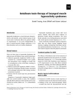
As the central venous hematocrit rises, there is increased viscosity and decreased blood
flow. When the hematocrit increases to Ͼ60%, there is decreased oxygen delivery
(1) (see Figure 46.1). Newborns have larger, irregularly shaped red blood cells (RBC)
with different membrane characteristics than the RBCs of adults (1–3). As viscosity
increases, there is impairment of tissue oxygenation and decreased glucose in plasma,
leading to increased risk of microthrombus formation. If these events occur in the
cerebral cortex, kidneys, or adrenal glands, significant damage may result. Hypoxia
and acidosis increase viscosity and deformity further. Poor perfusion increases the
possibility of thrombosis.
I.
DEFINITIONS
A. Polycythemia is defined as venous hematocrit of at least 65% (2,3). Hematocrit
measurements vary greatly with site of sample, and capillary hematocrit may be up
to 20% higher than venous (2). Hematocrit initially rises after birth from placental
transfer of RBCs, then decreases to baseline by approximately 24 hours (4). The
mean venous hematocrit of term infants is 53% in cord blood, 60% at 2 hours of
age, 57% at 6 hours of age, and 52% at 12 to 18 hours of age (2).
B. Hyperviscosity is defined as viscosity Ͼ2 standard deviations greater than the
mean (3). Blood viscosity, as described by Poiseuille, is the ratio of shear stress
to shear rate and is dependent on such factors as the pressure gradient along the
vessel, radius, length, and flow (4). The relationship between hematocrit and
viscosity is nearly linear below a hematocrit of 60%, but viscosity increases exponentially at a hematocrit of 70% or greater (Figure 46.1) (4,5).
Other factors affect blood viscosity, including plasma proteins such as fibrinogen, local blood flow, and pH (3,4). The hyperviscosity syndrome is usually seen
only in infants with venous hematocrits above 60%.
II. INCIDENCE. The incidence of polycythemia is 1% to 5% in term newborns
(1,3,6,7). Polycythemia is increased in babies that have intrauterine growth restriction
(IUGR), are small for gestational age (SGA), and are born postterm.
III.
CAUSES OF POLYCYTHEMIA
A. Placental red cell transfusion
1. Delayed cord clamping may occur either intentionally or in unattended
deliveries.
a. When the cord is clamped within 1 minute after birth, the blood volume
of the infant is approximately 80 mL/kg.
572
572-577_Cloherty_Ch46.indd 572
6/30/11 1:34 PM
Hematologic Disorders
573
Hypervolemic
Normovolemic
Oxygen Transport
Blood Flow
Viscosity
Relative
Change
40
50
60
Hematocrit (%)
30
30
40
50
60
Hematocrit (%)
Figure 46.1. Effect of hematocrit on viscosity, blood flow, and oxygen transport. (Adapted
from Glader B, Naiman JL. Erythrocyte disorders in infancy. In: Taeusch HW, Ballard RA,
Avery ME, eds. Diseases of the Newborn. Philadelphia: WB Saunders; 1991.)
b. When the cord is clamped 2 minutes after delivery, the blood volume of
the infant is 90 mL/kg.
c. In newborns with polycythemia, blood volume per kilogram of body
weight varies inversely in relation to birth weight (see Figure 46.2).
2. Cord stripping (thereby pushing more blood into the infant).
3. Holding the baby below the mother at delivery.
110
BV(mL/kg)
100
90
80
70
60
1,000
2,000 3,000 4,000
Birth Weight (g)
5,000
Figure 46.2. Nomogram designed for clinical use, correlating blood volume per kilogram
with birth weight in polycythemic neonates; BV ϭ blood volume. (From Rawlings JS, Pettett
G, Wiswell T, et al. Estimated blood volumes in polycythemic neonates as a function of birth
weight. J Pediatr 1982;101[4]:594–599.)
572-577_Cloherty_Ch46.indd 573
6/30/11 1:34 PM
574
P O LY C Y T H E M I A
4. Maternal-to-fetal transfusion is diagnosed with the Kleihauer-Betke stain
technique of acid elution to detect maternal cells in the circulation of the
newborn (see Chap. 45).
5. Twin-to-twin transfusion (see Chap. 11).
6. Forceful uterine contractions before cord clamping.
B. Placental insufficiency (increased fetal erythropoiesis secondary to chronic
intrauterine hypoxia)
1. SGA and IUGR infants.
2. Maternal hypertension syndromes (preeclampsia, renal disease, etc.).
3. Postterm infants.
4. Infants born to mothers with chronic hypoxia (heart disease, pulmonary disease).
5. Pregnancy at high altitude.
6. Maternal smoking.
C. Other conditions
1. Infants of diabetic mothers (increased erythropoiesis).
2. Some large-for-gestational-age (LGA) babies.
3. Infants with congenital adrenal hyperplasia, Beckwith-Wiedemann syndrome,
neonatal thyrotoxicosis, congenital hypothyroidism, trisomy 21, trisomy 13,
trisomy 18.
4. Drugs (maternal use of propranolol).
5. Dehydration of infant.
6. Sepsis (increase in fibrinogen, lower RBC deformability) (4).
IV. CLINICAL FINDINGS. Most infants with polycythemia are asymptomatic.
Clinical symptoms, syndromes, and laboratory abnormalities that have been described in association with polycythemia include the following:
A. Central nervous system (CNS). Poor feeding, lethargy, hypotonia, apnea, tremors, jitteriness, seizures, cerebral venous thrombosis.
B. Cardiorespiratory. Cyanosis, tachypnea, heart murmur, congestive heart failure,
cardiomegaly, elevated pulmonary vascular resistance, prominent vascular markings on chest x-ray.
C. Renal. Decreased glomerular filtration, decreased sodium excretion, renal vein
thrombosis, hematuria, proteinuria.
D. Other. Other thrombosis, thrombocytopenia, poor feeding, increased jaundice,
persistent hypoglycemia, hypocalcemia, testicular infarcts, necrotizing enterocolitis (NEC), priapism, disseminated intravascular coagulation.
All of these symptoms may be associated with polycythemia and hyperviscosity but may not be caused by it. They are common symptoms in many
neonatal disorders.
V. SCREENING. The routine screening of all newborns for polycythemia and/or hyperviscosity has been advocated by some authors (8,9). The timing and site of blood
572-577_Cloherty_Ch46.indd 574
6/30/11 1:34 PM
Hematologic Disorders
575
sampling alter the hematocrit value (3,10,11). We do not routinely screen well term newborns for this syndrome, because there are few data showing that treatment of asymptomatic patients with partial exchange transfusion is beneficial in the long term (3,11,12).
VI. DIAGNOSIS. The capillary blood or peripheral venous hematocrit level should
be determined in any baby who appears plethoric, who has any predisposing cause
of polycythemia, who has any of the symptoms mentioned in IV, or who is not well
for any reason.
A. Warming the heel before drawing blood for a capillary hematocrit determination
will give a better correlation with the peripheral venous or central hematocrit. If
the capillary blood hematocrit is above 65%, the peripheral venous hematocrit
should be determined.
B. Few hospitals are equipped to measure blood viscosity. If the equipment is available, the test should be done, because some infants with venous hematocrits under
65% will have hyperviscous blood (7).
VII.
MANAGEMENT
A. Once other causes of illness have been considered and excluded (e.g., sepsis, pneumonia, hypoglycemia), any child with symptoms that could be due to hyperviscosity should be considered for partial exchange transfusion if the peripheral
venous hematocrit is Ͼ65%.
B. Asymptomatic infants with a peripheral venous hematocrit between 60% and
70% can usually be managed by increasing fluid intake and repeating the
hematocrit in 4 to 6 hours.
C. Many neonatologists perform an exchange transfusion when the peripheral
venous hematocrit is Ͼ70% in the absence of symptoms, but this is a controversial issue (10–13).
D. The following formula can be used to calculate the exchange with normal saline that
will bring the hematocrit to 50% to 60%. In infants with polycythemia, the blood
volume varies inversely with the birth weight (see Fig. 46.2). Usually we take the
blood from the umbilical vein and replace it with normal saline in a peripheral
vein. Because randomized trials show no advantage with albumin and there is less
chance of infection, nonhuman products, such as saline, are preferred (14). There are
many methods of exchange (see Chap. 26).
Volume of exchange in mL
ϭ
(blood volume/kg ϫ weight in kg) ϫ (observed hematocrit Ϫ desired hematocrit)
observed hematocrit
Example: A 3-kg infant, hematocrit 75%, blood volume 80 mL/kg—to bring
hematocrit to 50%:
(80 mL ϫ 3 kg) ϫ (75 Ϫ 50)
75
240 mL ϫ 25
ϭ
75
ϭ 80-mL exchange
Volume of exchange (in mL) ϭ
572-577_Cloherty_Ch46.indd 575
6/30/11 1:34 PM
576
P O LY C Y T H E M I A
The total volume exchanged is usually 15 to 20 mL/kg of body weight.
This will depend on the observed hematocrit. (Blood volume may be up to
100 mL/kg in polycythemic infants.)
VIII.
OUTCOME
A. Infants with polycythemia and hyperviscosity who have decreased cerebral
blood flow velocity and increased vascular resistance develop normal cerebral
blood flow following partial exchange transfusion (12). They also have improvement in systemic blood flow and oxygen transport (2,5,11,13).
B. The long-term neurologic outcome in infants with asymptomatic polycythemia
and/or hyperviscosity, whether treated or untreated, remains controversial.
1. One trial with small numbers of randomized patients showed decreased IQ
scores in school-age children who had neonatal hyperviscosity syndrome, in
both treated and untreated newborns (10,15).
2. Another retrospective study, with small numbers of patients, showed no difference in the neurologic outcome of patients with asymptomatic neonatal
polycythemia, including both treated and untreated newborns (16).
3. A small prospective study showed no difference at follow-up between control
infants and those with hyperviscosity, between those with symptomatic and
asymptomatic hyperviscosity, and between asymptomatic infants treated with
partial exchange transfusion and those who were observed. Analysis revealed
that other perinatal risk factors and race, rather than polycythemia or partial
exchange transfusion, significantly influenced the long-term outcome (2,11).
4. An increased incidence of NEC following partial exchange transfusions by umbilical vein has been reported (15,17). NEC was not seen in one retrospective
analysis of 185 term polycythemic babies given partial exchange transfusions
with removal of blood from the umbilical vein and reinfusion of a commercial
plasma substitute through peripheral veins (18).
5. A larger prospective, randomized clinical trial comparing partial exchange
transfusion with symptomatic care (increased fluid intake, etc.) equally
balanced for risk factors and the etiologies of the polycythemia will be
necessary to give guidelines for treatment of the asymptomatic newborn with
polycythemia and/or hyperviscosity.
6. Partial exchange transfusion will lower hematocrit, decrease viscosity, and
reverse many of the physiologic abnormalities associated with polycythemia
and/or hyperviscosity but has not been shown to significantly change the longterm outcome of these infants (2).
REFERENCES
1. Glader B. Erythrocyte disorders in infancy. In: Taeusch HW, Ballard RA, Avery ME,
eds. Diseases of the newborn. 6th ed. Philadelphia: WB Saunders; 1991.
2. Werner EJ. Neonatal polycythemia and hyperviscosity. Clin Perinatol 1995;22(3):693–710.
3. Linderkamp O. Blood Viscosity of the Neonate. NeoReviews 2004;5:406–415.
4. Rosenkrantz TS. Polycythemia and hyperviscosity in the newborn. Semin Thromb
Hemost 2003;29(5):515–527.
5. Swetnam SM, Yabek SM, Alverson DC. Hemodynamic consequences of neonatal
polycythemia. J Pediatr 1987;110:443–447.
572-577_Cloherty_Ch46.indd 576
6/30/11 1:34 PM
Hematologic Disorders
577
6. Lindermann R, Haines L. Evaluation and treatment of polycythemia in the neonate.
In: Christensen RD, ed. Hematologic problems of the neonate. Philadelphia: WB
Saunders; 2000.
7. Wirth FH, Goldberg KE, Lubchenco LO. Neonatal hyperviscosity: I. Incidence.
Pediatrics 1979;63(6):833–886.
7.5. Ramamurthy RS, Berlanga M. Postnatal alteration in hematocrit and viscosity in normal
and polycythemic infants. J Pediatr 1987;110(6):929–934.
8. Drew JH, Guaran RL, Cichello M, et al. Neonatal whole blood hyperviscosity: the
important factor influencing later neurologic function is the viscosity and not the
polycythemia. Clin Hemorheol Microcirc 1997;17(1):67–72.
9. Wiswell TE, Cornish JD, Northam RS. Neonatal polycythemia: frequency of clinical
manifestations and other associated findings. Pediatrics 1986;78(1):26–30.
10. Delaney-Black VD, Camp BW, Lubchenco LO, et al. Neonatal hyperviscosity association
with lower achievement and IQ scores at school age. Pediatrics 1989;83(5):662–667.
11. Bada H, Korones SB, Pourcyrous M, et al. Asymptomatic syndrome of polycythemic
hyperviscosity: effect of partial plasma exhange transfusion. J Pediatr 1992;120(4 pt 1):
579–585.
12. Oski FA, Naiman JL. Hematologic problems in the newborn. 3rd ed. Philadelphia: WB
Saunders; 1982:87–96.
13. Phibbs RH, Clapp DW, Shannon KM. Hematologic problems. In: Klaus MH, Fanaroff
AA, Eds. Care of the high risk neonate. Philadelphia: WB Saunders; 1993:421.
14. de Waal KA, Baerts W, Offringa M. Systematic review of the optimal fluid for dilutional
exchange transfusion in neonatal polycythaemia. Arch Dis Child Fetal Neonatal Ed
2006;91(1):F7–F10.
15. Black VD, Lubchenco LO. Neonatal polycythemia and hyperviscosity. Pediatr Clin
North Am 1982;5:1137–1148.
16. Høst A, Ulrich M. Late prognosis in untreated neonatal polycythemia with minor or no
symptoms. Acta Paediatr Scand 1982;71(4):629–633.
17. Black VD, Rumack CM, Lubchenco LO, et al. Gastrointestinal injury in polycythemic
term infants. Pediatrics 1985;76(2):225–231.
18. Hein HA, Lathrop SS. Partial exchange transfusion in term, polycythemic neonates:
absence of association with severe gastrointestinal injury. Pediatrics 1987;80(1):75–78.
572-577_Cloherty_Ch46.indd 577
6/30/11 1:34 PM
47
Neonatal
Thrombocytopenia
Chaitanya Chavda, Matthew Saxonhouse, and
Martha Sola-Visner
I.
INTRODUCTION. Thrombocytopenia in neonates is traditionally defined as a
platelet count of less than 150 ϫ 103/mcL and is classified as mild (100–150 ϫ 103/
mcL), moderate (50–99 ϫ103/mcL), or severe (Ͻ50 ϫ 103/mcL). However, platelet counts in the 100–150 ϫ 103/mcL range are somewhat more common among
healthy neonates than among healthy adults. For that reason, careful follow-up and
expectant management in an otherwise healthy-appearing neonate with mild, transient thrombocytopenia is an acceptable approach, although lack of quick resolution,
worsening of thrombocytopenia, or changes in clinical condition should prompt further evaluation. The incidence of thrombocytopenia in neonates varies significantly,
depending on the population studied. Specifically, while the overall incidence of neonatal thrombocytopenia is relatively low (0.7%–0.9%) (1), the incidence among neonates admitted to the Neonatal Intensive Care Unit (NICU) is very high (22%–35%)
(2–4). Within the NICU, mean platelet counts are lower among preterm neonates
than among neonates born at or near term (5), and the incidence of thrombocytopenia is inversely correlated to the gestational age, reaching approximately 70% among
neonates born with a weight Ͻ1,000 g (6).
II.
APPROACH TO THE THROMBOCYTOPENIC NEONATE. When evaluating
a thrombocytopenic neonate, the first step to narrow the differential diagnosis is to
classify the thrombocytopenia as either early onset (within the first 72 h of life)
or late onset (after the initial 72 h of life), and to determine whether the infant is
clinically ill or well. Importantly, infection and sepsis should always be considered
near the top of the differential diagnosis (regardless of the time of presentation and the
infant’s appearance), as any delay in diagnosis and treatment can have life-threatening
consequences.
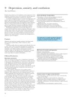
A. Early-onset thrombocytopenia (Figure 47.1). The most frequent cause of earlyonset thrombocytopenia in a well-appearing neonate is placental insufficiency,
as occurs in infants born to mothers with pregnancy-induced hypertension/preeclampsia or diabetes and in those with intrauterine growth restriction (IUGR)
(7,8). This thrombocytopenia is always mild to moderate, presents immediately
or shortly after birth, and resolves within 7 to 10 days. If an infant with a prenatal
history consistent with placental insufficiency and mild-to-moderate thrombocytopenia remains clinically stable and the platelet count normalizes within 10 days,
no further evaluation is necessary. However, if the thrombocytopenia becomes
severe and/or persists Ͼ10 days, further investigation is necessary.
Severe early-onset thrombocytopenia in an otherwise healthy infant should
trigger suspicion for an immune-mediated thrombocytopenia, either autoimmune
578
578-587_Cloherty_Ch47.indd 578
6/30/11 1:34 PM
Hematologic Disorders
579
Early Onset Thrombocytopenia
(Յ72 after birth)
Mild to Moderate
(PC 50,000–149,000/µL)
• Baby well
• Evidence of
placental
insufficiency
Evaluate for sepsis,
DIC and NAIT
• Baby ill
• No evidence
of placental
insufficiency
• PC rising
• PC normal
by 10 days
No further
evaluation
Severe
(PC Ͻ50,000/µL)
• No sepsis,
DIC, or NAIT
• Persistent
thrombocytopenia
• Evidence of
Sepsis, DIC, or NAIT
• PC improved with
treatment
• Mother with
thrombocytopenia?
• PE consistent with
TAR, Proximal
radio-ulnar synostosis,
Trisomy 13, 18, or 21,
Turners or Noonan
syndrome?
No further evaluation
If no to all questions, consider:
• TORCH infections
• Viral infections (HIV, enterovirus)
• Chromosomal abnormalities
• Inborn errors of metabolism
• Thrombosis (i.e., RVT)
• Congenital thrombocytopenias
Yes to any question:
Confirmatory tests
Evaluate for
sepsis, DIC
• Evidence of
sepsis, DIC
• PC improving
with treatment
• No evidence
of sepsis, DIC
• Persistent
thrombocytopenia
No further evaluation
• PC not rising
• PC not normal
by 10 days
Figure 47.1. Guidelines for the evaluation of neonates with early-onset thrombocytopenia
(Յ72 hours of life). PC ϭ platelet count; DIC ϭ disseminated intravascular coagulation;
NAIT ϭ neonatal alloimmune thrombocytopenia; RVT ϭ renal vein thrombosis.
(i.e., the mother is also thrombocytopenic) or alloimmune (i.e., the mother has
a normal platelet count). These varieties of thrombocytopenia are discussed in
detail below.
Early-onset thrombocytopenia of any severity in an ill-appearing term or
preterm neonate should prompt evaluation for sepsis, congenital viral or parasitic infections, or disseminated intravascular coagulation (DIC). DIC is most
frequently associated with sepsis but can also be secondary to birth asphyxia.
In addition to these considerations, the affected neonate should be carefully
examined for any radial abnormalities (suggestive of thrombocytopenia-absent radius (TAR) syndrome, amegakaryocytic thrombocytopenia with radioulnar synostosis (ATRUS), or Fanconi anemia). Although thrombocytopenia associated with
Fanconi almost always presents later (during childhood), neonatal cases have been
578-587_Cloherty_Ch47.indd 579
6/30/11 1:34 PM
580
N E O N AT A L T H R O M B O C Y T O P E N I A
reported (9). In these patients, thumb abnormalities are frequently found, and the
diepoxybutane test is nearly always diagnostic. If the infant has radial abnormalities with normal appearing thumbs, TAR syndrome should be considered (10).
The platelet count is usually Ͻ50 ϫ 103/mcL and the white cell count is elevated
in Ͼ90% of TAR syndrome patients, sometimes exceeding 100 ϫ 103/mcL and
mimicking congenital leukemia. Infants that survive the first year of life generally
do well, since the platelet count then spontaneously improves to low-normal levels
that are maintained through life (11). The inability to rotate the forearm on physical examination, in the presence of severe early-onset thrombocytopenia, suggests
the rare diagnosis of congenital ATRUS. Radiologic examination of the upper
extremities in these infants confirms the proximal synostosis of the radial and
ulnar bones (12). Other genetic disorders associated with early-onset thrombocytopenia include trisomy 21, trisomy 18, trisomy 13, Turner syndrome, Noonan
syndrome, and Jacobsen syndrome.
The presence of hepato- or splenomegaly is suggestive of a viral infection,
although it can also be seen in hemophagocytic syndrome and liver failure from
different etiologies. Other diagnoses, such as renal vein thrombosis, Kasabach–
Merritt syndrome, and inborn errors of metabolism (mainly propionic acidemia
and methylmalonic acidemia), should be considered and evaluated based on specific clinical indications (i.e., hematuria in renal vein thrombosis, presence of a
vascular tumor in Kasabach-Merritt syndrome).
B. Late-onset thrombocytopenia (Figure 47.2). The most common causes of
thrombocytopenia of any severity presenting after 72 hours of life are sepsis (bacterial or fungal) and necrotizing enterocolitis (NEC). Affected infants are usually
ill appearing and have other signs suggestive of sepsis and/or NEC. However,
thrombocytopenia can be the presenting sign of these processes and can precede
clinical deterioration. Appropriate treatment with antibiotics, fluid resuscitation,
and bowel rest (if NEC is considered) usually improves the platelet count in 1 to 2
weeks, although in some infants, the thrombocytopenia persists for several weeks.
The reasons underlying this prolonged thrombocytopenia are unclear.
If bacterial/fungal sepsis and NEC are ruled out, viral infections such as herpes simplex virus, CMV, or enterovirus should be considered. These are frequently
accompanied by abnormal liver enzymes. If the infant has or has recently had a
central venous or arterial catheter, thromboses should be part of the differential
diagnosis. Finally, drug-induced thrombocytopenia should be considered if the
infant is clinically well and is receiving heparin, antibiotics (penicillins, ciprofloxacin, cephalosporins, metronidazole, vancomycin, and rifampin), indomethacin, famotidine, cimetidine, phenobarbital, or phenytoin, among others (13,14).
Other less common causes of late-onset thrombocytopenia include inborn errors
of metabolism and Fanconi anemia (rare).
Novel tools to evaluate platelet production and aid in the evaluation of
thrombocytopenia have been recently developed and are likely to become widely
available to clinicians in the near future. Among those, the immature platelet
fraction (IPF) measures the percentage of newly released platelets (Ͻ24 hrs). The
IPF can be measured in a standard hematologic cell counter (Sysmex XE-2100
hematology analyzer) as part of the complete cell count and can help differentiate
thrombocytopenias associated with decreased platelet production from those with
increased platelet destruction in a manner similar to the use of reticulocyte counts
to evaluate anemia (15). Recent studies have shown the usefulness of the IPF
to evaluate mechanisms of thrombocytopenia and to predict platelet recovery in
578-587_Cloherty_Ch47.indd 580
6/30/11 1:34 PM
Hematologic Disorders
581
Late-onset Thrombocytopenia
(>72 hours after birth)
Evaluate for bacterial/fungal
sepsis and NEC
• Evidence of sepsis
and/or NEC
• PC normalizing
with treatment
No further
evaluation for
thrombocytopenia
No evidence of
bacterial/fungal
sepsis and/or NEC
Evaluate for:
• DIC
• Viral infection (i.e., HSV, acquired CMV)
• Thrombosis (especially if central line present)
• Consider drug-induced thrombocytopenia
• Consider inborn errors of metabolism
• Consider Fanconi anemia
Figure 47.2. Guidelines for the evaluation of neonates with late-onset thrombocytopenia
(Ͼ72 hours of life). PC ϭ platelet count; NEC ϭ necrotizing enterocolitis; HSV ϭ herpes
simplex virus; CMV ϭ cytomegalovirus.
neonates (16,17). The IPF should be particularly helpful to guide the diagnostic
evaluation of infants with thrombocytopenia of unclear etiology.
III.
IMMUNE THROMBOCYTOPENIA. Immune thrombocytopenia occurs due
to the passive transfer of antibodies from the maternal to the fetal circulation. There
are two distinctive types of immune mediated thrombocytopenia: (i) neonatal alloimmune thrombocytopenia (NAIT) and (ii) autoimmune thrombocytopenia. In
NAIT, the antibody is produced in the mother against a specific human platelet
antigen (HPA) present in the fetus but absent in the mother. The antigen is inherited from the father of the fetus. The anti-HPA antibody produced in the maternal
serum crosses the placenta and reaches the fetal circulation, leading to platelet destruction and thrombocytopenia. In autoimmune thrombocytopenia, the antibody
is directed against an antigen on the mother’s own platelets (autoantibody) as well as
on the baby’s platelets. The maternal autoantibody also crosses the placenta, resulting in destruction of fetal platelets and thrombocytopenia.
A. Neonatal alloimmune thrombocytopenia. NAIT should be considered in any
neonate who presents with severe thrombocytopenia at birth or shortly thereafter,
particularly in the absence of other risk factors, clinical signs, or abnormalities in
the physical exam or in the other blood cell counts. In a study of more than 200
neonates with thrombocytopenia, using a platelet count Ͻ50 ϫ 103/mcL in the first
578-587_Cloherty_Ch47.indd 581
6/30/11 1:34 PM
582
N E O N AT A L T H R O M B O C Y T O P E N I A
day of life as a screening indicator identified 90% of the patients with NAIT (18).
In addition, the combination of severe neonatal thrombocytopenia with a parenchymal (rather than intraventricular) intracranial hemorrhage is highly suggestive of
NAIT.
Laboratory Investigation: When NAIT is suspected, blood should be collected from the mother and father and submitted for confirmatory testing
(if accessible). The initial antigen screening should include HPA 1, 3, and 5.
This evaluation should identify approximately 90% of cases of NAIT. However,
if the diagnosis is strongly suspected and the initial evaluation is negative, further testing should be undertaken for HPA 9 and 15 (and HPA 4 if the parents
are of Asian descent) (19). If positive, these tests will reveal an antibody in the
mother’s plasma directed against the specific platelet antigen in the father. If
blood cannot be collected from the parents in a timely fashion, neonatal serum
may be screened for the presence of anti-platelet antibodies. However, a low
antibody concentration in the neonate coupled with binding of the antibodies
to the infant’s platelets can lead to false-negative results. Due to the complexity
of testing, evaluations should be performed in an experienced reference laboratory that has a large number of typed controls available for antibody detection
and the appropriate DNA-based technology to type multiple antigens.
Brain imaging studies should be performed as soon as NAIT is suspected,
regardless of the presence or absence of neurologic manifestations, because findings from these studies will dictate the aggressiveness of the treatment regimen for
the affected infant and for the mother’s future pregnancies. The clinical course
of NAIT is short in most cases, often resolving almost entirely within 2 weeks.
However, to confirm the diagnosis, it is important to follow the platelet count
frequently until a normal count is achieved.
Management: The management of NAIT differs depending on the specific
clinical scenario:
1. Suspected NAIT in an unknown pregnancy
2. Known case of NAIT
3. Antenatal management of pregnant woman with previous history of NAIT
a. Management of the neonate with suspected NAIT in an unknown
pregnancy. Based on recent data demonstrating that a large proportion of
infants with NAIT respond to random-donor platelet transfusions, this is
now considered the first line of therapy for infants in whom NAIT is
suspected (20).
i. If the patient is clinically stable and does not have evidence of an intracranial hemorrhage, platelets are usually given when the platelet count
is less than 30 ϫ 103/mcL, although this is arbitrary.
ii. If the patient has evidence of an intracranial hemorrhage, the goal is
to maintain a platelet count greater than 100 ϫ 103/mcL. This can be
challenging in neonates with NAIT.
iii. In addition to platelets, if the diagnosis of NAIT is confirmed or
strongly suspected, intravenous immune globulin (IVIG) (1 g/kg/day
for up to 2 consecutive days) may be infused to increase the patient’s
own platelets and potentially to protect the transfused platelets (21).
Because in NAIT the platelet count usually falls after birth, IVIG can
be infused when the platelet count is between 30 and 50 ϫ 103/mcL,
to try to prevent a further drop.
578-587_Cloherty_Ch47.indd 582
6/30/11 1:34 PM
Hematologic Disorders
583
iv. It is important to keep in mind that some infants with NAIT fail to respond to random-donor platelets and IVIG. For that reason, the blood
bank should be immediately alerted about any infant with suspected
NAIT, and arrangements should be made to secure a source of antigen-negative platelets (either from HPA-1b1b and 5a5a donors, which
should be compatible in Ͼ90% of cases, or from the mother) as soon
as possible if there is no response to the initial therapies. If maternal
platelets are used, they need to be concentrated to decrease the amount
of anti-platelet antibodies (present in the mother’s plasma) infused into
the infant. Platelets can also be washed to eliminate the plasma, but
this induces more damage to the platelets than concentrating them
(19). Of note, in some European countries, HPA-1b1b and 5a5a platelets are maintained in the blood bank inventory and are immediately
available for use. In those cases, these are preferable to random-donor
platelets and/or IVIG and should be the first line of therapy.
v. Methylprednisolone (1 mg/kg bid for 3–5 days) has also been used in
individual case reports and small series, but should only be considered
if the infant does not respond to random platelets and IVIG, there is no
suspicion of bacterial or viral infection, and antigen-matched platelets
are not readily available. Some experts recommend IV methylprednisolone at a low dose (1 mg q8h) on the days that IVIG is given (19).
b. Management of the neonate with known NAIT. When a neonate is born
to a mother who had a previous pregnancy affected by confirmed NAIT, genotypically matched platelets (e.g., HPA-1b1b platelets) should be available in
the blood bank at the time of delivery and should be the first line of therapy if
the infant is thrombocytopenic.
c. Antenatal management of pregnant women with previous history
of NAIT. Mothers who delivered an infant with NAIT should be followed
in high-risk obstetric clinics during all future pregnancies. The intensity of
prenatal treatment will be based on the severity of the thrombocytopenia
and the presence or absence of intracranial hemorrhage (ICH) in the previously affected fetus. This is particularly important to assess the risk of developing an ICH in the current pregnancy and to minimize this risk. Current
recommendations involve maternal treatment with IVIG (1–2 g/kg/wk) Ϯ
steroids, starting at 12 or at 20 to 26 weeks’ gestation, depending on whether
the previously affected fetus suffered an ICH, and if so, at what time during
pregnancy (19).
B. Autoimmune thrombocytopenia. The diagnosis of neonatal autoimmune thrombocytopenia should be considered in any neonate who has early-onset thrombocytopenia and a maternal history of either idiopathic thrombocytopenic purpura (ITP)
or an autoimmune disease (with or without thrombocytopenia). A retrospective
study of obstetric patients who had ITP (including a high number of mothers who
had thrombocytopenia during their pregnancies) demonstrated a relatively high incidence of affected babies: 25% of neonates exhibited thrombocytopenia at birth;
the thrombocytopenia was severe in 9%, and 15% received treatment for it (22).
Other large studies confirmed an incidence of severe neonatal thrombocytopenia in
this population ranging from 8.9% to 14.7%, with ICH occurring in 0% to 1.5%
of affected neonates (23–25). Based on these data, it is recommended that all neonates born to mothers who have autoimmune diseases undergo a screening platelet
578-587_Cloherty_Ch47.indd 583
6/30/11 1:34 PM
584
N E O N AT A L T H R O M B O C Y T O P E N I A
count at or shortly after birth. If the platelet count is normal, no further evaluation
is necessary. If the infant has mild thrombocytopenia, however, the platelet count
should be repeated in 2 to 3 days, since it usually reaches the nadir between days
2 and 5 after birth. If the platelet count is less than 30 ϫ 103/mcL, IVIG (1 g/kg,
repeated if necessary) is the first line of therapy. Random-donor platelets, in addition
to IVIG, should be provided only if the infant has evidence of active bleeding. Cranial imaging should be obtained in all infants with platelet counts Ͻ50 ϫ 103/mcL
to evaluate for intracranial hemorrhage. Importantly, neonatal thrombocytopenia
secondary to maternal ITP may last for months and requires long-term monitoring
and sometimes a second dose of IVIG at 4 to 6 weeks of life.
Maternal management. Even if the mother has true ITP, it appears that fetal
hemorrhage in utero is very rare compared with the small but definite risk of such
hemorrhage in alloimmune thrombocytopenia. Because of that, treatment of ITP
during pregnancy is mostly based on the risk of maternal hemorrhage (26). A small
prospective randomized trial of low-dose betamethasone (1.5 mg/day orally) failed
to prevent thrombocytopenia in newborns (27). IVIG given prenatally to the
mother with ITP has also not been clearly shown to affect the fetal platelet count.
There is in general little correlation between fetal platelet counts and either
maternal platelet counts, platelet antibody levels, or history of maternal splenectomy. However, attempts to measure the fetal platelet count before delivery are
not recommended due to the risk associated with such attempts. In regard to the
mode of delivery, there is no evidence that cesarean section is safer for the fetus
with thrombocytopenia than uncomplicated vaginal delivery. Given this fact,
combined with the difficulty predicting severe thrombocytopenia in neonates and
the very low risk of serious hemorrhage, the 2010 International Consensus Report
on the Investigation and Management of Primary Immune Thrombocytopenia
concluded that the mode of delivery in ITP patients should be determined by
purely obstetric indications (26).
IV. PLATELET TRANSFUSIONS IN THE NICU (see Chapter 42). Recent
studies have shown that there is great variability in neonatal transfusion practices in the
United States and worldwide (28,29). To a large extent, this is attributable to the paucity of scientific evidence in the field. Only one randomized trial has compared different
platelet transfusion thresholds in neonates, and it was limited to very low birth weight
(VLBW) infants in the first week of life (30). This study found no differences in the incidence or severity of intraventricular hemorrhages (IVHs) between a group of neonates
transfused for any platelet count less than 150 ϫ 103/mcL and a group transfused only
for counts below 50 ϫ 103/mcL. Based on these findings, the investigators concluded
that transfusing VLBW infants with platelet counts Ͼ50 ϫ 103/mcL did not reduce
the risk of IVH. A more recent retrospective study evaluated whether platelet counts
Ͻ50 ϫ 103/mcL could be safely tolerated in neonates. This study concluded that using
a platelet count of 30 ϫ 103/mcL as a transfusion threshold was a safe practice for stable
neonates with no prior hemorrhages (31). Based on this limited evidence, we currently
propose administering platelet transfusions to neonates according to the criteria shown
in Table 47.1.
There is more consensus in regard to the platelet product that should be transfused. Most experts agree that neonates should receive 10 to 15 mL/kg of a standard
platelet suspension, either a platelet concentrate (“random-donor platelets”) or apheresis platelets. Each random-donor platelet unit has approximately 50 mL of volume
and contains approximately 10 ϫ 109 platelets per 10 ml (32). There is no need to pool
578-587_Cloherty_Ch47.indd 584
6/30/11 1:34 PM
Hematologic Disorders
Table 47.1
585
Guidelines for Platelet Transfusion
Platelet Count
(؋ 103/mcL)
Ͻ30
Transfuse all
30–49
Transfuse if:
• BW Ͻ1,500g and Յ7 days old
• Clinically unstable
• Concurrent coagulopathy
• Previous significant hemorrhage (i.e., grade 3 or 4 IVH)
• Prior to surgical procedure
• Postoperative period (72 hours)
50–100
Transfuse if:
• Active bleeding
• NAIT with intracranial bleed
• Before or after neurosurgical procedures
BW ϭ birth weight; NAIT ϭ neonatal alloimmune thrombocytopenia.
more than one random-donor unit for a neonatal transfusion, a practice that (while
still somewhat prevalent) only increases donor exposures and induces platelet activation without any benefit. Two additional important considerations in neonatology
are the prevention of transfusion-transmitted CMV infections and graft-versus-host
disease (GVHD). Most blood banks provide either CMV-negative or leukoreduced
products to neonates, both of which significantly reduce (but do not eliminate) the
risk of transfusion transmitted CMV. GVHD is effectively prevented by irradiating
cellular blood products prior to transfusion. Of note, most neonatal cases of GVHD
have been reported in neonates with underlying immunodeficiencies, receiving intrauterine or large volume transfusions (i.e., double exchange transfusions) or receiving
blood products from a first-degree relative. These are all absolute indications for irradiating blood products (32).
When making platelet transfusion decisions, it is important for neonatologists to
be aware of the risks associated with these transfusions. In the case of platelet suspensions, the risk of bacterial contamination is higher than the combined risk of all viral
infections for which platelets are routinely tested. In addition, platelet transfusions
can induce transfusion-associated lung injury (TRALI), a process characterized by the
onset of hypoxemia and bilateral pulmonary infiltrates within 6 hours of a transfusion
(33). Given that neonates have frequent episodes of respiratory decompensation due
to different causes, TRALI is likely to be underrecognized in the NICU. Several recent
publications have also shown a strong association between the number of platelet
transfusions and the mortality rate among NICU patients (34–37). It is unclear from
these studies whether this association simply reflects sicker patients receiving more
platelets or whether platelet transfusions adversely affect outcomes. Nevertheless,
while we await for data from well-designed randomized controlled studies, platelet
transfusion decisions in neonates should be made thoughtfully, carefully balancing the
risks and benefits in each individual patient.
578-587_Cloherty_Ch47.indd 585
6/30/11 1:34 PM
586
N E O N AT A L T H R O M B O C Y T O P E N I A
REFERENCES
1. Burrows RF, Kelton JG. Fetal thrombocytopenia and its relation to maternal thrombocytopenia. N Engl J Med 1993;329(20):1463–1466.
2. Andrew M, Castle V, Saigal S, et al. Clinical impact of neonatal thrombocytopenia.
J Pediatr 1987;110(3):457–464.
3. Castle V, Andrew M, Kelton J, et al. Frequency and mechanism of neonatal thrombocytopenia. J Pediatr 1986;108(5 pt 1):749–755.
4. Mehta P, Vasa R, Neumann L, et al. Thrombocytopenia in the high-risk infant. J Pediatr
1980;97(5):791–794.
5. Wiedmeier SE, et al. Platelet reference ranges for neonates, defined using data from over
47,000 patients in a multihospital healthcare system. J Perinatol 2009;29(2):130–136.
6. Christensen RD, et al. Thrombocytopenia among extremely low birth weight neonates:
data from a multihospital healthcare system. J Perinatol 2006;26(6):348–353.
7. Murray NA, Roberts IA. Circulating megakaryocytes and their progenitors in early
thrombocytopenia in preterm neonates. Pediatr Res 1996;40(1):112–119.
8. Murray NA, Watts TL, Roberts IA. Endogenous thrombopoietin levels and effect of
recombinant human thrombopoietin on megakaryocyte precursors in term and preterm
babies. Pediatr Res 1998;43(1):148–151.
9. Gershanik JJ, Morgan SK, Akers R. Fanconi’s anemia in a neonate. Acta Paediatr Scand
1972;61(5):623–625.
10. Hedberg VA, Lipton JM. Thrombocytopenia with absent radii. A review of 100 cases.
Am J Pediatr Hematol Oncol 1998;10(1):51–64.
11. Geddis AE. Inherited thrombocytopenia: congenital amegakaryocytic thrombocytopenia
and thrombocytopenia with absent radii. Semin Hematol 2006;43(3):196–203.
12. Sola MC, Slayton WB, Rimsza LM, et al. A neonate with severe thrombocytopenia and
radio-ulnar synostosis. J Perinatol 2004;24(8):528–530.
13. Aster RH, Bougie DW. Drug-induced immune thrombocytopenia. N Engl J Med
2007;357(6):580–587.
14. Aster RH, Curtis BR, McFarland, et al. Drug-induced immune thrombocytopenia:
pathogenesis, diagnosis, and management. J Thromb Haemost 2009;7(6):911–918.
15. Abe Y, Wada H, Tomatsu H, et al. A simple technique to determine thrombopoiesis
level using immature platelet fraction (IPF). Thromb Res 2006;118(4):463–469.
16. Cremer M, Paetzold J, Schmalisch G, et al. Immature platelet fraction as novel laboratory parameter predicting the course of neonatal thrombocytopenia. Br J Haematol
2008;144(4):619–621.
17. Cremer M, Weimann A, Schmalisch G, et al. Immature platelet values indicate impaired
megakaryopoietic activity in neonatal early-onset thrombocytopenia. Thromb Haemost
2010;103(5):1016–1021.
18. Bussel JB, Zacharoulis S, Kramer K, et al. Clinical and diagnostic comparison of neonatal
alloimmune thrombocytopenia to non-immune cases of thrombocytopenia. Pediatr Blood
Cancer 2005;45(2):176–183.
19. Bussel JB, Sola-Visner M. Current approaches to the evaluation and management of the fetus and neonate with immune thrombocytopenia. Semin Perinatol
2009;33(1):35–42.
20. Kiefel V, Bassler D, Kroll H, et al. Antigen-positive platelet transfusion in neonatal
alloimmune thrombocytopenia (NAIT). Blood 2006;107(9):3761–3763.
21. Mueller-Eckhardt C, Kiefel V, Grubert A. High-dose IgG treatment for neonatal
alloimmune thrombocytopenia. Blut 1989;59(1):145–146.
22. Webert KE, Mittal R, Sigoun C, et al. A retrospective 11-year analysis of obstetric
patients with idiopathic thrombocytopenic purpura. Blood 2003;102(13):4306–4311.
23. Kaplan C, Daffos F, Forestier F, et al. Fetal platelet counts in thrombocytopenic
pregnancy. Lancet 1990;336(8721):979–982.
24. Samuels P, Bussel JB, Braitman LE, et al. Estimation of the risk of thrombocytopenia in
the offspring of pregnant women with presumed immune thrombocytopenic purpura.
N Engl J Med 1990;323(4):229–235.
578-587_Cloherty_Ch47.indd 586
6/30/11 1:34 PM
Hematologic Disorders
587
25. Burrows RF, Kelton JG. Pregnancy in patients with idiopathic thrombocytopenic purpura:
assessing the risks for the infant at delivery. Obstet Gynecol Surv 1993;48(12):781–788.
26. Provan D, Stasi R, Newland AC, et al. International consensus report on the investigation
and management of primary immune thrombocytopenia. Blood 2010;115(2):168–186.
27. Christiaens GC, Nieuwenhuis HK, von dem Borne AE, et al. Idiopathic thrombocytopenic purpura in pregnancy: a randomized trial on the effect of antenatal low dose
corticosteroids on neonatal platelet count. Br J Obstet Gynaecol 1990;97(10):893–898.
28. Josephson CD, Su LL, Christensen RD, et al. Platelet transfusion practices among
neonatologists in the United States and Canada: results of a survey. Pediatrics
2009;123(1):278–285.
29. Kahn DJ, Richardson DK, Billett HH. Inter-NICU variation in rates and management of
thrombocytopenia among very low birth weight infants. J Perinatol 2003;23(4):312–316.
30. Andrew M, Vegh P, Caco C, et al. A randomized, controlled trial of platelet transfusions
in thrombocytopenic premature infants. J Pediatr 1993;123(2):285–291.
31. Murray NA, Howarth LJ, McCoy MP, et al. Platelet transfusion in the management
of severe thrombocytopenia in neonatal intensive care unit patients. Transfus Med
2002;12(1):35–41.
32. Strauss RG. Blood banking and transfusion issues in perinatal medicine. In: Christensen
R, ed. Hematologic Problems of the neonate. Philadephia: WB Saunders; 2000:405–425.
33. Goldman M, Webert KE, Arnold DM, et al. Proceedings of a consensus conference:
towards an understanding of TRALI. Transfus Med Rev 2005;19(1):2–31.
34. Baer VL, Lambert DK, Henry E, et al. Do platelet transfusions in the NICU adversely
affect survival? Analysis of 1600 thrombocytopenic neonates in a multihospital
healthcare system. J Perinatol 2007;27(12):790–796.
35. Del Vecchio A, Sola MC, Theriaque DW, et al. Platelet transfusions in the neonatal
intensive care unit: factors predicting which patients will require multiple transfusions.
Transfusion 2001;41(6):803–808.
36. Garcia MG, Duenas E, Sola MC, et al. Epidemiologic and outcome studies of patients
who received platelet transfusions in the neonatal intensive care unit. J Perinatol
2001;21(7):415–420.
37. Stanworth SJ, Clarke P, Watts T, et al. Prospective, observational study of outcomes in
neonates with severe thrombocytopenia. Pediatrics 2009;124(5):826–834.
578-587_Cloherty_Ch47.indd 587
6/30/11 1:34 PM