Comparison of NIV-NAVA and NCPAP in facilitating extubation for very preterm infants
Bạn đang xem bản rút gọn của tài liệu. Xem và tải ngay bản đầy đủ của tài liệu tại đây (776.16 KB, 7 trang )
Lee et al. BMC Pediatrics
(2019) 19:298
/>
RESEARCH ARTICLE
Open Access
Comparison of NIV-NAVA and NCPAP in
facilitating extubation for very preterm
infants
Byoung Kook Lee1, Seung Han Shin2,3* , Young Hwa Jung2,4, Ee-Kyung Kim2,3 and Han-Suk Kim2,3
Abstract
Background: Various types of noninvasive respiratory modalities that lead to successful extubation in preterm
infants have been explored. We aimed to compare noninvasive neurally adjusted ventilatory assist (NIV-NAVA) and
nasal continuous positive airway pressure (NCPAP) for the postextubation stabilization of preterm infants.
Methods: This retrospective study was divided into two distinct periods, between July 2012 and June 2013 and
between July 2013 and June 2014, because NIV-NAVA was applied beginning in July 2013. Preterm infants of less
than 30 weeks GA who had been intubated with mechanical ventilation for longer than 24 h and were weaned to
NCPAP or NIV-NAVA after extubation were enrolled. Ventilatory variables and extubation failure were compared
after weaning to NCPAP or NIV-NAVA. Extubation failure was defined when infants were reintubated within 72 h of
extubation.
Results: There were 14 infants who were weaned to NCPAP during Period I, and 2 infants and 16 infants were
weaned to NCPAP and NIV-NAVA, respectively, during Period II. At the time of extubation, there were no
differences in the respiratory severity score (NIV-NAVA 1.65 vs. NCPAP 1.95), oxygen saturation index (1.70 vs. 2.09)
and steroid use before extubation. Several ventilation parameters at extubation, such as the mean airway pressure,
positive end-expiratory pressure, peak inspiratory pressure, and FiO2, were similar between the two groups. SpO2
and pCO2 preceding extubation were comparable. Extubation failure within 72 h after extubation was observed in
6.3% of the NIV-NAVA group and 37.5% of the NCPAP group (P = 0.041).
Conclusions: The data in the present showed promising implications for using NIV-NAVA over NCPAP to facilitate
extubation.
Keywords: Airway extubation, Continuous positive airway pressure, Neurally adjusted ventilator assist, Noninvasive
ventilation, Ventilator weaning
Background
Invasive mechanical ventilation (MV) is frequently required
in preterm infants after birth to maintain adequate alveolar
ventilation and effective gas exchange. However, tracheal
intubation and MV in preterm neonates can induce ventilator-induced lung injury (VILI) and airway inflammation [1,
2]. Prolonged MV in preterm infants also increases the risk
of ventilator-associated pneumonia, increasing the length of
* Correspondence:
2
Department of Pediatrics, Seoul National University College of Medicine,
Seoul, South Korea
3
Department of Pediatrics, Seoul National University Children’s Hospital, 101
Daehak-ro, Jongno-gu, Seoul 110-769, South Korea
Full list of author information is available at the end of the article
hospital stays, mortality, and neurologic impairment [3].
Therefore, noninvasive respiratory modalities have been
used in preterm infants to facilitate the transition to spontaneous breathing following extubation [4–7].
Nasal continuous positive airway pressure (NCPAP) maintains functional residual capacity while improving lung compliance and oxygenation. NCPAP has been widely used in
the neonatal intensive care unit (NICU) and has proven to
be effective in preventing failure of extubation in preterm infants [8]. However, studies have reported that extubation
failure rates ranged from 25 to 35% among preterm infants
who were given NCPAP after extubation [9, 10]. Nasal intermittent positive pressure ventilation (NIPPV) augments
© The Author(s). 2019 Open Access This article is distributed under the terms of the Creative Commons Attribution 4.0
International License ( which permits unrestricted use, distribution, and
reproduction in any medium, provided you give appropriate credit to the original author(s) and the source, provide a link to
the Creative Commons license, and indicate if changes were made. The Creative Commons Public Domain Dedication waiver
( applies to the data made available in this article, unless otherwise stated.
Lee et al. BMC Pediatrics
(2019) 19:298
NCPAP by superimposing ventilator inflation on NCPAP
[11]. Although synchronized (SNIPPV) or nonsynchronized
techniques can be used to supplement the infants’ own
breathing efforts, it is likely that more effective support can
be achieved with SNIPPV [12, 13]. To date, pneumatic capsules or flow sensors have been used to detect inspiration
for synchronization, but some limitations in clinical practice
have been reported [14–16].
Neurally adjusted ventilatory assist (NAVA) improves
synchrony in patients with respiratory support by detecting
the electrical activity of the diaphragm and may offer
potential benefits in neonatal ventilation [17–20]. Noninvasive ventilation using NAVA as a triggering modality (NIVNAVA) could be effective, as demonstrated in adult
populations [21, 22]. To date, few studies of NIV-NAVA in
preterm infants have been conducted. Patient-ventilator
synchrony and effective diaphragmatic unloading were
reported in preterm infants during NAVA-derived
noninvasive nasal ventilation [23]. Herein, we aimed to
compare NIV-NAVA and NCPAP for the postextubation
stabilization of very low birth weight infants.
Methods
This study used a retrospective approach and was approved
by the Institutional Review Board of Seoul National University Hospital. The study included preterm infants of less
than 30 weeks gestational age (GA) who were admitted to
the NICU of the Seoul National University Children’s Hospital (SNUCH) between July 2012 and June 2014 and survived more than 72 h. Infants who were on MV for longer
than 24 h and were weaned to NCPAP (Infant Flow system,
Viasys, Healthcare, Pennsylvania, United States) or NIVNAVA (SERVO-I, Maquet Critical Care AB, Solna,
Sweden) after extubation were eligible for the study. The
size of the Edi catheter used during the study period was 6
Fr/49 cm, which could be used for extremely preterm infants [20]. There were no postmenstrual age (PMA) criteria
for the use of NIV-NAVA during the study period if selfrespiration was well established in the baby. Infants who
had major congenital anomalies or who were intubated for
longer than 6 weeks were excluded from the study. The
study period was divided into two distinct periods, namely
between July 2012 and June 2013 (Period I) and between
July 2013 and June 2014 (Period II), because NIV-NAVA
was applied at SNUCH beginning in July 2013.
The respiratory severity score (RSS = mean airway
pressure (cmH2O) x FiO2) and oxygen saturation index
(OSI = MAP x FiO2 × 100 ÷ SpO2) were used to compare the pre-extubation respiratory conditions between
the two groups [24, 25]. The RSS has been used to predict extubation readiness or the length of mechanical
ventilation in preterm infants, and the OSI has been suggested to be a useful measurement to reliably assess the
severity of respiratory conditions in preterm infants
Page 2 of 7
when the oxygen index is not available [26, 27]. During
the study period, extubation was performed if the patient
remained stable with a SpO2 > 90% for at least 6 h while
on the following settings: mean airway pressure (MAP) ≤
9 cmH2O, positive end expiratory pressure (PEEP) ≤ 7
cmH2O and fraction of inspired oxygen (FiO2) ≤ 40%. In
infants who were mechanically ventilated for longer than
15 days, dexamethasone was administered to reduce airway edema. All infants included in the study population
were treated with caffeine. A capillary blood gas analysis
was performed within 1 h after extubation. Postextubation PEEP was initially set to 5~6 cmH2O both in the
NCPAP and NIV-NAVA groups, and was then adjusted
within a range of 4~8 cmH2O according to the clinician’s discrimination. The NAVA level was initially set
to 1.0~1.5 cmH2O/μV and adjusted to obtain pCO2 < 70
mmHg. In both ventilation strategies, binasal prongs and
masks were used alternatively every 24 h to minimize
nasal injury.
The primary outcome of the study was extubation failure within 72 h after extubation, which was defined according to a set of conditions for reintubation and the
reapplication of MV [28]. Infants with severe apnea requiring positive pressure ventilation (PPV), ≥ 4 apneic
episodes per hour needing moderate stimulation, FiO2 >
60%, or uncompensated respiratory acidosis (pH < 7.25)
were reintubated during the study period. Backup ventilation at a rate of 30/min and pressure of 10–15 cmH2O
above PEEP was applied if Edi was absent or apnea occurred for more than 5–10 s and the upper pressure
limit was set to 20–25 cmH2O [23].
All statistical analyses were performed with STATA
11.0 (Stata Corp, College Station, TX, USA) using the
independent t-test for continuous variables and the χ2test and Fisher’s exact test for categorical variables. For
all statistical analyses, P < 0.05 was considered statistically significant.
Results
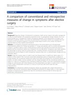
A total of 64 infants in Period I and 51 infants in Period
II who were born at less than 30 weeks of gestation and
survived greater than 72 h were admitted (Fig. 1). Two
infants from Period I were excluded: one infant had
Beckwith-Wiedemann syndrome, and the other infant
had Galen malformation of the brain. Sixteen infants in
Period I and 13 infants in Period II who were never intubated or intubated less than 24 h were also excluded.
After excluding infants who had been intubated for
greater than 6 weeks, those who were never extubated or
died before discharge, and those who were weaned to
other modalities, such as heated and humidified high
flow nasal cannula (HHHFNC), there were 14 infants
who were weaned to NCPAP during Period I and 16 infants who were weaned to NIV-NAVA during Period II.
Lee et al. BMC Pediatrics
(2019) 19:298
Page 3 of 7
Fig. 1 Selection of the study population during the study period
The 2 infants who were weaned to NCPAP during
Period II were categorized as the NCPAP group with the
infants from Period I.
The GA and birth weight of the NIV-NAVA group
and NCPAP group were not significantly different (27+ 1
vs. 26+ 5 weeks and 875 vs. 845 g, respectively) (Table 1).
The incidence of RDS, maternal histologic chorioamnionitis and antenatal steroid use were also not significantly
different between the two groups. At the time of extubation, PMA and weight exhibited no significant differences between the NIV-NAVA group and NCPAP
group (30 vs. 29+ 4 weeks and 1045 vs. 1205 g, respectively) (Table 2). No differences in RSS (NIV-NAVA 1.65
vs. NCPAP 1.95), OSI (1.70 vs. 2.09) or steroid use were
noted before extubation. Several ventilation parameters
at extubation, such as MAP, PEEP, PIP peak inspiratory
pressure (PIP), and FiO2, were similar between the two
groups. SpO2 and pCO2 preceding extubation were also
comparable.
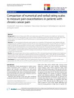
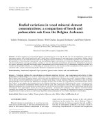
Extubation failure within 72 h after extubation was ascertained in 1 (6.3%) infant in the NIV-NAVA group and 6
(37.5%) infants in the NCPAP group (P = 0.041) (Table 3).
One infant in the NIV-NAVA group was reintubated 11 h
after extubation because of severe apnea requiring PPV. In
the NCPAP group, 3 infants were reintubated before 24 h
after extubation, 2 infants were reintubated 24–48 h after
extubation and one infant was reintubated 70 h after extubation (Fig. 2). Three infants were reintubated because of
severe apnea requiring PPV, two infants due to uncompensated respiratory acidosis (pH < 7.25) with pCO2 > 70
mmHg and one infant due to ≥4 apneic episodes per hour
needing moderate stimulation. The use of other respiratory
support parameters after extubation, such as PEEP and
FiO2, were comparable between the NCPAP and NIVNAVA groups with similar pCO2 and SpO2. Among those
who were reintubated in the study, GA at birth was 26.4
weeks in the NIV-NAVA group and 25.9 (25.3–28.1) weeks
in the NCPAP group. In the univariate logistic regression
analysis, GA at extubation and the duration of invasive
Table 1 Demographics of the study population
GA (weeks)
NIV-NAVA
(n = 16)
NCPAP
(n = 16)
P value
27+ 1 (26+ 5, 27+ 6)
26+ 5 (25+ 4, 27+ 6)
0.317
Birth weight (grams)
875 (677.5, 1145)
845 (700, 1030)
0.777
Male
11 (68.8)
7 (43.8)
0.143
C/S
8 (50.0)
7 (43.8)
0.500
Multiple births
12 (75.0)
10 (62.5)
0.352
PIH
4 (25.0)
1 (6.25)
0.166
hCAM
5 (31.3)
10 (62.5)
0.078
PPROM
7 (43.8)
6 (37.5)
0.500
Antenatal steroid
7 (43.8)
12 (75.0)
0.074
1-min AS
3 (2, 5)
3.5 (2, 4.5)
0.802
5-min AS
5.5 (4, 7)
7 (6, 7)
0.122
RDS
14 (87.5)
16 (100)
0.242
PDA
12 (75.0)
7 (73.3)
0.618
Values are presented as the median (interquartile range) or n (%)
NIV-NAVA Noninvasive neurally adjusted ventilatory assist, NCPAP Nasal
continuous positive airway pressure, GA Gestational age, C/S Cesarean section,
PIH Pregnancy induced hypertension, hCAM Histologic chorioamnionitis,
PPROM Preterm premature rupture of membrane, AS Apgar score, RDS
Respiratory distress syndrome, PDA Patent ductus arteriosus
(2019) 19:298
Lee et al. BMC Pediatrics
Page 4 of 7
Table 2 Clinical characteristics at the time of extubation
NIV-NAVA
(n = 16)
NCPAP
(n = 16)
P value
PMA at extubation (weeks)
30 (28+ 6, 31+ 4)
29+ 4 (27+ 3, 30+ 4)
0.282
Weight at extubation (grams)
1045 (800, 1325)
1025 (905, 1190)
0.651
Pre-extubation
Ventilator duration (days)
21.5 (11.5, 27)
9.5 (4.5, 34.5)
0.365
Systemic steroid use
7 (43.8)
5 (31.3)
0.358
RSS
1.65 (1.49, 2.28)
1.95 (1.68, 2.32)
0.317
OSI
1.70 (1.53, 2.39)
2.09 (1.76, 2.51)
0.274
MAP (cmH2O)
7 (7, 7.5)
8 (7, 8)
0.212
PEEP (cmH2O)
5 (5, 5)
5 (5, 6)
0.531
PIP (cmH2O)
13 (12, 14)
15 (12, 16)
0.180
FiO2 (%)
0.24 (0.21, 0.31)
0.25 (0.21, 0.30)
0.700
pCO2 (mmHg)
53.2 (45.0, 58.4)
49.1 (43.7, 65.3)
0.970
SpO2 (mmHg)
95.5 (94, 98.5)
96 (93.5, 97)
0.760
Values are presented as the median (interquartile range) or n (%)
NIV-NAVA Noninvasive neurally adjusted ventilatory assist, NCPAP Nasal continuous positive airway pressure, PMA Postmenstrual age, RSS Respiratory severity
score, OSI Oxygen saturation index, MAP Mean airway pressure, PEEP Positive end-expiratory pressure, PIP Peak inspiratory pressure
ventilation before extubation were not associated with reintubation (data not shown).
No differences were noted between the two groups regarding the other clinical outcomes, including the development of moderate to severe bronchopulmonary
dysplasia (BPD) (Table 4).
Discussion
Extubation failure is often observed in preterm infants
because the chest wall and upper airway collapses easily
and diaphragmatic strength is poor [29, 30]. The present
study revealed that NIV-NAVA facilitated extubation better than NCPAP. Following a period of endotracheal intubation and IPPV, NCPAP is effective for preventing
extubation failure in preterm infants [8]. This technique
appears to improve lung function and reduce apnea and
may therefore play a role in facilitating extubation in this
population. However, certain populations among preterm
Table 3 Post-extubation status of the study population
NIV-NAVA
(n = 16)
NCPAP
(n = 16)
P value
PEEP (cmH2O)
6 (5.5, 6)
6 (5, 7)
1.000
FiO2 (%)
0.30 (0.27, 0.35)
0.25 (0.21, 0.33)
0.109
pCO2 (mmHg)
48.5 (44.3, 53.6)
49.7 (40.7, 62.1)
0.695
SpO2 (mmHg)
96 (93, 97)
96.5 (94, 98)
0.597
Extubation failure ≤72 h
1 (6.3)
6 (37.5)
0.041
Values are presented as the median (interquartile range) or n (%). Postextubation status was checked 1 h after extubation
NIV-NAVA Noninvasive neurally adjusted ventilatory assist, NCPAP Nasal
continuous positive airway pressure, PMA Postmenstrual age, RSS Respiratory
severity score, OSI Oxygen saturation index, MAP Mean airway pressure, PEEP
Positive end-expiratory pressure, PIP Peak inspiratory pressure
infants who were subject to NCPAP experienced extubation failure [6, 31–33].
NIPPV augments NCPAP by delivering ventilator
breaths via nasal prongs or a mask. Although it did not
improve ventilation in infants who were able to maintain
their own ventilation on NCPAP, in infants with a
higher baseline PaCO2, ventilation was more effectively
increased by NIPPV than NCPAP [34]. Severe apnea and
increased PaCO2 were the most common causes of failure in infants receiving NCPAP, and NIPPV achieved a
comparative reduction in extubation failure in preterm
infants. A recent meta-analysis demonstrated that the incidence of extubation failure and the need for reintubation within 48 h to 1 week was reduced by NIPPV in
preterm infants [12]. However, synchronization and the
device used to deliver PPV may be important parameters
in NIPPV [13].
NAVA has been applied in clinical practice during the last
decade, but studies have rarely involved neonates, especially
the preterm infant population. However, a recent study
demonstrated the effectiveness and feasibility of NAVA in
this population [19]. Noninvasive support via NAVA improved patient-ventilator synchrony by reducing trigger
delay and the number of asynchrony events [35]. Previously,
we reported that NAVA improved patient-ventilator synchrony and diaphragmatic unloading in preterm infants during noninvasive nasal ventilation compared with pressure
support mode [23]. A recent physiologic study performed
by Gibu et al. compared NIV-NAVA and NIPPV and demonstrated that peak inspiratory pressure and FiO2 were lowered in NIV-NAVA than in NIPPV [36]. Furthermore, both
infant movement and caretaker’s work were lowered in
Lee et al. BMC Pediatrics
(2019) 19:298
Page 5 of 7
Fig. 2 Kaplan-Meier estimates for extubation success by post-extubation modality
NIV-NAVA, suggesting that NIV-NAVA was more effective
than NIPPV at increasing infant comfort. Because it has excellent synchronization, NIN-NAVA could serve as a substitute for NCPAP to facilitate extubation in preterm infants.
Most cases of reintubation in this study were the result of
severe apnea or uncompensated hypercapnia. When compared to NCPAP, apnea and hypercapnia were more preventable in NIPPV by generating higher airway pressure to
prevent obstructive apnea and triggering sigh in preterm infants [37, 38]. Although NIV-NAVA seemed to improve
ventilator synchrony and diaphragmatic unloading during
noninvasive ventilation compared to other NIPPV, there
was no evidence that NIV-NAVA is superior to other
NIPPV modalities after extubation [23, 39].
Even though there could be concerns regarding the
size of the baby when using NIV-NAVA, many studies
showed NIV-NAVA was feasible in extremely preterm
infants [23, 39]. In the present study, NIV-NAVA was
Table 4 Clinical outcomes of the study population
NIV-NAVA (n=16) NCPAP (n = 16) P value
Moderate to severe BPD
10 (62.5)
9 (60.0)
0.589
NEC ≥ stage 2
2 (12.5)
5 (33.3)
0.170
Retinopathy of prematurity
4 (25.0)
6 (40.0)
0.306
IVH ≥ grade 2
2 (12.5)
1 (6.7)
0.525
0 (0)
0.516
Periventricular leukomalacia 1 (6.3)
Values are presented as the median (interquartile range) or n (%)
NIV-NAVA Noninvasive neurally adjusted ventilatory assist, NCPAP Nasal
continuous positive airway pressure, BPD Bronchopulmonary dysplasia, NEC
Necrotizing enterocolitis, ROP Retinopathy of prematurity, IVH
Intraventricular hemorrhage
also found to be feasible in babies as small as 660 g at
extubation or 700 g at birth who were successfully
weaned to NIV-NAVA at PMA 28 weeks. A baby who
was 500 g at birth was also successfully weaned to NIVNAVA at 770 g. Moreover, Edi catheters can efficiently
serve as a feeding tube in these babies and thus an additional feeding tube did not need to be inserted for enteral feeding. NEC was comparable in both groups and
there were no intestinal perforations or air leaks after
the infants were weaned to NIV-NAVA or NCPAP. Although the rates of neonatal complications are lower in
noninvasive versus invasive MV, safety must be considered. Previously, it was suggested that neonates who
were mechanically ventilated with either a face mask or
nasal prongs had an increased risk of gastrointestinal
perforations. However, recent data has shown that
NIPPV does not appear to be associated with increased
gastrointestinal side effects, and the risk of air leaks was
lower in NIPPV than in NCPAP [40]. No differences in
the development of air leaks and NEC were observed between the two groups in the present study.
There are some limitations to the present study. This
study was a retrospective study with a small sample size,
thus making it difficult to draw robust conclusions. There
also was a period of overlap when both NIV-NAVA and
NCPAP were used as weaning modalities. The study population was highly selected because we analyzed only 50% of
the preterm infants born at < 30 weeks of gestation who
were intubated for more than 24 h and were extubated
thereafter during the study period. Furthermore, the duration of ventilation seemed to be shorter in the NCPAP
Lee et al. BMC Pediatrics
(2019) 19:298
group, although this result was not statistically significant.
While the sample size may have been too small to fully elucidate this difference, a logistic regression analysis for reintubation was performed ad hoc and showed that the
duration of ventilation before extubation was not associated
with reintubation (data not shown). The criteria for extubation were well-defined in our unit, and the pre-extubation
conditions in both groups including the PMA at extubation, RSS, OSI and the ventilation settings were comparable
in the present study. Despite these limitations, this is the
first study to compare the clinical responses between NIVNAVA and NCPAP when used to facilitate extubation in
preterm infants.
Conclusions
The data in the present study were not robust enough to
be conclusive due to small sample size, but showed promising implications for using NIV-NAVA over NCPAP to
facilitate extubation. NIV-NAVA could be an effective
modality for synchronized noninvasive ventilation following successful extubation from MV in preterm infants.
Abbreviations
HHHFNC: Humidified high flow nasal cannula; MAP: Mean airway pressure;
MV: Mechanical ventilation; NAVA: Neurally adjusted ventilatory assist;
NCPAP: Nasal continuous positive airway pressure; NICU: Neonatal intensive
care unit; NIPPV: Nasal intermittent positive pressure ventilation; NIVNAVA: Non-invasive ventilation using NAVA; OSI: Oxygen saturation index;
PEEP: Positive end expiratory pressure; PPV: Positive pressure ventilation;
RSS: Respiratory severity score; SNIPPV: Synchronized Nasal intermittent
positive pressure ventilation; VILI: Ventilator-induced lung injury
Acknowledgements
Not applicable.
Authors’ contributions
SHS, BKL and H-SK conceived and designed the study, collected and analyzed the data and drafted the manuscript. E-KK and YHJ revised the manuscript for critically important intellectual content. SHS, BKL and H-SK finalized
the manuscript. All authors read and approved the final manuscript.
Funding
This study was supported by a grant from the Seoul National University
Hospital Research Fund (04–2015-0430) and by the Basic Science Research
Program through the National Research Foundation of Korea (NRF) funded
by the Ministry of Education (2017R1D1A1B03036383). The funders did not
participate in the research, or in the preparation the manuscript.
Availability of data and materials
The dataset generated or analyzed during this study can be made available
to interested researchers by the authors of this article upon reasonable
request.
Ethics approval and consent to participate
Ethical approval to conduct this study was obtained from the Institutional
Review Board of Seoul National University Hospital. Written consent from the
caregivers of the neonates could not be obtained due to the retrospective
nature of the study. However, all the patient-related information was
anonymized.
Consent for publication
Not applicable.
Competing interests
The authors declare that they have no competing interests.
Page 6 of 7
Author details
1
Department of Pediatrics, Yonsei University Wonju College of Medicine,
Wonju, South Korea. 2Department of Pediatrics, Seoul National University
College of Medicine, Seoul, South Korea. 3Department of Pediatrics, Seoul
National University Children’s Hospital, 101 Daehak-ro, Jongno-gu, Seoul
110-769, South Korea. 4Department of Pediatrics, Seoul National University
Bundang Hospital, Seongnam, South Korea.
Received: 7 May 2019 Accepted: 21 August 2019
References
1. Donn SM, Sinha SK. Minimising ventilator induced lung injury in preterm
infants. Arch Dis Child Fetal Neonatal Ed. 2006;91(3):F226–30.
2. Carvalho CG, Silveira RC, Procianoy RS. Ventilator-induced lung injury in
preterm infants. Rev Bras Ter Intensiva. 2013;25(4):319–26.
3. Apisarnthanarak A, Holzmann-Pazgal G, Hamvas A, Olsen MA, Fraser VJ.
Ventilator-associated pneumonia in extremely preterm neonates in a
neonatal intensive care unit: characteristics, risk factors, and outcomes.
Pediatrics. 2003;112(6 Pt 1):1283–9.
4. Moretti C, Giannini L, Fassi C, Gizzi C, Papoff P, Colarizi P. Nasal flowsynchronized intermittent positive pressure ventilation to facilitate weaning
in very low-birthweight infants: unmasked randomized controlled trial.
Pediatr Int. 2008;50(1):85–91.
5. Khalaf MN, Brodsky N, Hurley J, Bhandari V. A prospective randomized,
controlled trial comparing synchronized nasal intermittent positive pressure
ventilation versus nasal continuous positive airway pressure as modes of
extubation. Pediatrics. 2001;108(1):13–7.
6. Dimitriou G, Greenough A, Kavvadia V, Laubscher B, Alexiou C, Pavlou V,
Mantagos S. Elective use of nasal continuous positive airways pressure
following extubation of preterm infants. Eur J Pediatr. 2000;159(6):434–9.
7. Peake M, Dillon P, Shaw NJ. Randomized trial of continuous positive airways
pressure to prevent reventilation in preterm infants. Pediatr Pulmonol. 2005;
39(3):247–50.
8. Davis PG, Henderson-Smart DJ. Nasal continuous positive airways pressure
immediately after extubation for preventing morbidity in preterm infants.
Cochrane Database Syst Rev. 2003;2:CD000143.
9. Ignacio L, Alfaleh K. High-flow nasal cannulae in very preterm infants after
extubation. J Clin Neonatol. 2014;3(1):11–3.
10. Davis P, Henderson-Smart D. Post-extubation prophylactic nasal continuous
positive airway pressure in preterm infants: systematic review and metaanalysis. J Paediatr Child Health. 1999;35(4):367–71.
11. De Paoli AG, Morley C, Davis PG. Nasal CPAP for neonates: what do we
know in 2003? Arch Dis Child Fetal Neonatal Ed. 2003;88(3):F168–72.
12. Bancalari E, Claure N. The evidence for non-invasive ventilation in the
preterm infant. Arch Dis Child Fetal Neonatal Ed. 2013;98(2):F98–F102.
13. Huang L, Mendler MR, Waitz M, Schmid M, Hassan MA, Hummler HD. Effects
of synchronization during noninvasive intermittent mandatory ventilation in
preterm infants with respiratory distress syndrome immediately after
extubation. Neonatology. 2015;108(2):108–14.
14. Hird MF, Greenough A: Comparison of triggering systems for neonatal
patient triggered ventilation. Arch Dis Child 1991, 66(4 Spec No):426–428.
15. John J, Bjorklund LJ, Svenningsen NW, Jonson B. Airway and body
surface sensors for triggering in neonatal ventilation. Acta Paediatr.
1994;83(9):903–9.
16. Courtney SE, Barrington KJ. Continuous positive airway pressure and
noninvasive ventilation. Clin Perinatol. 2007;34(1):73–92 vi.
17. Stein H, Firestone K. Application of neurally adjusted ventilatory assist in
neonates. Semin Fetal Neonatal Med. 2014;19(1):60–9.
18. Sinderby C, Navalesi P, Beck J, Skrobik Y, Comtois N, Friberg S, Gottfried SB,
Lindstrom L. Neural control of mechanical ventilation in respiratory failure.
Nat Med. 1999;5(12):1433–6.
19. Beck J, Reilly M, Grasselli G, Mirabella L, Slutsky AS, Dunn MS, Sinderby C.
Patient-ventilator interaction during neurally adjusted ventilatory assist in
low birth weight infants. Pediatr Res. 2009;65(6):663–8.
20. Lee J, Kim HS, Sohn JA, Lee JA, Choi CW, Kim EK, Kim BI, Choi JH.
Randomized crossover study of neurally adjusted ventilatory assist in
preterm infants. J Pediatr. 2012;161(5):808–13.
21. Schmidt M, Dres M, Raux M, Deslandes-Boutmy E, Kindler F, Mayaux J,
Similowski T, Demoule A. Neurally adjusted ventilatory assist improves
Lee et al. BMC Pediatrics
22.
23.
24.
25.
26.
27.
28.
29.
30.
31.
32.
33.
34.
35.
36.
37.
38.
39.
40.
(2019) 19:298
patient-ventilator interaction during postextubation prophylactic
noninvasive ventilation. Crit Care Med. 2012;40(6):1738–44.
Azoulay E, Kouatchet A, Jaber S, Lambert J, Meziani F, Schmidt M, Schnell D,
Mortaza S, Conseil M, Tchenio X, et al. Noninvasive mechanical ventilation
in patients having declined tracheal intubation. Intensive Care Med. 2013;
39(2):292–301.
Lee J, Kim HS, Jung YH, Shin SH, Choi CW, Kim EK, Kim BI, Choi JH. Noninvasive neurally adjusted ventilatory assist in preterm infants: a randomised
phase II crossover trial. Arch Dis Child Fetal Neonatal Ed. 2015;100(6):F507–13.
Iyer NP, Mhanna MJ. Non-invasively derived respiratory severity score
and oxygenation index in ventilated newborn infants. Pediatr Pulmonol.
2013;48(4):364–9.
Muniraman HK, Song AY, Ramanathan R, Fletcher KL, Kibe R, Ding L, et al.
Evaluation of oxygen saturation index compared with oxygenation index in
neonates with hypoxemic respiratory failure. JAMA Netw Open. 2019;2(3):191–79.
Malkar MB, Gardner WP, Mandy GT, Stenger MR, Nelin LD, Shepherd EG,
Welty SE. Respiratory severity score on day of life 30 is predictive of
mortality and the length of mechanical ventilation in premature infants
with protracted ventilation. Pediatr Pulmonol. 2015;50(4):363–9.
Mhanna MJ, Iyer NP, Piraino S, Jain M. Respiratory severity score and
extubation readiness in very low birth weight infants. Pediatr Neonatol.
2017;58(6):523–8.
Chawla S, Natarajan G, Gelmini M, Kazzi SN. Role of spontaneous breathing
trial in predicting successful extubation in premature infants. Pediatr
Pulmonol. 2013;48(5):443–8.
Gerhardt T, Bancalari E. Chestwall compliance in full-term and premature
infants. Acta Paediatr Scand. 1980;69(3):359–64.
Dransfield DA, Spitzer AR, Fox WW. Episodic airway obstruction in
premature infants. Am J Dis Child. 1983;137(5):441–3.
Ammari A, Suri M, Milisavljevic V, Sahni R, Bateman D, Sanocka U, RuzalShapiro C, Wung JT, Polin RA. Variables associated with the early failure of
nasal CPAP in very low birth weight infants. J Pediatr. 2005;147(3):341–7.
Reininger A, Khalak R, Kendig JW, Ryan RM, Stevens TP, Reubens L, D'Angio
CT. Surfactant administration by transient intubation in infants 29 to 35
weeks’ gestation with respiratory distress syndrome decreases the likelihood
of later mechanical ventilation: a randomized controlled trial. J Perinatol:
official journal of the California Perinatal Association. 2005;25(11):703–8.
Stefanescu BM, Murphy WP, Hansell BJ, Fuloria M, Morgan TM, Aschner JL. A
randomized, controlled trial comparing two different continuous positive
airway pressure systems for the successful extubation of extremely low birth
weight infants. Pediatrics. 2003;112(5):1031–8.
Ali N, Claure N, Alegria X, D'Ugard C, Organero R, Bancalari E. Effects of noninvasive pressure support ventilation (NI-PSV) on ventilation and respiratory
effort in very low birth weight infants. Pediatr Pulmonol. 2007;42(8):704–10.
Vignaux L, Grazioli S, Piquilloud L, Bochaton N, Karam O, Levy-Jamet Y,
Jaecklin T, Tourneux P, Jolliet P, Rimensberger PC. Patient-ventilator
asynchrony during noninvasive pressure support ventilation and neurally
adjusted ventilatory assist in infants and children. Pediatr Crit Care Med.
2013;14(8):e357–64.
Gibu C, Cheng P, Ward RJ, Castro B, Heldt GP. Feasibility and physiological
effects of non-invasive neurally-adjusted ventilatory assist (NIV-NAVA) in
preterm infants. Pediatr Res. 2017;82(4):650–7.
Lin CH, Wang ST, Lin YJ, Yeh TF. Efficacy of nasal intermittent positive
pressure ventilation in treating apnea of prematurity. Pediatr Pulmonol.
1998;26(5):349–53.
Bisceglia M, Belcastro A, Poerio V, Raimondi F, Medsuraca L, Crugliano C,
Corapi UP. A comparison of nasal intermittent versus continuous positive
pressure delivery for the treatment of moderate respiratory syndrome in
preterm infants. Minerva Pediatr. 2007;59(2):91–5.
Yonehara K, Ogawa R, Kamei Y, Oda A, Kokubo M, Hiroma T, Nakamura T.
Non-invasive neurally adjusted ventilatory assist versus nasal intermittent
positive-pressure ventilation in preterm infants born before 30 weeks’
gestation. Pediatr Int. 2018;60(10):957–61.
Lemyre B, Davis PG, De Paoli AG, Kirpalani H. Nasal intermittent positive
pressure ventilation (NIPPV) versus nasal continuous positive airway pressure
(NCPAP) for preterm neonates after extubation. Cochrane Database Syst
Rev. 2017;2:CD003212.
Publisher’s Note
Springer Nature remains neutral with regard to jurisdictional claims in
published maps and institutional affiliations.
Page 7 of 7