Baseline cardiac output and its alterations during ibuprofen treatment for patent ductus arteriosus in preterm infants
Bạn đang xem bản rút gọn của tài liệu. Xem và tải ngay bản đầy đủ của tài liệu tại đây (872.25 KB, 8 trang )
Hsu et al. BMC Pediatrics
(2019) 19:179
/>
RESEARCH ARTICLE
Open Access
Baseline cardiac output and its alterations
during ibuprofen treatment for patent
ductus arteriosus in preterm infants
Kai-Hsiang Hsu1,2* , Tai-Wei Wu3, I-Hsyuan Wu1, Mei-Yin Lai1,2, Shih-Yun Hsu1,4, Hsiao-Wen Huang1, Tze-Yee Mok1,
Cheng-Chung Lee1,2 and Reyin Lien1
Abstract
Background: Infants with hemodynamically significant patent ductus arteriosus (PDA) may physiologically
compensate with a supranormal cardiac output (CO). As such, a supranormal CO may be a surrogate marker for a
significant PDA or indicate a failed response to PDA closure by ibuprofen. Electrical cardiometry (EC) is an
impedance-based monitor that can continuously and non-invasively assess CO (COEC). We aimed to trend COEC
through ibuprofen treatment for PDA in preterm infants.
Methods: We reviewed our database of preterm infants receiving ibuprofen for PDA closure. Response to
ibuprofen was defined as no ductal flow in echocardiography ≤24 h after treatment. Responders were compared
with gestational age (GA) and postnatal age matched non-responders and their trends of COEC were compared.
Both groups’ baseline COEC were further compared to the reference infants without PDA.
Results: Eighteen infants (9 responders and 9 non-responders) with median (interquatile range) GA 27.5 (26.6–28.6)
weeks, birthweight 1038 (854–1218) g and age 3.5 (3.0–4.0) days were studied. There were positive correlations
between COEC and ductal diameter and left atrium/ aortic root ratio (r = 0.521 and 0.374, p < 0.001, respectively).
Both responders and non-responders had significantly higher baseline COEC than the reference. Although there was
no significant within-subject alteration of COEC during ibuprofen treatment, there was a between-subject difference
indicating non-responders had generally higher COEC.
Conclusions: The changes of COEC during pharmacological closure of PDA is less drastic compared to surgical
closure. Infants with PDA had higher baseline COEC compared to those without PDA, and non-responders had
higher COEC especially at baseline compared to responders.
Keywords: Cardiac output, Electrical cardiometry, Hemodynamic, Non-invasive monitor, Patent ductus arteriosus,
Preterm infant
Introduction
Patent ductus arteriosus (PDA) is common among preterm infants and failure of ductal closure is associated
with complications and poor outcomes [1]. Non-selective
cyclooxygenase (COX) inhibitor, such as ibuprofen, is the
pharmacological choice of treatment for PDA based on its
* Correspondence:
1
Division of Neonatology, Department of Pediatrics, Chang Gung Memorial
Hospital Linkou Branch, Taoyuan, Taiwan
2
Graduate Institute of Clinical Medical Science, Chang Gung University,
Taoyuan, Taiwan
Full list of author information is available at the end of the article
action of prostaglandin inhibition that promotes ductal
constriction. Both the intravenous and oral routes of ibuprofen administration appear comparably effective for
ductal closure [2]. However, successful PDA closure by
pharmacological treatment is not always definite or predictable [3]. The rate of ductal closure after COX inhibitors varies from 60 to 85% in preterm infants and is even
less effective in extremely premature infants [4–6].
Echocardiography is often used to evaluate hemodynamic
significance of PDA [7]. In general, pharmacological closure
of PDA is less successful in infants with ductal diameter > 2
mm [8]. Lower ductal maximum velocity, which is usually
© The Author(s). 2019 Open Access This article is distributed under the terms of the Creative Commons Attribution 4.0
International License ( which permits unrestricted use, distribution, and
reproduction in any medium, provided you give appropriate credit to the original author(s) and the source, provide a link to
the Creative Commons license, and indicate if changes were made. The Creative Commons Public Domain Dedication waiver
( applies to the data made available in this article, unless otherwise stated.
Hsu et al. BMC Pediatrics
(2019) 19:179
associated with a larger PDA or higher pulmonary pressure,
is another predictor of treatment failure [4, 8]. Furthermore,
an increase in left ventricular cardiac output (CO) has been
positively correlated with significant ductal shunting [7, 9, 10]
and PDA severity [11]. The underlying reason is that a PDA
with significant left-to-right flow may lead to a compensatory
increase in CO in order to maintain systemic blood flow [12,
13]. Indeed, following closure of ductus after COX inhibitor
therapy [12] or surgical ligation [10, 14], CO normalizes accordingly. We therefore hypothesized that a supranormal
CO in the first week of life in extreme premature infants
may indicate a hemodynamically significant PDA and that
we could observe CO changes during pharmacological treatment. However, the ability to perform neonatal functional
echocardiography requires practice, training and mentorship
[15]. Furthermore, the use of echocardiography to gather
meaningful hemodynamic data often necessitates serial assessments that can be tedious and labor-intensive.
Electrical cardiometry (EC) is a non-invasive, impedancebased monitor that provides absolute CO estimates in clinical practice [16]. Unlike echocardiography, EC is simple to
apply, continuous in measurements and not operatordependent. Comparisons between CO measured by EC
(COEC) and echocardiography have been studied in term
[17] and preterm [18–20] infants with and without PDA. Although CO values measured by EC and echocardiography
may not be interchangeable, it has been suggested that EC
can be useful in trending CO changes in the clinical setting
[20]. Hemodynamic reference by EC for neonates without
PDA and without invasive ventilation support has been
established, and COEC is positively correlated with gestational age (GA) and weight [21]. In addition, EC was used to
monitor the effects of ductal ligation on COEC, which revealed an initial decline in COEC followed by recovery [22].
Utilizing the ability of EC to continuously measure COEC,
we aimed to identify significant changes in COEC during
attempted pharmacological closure and compared COEC
characteristics in responders versus non-responders.
Page 2 of 8
excluded. Demographic data, serial echocardiographic findings and respiratory support at time of ibuprofen administration were collected.
Ibuprofen for PDA closure
The decision to initiate ibuprofen for PDA closure was made
based on individual’s clinical condition (e.g. increased respiratory support or hypotension) and echocardiographic
finding (e.g. large ductus > 2 mm or low peak systolic ductal
flow). Per unit policy, infants with right-to-left or bidirectional shunting PDA, intraventricular hemorrhage grade ≥ 3
or poor renal function (serum creatinine > 1.8 mg/dl or oligouria < 1 ml/kg/hr) were not candidates for ibuprofen treatment. The decision to treat with oral (ibuprofen oral
suspension, [Center Laboratories Inc., Taipei, Taiwan]) or
intravenous ibuprofen (Ibusine: Ibuprofen Lysine, [China
Chemical & Pharmaceutical Co., Taipei, Taiwan]) was also
made by the attending neonatologist. One course of treatment for both oral and intravenous ibuprofen consisted of
three consecutive doses of 10, 5, 5 mg/kg/dose given 24 h
apart. Responder to ibuprofen treatment was defined as absence of ductal flow in echocardiography within 24 h after
completion of treatment.
Echocardiography
Transthoracic echocardiography was performed using
Sonos 7500 (Philips, Andover, Massachusetts, USA) with
a 12 MHz transducers. Serial echocardiography was performed in relation to ibuprofen administration: within an
hour prior to dose #1 ibuprofen (baseline), 18–24 h after
dose #1 and #2 (during treatment), and 24 h after dose #3
of ibuprofen (treatment completion). This timeframe was
chosen to allow maximum effect of each dose. Echocardiographic parameters of the PDA were assessed, which
includes ductal size and shunt direction by color Doppler
mapping, maximum flow velocity by pulsed-wave Doppler,
and left atrium to aortic root ratio (LA/Ao) and left ventricular fractional shortening (FS) by M-mode.
Methods
Patients
Electrical Cardiometry (EC)
This study was conducted in the neonatal intensive care
unit of Chang Gung Memorial Hospital Linkou Branch
and was approved by the Institutional Review Board. As
part of a hemodynamic monitoring project in the unit,
echocardiographic findings and relevant hemodynamic
information were collected prospectively into a database.
We reviewed this database for very low birth weight (VLBW,
< 1500 g) preterm infants admitted between June 2015 to
June 2016 who received ibuprofen treatment for PDA closure. We enrolled infants who had both echocardiography
and EC data during the first treatment course. Infants with
chromosomal anomaly or structural heart defect other than
small patent foramen ovale or atrial septal defect were
EC (Aesculon, Osypka Medical, Berlin, Germany) was applied by attaching four standard surface electrocardiogram
electrodes over the forehead, left lower neck, left mid-axillary
line at the level of xiphoid process and lateral aspect of left
thigh. EC was placed at least 1 h prior to dose #1 ibuprofen
and kept in situ until 24 h after completing treatment.
Hemodynamic parameters by EC, including COEC, heart rate
(HREC) and stroke volume (SVEC) were captured every 10
min during the study period and subsequently exported into
a database using software Waveform Explorer by Osypka
Medical. The original data that 1 h before treatment and 18–
24 h after each ibuprofen dose were further averaged and analyzed (e.g. the baseline and 18–24 h following dose #1, #2
Hsu et al. BMC Pediatrics
(2019) 19:179
Page 3 of 8
and #3, respectively). Value of COEC and SVEC were weightadjusted as ml/kg/min and ml/kg.
Matching
In order to minimize confounders related to GA, weight
and post-natal age, we matched each responder to a nonresponder with comparable GA ± 1 week, weight ± 10% g
and post-natal age ± 7 days from the hemodynamic database. Furthermore, for comparison of COEC between infants with and without PDA, we also matched above
responders and non-responders respectively to our previously published reference [21] using the same criteria.
Statistics
Statistical analysis was performed using IBM SPSS Statistics version 20 (Armonk, NY, USA). Continuous variables in background demographics were tested using
Mann-Whitney U test, while hemodynamic parameters
by EC were tested with independent t-test between responders and non-responders or paired t-test was between two timing points. Repeated measures analysis of
variance (RM-ANOVA) was applied to compare trends
of hemodynamic parameters through the course. Categorical data were analyzed with Chi-square test or
Fisher’s exact test. Analysis of the relationship between
COEC and ductal diameter or LA/Ao was by Pearson
correlation coefficient. One-way ANOVA with Bonferroni correction was used to compare COEC among responders, non-responders and the reference. Statistical
significance was defined as two-sided p < 0.05.
Results
During the study period, 303 VLBW preterm infants
were admitted to our unit, of which 46 received ibuprofen treatment. There was complete data collection for both
echocardiography and EC in 36 infants, and 11 of them were
responders. After screening and matching, 9 out of 11 responders could be matched to 9 non-responders, and a total
of 18 preterm infants were included. Their median (interquartile range) GA, weight and post-natal age at initiation of ibuprofen were 27.5 (26.6–28.6) weeks, 1038 (854–1218) g and
3.5 (3.0–4.0) days old, respectively. There was no significant
difference in demographics, echocardiographic measurements,
post-natal age, route of ibuprofen or respiratory support between responders and non-responders (Table 1). None received vasopressor or inotrope during the treatment course.
Among 9 responders, 5 infants were found to have absence of
ductal flow after dose #1 ibuprofen, 2 infants after dose #2,
Table 1 Clinical characteristics for responders and non-responders for ibuprofen treatment for PDA
Responders
Non-Responders
(n = 9)
(n = 9)
Gestational age (weeks)
27.7 (27.1–29.9)
27.4 (26.1–27.9)
0.161
Weight (g)
1135 (913–1318)
1015 (830–1083)
0.222
Apgar at 1 min
7 (5–8)
6 (3–7)
0.077
Apgar at 5 min
9 (7–9)
9 (8–9)
0.931
Male
6 (67%)
2 (22%)
0.153
Cesarean section
6 (67%)
7 (78%)
1.000
Small for gestational age
2 (22%)
1 (11%)
1.000
2.05 (1.78–2.46)
2.20 (1.70–3.23)
0.666
Demographics
p
valuea
Echocardiography prior to ibuprofen treatment
PDA diameter (mm)
PDA diameter to weight (mm/kg)
2.04 (1.40–2.54)
2.26 (1.56–3.57)
0.340
PDA maximum flow velocity (m/s)
2.21 (1.59–2.60)
1.82 (1.30–2.52)
0.613
LA/Ao ratio
1.47 (1.27–1.76)
1.50 (1.44–1.87)
0.370
Fractional shortening (%)
41.0 (36.0–44.5)
39.0 (34.2–43.8)
0.661
Post-natal age at dose #1 (day)
3.0 (3.0–4.0)
4.0 (3.0–6.5)
0.222
Oral ibuprofen
8 (89%)
7 (78%)
1.000
Condition prior to ibuprofen treatment
Respiratory support
0.183
Non-invasive ventilation
5 (56%)
2 (22%)
Conventional ventilation
4 (44%)
5 (56%)
High frequency ventilation
0 (0%)
2 (22%)
PDA patent ductus arteriosus, LA/Ao left atrium to aortic root diameter
Data are median (interquartile range) or n (%)
a
A p value was tested by Mann-Whitney U test for continuous variables and Chi-square test or Fisher’s exact test for categorical data
Hsu et al. BMC Pediatrics
(2019) 19:179
Page 4 of 8
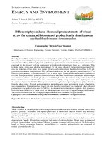
Fig. 1 Scatter diagrams of COEC and ductal diameter for preterm infants who responded (gray circles) and non-responded (black circles) to
ibuprofen treatment for PDA. Four timing points were plotted: 1 h prior to treatment (baseline, a) and 18–24 h post each dosage of ibuprofen (b,
c and d, respectively). COEC, cardiac output by electrical cardiometry; PDA, patent ductus arteriosus
and 2 infants after dose #3 (Fig. 1 a–d). Furthermore, there
was positive correlations between COEC and ductal diameter
(r = 0.521, p < 0.001) and LA/Ao (r = 0.374, p < 0.001).
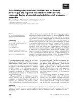
Non-responders had higher COEC compared to responders throughout the treatment course (RM-ANOVA
between-subject p = 0.005). This discrepancy was most
significant prior to ibuprofen treatment (282 ± 21 vs.
250 ± 31 ml/kg/min, p = 0.022), at 24 h post dose #2
(257 ± 33 vs. 226 ± 23 ml/kg/min, p = 0.034), and 24 h
post dose #3 (270 ± 39 vs. 232 ± 14 ml/kg/min, p = 0.022)
(Fig. 2a). No significant differences in HREC or SVEC
were found between the two groups (Fig. 2b and c).
When analyzing within-subject changes throughout
the treatment course, there were no significant changes
of COEC, HREC or SVEC in either responders or nonresponders (RM-ANOVA). The average alteration of
COEC was − 7% ± 12% for responders and − 6% ± 16% for
non-responders. On the other hand, when comparing
baseline COEC to the earliest time point when no ductal
flow was visualized by echocardiography, there was a
significant but small-scale reduction in COEC by 25 ml/
kg/min or 10% (250 ± 31 vs. 225 ± 17 ml/kg/min, paired
t-test p = 0.031) (Table 2). However, we found 4/9 (44%)
of non-responders had > 10% reduction of COEC at some
timing points as well.
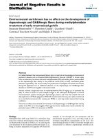
Another 18 infants without PDA were matched for
baseline COEC comparison. Their median GA and weight
were 28.6 (28.0–30.2) weeks and 1175 (1005–1312) g, respectively, and were all 3–4 days old. No demographic difference existed among these three groups (responders,
non-responders and the reference). There was a significant stepwise increment in baseline COEC from infants
with no PDA (207 ± 28 ml/kg/min), to infants with PDA,
responders (250 ± 31 ml/kg/min), to infants with PDA,
non-responders (282 ± 21 ml/kg/min, p < 0.001) (Fig. 3).
Discussions
In this study, we showed the potential of EC to continuously monitor changes in COEC among preterm infants.
By carefully matching target infants, we demonstrated
that infants with PDA had higher baseline COEC and
there was no significant COEC alteration during ibuprofen treatment for ductal closure.
Our finding indicated that preterm infants with PDA
have significantly higher baseline COEC compared to agematched reference, and that baseline COEC is positively
Hsu et al. BMC Pediatrics
(2019) 19:179
Page 5 of 8
Fig. 2 Trends charts of COEC, HREC and SVEC for responders (gray line) and non-responders (black line) through ibuprofen treatment. Three gray
bands indicate the time of each ibuprofen administration. Although there was no remarkable alteration of COEC, HREC and SVEC within each
group, non-responders had significantly higher COEC than responders through the course (between-subject p = 0.005) (¶), especially at the timing
prior to dose #1 ibuprofen, 18–24 h post dose #2 and 18–24 h post dose #3, respectively (*). COEC, cardiac output; HREC, heart rate; SVEC, stroke
volume; all measured by electrical cardiometry
Table 2 Hemodynamic changes at specific timing points
COEC (ml/kg/min)
Prior to dose #1
a
HREC (beats/min)
SVEC (ml/kg)
Responders (n = 9)
Non-responders (n = 9)
p value
250 ± 31
282 ± 21
0.022b
c
No ductal flow
225 ± 17
N/A
N/A
18–24 h after dose #3
232 ± 15
270 ± 39
0.021b
Prior to dose #1
157 ± 7
160 ± 8
0.394
a
No ductal flow
151 ± 7
N/A
N/A
18–24 h after dose #3
153 ± 8
160 ± 6
0.077
Prior to dose #1
1.59 ± 0.23
1.77 ± 0.30
0.165
No ductal flowa
1.50 ± 0.15
N/A
N/A
18–24 h after dose #3
1.63 ± 0.29
1.63 ± 0.25
0.926
CO cardiac output, HR heart rate, SV stroke volume, EC electrical cardiometry, N/A not applicable
Data are mean (± SD)
a
Five infants’ ductal flow disappeared in color Doppler post dose #1, two post dose #2 and two post dose #3
b
indicates statistical significance between responders and non-responders (independent t-test)
c
indicates statistical significance comparing to baseline value prior to dose #1 (paired t-test)
Hsu et al. BMC Pediatrics
(2019) 19:179
Page 6 of 8
Fig. 3 Box plot of baseline COEC for responders, non-responders and matched reference. The horizontal lines are median COEC and the diamond
marks are mean of COEC for respective group. Mean COEC of three groups were statistically different, especially non-responders had the highest
COEC. COEC, cardiac output by electrical cardiometry
correlated to PDA diameter and LA/Ao. The positive correlation suggests that infants with greater COEC have a
higher likelihood of more significant ductal shunting. It
was interesting to find that only the baseline COEC, but
not ductal diameter, maximum ductal flow or LA/Ao, was
significantly different between responders and nonresponders in our study. It can be reasoned that with high
left-to-right ductal shunting, CO represents the sum of
systemic flow plus ductal shunting, and hence increases in
CO is a compensation and proportional to ductal shunting
[7, 13]. Furthermore, only COEC but not HREC or SVEC
was significantly different between responders and nonresponders. This may indicate that CO represents the sum
of left ventricular work, i.e., HR and SV, to compensate for
the ductal steal effect. It also suggests that CO may be a
more comprehensive surrogate in determining the degree
of ductal shunting. The difference in baseline COEC between responders and non-responders is compatible with
previous studies that infants with larger ductal shunting
may response poorly to COX inhibitor [4, 8].
We observed no significant COEC alteration through ibuprofen treatment for PDA closure. Although there was a
mean decrease of COEC by 10% on initial ductal closure, this
reduction of COEC cannot be an indicator for ductal closure
because non-responders may also had > 10% reduction of
COEC through the course. Moreover, the small-scale decline
is unlike our previous study that a 26% decrease in COEC at
time of ductal ligation [22]. We speculate that the effect of
ibuprofen in inducing ductal closure was progressive or
intermittent while allowing time for the myocardium to
adapt to the hemodynamic changes. This is further supported by the fact that no infant in our study required inotropic support, which is needed in infants with post-ligation
hemodynamic instability.
There is a similar study utilizing EC to monitor CO during
attempted pharmacological closure of PDA by intravenous
ibuprofen in preterm infants [23], of which, a fall in median
COEC from 290 to 240 ml/kg/min (17%) 72 h after the initiation of treatment was found. However, the study is limited
by its small case number (6 responders) and a wide overlap
of COEC between baseline and 72 h after the first dose ibuprofen. In addition, 2 out of 6 infants in this study received
dopamine infusion before ibuprofen treatment and dopamine was tapered off at the end of ibuprofen treatment,
which can confound the baseline and post-treatment COEC
measurements [24]. The dopamine infusion may have contributed to the larger discrepancy between baseline and posttreatment COEC in this study.
Some limitations should be addressed. Firstly, the sample size of current study was small. The number of responders limited the power to demonstrate exact COEC
changes and to detect a confident cut-off COEC to assess
treatment response. Secondly, using echocardiography to
detect the exact timing of ductal closure during ibuprofen treatment is clinically complex. We are only able to
use the earliest available echocardiography data that indicates no ductal flow to assess COEC alteration. This
also limited the ability to estimate short-term alteration
Hsu et al. BMC Pediatrics
(2019) 19:179
following ductal closure. We also lacked other echocardiographic markers for PDA severity such as superior
vena cava flow for systemic blood flow [25] or left pulmonary artery end-diastolic flow for pulmonary overcirculation [26]. Thirdly, some demographic information
was not included into analysis. Closure of PDA is a multifactorial interaction, complete respiratory evaluation inclusive of arterial blood gas analysis, inhaled oxygen
fraction and mean airway pressure, and even genetic disposition or pharmacokinetic difference should be considered. Lastly, we merely analyzed infants who received the
first treatment course. Since it is known that the ibuprofen
response is accumulative, it is warranted to enroll those
receiving repeated courses in a future study.
Conclusions
The decrease in COEC during pharmacological closure of
PDA is less drastic. Baseline CO measured by EC is higher
in infants with PDA compared to those without PDA, especially non-responders had higher COEC at baseline compared
to responders. Monitoring COEC is clinically applicable in
bedside hemodynamic trending; however, a detailed assessment of hemodynamic compensation to a significant ductal
shunt and to estimate pharmacological closure of the duct
requires further studies.
Abbreviations
CO: Cardiac output; COX: Cyclooxygenase; EC: Electrical cardiometry;
HR: Heart rate; PDA: Patent ductus arteriosus; RM-ANOVA: Repeated
measures analysis of variance; SV: Stroke volume; VLBW: Very low birth
weight
Acknowledgments
Not applicable.
Authors’ contributions
KH has contributed to the design of the study, measurements, statistical
analysis, has drafted the initial and the revised version of the manuscript. TW
has contributed to the statistical analysis, writing of the manuscript and
critically reviewed the manuscript. IW, ML, SH, HH, TM and CL participated in
the design of the study and measurements, coordination and helped to
draft the manuscript. RL participated in the design of the study and critically
reviewed the manuscript. All authors read and approved the final
manuscript.
Funding
This study was supported by the Ministry of Health and Welfare of Taiwan
aiming to improve quality of pediatric critical care. The funding body had no
role in designing the study, collection, analysis, and interpretation of data, or
in writing the manuscript.
Availability of data and materials
The dataset supporting the conclusions of this article is available by inquiring
to
Ethics approval and consent to participate
This study was approved by the Institutional Review Board (IRB) of Chang
Gung Memorial Hospital Linkou Branch (project number: 104 - 9357A).
Consent for publication
Not applicable.
Page 7 of 8
Competing interests
The authors declare that they have no competing interests.
Author details
1
Division of Neonatology, Department of Pediatrics, Chang Gung Memorial
Hospital Linkou Branch, Taoyuan, Taiwan. 2Graduate Institute of Clinical
Medical Science, Chang Gung University, Taoyuan, Taiwan. 3Center for Fetal
and Neonatal Medicine, Division of Neonatology, Children’s Hospital Los
Angeles and Keck School of Medicine, University of Southern California, Los
Angeles, CA, USA. 4Division of Neonatology, Department of Pediatrics, Chang
Gung Memorial Hospital Keelung Branch, Keelung, Taiwan.
Received: 16 February 2019 Accepted: 28 May 2019
References
1. Noori S, Michael M, Friedlich P, Bright B, Gottipati V, Seri I, et al. Failure of
ductus arteriosus closure is associated with increased mortality in preterm
infants. Pediatrics. 2009;123(1):e138–44.
2. Neumann R, Sm S, Buhrer C. Oral ibuprofen versus intravenous ibuprofen or
intravenous indomethacin for the treatment of patent ductus arteriosus in preterm
infants: a systematic review and meta-analysis. Neonatology. 2012;102(1):9–15.
3. Koch J, Gaynelle H, Roy L, Brown S, Ramaciotti C, Rosenfeld CR. Prevalence
of spontaneous closure of the ductus arteriosus in neonates at a birth
weight of 1000 grams or less. Pediatrics. 2006;117(4):1113–21.
4. Van Overmeire B, Smets K, Lecoutere D, Van de Broek H, Weyler J, De
Groote K, et al. A comparison of ibuprofen and indomethacin for closure of
patent ductus arteriosus. N Engl J Med. 2000;343(10):674–81.
5. Chorne N, Jegatheesan P, Lin E, Shi R, Clyman RI. Risk factors for persistent ductus
arteriosus patency during indomethacin treatment. J Pediatr. 2007;151(6):629–34.
6. Heuchan AM, Clyman RI. Managing the patent ductus arteriosus: current
treatment options. Arch Dis Child Fetal Neonatal Ed. 2014;99(5):F431–6.
7. El Hajjar M, Vaksmann G, Rakza T, Kongolo G, Storme L. Severity of the
ductal shunt: a comparison of different markers. Arch Dis Child Fetal
Neonatal Ed. 2005;90(5):F419–22.
8. Desandes R, Jellimann JM, Rouabah M, Haddad F, Desandes E, Boubred F, et
al. Echocardiography as a guide for patent ductus arteriosus ibuprofen
treatment and efficacy prediction. Pediactr Crit Care Med. 2012;13(3):324–7.
9. Sehgal A, McNamara PJ. Does echocardiography facilitate determination of
hemodynamic significance attributable to the ductus arteriosus? Eur J
Pediatr. 2009;168(8):907–14.
10. Walther FJ, Kim D, Ebrahimi M, Siassi B. Pulsed Doppler measurement of left
ventricular output as early predictor of symptomatic patent ductus
arteriosus in very preterm infants. Biol Neonate. 1989;56(3):121–8.
11. Hirsimäki H, Kero P, Wanne O, Erkkola R, Makoi Z. Doppler-derived cardiac
output in healthy newborn infants in relation to physiological patency of
the ductus arteriosus. Pediatr Cardiol. 1988;9(2):79–83.
12. Shimada S, Kasai T, Konishi M, Fujiwara T. Effects of patent ductus arteriosus on left
ventricular output and organ blood flows in preterm infants with respiratory distress
syndrome treated with surfactant. J Pediatr. 1994;125(2):270–7.
13. Lindner W, Seidel M, Versmold HT, Dohlemann C, Riegel KP. Stroke volume
and left ventricular output in preterm infants with patent ductus arteriosus.
Pedaitr Res. 1990;27(3):278–81.
14. El-Khuffash A, Jain A, McNamara P. Ligation of the patent ductus arteriosus in
preterm infants: understanding the physiology. J Pediatr. 2013;162(6):1100–6.
15. Mertens L, Seri I, Marek J, Arlettaz R, Barker P, McNamara P, et al. Targeted
neonatal echocardiography in the neonatal intensive care unit: practice
guidelines and recommendations for training. Writing group of the
American Society of Echocardiography (ASE) in collaboration with the
European Association of Echocardiography (EAE) and the Association for
European Pediatric Cardiologists (AEPC). J Am Soc Echocardiogr. 2011;
24(10):1057–78.
16. de Boode WP. Cardiac output monitoring in newborns. Early Hum Dev.
2010;86(3):143–8.
17. Noori S, Drabu B, Soleymani S, Seri I. Continuous non-invasive cardiac
output measurements in the neonate by electrical velocimetry: a
comparison with echocardiography. Arch Dis Child Fetal Neonatal Ed. 2012;
97(5):F340–3.
18. Song R, Rich W, Kim JH, Finer NN, Katheria AC. The use of electrical
cardiometry for continuous cardiac output monitoring in preterm neonates:
a validation study. Am J Perinatol. 2014;31(12):1105–10.
Hsu et al. BMC Pediatrics
(2019) 19:179
19. Grollmuss O, Gonzalez P. Non-invasive cardiac output measurement in low and
very low birth weight infants: a method comparison. Front Pediatr. 2014;2:16.
20. Hsu KH, Wu TW, Wu IH, Lai MY, Hsu SY, Huang HW, et al. Electrical cardiometry
to monitor cardiac output in preterm infants with patent ductus arteriosus: a
comparison with echocardiography. Neonatology. 2017;112(3):231–7.
21. Hsu KH, Wu TW, Wang YC, Lim WH, Lee CC, Lien R. Hemodynamic reference
for neonates of different age and weight: a pilot study with electrical
cardiometry. J Perinatol. 2016;36(6):481–5.
22. Lien R, Hsu KH, Chu JJ, Chang YS. Hemodynamic alterations recorded by
electrical cardiometry during ligation of ductus arteriosus in preterm infants.
Eur J Pediatr. 2015;174(4):543–50.
23. Rodriguez Sanchez de la Blanca A, Sanchez Luna M, Gonzalez Pacheco N,
Arriaga Redondo M, Navarro Patino N. Electrical velocimetry for noninvasive monitoring of the closure of the ductus arteriosus in preterm
infants. Eur J Pediatr. 2018;177(2):229–35.
24. Padbury JF, Agata Y, Baylen BG, Ludlow JK, Polk DH, Goldblatt E, et al. Dopamine
pharmacokinetics in critically ill newborn infants. J Pediatr. 1987;110(2):293–8.
25. Kluckow M, Evans N. Superior vena cava flow in newborn infants: a novel marker of
systemic blood flow. Arch Dis Child Fetal Neonatal Ed. 2000;82(3):F182–7.
26. Suzmura H, Nitta A, Tanaka O. Diastolic flow velocity of left pulmonary artery of
patent ductus arteriosus in preterm infants. Pediatr Int. 2001;43:146–51.
Publisher’s Note
Springer Nature remains neutral with regard to jurisdictional claims in
published maps and institutional affiliations.
Page 8 of 8