Novel mutations of the POLR3A gene caused POLR3-related leukodystrophy in a Chinese family: A case report
Bạn đang xem bản rút gọn của tài liệu. Xem và tải ngay bản đầy đủ của tài liệu tại đây (1.71 MB, 6 trang )
Wu et al. BMC Pediatrics
(2019) 19:289
/>
CASE REPORT
Open Access
Novel mutations of the POLR3A gene
caused POLR3-related leukodystrophy
in a Chinese family: a case report
Shuiyan Wu1, Zhenjiang Bai1, Xingqiang Dong1, Daoping Yang1, Hongmei Chen1, Jun Hua1, Libing Zhou1 and
Haitao Lv2*
Abstract
Background: POLR3-related leukodystrophy is an autosomal recessive neurodegenerative disorder characterized by
onset time ranging from the neonatal period to late childhood, progressive motor decline that manifests as
spasticity, ataxia, tremor, and cerebellar symptoms, as well as mild cognitive regression and hypodontia. POLR3related leukodystrophy belongs to the family of RNA polymerase III-related leukodystrophy, which are caused by
biallelic mutations in the POLR3A, POLR3B, POLRC1, or POLR3K genes.
Case presentation: In this study, we report a female child with POLR3-related leukodystrophy manifesting as
cognitive decline, moderate dysarthria, motor decline, cerebellar syndrome, short stature, dysphagia, hypodontia,
and mild delayed myelination by brain imaging. Interestingly, polytrichia and bronchodysplasia were first observed
in a POLR3-related leukodystrophy patient. Medical exome sequencing with high coverage depth was employed to
identify potential genetic variants in the patient. Novel compound heterozygous mutations of the POLR3A gene,
c.1771-6C > G and c.2611del (p.M871Cfs*8), were detected. One of them is an uncommon splice site mutation, and
this is the first report of this mutation in a Chinese family. The father was determined to be a heterozygous carrier
of the c.2611del (p.M871Cfs*8) mutation and the mother a heterozygous carrier of the c.1771-6C > G mutation.
Conclusion: The patient’s newly emerged clinical features and mutations provide useful information for further
exploration of genotype-phenotype correlations of POLR3-related leukodystrophy.
Keywords: POLR3-related leukodystrophy, POLR3A gene, Polytrichia, Bronchodysplasia
Background
POLR3-related leukodystrophy, which includes hypomyelination, hypodontia, and hypogonadotropic hypogonadism (4H syndrome); ataxia, delayed dentition, and
hypomyelination (ADDH); tremor-ataxia with central
hypomyelination (TACH); leukodystrophy with oligodontia (LO), and hypomyelination with cerebellar
atrophy and hypoplasia of the corpus callosum (HCAH
C), is an autosomal recessive neurodegenerative disorder
characterized by onset time ranging from the neonatal
period to late childhood and a wide range of severities
relating to many systems [1]. The primary clinical
* Correspondence:
2
Department of Cardiovascular Medicine, Children’s Hospital of Soochow
University, No.92, Zhongnan street, Suzhou Industrial Park, Suzhou, Jiangsu,
China
Full list of author information is available at the end of the article
features include cerebellar symptoms (i.e., spasticity, ataxia,
tremor, and cognitive regression); dental abnormalities
(i.e., tooth delay, tooth agenesis, fewer teeth, and abnormal tooth form and arrangement), short stature, dysphagia, hypogonadotropic hypogonadism, and progressive
eye abnormalities (e.g., myopia and optic atrophy) [1].
Some rare features have also been reported in other
studies (Table 1) [1–4]. Myopia is seen in almost all
patients and short stature occurs in 50% of patients with
POLR3-related leukodystrophy. However, dental issues,
difficulty swallowing, endocrine features, and aberrant
tooth and hormonal abnormities are not always present
[2]. Systematic magnetic resonance imaging (MRI)
revealed that the combination of hypomyelination with
relative T2 hypointensity of the ventrolateral thalamus,
© The Author(s). 2019 Open Access This article is distributed under the terms of the Creative Commons Attribution 4.0
International License ( which permits unrestricted use, distribution, and
reproduction in any medium, provided you give appropriate credit to the original author(s) and the source, provide a link to
the Creative Commons license, and indicate if changes were made. The Creative Commons Public Domain Dedication waiver
( applies to the data made available in this article, unless otherwise stated.
Wu et al. BMC Pediatrics
(2019) 19:289
Page 2 of 6
Table 1 Clinical manifestations of POLR3-related leukodystrophy patients
Neurology
Classical manifestations
Rare manifestation
Cerebellar features: gait ataxia, dysarthria,
dysmetria, tremor, nystagmus, swallowing
deterioration; cognitive degression;
pyramidal signs
Microcephaly; seizures; extrapyramidal signs;
dystonia
Non-neurology
Dental
natal teeth, delayed dentition, abnormal
order of teeth eruption, hypodontia
Endocrine
hypogonadotropic hypogonadism with
delayed, arrested or absent puberty; short
stature
late-onset GH deficiency
Ocular
myopia
Cataract; optic atrophy
Bone
short status
Osteosclerosis; hyperostosis frontalis; thick
frontal bones; Vertebral Anomalies
Bladder
chronic bladder dysfunction
Brain MRI imaging
Hypomyelination
ventrolateral thalamus, optic radiation,
globus pallidus, pyramidal tracts within the
posterior limb of the internal capsule and
dentate nucleus
selective hypomyelination of the
corticospinal tracts; cerebellar atrophy with
or without focal hypomyelination;
Involvement of the striata and red nuclei;
supratentorial and infratentorial; peripheral
hypomyelination
Atrophy
Cerebellar; thinning of the corpus callosum
cortical
MR spectroscopic abnormality
optic radiation, globus pallidus, dentate nucleus, cerebellar atrophy, and thinning of the corpus callosum indicate
POLR3-related leukodystrophy. Rare characteristics were
found in other cases as well (Table 1) [4, 5]. MRI characteristics are the main supporting evidence for diagnosis
of POLR3-related leukodystrophy, especially if classic
non-neurological features are absent [2, 3, 6–8].
POLR3-related leukodystrophy is caused by biallelic
mutations in POLR3A, POLR3B, POLR1C, and POLR3K
(through interaction with POLR3B) genes. These genes
are responsible for encoding the two largest subunits of
RNA polymerase III (Pol III), which has been hypothesized to be crucial for the synthesis of small RNAs, such
as 5SrRNA and transfer RNAs (tRNAs). Mutations of
these genes cause abnormal tRNA and non-coding
RNA transcription in a cell type and growth state
dependent manner, and can impact cellular growth,
differentiation, and apoptosis [9, 10]. Patients with
POLR3A mutations have a more severe disease course
and an unfavorable prognosis compared to cases with
POLR3B mutations [2]. For this reason, Bernard et al.
hypothesized that POLR3A mutations lead to dysregulation of Pol III and its targets, resulting in decreased
expression of certain tRNAs during development and
impaired protein synthesis [11]. Previous studies have
shown that 14 recessive mutations in the POLR3A
gene were found in 19 French-Canadian, Caucasian,
and Syrian individuals [11]. However, cases among
decrease of choline-containing compounds;
increased myoinositol
the Chinese population are still unclear. Most published mutations of POLR3A associated with POLR3related leukodystrophy [2, 6, 7, 9, 12] have focused
on mutations that cause a change of amino acid;
studies of splice site mutations and copy number variants are rare. In the present study, we report a female
patient with a novel compound heterozygous mutation with an uncommon splice site mutation, c.17716C > G and c.2611del of POLR3A. The present study
has expanded the current evidence concerning mutations associated with POLR3-related leukodystrophy.
Case presentation
The case was obtained from the Children’s Hospital of
Soochow University. The parents were nonconsanguineous and both appeared normal. The little girl had a
history of recurrent pneumonia and was the first birth of
the parents with a full-term normal delivery and a birth
weight of 3000 g. There was no history of asphyxia or injury in the parturition period. Her motor development
before 6 months of age appeared to be normal. At 9
months old, she presented with reduced motor ability
and required assistance to sit. At the same time, the patient started to show prominent cerebellar signs, including nystagmus, motor ataxia, dysarthria, and spastic
tetraplegia. Delayed dentition and development figures,
prominent body hair, and hypertonia of both the upper
and lower limbs were also observed at 1 year of age.
Wu et al. BMC Pediatrics
(2019) 19:289
Two febrile seizures with fever occurred at the ages of
1.5 and 2 years. Before 2 years of age, she communicated
with her families using facial expressions, gestures, and
simple sounds as there were no visual or hearing impairments. When evaluated at the age of 2.5 years, she was
admitted to hospital because of severe pneumonia for
hyper-breath and poor appetite for 2 days, with aggravated symptoms for a half-day period. The patient
underwent a careful physical examination. Short stature
was found with a height of 80 cm (≤ − 3 SD), while nutrition and development were within the normal range
with a body weight of 15 kg (+ 1 SD). She presented with
dysarthria without simple word speaking. In addition,
cognitive decline was apparent as she was sometimes
not able communicate with her family and neuropsychologic testing also indicated a worsening of her global
intelligence quotient (according to the Wechsler
Intelligence Scale for Children-Revised, an intelligence
quotient of 52 at that time). In addition, spastic tetraplegia, nystagmus, dysarthria, and motor disability were
increasing in severity. She could not attain complete
head control. Another striking observation was dysphagia. Gastro-esophageal reflux often occurs with tube
feeding, indicating decreased visceral smooth muscle
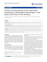
mobility. Body examination indicated nystagmus, hypodontia, polytrichia (Fig. 1a and b), ataxia, and spastic
tetraplegia. In a previous brain image, we identified an
extracerebral space widening at the age of 6 months (Fig.
1c) and further frontotemporal space widening at the
age of 11 months with delayed myelination or hypomyelination of white matter in the focal area around the posterior horn of the bilateral lateral ventricles (Fig. 1d–f).
Laboratory examination indicated that plasma ammonia,
lactate, serum antibody tests for toxoplasma, rubella
Page 3 of 6
virus, cytomegalovirus, and herpes simplex virus
(TORCH), vitamin B, trace elements, creatine kinase,
and thyroid function were normal. Electroencephalogram and electrocardiogram results were negative. The
value of auditory brain-stem responses was greater
than the threshold line (50 dbnnl) (Table 2). Chest Xray showed bilateral lung inflammation. Because of
recurrent pneumonia, tracheobronchoscopy was
performed and an orifice of the right middle bronchus was found to be absent (Fig. 1g), which was first
observed in POLR3-related leukodystrophy. Genetic metabolic screening of blood and urine were performed twice
and parameters were determined to be within normal
range. The results of the abdomen ultrasound examination were negative. Fundus examination was normal
without optic atrophy and cataract. Visual acuity was also
measured and no myopia was found. The endocrinal profile was not detected because the patient was too young;
data regarding motor conduction velocity was also not
available. Conventional karyotype analysis revealed a normal 46 XX karyotype.
To achieve an accurate genetic diagnosis, medical
exome sequencing was carried out with a Trio sample
strategy. A peripheral blood sample was collected from
the proband and her parents and genomic DNA was
isolated using the High Pure PCR Template Preparation
Kit (Roche, Basel, Switzerland) according to the manufacturer’s instructions. The medical exome including
coding regions and known pathogenic non-coding regions of over 4000 disease-related genes was captured
before next-generation sequencing (Amcare Genomic
Laboratory, Guangzhou, China). The potential pathogenic variants were filtered by bioinformatics analysis as
described previously [12]. Sequencing of 50,902 genomic
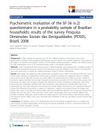
Fig. 1 Clinical pictures of this patient. a: Tooth delay or tooth agenesis was found at the age of 2 years and 6 months old; b: Body examination
indicated manifestation of polytrichia; c: Brain MRI showed the extra cerebral space widening at six months old; d-f: Frontotemporal space
widening, delayed myelination or hypomyelination of white matter in the focal area around the posterior horn of the bilateral lateral ventricles at
the age of eleven months. g: Fiberoptic bronchoscopy presented the absence of right middle bronchus orifice
Wu et al. BMC Pediatrics
(2019) 19:289
Table 2 Laboratory results
Test
Results
Chromosome karyotype
46 XX, normal
Plasma ammonia
Normal
Lactate
Normal
TORCH
Negative
Genetic Metabolic Screening
Negative
Electroencephalogram EEG
Normal
Auditory brain-stem responses, ABR
Over than threshold (50dbnnl)
Vitamin B
Normal
Trace elements
Normal
Creatine kinase
Normal
Thyroid function
Normal
regions spread over 8,591,731 bp with an average coverage of 274+/− 164× was obtained; the coverage of 99.4%
of the sequenced regions exceeded 10× and the coverage
of 99.2% of the sequenced regions exceeded 20×. Further
analysis revealed two novel mutations of POLR3A in the
patient: c.1771-6C > G (NM_007055) adjacent to the
mRNA splicing site and c.2611del, which results in early
termination of translation (p.M871Cfs*8). The c.17716C > G mutation occurs at very low frequency in the
population (< 0.001), while the c.2611del mutation is not
Page 4 of 6
listed in 1000 Genomes (The 1000 Genome Project
Consortium) or The Genome Aggregation Database
(gnomAD, Broad Institute). Co-segregation analysis
confirmed that the two mutations were inherited from
the heterozygous parents of the proband. The father was
determined to be the carrier of the c.2611del
(p.M871Cfs*8) mutation and the mother was determined
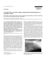
to be the carrier of the c.1771-6C > G mutation. Collectively, we identified novel compound heterozygous mutations of the POLR3A gene that caused POLR3-related
leukodystrophy in the patient combined with the clinical
presentation, MRI brain pattern, and medical exome
sequencing (Figs. 1 and 2).
Discussion and conclusion
Our case from the southern district of China displayed severe neurological manifestations and presented with typical childhood onset with various features such as
cerebellar symptoms (spasticity and ataxia), cognitive regression, motor decline, and delayed dentition. Brain MRI
indicated delayed myelination or hypomyelination of
white matter in the focal area around the posterior horn
of the bilateral lateral ventricles. Takanashi et al. reported
that hypomyelination of the brain often indicates POLR3A
mutation, which is associated with leukodystrophy
disorders [6]. To verify this, we performed medical exome
Fig. 2 Identification of novel POLR3A mutations in the family by next-generation sequencing
Wu et al. BMC Pediatrics
(2019) 19:289
sequencing and found novel compound heterozygous mutations of the POLR3A gene, reminiscent of other patients.
According to the clinical manifestations, we concluded
the diagnosis and identified the compound heterozygous
variants as the causative variants for the disease in this
patient. It is noteworthy that this disease has mostly been
reported in European populations, including FrenchCanadian, Caucasian, and Syrian individuals [2, 7]. Occasional cases have been reported in the Indian population
[13–15]. However, this is the first case reported in a
Chinese family.
Neurological impairment of our case started in the
infantile period with a decline in motor ability, cognitive
impairment, and cerebellar features. Although cerebellar
signs of this case became progressively obvious, cerebellar
atrophy was not observed, which is likely related to the
molecular basis or other factors. Previous studies have
found that cerebellar anomalies were more severe in
patients with POLR3B defects while the pattern of hypomyelinization was more evident in the MRI of patients
with POLR3A mutations [2, 6]. This may be another explanation for our case. Our patient also showed classical
extraneurologic features, characterized by hypodontia with
delayed tooth eruption and short stature. She also displayed polytrichia, an atypical feature of POLR3-related
leukodystrophy, which may be due to aberrant endocrine
hormone levels or other reasons. Hypogonadotropic hypogonadism was not detected because she was too young.
Previous studies have also shown that the syndrome may
or may not be associated with hypodontia and/or hypogonadotrophic hypogonadism in many cases [8, 11]. The
case did not show myopia and optic atrophy. This is inconsistent with most cases, which are usually accompanied by myopia [2]. Her dysphagia phenotype was striking.
She had obvious difficulty with tube feeding and forceful
vomiting occurred frequently. This is likely due to the
incoordination of swallowing of cerebellar syndrome, or
due to other unpredictable reasons. Bronchodysplasia is
another feature first observed in POLR3-related leukodystrophy, suggesting that it was not recognized previously in
the POLR3-related leukodystrophy spectrum. Thus, in
addition to the classical extraneurological features,
abnormal body hair and visceral smooth muscle features
should be carefully looked for in patients with POLR3related. When classical features do not exist, rare
manifestations will a clue in the diagnosis of this disorder.
Although there is no cure for this disease to date,
treatment of manifestations such as seizures, hypogonadotropic hypogonadism, dystonia, and dysphagia can be
managed on an individual basis for an improved quality of
life and the prevention of complications.
Our case presented with severe manifestation at early
onset and diverse manifestations among those of patients
with POLR3-related leukodystrophy, which may be a result
Page 5 of 6
of the genotype identified in this patient; further analysis is
necessary. To date, four genes (POLR3A, POLR3B,
POLR1C, and POLR3K) have been reported to be associated with POLR3-related leukodystrophy [11, 16]. Most of
the identified mutations are point mutations in the codon
region; however, non-coding DNA variants are suspected
to account for a substantial portion of undiscovered causes
of rare diseases [17, 18]. Minnerop et al. identified mutations in deep intronic regions of POLR3A as a common
cause of hereditary spastic paraplegia and cerebellar ataxia,
and > 80% of POLR3A mutation carriers presented the
same deep intronic mutation (c.1909 + 22G > A), which
leads to a novel, distinct, uniform, and severe phenotype
[17]. Jay et al. also reported alteration of mRNA splicing in
POLR3A causing neonatal progeroid syndrome with severe
clinical manifestations [23]. In this study, we identified the
c.1771-6C > G (NM_007055) mutation adjacent to the
mRNA splice site demonstrating that exploring noncoding genomic regions was helpful in revealing the causes
of related hereditary diseases.
The complexity of clinical phenotypes and the heterogeneity of genotypes raise new challenges in genetic diagnoses. In the present study, medical exome sequencing
was used to explore the possible genetic defects resulting
in the disease of the patient. Compared to whole genome
and whole exome sequencing, medical exome sequencing
focuses on clinical interpretable regions of genes; less
variants of uncertain significance in medical exome sequencing greatly improve the diagnostic yield and increase
the coverage depth of sequencing, improving the accuracy
of sequencing and broadening the spectrum of variants. In
the present study, we identified novel heterozygous
mutations of POLR3A that caused POLR3-related
leukodystrophy disease for the first time in a Chinese
family. This study will further our understanding of the
molecular mechanisms of POLR3-related leukodystrophy
and contribute to further analysis of phenotype–genotype
correlations of related disorders.
Abbreviations
ADDH: Ataxia, delayed dentition, and hypomyelination;
HCACH: Hypomyelination with cerebellar atrophy and hypoplasia of the
corpus callosum; LO: Leukodystrophy with oligodontia; MRI: Magnetic
resonance imaging; TACH: Tremor-ataxia with central hypomyelination;
TORCH: Serum antibody tests for toxoplasma, rubella virus, cytomegalovirus,
and herpes simplex virus
Acknowledgements
We thank International Science Editing (ernationalscienceediting.
com) for editing this manuscript.
Authors’ contributions
SW: Designed the research, analyzed the data and drafted the manuscript;
ZB: Participated in analyzing the part of data; XD: Collected clinical data; HC,
DY and JH: Participated in the communicate with patients’ guardians; LZ:
Collected clinical data; HL: Participated to the in discussion and
interpretation of the data and results, involved in the critical revision of this
manuscript and take the primary responsibility of this research; All authors
have read and approved this manuscript and ensure that this is the case.
Wu et al. BMC Pediatrics
(2019) 19:289
Funding
Design of the study and collection, analysis, and interpretation of data and in
writing the manuscript were funded by Suzhou Science and Technology
Development Project (project code SYS 201757) and Natural science fund for
colleges and universities of Jiangsu Province (project code 18KJB320022).
Page 6 of 6
12.
Availability of data and materials
The datasets used and/or analysed during the current study are available
from the corresponding author (Haitao Lv) on reasonable request.
13.
Ethics approval and consent to participate
Ethical approval for this study was obtained from the local ethics committee.
Informed consent informed consent was obtained from the patient’s parents.
14.
Consent for publication
The guardians have written informed consent to publish this information
and the proof of consent can be requested at any time.
Competing interests
The authors declare that they have no conflict of interest.
Author details
1
Department of Intensive Care Unit, Children’s Hospital of Soochow
University, Suzhou, Jiangsu, China. 2Department of Cardiovascular Medicine,
Children’s Hospital of Soochow University, No.92, Zhongnan street, Suzhou
Industrial Park, Suzhou, Jiangsu, China.
15.
16.
17.
18.
encoding RNA polymerase III subunits cause an autosomal-recessive
hypomyelinating leukoencephalopathy. Am J Hum Genet. 2011;89(5):644–51.
Azmanov DN, Siira SJ, Chamova T, Kaprelyan A, Guergueltcheva V,
Shearwood AJ, Liu G, Morar B, Rackham O, Bynevelt M, et al. Transcriptomewide effects of a POLR3A gene mutation in patients with an unusual
phenotype of striatal involvement. Hum Mol Genet. 2016;25(19):4302–14.
McKenna A, Hanna M, Banks E, Sivachenko A, Cibulskis K, Kernytsky A,
Garimella K, Altshuler D, Gabriel S, Daly M, et al. The genome analysis
toolkit: a MapReduce framework for analyzing next-generation DNA
sequencing data. Genome Res. 2010;20(9):1297–303.
Jauhari P, Sahu JK, Singhi P, Dayal D, Khandelwal N. An Indian boy with a
novel leukodystrophy: 4H syndrome. J Child Neurol. 2014;29(1):135–8.
Muthusamy K, Sudhakar SV, Yoganathan S, Thomas MM, Alexander M.
Hypomyelination, Hypodontia, hypogonadotropic hypogonadism (4H)
syndrome with vertebral anomalies: a novel association. J Child Neurol.
2015;30(7):937–41.
Dumay-Odelot H, Durrieu-Gaillard S, Da Silva D, Roeder RG, Teichmann M.
Cell growth- and differentiation-dependent regulation of RNA polymerase III
transcription. Cell Cycle. 2010;9(18):3687–99.
Minnerop M, Kurzwelly D, Wagner H, Soehn AS, Reichbauer J, Tao F, Rattay
TW, Peitz M, Rehbach K, Giorgetti A, et al. Hypomorphic mutations in
POLR3A are a frequent cause of sporadic and recessive spastic ataxia. Brain.
2017;140(6):1561–78.
Baralle D, Buratti E. RNA splicing in human disease and in the clinic. Clin Sci.
2017;131(5):355–68.
Publisher’s Note
Received: 1 June 2019 Accepted: 31 July 2019
References
1. Bernard G, Vanderver A: POLR3-related Leukodystrophy. In:
GeneReviews((R)). Edn. Edited by Adam MP, Ardinger HH, Pagon RA,
Wallace SE, Bean LJH, Stephens K, Amemiya A. Seattle (WA); 1993.
2. Wolf NI, Vanderver A, van Spaendonk RM, Schiffmann R, Brais B, Bugiani M,
Sistermans E, Catsman-Berrevoets C, Kros JM, Pinto PS, et al. Clinical
spectrum of 4H leukodystrophy caused by POLR3A and POLR3B mutations.
Neurology. 2014;83(21):1898–905.
3. Sato I, Onuma A, Goto N, Sakai F, Fujiwara I, Uematsu M, Osaka H, Okahashi
S, Nonaka I, Tanaka S, et al. A case with central and peripheral
hypomyelination with hypogonadotropic hypogonadism and hypodontia
(4H syndrome) plus cataract. J Neurol Sci. 2011;300(1–2):179–81.
4. Bekiesinska-Figatowska M, Mierzewska H, Kuczynska-Zardzewialy A,
Szczepanik E, Obersztyn E. Hypomyelination, hypogonadotropic
hypogonadism, hypodontia - first polish patient. Brain Dev. 2010;32(7):574–8.
5. Timmons M, Tsokos M, Asab MA, Seminara SB, Zirzow GC, Kaneski CR, Heiss
JD, van der Knaap MS, Vanier MT, Schiffmann R, et al. Peripheral and central
hypomyelination with hypogonadotropic hypogonadism and hypodontia.
Neurology. 2006;67(11):2066–9.
6. Takanashi J, Osaka H, Saitsu H, Sasaki M, Mori H, Shibayama H, Tanaka M,
Nomura Y, Terao Y, Inoue K, et al. Different patterns of cerebellar
abnormality and hypomyelination between POLR3A and POLR3B mutations.
Brain Dev. 2014;36(3):259–63.
7. Daoud H, Tetreault M, Gibson W, Guerrero K, Cohen A, Gburek-Augustat J,
Synofzik M, Brais B, Stevens CA, Sanchez-Carpintero R, et al. Mutations in
POLR3A and POLR3B are a major cause of hypomyelinating
leukodystrophies with or without dental abnormalities and/or
hypogonadotropic hypogonadism. J Med Genet. 2013;50(3):194–7.
8. Thiffault I, Wolf NI, Forget D, Guerrero K, Tran LT, Choquet K, Lavallee-Adam
M, Poitras C, Brais B, Yoon G, et al. Recessive mutations in POLR1C cause a
leukodystrophy by impairing biogenesis of RNA polymerase III. Nat
Commun. 2015;6:7623.
9. Potic A, Brais B, Choquet K, Schiffmann R, Bernard G. 4H syndrome with
late-onset growth hormone deficiency caused by POLR3A mutations.
Arch Neurol. 2012;69(7):920–3.
10. Dorboz I, Dumay-Odelot H, Boussaid K, Bouyacoub Y, Barreau P, Samaan S,
Jmel H, Eymard-Pierre E, Cances C, Bar C, et al. Mutation in POLR3K causes
hypomyelinating leukodystrophy and abnormal ribosomal RNA regulation.
Neurology Genetics. 2018;4(6):e289.
11. Saitsu H, Osaka H, Sasaki M, Takanashi J, Hamada K, Yamashita A, Shibayama H,
Shiina M, Kondo Y, Nishiyama K, et al. Mutations in POLR3A and POLR3B
Springer Nature remains neutral with regard to jurisdictional claims in
published maps and institutional affiliations.