Exploratory laparoscopy combined with pathological examination in the diagnosis of obscure gastrointestinal bleeding in a child: A case report
Bạn đang xem bản rút gọn của tài liệu. Xem và tải ngay bản đầy đủ của tài liệu tại đây (1.49 MB, 4 trang )
Chen et al. BMC Pediatrics
(2018) 18:371
/>
CASE REPORT
Open Access
Exploratory laparoscopy combined with
pathological examination in the diagnosis
of obscure gastrointestinal bleeding in a
child: a case report
Jiande Chen1†, Bin Zhang2†, Zhilong Yan3, Huaying Zhao3, Kaihua Yang2, Yong Yin1* and Lirong Jiang2*
Abstract
Background: The diagnosis of obscure gastrointestinal bleeding (OGIB) which is defined as bleeding of unknown
origin of the small bowel by routine evaluation in childhood is a challenge.
Case presentation: Here we report a one-year-old Chinese girl who was suspected with idiopathic pulmonary
haemosiderosis (IPH) and referred to our department for further diagnosis. Finally she was diagnosed with vascular
malformations (VM) by exploratory laparoscopy combined with pathological examination.
Conclusions: Children OGIB could be easily misdiagnosed in the beginning, and OGIB children with active ongoing
bleeding may benefit from proceeding directly to exploratory laparoscopy, followed by pathological confirmation
of the diagnosis.
Keywords: Iron-deficiency anemia, Melena, Vascular malformations
Background
Obscure gastrointestinal bleeding (OGIB) is defined as
bleeding of unknown origin that persists or recurs after
bidirectional endoscopy and radiologic evaluation of the
small bowel [1]. It could be categorized into obscure
overt and obscure occult bleeding based on the presence
or absence of clinically evident bleeding [2]. Causes of
OGIB may potentially include lesions that are overlooked in the esophagus, stomach, and colon during initial workup or lesions in the small intestine that are
difficult to visualize with conventional endoscopy and
radiologic imaging [1]. After negative endoscopy and
colonoscopy, performing small bowel endoscopic investigation by capsule endoscopy (CE) and balloon-assisted
enteroscopy (BAE) has a very good diagnostic yield [3].
* Correspondence: ;
†
Jiande Chen and Bin Zhang contributed equally to this work.
1
Department of Respiratory Medicine, Shanghai Children’s Medical Center
Affiliated to Shanghai Jiao Tong University School of Medicine, No.1678
Dongfang Road, Pudong 200127, Shanghai, China
2
Department of Gastroenterology, Shanghai Children’s Medical Center
Affiliated to Shanghai Jiao Tong University School of Medicine, No.1678
Dongfang Road, Pudong 200127, Shanghai, China
Full list of author information is available at the end of the article
Intraoperative enteroscopy is currently reserved as a last
option, for when other measures cannot identify a bleeding source in selected patients.
This paper presents an unusual case study of a
one-year-old girl who presented with OGIB, the subsequent diagnostic challenges encountered and how these
were addressed.
Case presentation
A nine-month-old Chinese girl presented with one-week
history of pallor at a referral hospital where she received a
red blood cell transfusion for severe anemia (Hb 3.4 g/dL)
and started to treat for iron-deficiency anemia (IDA) after
microcytosis (mean corpuscular volume 74.6 fl), hypochromia (mean cell Hb 21.5 pg), and low serum iron concentration (1.28umol/L) were confirmed. On discharge
after 1 week of treatment, anemia was corrected (Hb 12.4
g/dL). However, recurrent anemia was observed over a
six-month period, even another red blood cell transfusion
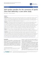
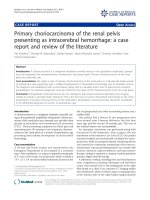
was given in this period. Positive fecal occult blood test results were intermittent. A chest computed tomography
(CT) scan showed the increase of patch density in the left
© The Author(s). 2018 Open Access This article is distributed under the terms of the Creative Commons Attribution 4.0
International License ( which permits unrestricted use, distribution, and
reproduction in any medium, provided you give appropriate credit to the original author(s) and the source, provide a link to
the Creative Commons license, and indicate if changes were made. The Creative Commons Public Domain Dedication waiver
( applies to the data made available in this article, unless otherwise stated.
Chen et al. BMC Pediatrics
(2018) 18:371
lower lobe (Fig. 1a) and right upper lobe (Fig. 1b) of the
lung. Although she had no history of repetitive haemoptysis, chronic cough and dyspnoea, idiopathic pulmonary
haemosiderosis (IPH) was entertained and the IDA therapy was discontinued.
Patient was referred to our hospital for further management. Flexible bronchoscopy was performed, but
bronchoalveolar lavage examination of blood-stained
fluid and hemosiderin-laden macrophages from involved
areas was negative. Review of the chest CT scan showed
no extensive ground glass opacities and reticular
shadows. Therefore, diffuse alveolar haemorrhage was
ruled out. Review of the patient’s history found an episode of intermittent melena 1 month after the IDA treatment, and that was considered to be the side effect of
the drug by the outpatient doctor. No related family
genetic history. Physical exam demonstrated a girl of
normal appearance consistent with her ethnicity except
pallor. The diagnostic approach for gastrointestinal
bleeding was started. However, the patient underwent
both upper and lower endoscopy with negative findings
in all of the endoscopic examinations. Plain and enhanced CT of abdomen and the technetium-99 m–labeled red blood cell scans were performed. Again, they
were all negative. Her symptoms persisted and one red
blood cell transfusion was needed each week.
The department of general surgery was involved in the
management and a decision to do surgical exploration
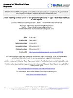
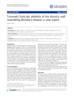
with laparoscopy was taken. A 3 cm lesion with dense
blistered protrusions on the surface was found within
the wall of jejunum (Fig. 2), acting as a lead point, so a
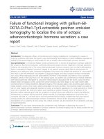
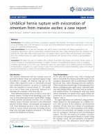
jejunal segment was resected and an end to end jejunojejunostomy was performed. Pathological examination
indicated a vascular malformations (VM) (Fig. 3). Postoperative period was uneventful and she was discharged
Fig. 1 Chest CT. Increased patch density in the left lower lobe (a)
and the right upper lobe (b) of the lung
Page 2 of 4
Fig. 2 Lesion within the wall of jejunum. A 3 cm lesion with dense
blistered protrusions on the surface within the wall of jejunum
home with no complications. There was no recurrence
during follow-ups.
Discussion and conclusions
OGIB from VM in this case affected the delay in diagnosis because of its rarity and limitations in the diagnostic
approach in pediatric patients.
Massive gastrointestinal haemorrhage in a child due to
VM of the jejunum is very uncommon [4]. To our
knowledge this is the second case of an acute gastrointestinal haemorrhage in a child due to VM of the jejunum. Most VM cases may lay a false trail for the
clinician because of accompanied IDA with no gastrointestinal symptoms at the initial time [5]. In our case,
IDA combined with asymptomatic pulmonary infection
misled the diagnosis as IPH. The clinical conditions of
our case are reported for the first time.
Syndromes such as the Klippel-Trenaunay syndrome and
the blue rubber blebnevus syndrome usually encompass
VM as a skin manifestation, so the possibility of visceral lesion may be suspected. In this case, however, the malformations were a unique manifestation without any associated
syndrome, which increased the difficulty of diagnosis.
Angiography may detect OGIB lesions and also offers
a therapeutic option with embolization if a bleeding lesion is identified. In OGIB patients, the bleeding rate
may be slow or intermittent, thereby not allowing identification by either angiography or bleeding scan [6]. A
small case series also suggests that the overall yield of
provocative angiography is low [7].
CE is currently the preferred test for the initial investigation in patients with OGIB due to its high diagnostic yield
[1]. However, this technology requires precision instruments and skilled endoscopic images interpreters. In
addition, the increase of the cost-effectiveness, imprecise
localization, the risk of capsule retention and a lack of
Chen et al. BMC Pediatrics
(2018) 18:371
Page 3 of 4
Fig. 3 Gross view and microscopic features of intestinal wall VM. Macroscopically, there was a lesion with dense blistered protrusions on the
surface within the intestinal wall (a). On microscopy, abnormal collections of dilated vascular structures of variable sizes were observed in the
lesion (b, original magnification × 10)
therapeutic capability also restrict the wide application of
CE among children patients, particularly in acute cases.
OGIB was a common indication for small bowel endoscopy. The development of BAE represents a decisive
breakthrough in the diagnosis and management of small
bowel diseases. The overall diagnostic yield of BAE was
about 70% [8, 9]. The approach of CE followed by BAE
might show a diagnostic yield over 90% [10]. However,
this technology has not been widely used in children’s hospitals for concerns regarding safety, design of instruments,
training, availability, and a lack of knowledge about its use
and relative indications.
The safety and effectiveness of using laparoscopy as the
diagnostic and therapeutic tool for OGIB in children have
been well established by pediatric literature [11–14]. In
the cases of difficult-to-manage or acute bleeding, we may
directly resort to laparoscopy for difficult-to-access lesions. Pathological examination should be performed to
make a definite diagnosis after lesions resection.
Our experience of successful management of this case
suggested that children OGIB combined with asymptomatic pulmonary infection could be easily misdiagnosed as IPH in the beginning, and OGIB children with
active ongoing bleeding may benefit from proceeding
directly to exploratory laparoscopy, followed by pathological confirmation of the diagnosis.
Abbreviations
BAE: Balloon-assisted enteroscopy; CE: Capsule endoscopy; CT: Computed
tomography; IDA: Iron-deficiency anemia; IPH: Idiopathic pulmonary
haemosiderosis; OGIB: Obscure gastrointestinal bleeding; VM: Vascular
malformations
Acknowledgments
We thank the patient and her family. We also thank pathology department,
radiology department, rheumatism department, hematology-oncology
department and other departments of Shanghai Children’s Medical Center
Affiliated to Shanghai Jiao Tong University School of Medicine for their assistance.
Funding
Not applicable.
Availability of data and materials
The data and materials used and/or analysed during the current study were
presented within the manuscript.
Authors’ contributions
JC and BZ interpreted the results for the case report, drafted, wrote and
revised the report, and provided important intellectual review. ZY and HZ
carried out the surgical exploration with laparoscopy and helped to draft the
manuscript. KY collected the data from our hospital work system and critically
reviewed the manuscript. YY and LJ conceived of the study, and participated in
its design and coordination and helped to review the manuscript. All authors
read and approved the final manuscript.
Ethics approval and consent to participate
This study was approved by the Ethics Committee of Shanghai Children’s
Medical Center Affiliated to Shanghai Jiao Tong University School of
Medicine and was conducted in accordance with the Declaration of Helsinki.
Consent for publication
Written informed consent was obtained from the parent for the publication
of this case report.
Competing interests
The authors declare that they have no competing interests.
Publisher’s Note
Springer Nature remains neutral with regard to jurisdictional claims in published
maps and institutional affiliations.
Author details
1
Department of Respiratory Medicine, Shanghai Children’s Medical Center
Affiliated to Shanghai Jiao Tong University School of Medicine, No.1678
Dongfang Road, Pudong 200127, Shanghai, China. 2Department of
Gastroenterology, Shanghai Children’s Medical Center Affiliated to Shanghai
Jiao Tong University School of Medicine, No.1678 Dongfang Road, Pudong
200127, Shanghai, China. 3Department of General Surgery, Shanghai
Children’s Medical Center Affiliated to Shanghai Jiao Tong University School
of Medicine, No.1678 Dongfang Road, Pudong 200127, Shanghai, China.
Received: 11 April 2018 Accepted: 12 November 2018
References
1. Raju GS, Gerson L, Das A, Lewis B. American gastroenterological association
(AGA) institute technical review on obscure gastrointestinal bleeding.
Gastroenterology. 2007;133(5):1697–717.
2. American Gastroenterological Association medical position statement:
evaluation and management of occult and obscure gastrointestinal
bleeding. Gastroenterology. 2000;118(1):197–201.
3. Romano C, Oliva S, Martellossi S, Miele E, Arrigo S, Graziani MG, Cardile S,
Gaiani F, de'Angelis GL, Torroni F. Pediatric gastrointestinal bleeding:
perspectives from the Italian Society of Pediatric Gastroenterology. World J
Gastroenterol. 2017;23(8):1328–37.
4. Kimpton JA, Bowen JC, Craigie RJ. Paediatric angiodysplasia of the jejunum:
a case report and review of the literature. Scott Med J. 2012;57(4):247.
Chen et al. BMC Pediatrics
5.
6.
7.
8.
9.
10.
11.
12.
13.
14.
(2018) 18:371
Kim SH, Cho YH, Kim HY. Vascular malformations of the small intestine
manifesting as chronic anemia: two pediatric cases managed by single-site
umbilical laparoscopic surgery. Int J Surg Case Rep. 2017;31:233–6.
Rantis PCJ, Harford FJ, Wagner RH, Henkin RE. Technetium-labelled red
blood cell scintigraphy: is it useful in acute lower gastrointestinal bleeding?
Int J Color Dis. 1995;10(4):210–5.
Bloomfeld RS, Smith TP, Schneider AM, Rockey DC. Provocative angiography
in patients with gastrointestinal hemorrhage of obscure origin. Am J
Gastroenterol. 2000;95(10):2807–12.
Hong SN, Kim ER, Ye BD, Jang HJ, Jeon SR, Park SJ, Im JP, Kim JH, Choi CH, Choi
H, et al. Indications, diagnostic yield, and complication rate of balloon-assisted
enteroscopy (BAE) during the first decade of its use in Korea. Dig Endosc. 2016;
28(4):443–49.
Ma JJ, Wang Y, Xu XM, Su JW, Jiang WY, Jiang JX, Lin L, Zhang DQ, Ding J,
Chen L, et al. Capsule endoscopy and single-balloon enteroscopy in small
bowel diseases: competing or complementary? World J Gastroenterol. 2016;
22(48):10625–30.
Oliva S, Pennazio M, Cohen SA, Aloi M, Barabino A, Hassan C, Pession A,
Lima M, Frediani S, Di Nardo G. Capsule endoscopy followed by single
balloon enteroscopy in children with obscure gastrointestinal bleeding: a
combined approach. Dig Liver Dis : official journal of the Italian Society of
Gastroenterology and the Italian Association for the Study of the Liver.
2015;47(2):125–30.
Lee KH, Yeung CK, Tam YH, Ng WT, Yip KF. Laparascopy for definitive
diagnosis and treatment of gastrointestinal bleeding of obscure origin in
children. J Pediatr Surg. 2000;35(9):1291–3.
Pal K, El Shafei H, Al Buainain H, Mitra DK. Successful laparoscopic treatment
of hemorrhage from ileal duplication cyst in a 10-year-old Saudi boy. J
Laparoendosc Adv Surg Tech Part A. 2010;20(1):99–101.
Loh DL, Munro FD. The role of laparoscopy in the management of lower
gastro-intestinal bleeding. Pediatr Surg Int. 2003;19(4):266–7.
Craigie RJ, Forrest N, Nanthakumaran S, Mahomed AA. Laparoscopy in
diagnosis and management of Meckel's diverticulum. J Laparoendosc Adv
Surg Tech Part A. 2006;16(1):70–3.
Page 4 of 4