Repeatability of echocardiographic parameters to evaluate the hemodynamic relevance of patent ductus arteriosus in preterm infants: A prospective observational study
Bạn đang xem bản rút gọn của tài liệu. Xem và tải ngay bản đầy đủ của tài liệu tại đây (634.02 KB, 5 trang )
Schwarz et al. BMC Pediatrics (2016) 16:18
DOI 10.1186/s12887-016-0552-7
RESEARCH ARTICLE
Open Access
Repeatability of echocardiographic
parameters to evaluate the hemodynamic
relevance of patent ductus arteriosus in
preterm infants: a prospective
observational study
Christoph E. Schwarz1*, Antonio Preusche1, Winfried Baden2, Christian F. Poets1 and Axel R. Franz1,3
Abstract
Background: The hemodynamically relevant patent ductus arteriosus in preterm infants is not well defined.
Different clinical and echocardiographic parameters are used and the diagnostic accuracy is unknown because
of the lack of a gold standard definition. Our study evaluates the inter-observer repeatability of echocardiographic and
Doppler-ultrasound parameters.
Methods: This prospective observational study included 19 very low birth weight preterm infants (median
[interquartile range]: gestational age 28.0 (28.0–29.0) weeks, birth weight 1130 (905–1321) g, postnatal age at
measurement 8.7 (4.8–23.5) d) with a clinical suspicion of ductal patency in whom 27 repeated echocardiographic and
Doppler-ultrasound examinations were performed within 30 min by 2 of 3 independent observers (54 measurements
overall). The repeatability index (=2 times the standard deviation of the differences/mean of all measurements)
according to Bland and Altman was used to assess repeatability of different parameters.
Results: The repeatability indices of the echocardiographic parameters (left Atrium-to-Aortic root-ratio, diameter
of the patent ductus arteriosus at its narrowest part, the left-ventricular-preejection-period-to-ejection-time-ratio
and the ratio of the velocity time integrals in the large vessels were 16, 21, 23 and 26 % respectively. The repeatability
indices of Doppler-ultrasound measurements (resistance index in celiac artery and anterior cerebral artery) were 11 and
14 %, respectively.
Conclusions: The inter-observer repeatability of all echocardiographic parameters was poor compared to that of
resistance indices in peripheral vessels. Therefore, interventions for ductal patency should be indicated based on
averaged repeated rather than single measurements, especially when measured values are close to their cut-off
value - both in clinical routine and for study purposes.
Keywords: Reproducibility, Doppler-ultrasound, Inter-observer
* Correspondence:
1
Department of Neonatology, University Children’s Hospital of Tuebingen,
University of Tuebingen, Calwerstr. 7, 72076 Tuebingen, Germany
Full list of author information is available at the end of the article
© 2016 Schwarz et al. Open Access This article is distributed under the terms of the Creative Commons Attribution 4.0
International License ( which permits unrestricted use, distribution, and
reproduction in any medium, provided you give appropriate credit to the original author(s) and the source, provide a link to
the Creative Commons license, and indicate if changes were made. The Creative Commons Public Domain Dedication waiver
( applies to the data made available in this article, unless otherwise stated.
Schwarz et al. BMC Pediatrics (2016) 16:18
Background
The patent ductus arteriosus (PDA) in preterm infants is
associated with increased mortality and morbidity [1–7].
However, there is little evidence as to which parameters
define a PDA that requires treatment. Zonnenberg and
de Waal showed that, besides clinical parameters, echocardiographic and Doppler-ultrasound measurements
are used to evaluate the magnitude and clinical relevance
of the left-to-right shunt through a PDA, and hence the
need for treatment: In a systematic review of 67 randomised controlled trials (RCTs) they described the following most frequently used parameters and applied cut-off
values: Left-atrium-to-aortic-root-ratio (LA/Ao-ratio)
used in 34 trials, median cut-off >1.3 (range: 1.15–1.70);
diastolic reverse flow in peripheral vessels (21 trials); and
PDA-diameter (8 trials), cut-off >1.5 (1.5–2.0) mm [8].
McNamara and Sehgal suggested a scoring system including clinical and echocardiographic criteria to define
hrPDA [9]. The echocardiographic part of this staging
seems to be predictive for neonatal morbidity and can
serve as a guide to clinical decisions [10], whereas the
clinical criteria comprise unspecific respiratory signs.
Prospective data suggesting that application of the echocardiographic parameters summarized by Zonnenberg
and de Waal or the score by McNamara and Sehgal results in improved outcome is lacking. However, recent
retrospective data suggest that echocardiographic screening for PDA within the first 3 postnatal days may reduce
mortality in infants born at <29 weeks gestation [11].
To inform future studies and clinical guidelines on PDA
treatment, this study aimed to evaluate the inter-observer
repeatability of echocardiographic and Doppler-ultrasound
parameters, which are frequently determined to assess the
need for PDA treatment.
Methods
This prospective observational cohort study was approved
by the research ethics committee at the facutly of
medicine and the university hospital of the Eberhard
Karls University Tuebingen and written informed parental consent obtained. To assess the repeatability of
echocardiographic parameters commonly used to determine the magnitude of the left-to-right shunt, a
convenience sample of preterm infants with suspected
PDA was analysed. Inclusion criteria were: birth
weight ≤1500 g and clinical suspicion of PDA such as
cardiac murmur, bounding pulses, ventilator dependency and increased oxygen demand. Syndromal anomalies and congenital heart defects except persisting
foramen ovale or atrial septal defect were exclusion
criteria. The period of recruitment was between June
2012 and May 2013 at the Department of Neonatology, University Children’s Hospital of Tuebingen,
University of Tuebingen, Germany.
Page 2 of 5
Within 30 min (to minimize fluctuations in hemodynamic
status), 2 of 3 investigators (with more than 20, 10,
or 3 years, respectively, of experience in neonatal
echocardiography, everyday skills or every week, respectively, 2 were board certified paediatric cardiologists, one investigator is attending physician at the
NICU) prospectively and independently performed
repeated echocardiographic and Doppler-sonographic
measurements including the following parameters:
LA/Ao-ratio [12]; resistance index (RI) in celiac artery (CA) [13] and anterior cerebral artery (ACA)
[14]; diameter of the PDA at its narrowest part [15];
the left-ventricular-preejection-period-to-ejection-time-ratio, calculated by including 3–4 cardiac cycles (LVPEP/
LVET) [16]; and the ratio of the velocity time integrals in
the large vessels (VTI_Ao/VTI_Pa). The VTI_Ao was
measured from an apical-5-chamber-view, the VTI_Pa
was measured in a parasternal short axis calculated automatically with built-in software. We assumed that, in the
absence of congenital heart defects, this ratio correlates
with the ductal left-to-right shunt. The PDA-diameter was
measured at its narrowest part (identified via colour
Doppler) and measured in B-Mode to avoid the influence of gain-settings on the PDA-width if assessed on
colour Doppler images.
All measurements were done with a Toshiba “Aplio”
using a 6.5 MHz phased array transducer.
Statistical analyses involved repeatability coefficient
(RepC = 2 times the standard deviation of the differences) and repeatability index (RepI = RepC/mean of all
measurements) according to Bland and Altman [17] and
a confidence-step-analysis (CSA) [18]. The RepC represents the upper limit of the 95 % confidence interval of
the absolute differences between two measurements performed by two independent observers. The RepI describes the relation between RepC and the mean value of
the measurements. This allows comparison of repeatability between different measures. A high CSA value
(CSA = difference between lowest and highest value /
RepC) indicates that differences between low and high
values in this parameter observed in a given population
are unlikely related to inter-/intra-observer variability,
whereas a CSA of ≤1 indicates that observed differences
between low and high values in this population are likely
due to inter-/intra-observer variability.
For unexpected RepI differences, 95 % confidence intervals (CI) were exploratorily calculated post-hoc and
differences between echo-parameters classified as ‘significant’ if these 95 %-CI did not overlap.
Data are shown as median (interquartile range).
Results
Twenty-seven repeated measurements were performed
in 19 preterm infants. One infant with a birth weight of
Schwarz et al. BMC Pediatrics (2016) 16:18
Page 3 of 5
1550 g was included inadvertently due to a weight at the
time of measurement of 1465 g.
Gestational age at birth was 28.0 (28.0–29.0) weeks,
birth weight 1130 (905–1321) g, postnatal age at measurement 8.7 (4.8–23.5) d and weight at the time of
measurements 1243 (1024–1528) g. The mean difference in time between the first measurements of the repeated echocardiographic examinations was 12 min
with a standard deviation (SD) of 4 min. The mean
heart rate while recording left ventricular time intervals
during all examinations was 167/min, and mean heart
rate difference between repeated examinations was 2/min
with a SD of 10/min. Arterial oxygen saturation was
targeted at 90–95 % if on supplemental oxygen. Supplemental oxygen was necessary in 8 patients (respiratory support: 4 intubated and ventilated, 4 on binasal
CPAP). Of 19 infants in room air, 5 were without respiratory support and 14 on binasal CPAP. No infant
required catecholamines, and 3 had indomethacin
within 24 h prior to measurements (0.1/0.2/0.4 mg/kg
bodyweight/day, respectively).
A left-to-right shunt was identified by colour-Dopplerultrasound in 15/27 measurements. PDA-diameter at
the narrowest part could rarely be measured by both investigators (n = 6) because of difficulties visualizing the
PDA in its complete course in B-mode (not colour
Doppler-mode). The results are presented in Table 1.
Discussion
In general, a good diagnostic parameter can easily and
quickly be determined and has high repeatability, sensitivity and specificity. Neonatal echocardiography can be
performed easily and quickly to determine the need for
treatment in preterm infants with PDA, however, the
Table 1 Repeatability Index (RepI), Repeatability Coefficient (RepC)
and Confidence-Step-Analysis (CSA) values for Echocardiographic
Parameters in Preterm Infants with Suspected Patent Ductus
Arteriosus
Parameter
N
CSA Repeatability Coefficient Repeatability Index
RepC
95 % CI
RepI [%] 95 % CI
RI_CA
23 6.5
0.09
0.07–0.13
11
9–17*
RI_ACA
23 4.1
0.11
0.08–0.16
14
11–20**
LA/Ao
23 4.9
0.23
0.17–0.33
16
12–23
LVPEP/LVET
27 3.3
0.08
0.06–0.11
23
18–32
VTI_Ao/VTI_Pa
23 8.2
PDA - diameter 6
7.6
0.28
0.21–0.40
26
20–38
0.28
0.15–1.47
21
12–112
RI (resistance index) in CA (celiac artery) and ACA (anterior cerebral artery), LA/
Ao-ratio (Left-atrium-to-aortic-root-ratio), LVPEP/LVET (left-ventricularpreejection-period-to-ejection-time-ratio), VTI_Ao/VTI_Pa (ratio of the velocity
time integrals in the large vessels) and PDA diameter (patent ductus
arteriosus); 95 % CI (95 % confidence interval) significantly smaller than RepI
of LVPEP/LVET and VTI_Ao/VTI_Pa marked with “ * ”, “significantly” smaller
than RepI of VTI_Ao/VTI_Pa marked with “ ** ”
diagnostic accuracy is unknown because of the lack of a
gold standard definition of a hrPDA.
This work on the largest cohort reported to date
shows that repeatability of neonatal echocardiographic
and Doppler-ultrasound parameters in preterm infants
with suspected PDA is far from optimal. This is not due
to a lack of expertise because our results are in the range
of those few reports that previously addressed the issue
of the repeatability of echocardiographic parameters in
smaller cohorts (Table 2) [18–22]. However, as summarised in Table 2, of the parameters elected here, only
the RI_ACA has previously been addressed in a repeatability study.
In fact, our protocol simulated a “best case scenario”,
as it evaluated repeated measurements by experienced
investigators using the same ultrasound device within a
short time interval on the same patient. Our study adds
that the concerns regarding repeatability raised in the
1990s [18–22] are still relevant today despite improved
ultrasound technology. Nevertheless, knowledge about
the poor repeatability has not yet been taken into
account in clinical treatment guidelines or current
study protocols. The comparability and generalizability of
results of data on echocardiography-guided PDA treatment are limited because of differences in the parameters applied and the poor reproducibility of all these
parameters.
A large number of echocardiographic and Dopplerultrasound parameters are used to quantify left-to-right
shunt through, and hemodynamic relevance of, a PDA
(summarised in [8]). These may include ductal flow pattern and velocity, absent or reverse diastolic flow in superior mesenteric artery, diastolic flow velocity in left
pulmonary artery, reverse flow in descending aorta, and
LVO/SVC-flow ratio (left ventricular output/superior
vena cava-flow ratio). Some of these parameters may include redundant information [23], others, such as LVO/
SVC-flow ratio, may not be trivial to measure, because
of the complex cross sectional area of the SVC. Our selection of parameters reflects local preferences and was
limited to reduce examination time and hence studydriven burden on the infants and subsequent fluctuations of their hemodynamic status in time.
In general, precision of a measurement with poor
repeatability can be increased by averaging results of
repeated measurements. In the context of this study,
the effect of averaging measurements was cut-off
dependent: Choosing, for example, cut-offs of >1.15,
1.3, 1.5, and 1.7 for the LA/Ao-ratio as the most frequently used parameter (i.e., cut-offs previously reported
[8]) would result in n = 22, 16, 10 and 5, respectively, of
the 27 episodes with at least one single-observermeasurement above the cut-off. In contrast, if only the
mean of 2 measurements were considered, LA/Ao would
Schwarz et al. BMC Pediatrics (2016) 16:18
Page 4 of 5
Table 2 Repeatability Index (RepI) of Echocardiographic Parameters in Paediatric Patients According to the Literature, [18–22]
author, year
N
Gestational age/Postnatal age
Birthweight
Mean (SD)/Median
Range
Mean (SD)/Median
Range
Groves, 2008
9
28 w
27–30 w
1250 g
910–1900 g
Skinner, 1996
26
34 w
26–40 w
2406 g
975–4480 g
Hudson, 1990
20
NR
27–43 w
NR
2380–4020 g
Moorthy, 1990
12
“preterm”
van Dijk, 1996
44
8.4 (4.7) y
Parameter
RepI
diameter Aorta descendens
31 %
VTI Aorta descendens
57 %
PDA Vmax sys.
28 %
PDA Vmean
36 %
Ao Leading Edge to Leading Edge
10 %
NR
RI_ACA
20 %
NR
RVPEP
17 %
RVET
57 %
Abbreviations: VTI velocity time integral, PDA patent ductus arteriosus, Ao Diameter Aorta, RI_ACA resistance index in anterior cerebral artery, RVPEP right
ventricular preejection period, RVET right ventricular ejection time, w weeks, y years, NR not reported
have been above the cut-off in 20, 15, 8 and 2 episodes, indicating that in 4–11 % of cases a treatment decision
based on LA/Ao-ratio would have been changed by averaging results of only 2 repeated measurements.
Before embarking on this study, we assumed that
the VTI_Ao/VTI_Pa-ratio might be another easily determined parameter suitable for quantifying ductal
left-to-right shunt. Unfortunately, repeatability was
similarly poor, presumably because this parameter required measurements in two different views (parasternal short axis and apical 5-chamber view) and
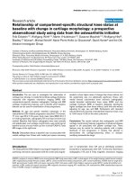
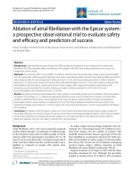
VTI_PA was corrupted by the ductal jet (Fig. 1). It is
also important to note that VTI_Ao/VTI_Pa-ratio
may not accurately reflect the degree of shunt
through a PDA because of inter-atrial shunting which
is commonly observed in VLBW infants just like in
our cohort (only 1 out of 19 infants had no inter-atrial
shunting, no infant had a ventricular septal defect). This
latter limitation also applies to more commonly used
parameters such as the LA/Ao ratio. Furthermore, the assumption underlying the determination of VTI_Ao/
VTI_Pa-ratio that the cross-sectional areas of P- and
Ao-valve are similar may not applicable to all infants.
However, despite poor repeatability, VTI_Ao/VTI_Pa
had a high CSA-value, indicating a high potential in
identifying inter-individual differences and consequently
permitting accurate classification (Table 1). Similarly,
determination of the PDA-diameter was challenging,
Fig. 1 Measurement of VTI_Pa in a parasternal short axis view. The pulsed-wave Doppler-sonographic measurement of VTI_Pa in a parasternal
short axis view is corrupted by ductal jet extending to the pulmonary valve
Schwarz et al. BMC Pediatrics (2016) 16:18
Page 5 of 5
because it requires visualisation of the PDA from the aorta
to the pulmonary artery.
A limitation of our study is that extremely immature
preterm infants with the highest risk of PDA are underrepresented because we were hesitating to subject these
most vulnerable infants during their first postnatal days
to repeated measurements. Future studies need to assess
intra-observer repeatability.
3.
Conclusions
The repeatability of echocardiographic parameters to
evaluate ductal left-to-right shunt is poor. The highest
repeatability was achieved by RIs in ACA and CA. This
has implications for clinical practice as well as the design
of future studies on PDA treatment. In both settings, repeated measurements and averaging of results should be
implemented, especially when measured values are close
to their cut-off value.
7.
Abbreviations
ACA: anterior cerebral artery; CA: celiac artery; CI: confidence interval;
CPAP: continuous positive airway pressure; CSA: confidence-step-analysis;
LA/Ao-ratio: left-atrium-to-aortic-root-ratio; LVO/SVC-flow: left -ventricular
-output-to-superior -vena -cava-flow - ratio; LVPEP/LVET: left-ventricularpreejection-period-to-ejection-time-ratio; PDA: patent ductus arteriosus;
RepC: repeatability coefficient; RepI: repeatability index; RI: resistance
index; SD: standard deviation; VLBW: very low birth weight;
VTI_Ao: velocity time integral ascending Aorta; VTI_Pa: velocity time
integral pulmonary artery.
Competing interests
The authors declare that they have no competing interests.
Authors’ contributions
CES has contributed to the design of the study, measurements, statistical
analysis, has drafted the initial and the revised version of the manuscript.
AP has contributed to the measurements, statistical analysis and writing of
the manuscript. WB participated in the measurements and writing of the
manuscript. CFP participated in the design of the study and critically
reviewed the manuscript. ARF conceived of the study, and participated in
its design, measurements, statistical analysis and coordination and helped
to draft the manuscript. All authors read and approved the final manuscript.
4.
5.
6.
8.
9.
10.
11.
12.
13.
14.
15.
16.
17.
18.
Acknowledgements
We like to thank the “Else Kröner-Fresenius-Stiftung” for supporting this
study.
19.
Author details
1
Department of Neonatology, University Children’s Hospital of Tuebingen,
University of Tuebingen, Calwerstr. 7, 72076 Tuebingen, Germany.
2
Department of Pediatric Cardiology, University Children’s Hospital of
Tuebingen, University of Tuebingen, Tuebingen, Germany. 3Center for
Pediatric Clinical Studies, University Children’s Hospital of Tuebingen,
University of Tuebingen, Tuebingen, Germany.
20.
21.
22.
Received: 3 August 2015 Accepted: 19 January 2016
References
1. Cassady G, Crouse DT, Kirklin JW, Strange MJ, Joiner CH, Godoy G, et al. A
randomized, controlled trial of very early prophylactic ligation of the ductus
arteriosus in babies who weighed 1000 g or less at birth. N Engl J Med.
1989;320(23):1511–6.
2. Shortland DB, Gibson NA, Levene MI, Archer LN, Evans DH, Shaw DE. Patent
ductus arteriosus and cerebral circulation in preterm infants. Dev Med Child
Neurol. 1990;32(5):386–93.
23.
Rojas MA, Gonzalez A, Bancalari E, Claure N, Poole C, Silva-Neto G. Changing
trends in the epidemiology and pathogenesis of neonatal chronic lung
disease. J Pediatr. 1995;126(4):605–10.
Evans N, Kluckow M. Early ductal shunting and intraventricular haemorrhage
in ventilated preterm infants. Arch Dis Child Fetal Neonatal Ed. 1996;75(3):
F183–6.
Kluckow M, Evans N. Ductal shunting, high pulmonary blood flow, and
pulmonary hemorrhage. J Pediatr. 2000;137(1):68–72.
Oh W, Poindexter BB, Perritt R, Lemons JA, Bauer CR, Ehrenkranz RA, et al.
Association between fluid intake and weight loss during the first ten days
of life and risk of bronchopulmonary dysplasia in extremely low birth
weight infants. J Pediatr. 2005;147(6):786–90.
Noori S, McCoy M, Friedlich P, Bright B, Gottipati V, Seri I, et al. Failure of
ductus arteriosus closure is associated with increased mortality in preterm
infants. Pediatrics. 2009;123(1):e138–44.
Zonnenberg I, de Waal K. The definition of a haemodynamic significant
duct in randomized controlled trials: a systematic literature review. Acta
Paediatr. 2012;101(3):247–51.
McNamara PJ, Sehgal A. Towards rational management of the patent
ductus arteriosus: the need for disease staging. Arch Dis Child Fetal
Neonatal Edition. 2007;92(6):F424–7.
Schena FFG, Cappelleri A, Picciolli I, Mayer A, Mosca F, Fumagalli M.
Association between hemodynamically significant patent ductus arteriosus
and bronchopulmonary dysplasia. J Pediatr. 2015;166(6):1488–92.
Rozé JC, Cambonie G, Marchand-Martin L, Gournay V, Durrmeyer X, Durox
M, et al. Association between early screening for patent ductus arteriosus
and in-hospital mortality among extremely preterm infants. JAMA. 2015;
313(24):2441–8.
Green TP, Thompson TR, Johnson DE, Lock JE. Furosemide promotes patent
ductus arteriosus in premature infants with the respiratory-distress
syndrome. N Engl J Med. 1983;308(13):743–8.
Shimada S, Kasai T, Konishi M, Fujiwara T. Effects of patent ductus arteriosus
on left ventricular output and organ blood flows in preterm infants with
respiratory distress syndrome treated with surfactant. J Pediatr. 1994;125(2):
270–7.
Martin CG, Snider AR, Katz SM, Peabody JL, Brady JP. Abnormal cerebral
blood flow patterns in preterm infants with a large patent ductus arteriosus.
J Pediatr. 1982;101(4):587–93.
Kluckow M, Evans N. Early echocardiographic prediction of symptomatic
patent ductus arteriosus in preterm infants undergoing mechanical
ventilation. J Pediatr. 1995;127(5):774–9.
Robel-Tillig E, Knupfer M, Pulzer F, Vogtmann C. Dopplersonographic
findings in neonates with significant persistent ductus arteriosus.
Z Geburtshilfe Neonatol. 2002;206(2):51–6.
Bland JM, Altman DG. Statistical methods for assessing agreement between
two methods of clinical measurement. Lancet. 1986;1(8476):307–10.
Skinner JR, Boys RJ, Heads A, Hey EN, Hunter S. Estimation of pulmonary
arterial pressure in the newborn: study of the repeatability of four Doppler
echocardiographic techniques. Pediatr Cardiol. 1996;17(6):360–9.
Groves AM, Kuschel CA, Knight DB, Skinner JR. Echocardiographic
assessment of blood flow volume in the superior vena cava and
descending aorta in the newborn infant. Arch Dis Child Fetal Neonatal Ed.
2008;93(1):F24–8.
Hudson I, Houston A, Aitchison T, Holland B, Turner T. Reproducibility of
measurements of cardiac output in newborn infants by Doppler ultrasound.
Arch Dis Child. 1990;65(1 Spec No):15–9.
Moorthy B, Colditz PR, Ives KN, Rees DG, van’t Hoff WG, Hope PL.
Reproducibility of cerebral artery Doppler measurements. Arch Dis Child.
1990;65(7 Spec No):700–1.
van Dijk AP, Hopman JC, Klaessens JH, van der Werf T, Daniels O. Is
noninvasive determination of pulmonary artery pressure feasible using
deceleration phase Doppler flow velocity characteristics in mechanically
ventilated children with congenital heart disease? Am J Cardiol. 1996;78(12):
1394–9.
Carmo KB, Evans N, Paradisis M. Duration of indomethacin treatment of the
preterm patent ductus arteriosus as directed by echocardiography. J
Pediatr. 2009;155:819–22.