Retinoblastoma in a pediatric oncology reference center in Southern Brazil
Bạn đang xem bản rút gọn của tài liệu. Xem và tải ngay bản đầy đủ của tài liệu tại đây (557.07 KB, 9 trang )
Selistre et al. BMC Pediatrics (2016) 16:48
DOI 10.1186/s12887-016-0579-9
RESEARCH ARTICLE
Open Access
Retinoblastoma in a pediatric oncology
reference center in Southern Brazil
Simone G. A. Selistre1,2*, Marcelo K. Maestri3, Patricia Santos-Silva1,4, Lavinia Schüler-Faccini5,6,7,8,
Luis S. P. Guimarães9,10, Juliana Giacomazzi4,6,11, Mario C. Evangelista Júnior1,2 and Patricia Ashton-Prolla1,4,5,6,7,8,9
Abstract
Background: Retinoblastoma (Rb) is the most common intraocular tumor diagnosed in children in Brazil. However,
detailed information is lacking regarding patient clinical demographics. This study aimed to determine the clinical
profile of patients with Rb who were treated in a public university hospital in southern Brazil from 1983 to 2012.
Methods: Patients’ medical records were reviewed to retrospectively identify patients with a principal diagnosis of
Rb. Rb was classified as hereditary or non-hereditary. Clinical staging was reviewed by an ophthalmologist. Statistical
analysis was performed using SPSS.
Results: Of 165 patients with a diagnosis of Rb during this period, 140 were included in the study. Disease was
unilateral in 65.0 % of patients, bilateral in 32.9 %, and trilateral in 2.1 %. The mean age at onset of the first sign/
symptom was 18.1 month, and 35.7 % of patients were diagnosed during the first year of life. The most common
presenting signs were leukocoria (73.6 %) and strabismus (20.7 %). The mean age at diagnosis was 23.5 months,
and time to diagnosis was 5.4 months. In patients with clinical features of hereditary Rb, both onset of the first
sign/symptom and diagnosis were at an earlier age than in patients without these features (12.3 vs 21.6 months
[P = 0.001] and 15.9 vs 28.0 months [P < 0.001], respectively). However, there was no significant difference in overall
survival between the two groups. Ocular stage at diagnosis was advanced in 76.5 % (Reese V) and 78.1 %
(International Classification D or E). Of patients with unilateral and bilateral disease, 35.2 % and 34.8 %, respectively,
had extraocular disease at diagnosis; 10.7 % had metastatic disease at diagnosis. Enucleation was observed in
88.1 % and exenteration in 11.9 % of patients; 93.6 % patients were followed until 2012, and 22.9 % relapsed.
Overall survival was 86.4 %.
Conclusions: Most Rb diagnoses are still diagnosed in advanced stages of the disease, considerably reducing
overall survival time and the rate of eye and vision preservation.
Keywords: Retinoblastoma, Malignant tumors of the retina, Intraocular malignancies, Hereditary retinoblastoma,
Pediatric tumors
Background
In Brazil, cancer is the leading cause of death by disease
among children and adolescents aged ≤ 19 years, with an
incidence of 11,530 new cases in 2012 [1, 2]. Retinoblastoma (Rb) is the most common primary intraocular malignancy of childhood, and most cases are diagnosed before
five years of age [3–5].
* Correspondence:
1
Post-Graduate Program in Medicine: Medical Sciences, Universidade Federal
do Rio Grande do Sul (UFRGS), Porto Alegre, Brazil
2
Pediatric Oncology Service, Hospital de Clinicas de Porto Alegre (HCPA),
Porto Alegre, Brazil
Full list of author information is available at the end of the article
Rb is considered a rare tumor in developed countries,
accounting for approximately 3 % of all childhood malignancies and 11 % of all tumors that develop during the
first year of life [6–8]. Its global incidence is estimated at
1:12, 500–25,000 live births (1:16,000 in France) [9]. The
annual incidence rate of Rb in the United Stated is 3.4
to 4.0 per million children aged 0–15 years [4, 10].
There is indirect evidence that its incidence increases in
developing countries, including those in Latin America,
Africa, India, and Asia (excluding Japan). Therefore, in
these areas Rb is considered one of the most frequent
pediatric solid tumors [8, 11].
© 2016 Selistre et al. Open Access This article is distributed under the terms of the Creative Commons Attribution 4.0
International License ( which permits unrestricted use, distribution, and
reproduction in any medium, provided you give appropriate credit to the original author(s) and the source, provide a link to
the Creative Commons license, and indicate if changes were made. The Creative Commons Public Domain Dedication waiver
( applies to the data made available in this article, unless otherwise stated.
Selistre et al. BMC Pediatrics (2016) 16:48
The most common presenting sign of Rb is leukocoria
(75 % of cases), followed by strabismus (25 % of cases)
[12–14]. Although evidence of sex predominance is inconclusive, a few studies have shown a higher prevalence
of Rb in boys (1.1–1.4:1) [4, 15]. Approximately 40 % of
all Rb cases are hereditary, caused by germline mutations in the RB1 gene [4, 7, 16]. The retinoblastoma
phenotype in addition to presence or absence of family
history is important features to determine the probability
of hereditary predisposition. Thus, the probability of
hereditary retinoblastoma in patients with bilateral, trilateral or unilateral Rb with a positive Rb family history
is 90, 100 and 15 %, respectively [4, 10].
Different treatment modalities are available for patients
with Rb, including cryotherapy, laser therapy, enucleation,
radiotherapy, high-dose systemic chemotherapy, intraarterial chemotherapy, intravitreal chemotherapy, and
autologous stem cell transplantation. Treatment should be
tailored to the patient’s needs, taking into account
laterality, ocular stage, presence of extraocular disease,
the child’s age, and visual acuity [17–19]. Overall, the
prognosis is favorable for patients with early-stage intraocular Rb, with a 5-year survival rate of 93 %. However, when
there is extraocular extension, more aggressive treatment
is required and the 5-year overall survival decreases dramatically to approximately 30 % [8, 11, 18, 20–22].
Although Rb is the most common intraocular tumor in
children, there are little published data regarding the general characteristics of patients diagnosed and treated in
Brazil. Most data are obtained from online databases of
cancer registries located mainly in the southeastern region
of the country. According to the 14 registries in this region,
the incidence rate of Rb, in 2010, ranged from 2.40 to 9.80
per million children and adolescents aged ≤ 19 years [23].
However, detailed information is lacking regarding patient
demographics and clinical characteristics, especially in the
southern region [23]. The aim of the present study was to
characterize patients with a diagnosis of Rb who were
treated in a public university hospital in southern Brazil,
providing additional data that may contribute to improving
the diagnosis and management of these patients.
Methods
This was a retrospective cohort study of patients with
Rb who were treated in the Departments of Pediatric
Oncology, Ophthalmology and Medical Genetics at Hospital de Clínicas de Porto Alegre (HCPA) from 1983 to
2012. HCPA is a tertiary care teaching hospital located in
Porto Alegre, city and capital of Rio Grande do Sul, the
southernmost state of Brazil. The study was approved
by the Institutional Review Board – Hospital de Clínicas
de Porto Alegre (IRB number 100521). The need for
informed consent was waived by this IRB for this
retrospective and epidemiologic study.
Page 2 of 9
The patients’ medical records were reviewed to retrospectively identify patients with a principal diagnosis of
Rb according to the International Classification of
Diseases, 10th Revision, from codes designating malignant
neoplasms of the eye (codes from C69.0 to C69.9); more
specifically code C69.2 (malignant neoplasms of the
retina). Clinical and demographic data were also collected
at this stage using a protocol developed by the authors.
Rb was classified as hereditary or non-hereditary according to the clinical guidelines proposed by Lohmann
and Gallie [3].
Except for patients who had undergone enucleation at
another hospital before transfer, the diagnosis was confirmed by an ophthalmologist (MKM)based on data
from medical records, presence of typical signs, such as
leukocoria, and either (a) by binocular indirect ophthalmoscopy, with visualization of a characteristic yellowwhite mass, or (b) by ocular ultrasound and computed
tomography (CT) of the eye and orbit for identification
of intratumoral calcification in eyes with turbid media,
which prevented direct visualization of the retina. Although it is our understanding that CT-scans should be
avoided in patients with retinoblastoma, for many years
alternative imaging (i.e., magnetic resonance imaging)
was not available in the institution and CT scans were
often used. In addition, CT scans were performed in all
patients for evaluation of possible metastatic disease at
diagnosis. Histopathologic specimens of enucleated or
exenterated eyes were further analyzed by a pathologist.
Clinical staging was reviewed by same ophthalmologist
(MKM) based on the Reese-Ellsworth classification
and the International Classification of Intraocular Rb
(ABCDE groups) [6, 22, 24]. Extraocular disease was
classified according to the system used by the Children’s
Cancer Group [24, 25]. Systemic staging was performed as
previously described [6, 22, 26, 27]. All therapeutic procedures performed for management of Rb were recorded for
each patient. The choice of initial treatment was based
on protocols established by international reference
centers or on the 2009 Brazilian Protocol for Rb Treatment developed by the Brazilian Society of Pediatric
Oncology [26, 27]. In brief, we identified 3 different
protocols of treatment used in different years along the
overall study period. This information is summarized
in Additional file 1: Table S1.
The following outcomes were assessed: (a) time to
diagnosis, defined as the time between onset of the first
sign or symptom and the actual diagnosis;(b) duration of
follow-up, calculated as the difference between the patient’s age at diagnosis and their age at the time of the
last consultation (if alive) or death; (c) diagnosis of a second or third neoplasm; and (d) death. Loss to follow-up
was defined as no recorded consultation with a physician
at HCPA for more than five years.
Selistre et al. BMC Pediatrics (2016) 16:48
Page 3 of 9
Statistical analysis was performed using SPSS, version
18.0, and the level of significance was set at P < 0.05.
Continuous variables were expressed as mean (minimum-maximum), median and interquartile range (IQR),
with a 95 % confidence interval. Categorical variables
were expressed as absolute and relative frequencies. The
Kaplan-Meier method was used to estimate survival as a
function of time, and the log-rank test was used for
comparison of survival curves according to clinical characteristics. The patient’s age and time to diagnosis, in association with other characteristics, were analyzed using
the Kruskal-Wallis test, followed by Dunn’s multiplecomparison test when P-values were less than 0.05.
Fisher’s exact test was used to analyze the association of
disease extension (intra- or extraocular) and laterality.
Table 2 Characteristics of patients with a diagnosis of
retinoblastoma (Rb) (n = 140)
N
%
Leukocoria
103
73.6
Strabismus
29
20.7
Glaucoma
4
2.9
Buphthalmos
4
2.9
Proptosis
4
2.9
a
First sign or symptom
Hyperemia
4
2.9
Ocular pain
3
2.1
Anisocoria
3
2.1
Blindness
2
1.4
Orbital edema
2
1.4
Results
Hyphema
2
1.4
Demographics
Visual deficiency
1
0.7
Cervical adenopathy
1
0.7
Ecchymosis
1
0.7
Total eyes involved
187
66.8
Unilateral Rb
91
65.0
Right eye
50
54.9
Left eye
41
45.1
Bilateral Rb
46
32.9
Trilateral Rb
3
2.1
Of 165 patients with a diagnosis of Rb who were treated
at HCPA from 1983 to 2012, 140 (86.4 %) were included
in the study. The medical records of25 patients who
were diagnosed during the first two decades of the study
were not available. Most patients (95.0 %) were born and
lived in the state of Rio Grande do Sul, and 21.8 % were
from the capital of the state, Porto Alegre. Tables 1 and 2
shows the main clinical characteristics of patients with Rb
included in the study. There was a slight predominance of
male over female patients (n = 87; 62.1 %).
Systemic dissemination at diagnosis
Diagnosis
Most tumors were unilateral at diagnosis (n = 91;
65.0 %). In most cases, unilateral tumors were diagnosed
at an advanced stage (n = 88,96.7 %; 4 at IVb stage and
84 at Va or Vb stage), and all of them were considered
unifocal because of their large size. The few unilateral
tumors diagnosed at an early stage (n = 3, IIb stage) were
also unifocal. Forty-six patients (32.9 %) had bilateral
lesions at diagnosis, most of which (80.4 %) were
multifocal (P = 0.015).
There was no association between sex and disease
laterality (P = 0.351). Similarly, there was no association between the clinical presentation of leukocoria
or strabismus and poor prognosis (P = 0.612) or between time to diagnosis > 6 months and poor prognosis (P = 0.052).
Table 1 Characteristics of patients with a diagnosis of
retinoblastoma (Rb) (n = 140)
Characteristics (months)
Mean median
18.1
12.0
0–129.0
Age at diagnosis
23.5
16.5
1.0–206.0
5.4
3.0
Duration of follow-up
323.2
Non-metastatic disease
125
89.3
Metastatic disease
15
10.7
Metastatic sites at diagnosis
Orbit
12
80.0
CNS
8
53.3
Bone
4
26.7
Bone marrow
3
20.0
Cerebrospinal fluid
2
13.3
Cervical lymph nodes
1
6.7
Legend: Ages, time to diagnosis and duration of follow-up are expressed in
months; Kruskal-Wallis test was used for analysis of ages and time to diagnosis;
Log-rank test was used to estimate duration of follow-up; time to diagnosis:
time between onset of the first sign or symptom and diagnosis; duration of
follow-up: difference between the patient’s age at diagnosis and their age at
the time of the last consultation (if alive) or date of death
CNS central nervous system
a
some patients had more than one sign or symptom; more than one site
per patient
95 % CI
Age at first sign or symptom
Time to diagnosis
Ocular laterality
0–77.0
300.3–346.1
Bilateral and trilateral tumors were diagnosed at an
earlier age than unilateral tumors (P < 0.001). Fifty
patients (35.7 %) were diagnosed before 12 months of age.
Of these, 44.0 % had unilateral tumors and 56.0 % had
bilateral tumors; 6.0 % had metastatic disease.
Selistre et al. BMC Pediatrics (2016) 16:48
Page 4 of 9
Ocular staging at diagnosis is shown in Tables 3 and 4.
Extraocular extension of disease in at least one eye at
diagnosis was present in 32 of 91 patients (35.2 %)
with unilateral Rb, in 16 of 46 patients (34.8 %) with
bilateral Rb, and in all three patients with trilateral
Rb, totaling 51 patients (36.4 %). Considering the
total number of eyes involved (n = 187), 28.9 % had
extraocular disease at diagnosis. Over the years, a decrease
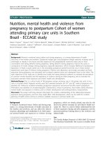
was observed in the proportion of patients with extraocular disease (Fig. 1).
All patients were evaluated for features suggestive of
hereditary Rb (Table 5). The presence of at least one criterion suggestive of hereditary Rb was observed in 52
patients (37.1 %) from 50 different families. One- and
two-generation family history of Rb was positive for cancer in 23 patients (16.4 %), and 10 of these patients
(43.5 %) had an affected parent. The mean age at onset
of the first sign or symptom was 12.3 months in the
group with probable hereditary predisposition to Rb and
21.6 months in the non-hereditary group (P = 0.001).
The mean age at diagnosis was 15.9 months in the hereditary group and 28.0 months in the non-hereditary
group (P < 0.001). However, there was no significant
difference in overall survival between the hereditary
and non-hereditary groups (84.6 % vs. 87.5 %, respectively; P = 0.844). Survival data are summarized in
Additional file 2: Figure S1.
Data on mean age at onset of the first signs and symptoms of Rb, mean age at diagnosis and time relapsed between onset of signs and symptoms and diagnosis are
shown in Tables 6 and 7. Additional file 3: Figure S2 is a
graphical representation of the overall survival data
described in Tables 6 and 7.
Treatment
Several treatment modalities were used in the present
cohort. Among 134 patients (95.7 %) who underwent
surgery, enucleation was performed in 118 (88.1 %) and
exenteration in 16 (11.9 %). Fifty-seven patients (42.5 %)
were treated with enucleation alone, while 77(57.5 %)
were treated with enucleation combined with some
other form of treatment. There has not been a significate
Table 4 Ocular staging at diagnosis
International Classification of Intraocular Rb (ABCDE)
Ocular staging
N
%
A
3
1.6
B
30
16.0
C
8
4.3
D
32
17.1
E
89
47.6
Presumed Da
5
2.7
Presumed Ea
20
10.7
a
Patients evaluated after enucleation performed at another hospital
decline in the number of enucleations related to the different chemotherapy regimens for retinoblastoma.
Only six patients (4.3 %) did not undergo enucleation
and were treated with multimodal therapy, including
chemotherapy, brachytherapy, thermotherapy, and cryotherapy. Among the 77 patients treated with enucleation
and some other form of therapy, 74 (96.1 %) received systemic chemotherapy, followed by orbital external beam
radiotherapy alone (2.6 %) and cryotherapy alone (1.3 %).
Among the 74 patients treated with enucleation and systemic chemotherapy, 48 (64.9 %) also received radiotherapy. Of all 140 patients, 80 (57.1 %) received systemic
chemotherapy and 52 (37.1 %) received radiotherapy. Two
patients (1.4 %) underwent autologous stem cell transplantation. The treatment modalities used in the present
cohort are described in detail in Additional files 4, 5 and 6:
Tables S2, S3, and S4.
Six patients (4.3 %) receiving ionizing radiation were diagnosed with a secondary malignancy: four with a soft tissue sarcoma (three of the mat sites that had been
previously irradiated), one with osteosarcoma, and one with
acute lymphoblastic leukemia (ALL). The patient with a
diagnosis of ALL developed a third malignant neoplasm
(osteosarcoma of the femur) and was the only patient
Table 3 Ocular staging at diagnosis
Reese-Ellsworth classification
%
B
%
Total
Ocular staging
A
I
1
0.5
3
1.6
4
2.1
II
12
6.4
8
4.3
20
10.7
III
7
3.8
3
1.6
10
5.4
IV
5
2.7
5
2.7
10
5.4
V
92
49.2
23
12.0
115
61.5
26
13.9
2
1.1
28
15.0
a
Presumed V
a
Patients evaluated after enucleation performed at another hospital
(%)
Fig. 1 Proportion of cases of extraocular disease at diagnosis in
patients with unilateral and bilateral Rb from 1983 to 2012. Legend:
At the point where the two lines meet (between 1993 and 1997),
33 % refers to unilateral cases and 31 % refers to bilateral cases.
*Line red: bilateral extraocular
*Line blue: unilateral extraocular
Selistre et al. BMC Pediatrics (2016) 16:48
Page 5 of 9
Table 5 Distribution of patients with criteria for hereditary Rb at diagnosis
Criteria for hereditary Rb
Number
Percent
Total of families with at least one criterion for hereditary Rba
50
36.2
Bilateralb
39
75.0
Trilateralc
3
5.8
3
5.8
Only one criterion present:
Family history of Rb
ac
Two criteria present:
Bilateral and family historyc
7
13.4
Samples collected for mutation analysis based on criteriad
25
48.1
RB1 mutation identified
13
52.0
e
Patients with secondary malignant neoplasmf
a
2
15.4
b
c
Two families with two patients with Rb; Three cases of unilateral Rb with family history of Rb (all diagnosed before 12 months of age); Total cases with family
history of Rb = 10; dTotal patients who collected samples for molecular genetic testing = 32 (22.9 %); RB1 mutation was detected in one patient who had no
criteria for hereditary Rb at diagnosis (unilateral and unifocal); normal results (n = 12); no results available (n = 6). This percentage (48.1 %) refers to the proportion
25/52; eThis percentage (52.0 %) refers to the proportion 13/25; fThis percentage (15.4 %) refers to the proportion 2/13
treated with brachytherapy in this group. Presence of germline mutations in the RB1 gene was investigated by wholegenome sequencing and multiplex ligation-dependent
probe amplification (MLPA) in two patients with multiple
solid tumors. A pathogenic mutation (p.R320X) was
identified in only one of the patients. Details of these
six patients that developed a secondary malignancy were:
all 6 patients who had a second primary tumor treatment
received systemic chemotherapy (4 with cisplatin, teneposide, vincristine, doxorubicin, cyclophosphamide, and
methotrexate, cytarabine intrathecal dexamethasone
and 2 with vincristine, etoposide and carboplatin. The
age at diagnosis of these patients varied from 3 to
26 months and the age who received systemic chemotherapy ranged from 4 to 28 months. Five of these
patients also received radiation therapy between 5 and
29 months and of those, 3 had a second primary tumor in
previously irradiated area. The age of the irradiated patients who had a second primary tumor ranged between
134 and 221 months, and the patient who did not undergo
radiation therapy had a second primary tumor at
24 months.
Table 6 Follow-up characteristics of patients with Rb according to subgroups
Laterality (n = 140 patients)
Age at first signs and symptoms
Unilateral (n = 91) Age;
Age dx (min-max) (months)
Bilateral (n = 46) Age;
Age dx (min-max) (months)
Trilateral (n = 3)Age;
Age dx (min-max) (months)
P
21.7; 15.0 (0-129.0)
10.3; 6.0 (0-84.0)
29.0; 24.0 (3.0-60.0)
<0.001
13.4; 8.0 (1.0-84.0)
40.3; 26.0 (15.0-80.0)
<0.001
3.1; 2.0 (0-14.0)
11.3; 12.0 (2.0-20.0)
0.029
334.9 (299.3-370.5)
22.3 (2.9-42.7)
<0.001
Age at diagnosis
Time to diagnosis
Duration of follow-up (months)
28.1; 22.0 (1.0-206.0)
[24.0]
6.4; 3.0 (0-77.0)
275.6 (253.2-297.9)
Laterality for each eye (n = 187 eyes involved)
Time to diagnosis between IO and
EO in each subgroup
Duration of follow-up between IO
and EO in each subgroup (months)
Unilateral (n = 91) Age;
Age dx (min-max) (months)
Bilateral (n = 92) Age;
Age dx (min-max) (months)
Trilateral (n = 4) Age; Age dx
(min-max) (months)
IO: 4.0; 2.0 (0-20.0)
IO: 2.9; 2.0 (0-14.0)
IO: 2.0; 2.0 (2.0-2.0)
EO: 10.8; 4.5 (1.0-77.0)
EO: 4.2; 4.0 (0-12.0)
EO: 11.3; 12.0 (2.0-20.0)
P= 0.003
P = 0.147
P = 0.346
IO: 293.0 (273.3-312.8)
IO: 355.7 (334.6-376.9)
IO: 4.0 (4.0-4.0)
EO: 230.9 (180.4-281.4)
EO: 255.5 (174.8-336.3)
EO: 22.3 (2.9-41.7)
P= 0.002
P = 0.001
P = 0.317
dx diagnosis, CI confidence interval; age, time to diagnosis and duration of follow-up are expressed in months. Kruskal-Wallis test was used for analysis of ages
and time to diagnosis; Log-rank test was used to estimate duration of follow-up. Disease extension, IO intraocular, EO extraocular. Time to diagnosis: time between
onset of the first sign or symptom and diagnosis (months); duration of follow-up: difference between the patient’s age at diagnosis and their age at the time of
the last consultation (if alive) or date of death (months)
Selistre et al. BMC Pediatrics (2016) 16:48
Page 6 of 9
Table 7 Follow-up characteristics of patients with Rb according to subgroups
Systemic dissemination (n= 140 patients)
Metastatic (n = 15) Age;
Age dx (min-max) (months)
Non-metastatic (n = 125) Age;
Age dx (min-max) (months)
P
Age at first signs and symptoms
32.1; 24.0 (1.0-129.0)
16.4; 11.0 (0-84.0)
0.107
Age at diagnosis
46.5; 26.0 (2.0-206.0)
20.8; 16.0 (1.0-84.0)
0.024
Time to diagnosis
14.3; 4.0 (1.0-77.0)
4.4; 3.0 (0-26.0)
0.123
Duration of follow-up (months)
77.7 (20.6-134.7)
345.2 (325.6-364.7)
<0.001
Hereditary criteria (n= 140 patients)
Hereditary (n = 52) Age;
Age dx (min-max) (months)
Non-hereditary (n = 88) Age;
Age dx (min-max) (months)
P
Age at first signs and symptoms
12.3; 6.5 (0-84.0)
21.6; 14.0 (0-129.0)
0.001
Age at diagnosis
15.9; 10.5 (1.0-84.0)
28.0; 21.5 (1.0-206.0)
<0.001
Time to diagnosis
3.7; 2.0 (0-20.0)
6.5; 3.0 (0-77.0)
0.074
Duration of follow-up (months)
318.2 (280.2-356.2)
274.0 (250.8-297.2)
0.844
Disease extension for each eye (n= 187 eyes involved)
Intraocular (n = 133) Age;
Age dx (min-max) (months)
Extraocular (n = 54) Age;
Age dx (min-max) (months)
P
Age at first signs and symptoms
13.6; 8.0 (0-84.0)
22.7; 12.0 (0-129.0)
0.001
Age at diagnosis
16.9; 13.0 (1.0-84.0)
31.3; 24.0 (1.0-206.0)
<0.001
Time to diagnosis
3.4; 2.0 (0-20.0)
8.5; 4.0 (0-77.0)
<0.001
Duration of follow-up
352.9 (336.0-369.8)
252.7 (204.7-301.3)
<0.001
Mortality
Overall survival was 86.4 % for the entire cohort, 92.0 %
for patients with non-metastatic disease, and 40.0 % for
patients with metastatic disease. Overall survival for patients with intraocular disease was 94.0 % vs 68.5 % for
patients with extraocular disease. Patients with unilateral
and bilateral tumors had a similar overall survival rate
(approximately 88.0 %), whereas all three patients with
trilateral disease died.
Among patients with unilateral Rb, overall survival was
94.9 % for those with intraocular tumors and 75.0 % for
those with extraocular tumors. Among patients with bilateral Rb, overall survival was 94.5 % for those with intraocular tumors and 68.4 % for those with extraocular tumors.
Follow up
Of 140 patients treated at the institution, 131 (93.6 %)
were followed until the end of 2012 or until death; only
nine patients (6.4 %) were lost to follow up. During the
follow up period, 32 patients (22.9 %) relapsed (20 were
non-metastatic and 12 were metastatic at diagnosis),
leading to 19 deaths. Of these, 16 (84.2 %) were due to
disease progression (10 were non-metastatic and nine
were metastatic at diagnosis).
Mean follow up time was 323.2 months (minimum,
300.3 months; maximum, 346.1 months). The mean age at
diagnosis of patients with trilateral disease was 40.3 months
(median, 26.0 months; IQR, 15.0–80.0 months), and mean
follow up time was 22.3 months (IQR, 2.9–41.7 months).
Discussion
The main purpose of the present study was to determine
the clinical profile of patients diagnosed with Rb and
treated in a public university hospital in southern Brazil
over a 30-year period. Most patients were from Rio
Grande do Sul, the southernmost state of Brazil. Consistent with previous findings, there was a slight predominance of male children in the population studied.
However, there was no significant difference between
sexes regarding laterality of the affected eye. The most
common presenting signs and symptoms were leukocoria
and strabismus, which are consistent with the literature
[12–14]. However, there was no significant association of
patients with strabismus or leukocoria at diagnosis with
time to diagnosis > 6 months, and no association of any of
these factors with poor prognosis as previously reported
[18, 28, 29]. Since this is a retrospective study, we were
unable to obtain detailed information on the exact dates
of medical consultations before the diagnosis, although we
expect to exist significant heterogeneity in the individual
times to seek medical care.
Although most patients had unilateral disease at diagnosis, almost all patients were diagnosed at a very advanced
stage (stage IVb, Va, or Vb). Early diagnosis is directly
Selistre et al. BMC Pediatrics (2016) 16:48
related to how easily patients are able to get needed care
through the public health system and how readily health
care professionals can recognize the signs and symptoms
of Rb. Despite a growing economy, in Brazil, the number
of patients presenting with locally advanced or metastatic
disease is similar to the numbers reported for lowerincome countries, with an average per capita income of
USD 935.00 [30]. Conversely, about one-third of patients
had bilateral disease at diagnosis, and most of these lesions were multifocal and diagnosed at an earlier age, as
expected is cases of hereditary disease. However, although
prevalent, the cases of hereditary Rb were not referred for
genetic risk assessment. The average age at diagnosis of
patients with hereditary Rb according to our clinical criteria (12.3 months) was similar to the average age of diagnosis in studies from developed countries (12 months) [6].
Only a proportion of the patients with probable hereditary
Rb were offered genetic tests to evaluate RB1 mutations
treatment options. Only a few patients with probable hereditary Rb were offered genetic testing for RB1 mutations,
and several patients were treated with radiotherapy because of the lack of other treatment options. This may be
due to difficulties in accessing public health services, but
at least in part it also reflects a lack of knowledge about
how hereditary Rb manifests itself. It is important to note
that molecular genetic testing is not available to patients
receiving care through the public health system in Brazil.
Such tests are performed only at the Brazilian National
Cancer Institute for patients who are currently enrolled in
clinical trials or research protocols [1].
Based on tumor characteristics, ocular stage was
advanced in a large number of patients (stage V in
76.5 %; D or E in 78.1 %) and metastatic disease was
present at diagnosis in 10.7 % of cases. These figures
are much higher than those expected for developing
countries with a socioeconomic status equivalent to that
of Brazil [8, 31, 32]. Possible explanations for this result include inability of health care professionals to
recognize the signs of Rb and difficult access to specialized
pediatric oncology centers for Rb diagnosis and treatment
[8, 30–33]. In another Brazilian study, conducted in São
Paulo, 83 pediatric patients with a diagnosis of extraocular
Rb admitted for treatment between 1987 and 2000 were
treated with two different chemotherapeutic regimens. The
authors reported an average age at diagnosis of 32.9 months,
with unilateral disease presented with a median age of
33.2 months and those with bilateral disease had a median
age of 23.7 months and a latency of 10.5 months between
the appearance of the first sign and confirmation of
diagnosis [33]. Comparatively, in the present study, the
mean age at diagnosis was 23.5 months, but with a time of
34.1 months which may be due to the unilateral cases
(unilateral 41.3 vs. bilateral 10.6 vs. trilateral 11.3 months)
[33]. A delayed diagnosis can be expected in atypical cases
Page 7 of 9
where these usual signs of presentation, leukocoria, strabismus and posterior pole tumors are not present.
Only 16.4 % of patients in the present cohort had a
positive family history of cancer, which is below the estimated rate of 30 % [1, 16, 34]. However, 37.1 % of patients had at least one criterion for hereditary Rb, which
is consistent with the rate of 40 % described in the literature [4, 7, 10, 16]. As expected, patients with criteria for
hereditary Rb showed typical signs and were diagnosed
at an earlier age, the germline mutation in the RB1 gene
[34]. Thus, early detection could also be attributed in
part to a closer observation by parents who were
aware of the family history. There was no difference
in overall survival between the hereditary and nonhereditary subgroups, which may be due to prompt
and appropriate intervention.
Late diagnosis at an advanced stage, which was predominant in the present cohort was probably the main
determining factor in the choice of enucleation as the
primary treatment, with a large proportion of cases
indicated for surgical procedure: approximately 60 %
of the eyes were enucleated, and 12 % exenterated.
Even considering modern globe salvage treatment options
in retinoblastoma, enucleations is still high in unilateral
cases, because the majority was diagnosed in advanced D
and E stages. It is estimated that 30 % of all Rb cases
worldwide occur in sub-Saharan Africa, where the population faces considerable barriers to access health care. Two
African studies, conducted over the past 5 years, reported
enucleation rates of 67 and 64 %, with overall 30-month
survival rates of 56 and 36 %, respectively [9, 35]. Several
lines of evidence show a clear correlation between a country’s socioeconomic status and the incidence of extraocular disease, metastatic disease, and overall survival. In this
sense, the present findings regarding disease stage at diagnosis and overall survival are comparable to those for patients from low-income countries. Although a slight
decrease in the frequency of extraocular disease at diagnosis was observed over time, the current scenario is still far
from what is observed in developed countries (5-year
overall survival of 95 %, and metastatic disease present in
less than 10 % of patients) [31, 36]. As an illustration, the
chart below comparatively shows the rates of intraocular
disease, extraocular disease, and metastatic disease in
three countries with different socioeconomic profiles [36]
and the results obtained in the present. Tanzania, India
and Argentina are considered countries with a low, average and high socioeconomic status, respectively, according
to the World Bank [36].
The most important prognostic variable observed in this
study is late diagnosis and the advanced tumor staging is
the main determinant for choosing which treatment will
be employed. Enucleation in D unilateral disease classified
patient is a reasonable treatment option to avoid prolonged
Selistre et al. BMC Pediatrics (2016) 16:48
systemic chemotherapy and its relevant potential sequelae
we still observe an elevated percentage of cases treated
with radiotherapy (37.1 %), even in patients with a clinical
presentation suggestive of hereditary Rb, for whom the
treatment of choice should be systemic chemotherapy [31].
Radiotherapy is an option of conservative treatment for
retinoblastoma vitreous seeding, mainly in bilateral disease, but this clinical sign is related to bad prognosis
regarding globe preservation. So, in such situation,
enucleation is an ultimate safe treatment option and
must not be considered as a failure.
An additional barrier is that only a few centers in Brazil
have access to other therapeutic procedures, such as
brachytherapy and intra-arterial chemotherapy, which are
indicated as a treatment for early-stage tumors and allow
preservation of the globe [7, 31, 37]. Moreover, in the
present cohort, only two patients underwent autologous
stem cell transplantation, although several other patients
were good candidates for this procedure [18, 38–41].
According to the characteristics observed at diagnosis,
the overall survival rate (86.4 %) and survival rate within
the non-metastatic (92 %) and metastatic (40 %) subgroups were similar to those reported in the literature
[1, 5, 18, 31]. No clear, systematic treatment was offered
during the first 15 years of the study. However, this behavior changed with the implementation of treatment protocols established by Brazilian cooperative groups in the last
decade [26, 32]. This initiative is in accordance with the
guidelines of the International Society of Pediatric Oncology, which published in 2013 the recommendations for
staging and treatment of unilateral and bilateral Rb in developing countries [32]. Other similar initiatives have been
implemented in specific regions, including Latin America,
aiming to reduce mortality by education, early diagnosis,
and proper treatment of Rb [18, 19, 28, 42].
Conclusion
Considering that Rb is the most common intraocular
neoplasm in children in Brazil and that most Rb diagnoses are still made at advanced stages of the disease,
resulting in a considerable reduction in overall survival
time and in the rate of eye and vision preservation, several initiatives are needed to change this scenario. These
include raising public awareness and training of health
care professionals for early recognition of the signs and
symptoms of Rb, improving access to health care, such
as prompt consultation with an ophthalmologist and a
pediatric oncologist, and facilitating access to pediatric
oncology centers, where specialized treatment can be
provided. Even in the absence of suspicious signs or
symptoms, parents should take their children to an ophthalmologist every three months during their first year
of life, as recommended by the Brazilian Society of Ophthalmology and the Brazilian Society of Pediatrics.
Page 8 of 9
Early diagnosis of Rb is the main goal and challenge
for health care professionals in order to offer these patients a chance to achieve cure, similar to what is currently observed in developed countries.
Additional files
Additional file 1: Table S1. Chemotherapy protocols used during the
period of study. (DOCX 14 kb)
Additional file 2: Figure S1. Overall survival of patients with presumed
hereditary and non-hereditary Rb (TIFF 36 kb)
Additional file 3: Figure S2. Overall survival (2A, 2B, 2C) of patients
with Rb divided into groups according to laterality, disease extension,
and systemic dissemination. (TIFF 62 kb)
Additional file 4: Table S2. Surgical treatments performed in patients
with retinoblastoma (Rb). (N = 140 patients). (DOC 29 kb)
Additional file 5: Table S3. Local treatments performed in patients
diagnosed with retinoblastoma (Rb) - (N = 140 patients). (DOCX 15 kb)
Additional file 6: Table S4. Systemic treatments performed in patients
with retinoblastoma (Rb). (N = 140 patients). (DOCX 14 kb)
Abbreviations
ALL: Acute lymphoblastic leukemia; CT: computed tomography;
HCPA: Hospital de Clínicas de Porto Alegre; IQR: interquartile range;
MLPA: multiplex ligation-dependent probe amplification; Rb: retinoblastoma;
SPSS: statistical package for the social sciences.
Competing interests
The authors declare that they have no competing interests.
Authors’ contributions
SGAS, MKM, JG and PAP were responsible for the design of the study. SGAS,
MKM, PSS, LSF, MCEJ were responsible for selecting the patients for inclusion
in the study. All authors participated in the statistical and epidemiological
analyses, and helped to draft the manuscript. All authors read and approved
the final manuscript.
Acknowledgements
This study was supported in part by the Fund for Research and Event
Promotion of the Hospital de Clínicas de Porto Alegre (FIPE-HCPA). PS-S and
JG received research grants from the Medical Foundation of Rio Grande do
Sul. PA-P and LS-F are CNPq researchers. The authors thank Dr. Clarice Franco
Meneses, and dedicate this work to all patients with retinoblastoma.
Author details
1
Post-Graduate Program in Medicine: Medical Sciences, Universidade Federal
do Rio Grande do Sul (UFRGS), Porto Alegre, Brazil. 2Pediatric Oncology
Service, Hospital de Clinicas de Porto Alegre (HCPA), Porto Alegre, Brazil.
3
Ophthalmology Service, HCPA, Porto Alegre, Brazil. 4Genomic Medicine
Laboratory, Experimental Research Center, HCPA, Porto Alegre, Brazil.
5
Post-Graduate Program in Genetics and Molecular Biology, UFRGS, Porto
Alegre, Brazil. 6Genetics Department, Biosciences Institute, UFRGS, Porto
Alegre, Brazil. 7Instituto de Genética Médica Populacional (INAGEMP), Porto
Alegre, Brazil. 8Medical Genetics Service, HCPA, Porto Alegre, Brazil. 9Statistics
and Epidemiology, UFRGS, Porto Alegre, Brazil. 10Statistics and Epidemiology,
HCPA, Porto Alegre, Brazil. 11Hospital Tacchini, Bento Gonçalves, Brazil.
Received: 4 June 2015 Accepted: 9 March 2016
References
1. Instituto Nacional do Câncer (INCA), 2012. Estimativa de Câncer 2012.
[Internet]. />homepage/estimativas-de-incidencia-decancer-2012/estimativas_incidencia_
cancer_2012.pdf. Accessed 14 October 2013.
2. Dimaras H, Kimani K, Dimba EA, Gronsdahl P, White A, Chan HS, et al.
Retinoblastoma. Lancet. 2012; 379:1436–46.
Selistre et al. BMC Pediatrics (2016) 16:48
3.
4.
5.
6.
7.
8.
9.
10.
11.
12.
13.
14.
15.
16.
17.
18.
19.
20.
21.
22.
23.
24.
25.
26.
27.
28.
Lohmann, DR, Gallie BL. Retinoblastoma. GeneReviews™. 2000.
Accessed 28 March 2013.
National Cancer. Institute. />php?sectionSEL=29&pageSEL=sect_29_zfig.01.html. Accessed 15 October 2013.
Murphree AL. Intraocular retinoblastoma: the case for a new group
classification. Ophthalmol Clin North Am. 2005;18:41–53.
Aerts I, Lumbroso-Le Rouic L, Aerts I, Lumbroso-Le Rouic L, Gauthier-Villars M,
Brisse H, Doz F, Desjardins L. Retinoblastoma. Orphanet J Rare Dis. 2006;1:31.
Chantada G, Fandiño A, Manzitti J, Urrutia L, Schvartzman E. Late diagnosis
of retinoblastoma in a developing country. Arch Dis Child. 1999;80:171–4.
Ali AA, Elsheikh SM, Elhaj A, Osman N, Abuidris D, Eltayeb EA, et al. Clinical
presentation and outcome of retinoblastoma among children treated at the
National Cancer Institute (NCI) in Gezira, Sudan: a single Institution
experience. Ophthalmic Genet. 2011;32:122–25.
Lindor NM, McMaster ML, Lindor CJ, Greene MH, National Cancer Institute,
Division of Cancer Prevention, Community Oncology and Prevention Trials
Research Group. Concise handbook of familial cancer susceptibility
syndromes - second edition. J Natl Cancer Inst Monogr. 2008;1–93. http://
seer.cancer.gov/csr/1975_2010/browse_csr.
php?sectionSEL=29&pageSEL=sect_29_zfig.01.html.
MacCarthy A, Draper GJ, Steliarova-Foucher E, Kingston JE. Retinoblastoma
incidence and survival in European children (1978–1997). Report from the
Automated Childhood Cancer Information System project. Eur J Cancer.
2006;42:2092–102.
Shields CL, Meadows AT, Leahey AM, Shields JA. Continuing challenges
in the management of retinoblastoma with chemotherapy. Retina.
2004;24:849–62.
Pizzo PA, Poplack DG, editors. Principles and Practice of Pediatric Oncology.
5th ed. Philadelphia: Lippincott-Raven; 2006. p. 1780.
Singh AD, Damato BE, Pe’er J, Murphree AL, Perry JD, editors. Clinical
Ophthalmic Oncology. Philadelphia: Saunders Elsevier; 2007. p. 611.
Akhiwu WO, Igbe AP, Aligbe JU, Eze GI, Akang EE. Malignant childhood
solid tumours in Benin City, Nigeria. West Afr J Med. 2009;28:222–26.
Kleinerman RA, Tucker MA, Tarone RE, Abramson DH, Seddon JM, Stovall M,
et al. Risk of new cancers after radiotherapy in long-term survivors of
retinoblastoma: an extended follow-up. J Clin Oncol. 2005;23:2272–9.
Melamud A, Palekar R, Singh A. Retinoblastoma. Am Fam Physician.
2006;73:1039–44.
Rodriguez-Galindo C, Wilson MW, Haik BG, Lipson MJ, Cain A, Merchant TE, et al.
Treatment of metastatic retinoblastoma. Ophthalmology. 2003;110:1237–40.
Rodriguez-Galindo C, Friedrich P, Morrissey L, Frazier L. Global challenges in
pediatric oncology. Curr Opin Pediatr. 2013;25:3–15.
Leander C, Fu LC, Peña A, Howard SC, Rodriguez-Galindo C, Wilimas JA,
et al. Impact of an education program on late diagnosis of retinoblastoma
in Honduras. Pediatr Blood Cancer. 2007;49:817.
Bowman RJ, Mafwiri M, Luthert P, Luande J, Wood M. Outcome of
retinoblastoma in east Africa. Pediatr Blood Cancer. 2006;50:160–2.
Gallie BL, Truong TV, Heon E, et al. Retinoblastoma ABC classification survey.
In: 11th International symposium; 2003; Paris, France; 2003.
Mirra AP, Latorre MRDO, Veneziano DB. Incidência, Mortalidade e Sobrevida
do Câncer da Infância no Município de São Paulo. São Paulo: Registro de
Câncer de São Paulo; 2004.
de Camargo B, de Oliveira Santos M, Rebelo MS, de Souza Reis R, Ferman S,
Noronha CP, et al. Cancer incidence among children and adolescents in
Brazil: first report of 14 population-based cancer registries. Int J Cancer.
2010;126:715–20.
Chantada G, Doz F, Antoneli CB, Grundy R, Clare Stannard FF, Dunkel IJ,
et al. A proposal for an international retinoblastoma staging system. Pediatr
Blood Cancer. 2006;47(6):801–5.
Wolf JA, Boesel C, Ellsworth R, et al. Extraocular retinoblastoma. Children
Cancer Study Group – Protocol CCSG 962. New York: Children Cancer
Group; 1978.
Grupo Cooperativo Brasileiro de Tratamento de Retinoblastoma da
Sociedade Brasileira de Oncologia Pediátrica. Protocolo Brasileiro de
Retinoblastoma. 2009.
Antoneli CB, Steinhorst F, Ribeiro KC, Chojniak MM, Novaes PE, Arias V, et al.
The Pediatrician's ability to recognize the presenting signs and symptoms
of retinoblastoma. Rev Assoc Med Bras. 2004;50:400–2.
Rodrigues KES, Latorre MRDO, Camargo B. Delayed diagnosis in
retinoblastoma. J Pediatr. 2004;80:511–6.
Page 9 of 9
29. Ali MJ, Honavar SG, Reddy VA. Orbital retinoblastoma: present status and
future challenges – a review. Saudi J Ophthalmol. 2011;25:159–67.
30. Chantada GL, Fandiño AC, Schvartzman E, Raslawski E, Schaiquevich P,
Manzitti J. Impact of chemoreduction for conservative therapy for
retinoblastoma in Argentina. Pediatr Blood Cancer. 2014;61:821–6.
31. Canturk S, Qaddoumi I, Khetan V, Ma Z, Furmanchuk A, Antoneli CB, et al.
Survival of retinoblastoma in less-developed countries impact of
socioeconomic and health-related indicators. Br J Ophthalmol. 2010;94:1432–6.
32. Chantada GL, Luna-Fineman S, Sitorus RS, Kruger M, Israels T, Leal-Leal C, et
al. SIOP-PODC recommendations for graduated-Intensity treatment of
retinoblastoma in developing countries. Pediatr Blood Cancer. 2013;60:719–27.
33. Antoneli CB, Steinhorst F, de Cássia Braga Ribeiro K, Novaes PE, Chojniak
MM, Arias V, et al. Extraocular retinoblastoma: a 13-year experience. Cancer.
2003;98:1292–8.
34. Barbosa RH, Aguiar FC, Silva MF, Costa RA, Vargas FR, Lucena E, et al.
Screening of RB1 alterations in Brazilian patients with retinoblastoma and
relatives with retinoma: phenotypic and genotypic associations. Invest
Ophthalmol Vis Sci. 2013;54:3184–94.
35. Chantada GL, Qaddoumi I, Canturk S, Khetan V, Ma Z, Kimani K, et al.
Strategies to manage retinoblastoma in developing countries. Pediatr Blood
Cancer. 2011;56:341–48.
36. Abramson DH, Dunkel IJ, Brodie SE, Marr B, Gobin YP. Superselective
ophthalmic artery chemotherapy as primary treatment for retinoblastoma
(chemosurgery). Ophthalmology. 2010;117:1623–9.
37. Dunkel IJ, Aledo A, Kernan NA, Kushner B, Bayer L, Gollamudi SV, et al.
Sucessful treatment of metastatic retinoblastoma. Cancer. 2000;89:2117–21.
38. Wright KD, Qaddoumi I, Patay Z, Gajjar A, Wilson MW, Rodriguez-Galindo C.
Successful treatment of early detected trilateral retinoblastoma using
standard infant brain tumor therapy. Pediatr Blood Cancer. 2010;55:570–2.
39. Rodriguez-Galindo C, Chantada GL, Haik BG, Wilson MW. Treatment of
retinoblastoma: current status and future perspectives. Curr Treat Options
Neurol. 2007;9:294–307.
40. Antoneli CBG, Ribeiro KCB, Sakamoto LH, Chojniak MM, Novaes PE, Arias VE.
Trilateral retinoblastoma. Pediatr Blood Cancer. 2007;48:306–31.
41. Boubacar T, Fatou S, Fousseyni T, Mariam S, Fatoumata DT, Toumani S, et al.
A 30-month prospective study on the treatment of retinoblastoma in the
Gabriel Toure Teaching Hospital, Bamako, Mali. Br J Ophthalmol. 2010;94:
467–69.
42. Palazzi MA, Stephan C, Brandalise Sdos SR, Aguiar S. Retinoblastoma
diagnosis: a proposal based on the experience of Centro Infantil Boldrini,
Brazil. Pediatr Hematol Oncol. 2013;30:379–85.
Submit your next manuscript to BioMed Central
and we will help you at every step:
• We accept pre-submission inquiries
• Our selector tool helps you to find the most relevant journal
• We provide round the clock customer support
• Convenient online submission
• Thorough peer review
• Inclusion in PubMed and all major indexing services
• Maximum visibility for your research
Submit your manuscript at
www.biomedcentral.com/submit