Unusual presentation of Rosai-Dorfman disease in a 14-month-old Italian child: A case report and review of the literature
Bạn đang xem bản rút gọn của tài liệu. Xem và tải ngay bản đầy đủ của tài liệu tại đây (630.75 KB, 7 trang )
di Dio et al. BMC Pediatrics (2016) 16:62
DOI 10.1186/s12887-016-0595-9
CASE REPORT
Open Access
Unusual presentation of Rosai-Dorfman
disease in a 14-month-old Italian child: a
case report and review of the literature
Francesco di Dio, Ilaria Mariotti, Elena Coccolini, Patrizia Bruzzi, Barbara Predieri and Lorenzo Iughetti*
Abstract
Background: Rosai-Dorfman disease (RDD) is a rare form of histiocytosis characterized by histiocyte proliferation
within lymph nodes and extranodal tissue. Here we report an unusual presentation of RDD in an Italian toddler.
Moreover, we reviewed the pediatric case reports published between 2004 and 2014, focusing in particular on
medical therapy.
Case presentation: We report the case of a 14-month-old child who developed a progressive swelling of the right
parotid, associated with systemic symptoms and abnormal blood tests. During diagnostic work-up, cervical,
intraparotid, and unilateral hilar lymphadenopathies were found. Histopathological and immunohistochemistry
studies of a cervical lymph node biopsy established the diagnosis of RDD, with positive PCR for Epstein - Barr virus
on the biopsy specimen. Oral steroid therapy was started with progressive reduction in size of all lesions, resolution
of systemic symptoms, and normalization of blood tests.
Conclusion: RDD is generally considered a benign and self-limiting form of histiocytosis, usually associated with
favorable prognosis. However, complications are not infrequent and fatal cases were reported even in children.
Efforts should be made to establish the best therapeutic strategy for this disease, as no well-defined guidelines
exist. Finally, RDD should be included in differential diagnosis of lymphadenopathy and parotid swelling even in
very young children.
Keywords: Rosai-Dorfman disease, Histiocytosis, Lymphadenopathy, Parotid swelling, Steroid therapy
Background
Rosai-Dorfman disease (RDD), also known as sinus histiocytosis with massive lymphadenopathy, is a form of
class II histiocytosis typically characterized by massive
bilateral and painless cervical lymphadenopathy associated with systemic symptoms such as fever and weight
loss. This is the most typical presentation, however, few
diseases can present with such a diversity of signs and
symptoms as RDD.
It is possible to distinguish two clinical forms of the
disease: a systemic form, which the previous definition
refers to, and an exclusively cutaneous one, sharing the
same histopathological pattern as the “classical” form,
but with different epidemiological characteristics, and
with no involvement of tissues other than the skin [1, 2].
The systemic form, first described by Destombes in
1965, was recognized as a specific pathological entity in
1969 by Juan Rosai and Ronald Dorfman [3].
It is a rare disease, having a reported prevalence of
1:200.000 [4]. The disease affects mainly males (58 % vs
42 %) [5]. It is more frequent in subjects of African descent and in young adults, even if cases in individuals
ranging from 1 to 74 years of age were reported [4]. The
cutaneous form is even rarer, accounting for 3 % of the
total cases. Furthermore, it appears to be more frequent
in females (2 to 1 ratio) and in adults (there are no reported cases in children under 15 years of age) of Asian
ethnicity [1, 2].
* Correspondence:
Pediatric Unit, Department of Medical and Surgical Sciences for Mothers,
Children and Adults, University of Modena & Reggio Emilia, Via del Pozzo, 71,
41124 Modena, Italy
© 2016 di Dio et al. Open Access This article is distributed under the terms of the Creative Commons Attribution 4.0
International License ( which permits unrestricted use, distribution, and
reproduction in any medium, provided you give appropriate credit to the original author(s) and the source, provide a link to
the Creative Commons license, and indicate if changes were made. The Creative Commons Public Domain Dedication waiver
( applies to the data made available in this article, unless otherwise stated.
di Dio et al. BMC Pediatrics (2016) 16:62
We describe an unusual presentation of systemic RDD
in an Italian toddler. Moreover, we analyze recent case
reports published in literature, focusing in particular on
medical therapy of RDD.
Case presentation
A 14-month-old child first presented to medical care at an
outside Hospital for a swelling of the right parotid, without any other signs or symptoms. The swelling was noted
by parents 1 month before, becoming more evident during
the last 10 days. Past clinical and family histories were unremarkable. Clinical examination revealed the presence of
a hard, painless swelling in the right parotid area, a palpable bilateral cervical lymphadenopathy (more evident on
the right side of the neck), and a mild upper respiratory
tract infection. The palpable lymph nodes were described
as mobile and painless, having a maximum diameter of
approximately 2 centimeters. First-line blood tests were
negative, with no increase in C-reactive protein (CRP) or
leukocytosis. Mononuclear spot test for Epstein - Barr
virus (EBV) and other serological blood tests allowed us
to exclude ongoing infections. Right parotid gland’s ultrasonography (US) showed cystic and solid nodular lesions
with diameters between 6.5 mm and 24.6 mm. Similar formations were also noted within the left parotid, although
of smaller dimensions. Bilateral cervical lymphadenopathy
was confirmed by ultrasound, especially on the right side
of the neck, with maximum lymph node diameter of
15 mm. The patient was discharged with oral antibiotic
therapy (Amoxicillin plus Clavulanic acid). After 11 days,
the child was referred to our Pediatric Department. Parents reported the onset of low-grade fever, an increased
swelling and local pain. Blood tests were repeated, showing mild neutrophilic leukocytosis (white blood cells
14.61 × 10⁹/L, 68 % neutrophils), high erythrocyte sedimentation rate (ESR; 77 mm/hr), and CRP of 9.14 mg/dl
(upper normal limit 0.8 mg/dl). Asides from the
leukocytosis, full blood count was normal. US showed an
enlargement of the previously reported lymph nodes in
the right parotid, indicative for lymph node conglomerates
(maximum diameter 40 mm).
Suspecting a bacterial infection, a broad-spectrum intravenous antibiotic was started, as well as Ibuprofen as antiinflammatory therapy. Extended serological analysis were
performed, including those for Mycoplasma pneumoniae,
Toxoplasmosis, EBV, Cytomegalovirus (CMV), rubella
virus, mumps virus, measles virus, type 1 Herpes simplex
virus, human Herpesvirus 6 (HHV-6), varicella-zoster
virus, Bartonella henselae, Adenovirus, Enterovirus and
Parvovirus B19. All of these resulted negative for ongoing
infections. However, an interesting finding was that
EBNA-IgG for EBV were negative, with highly positive
VCA IgG (121 U/ml; laboratory cut-off at 11,5 U/ml) and
negative IgM, suggesting a recent infection. Blood PCR
Page 2 of 7
for EBV was not performed. HIV and tuberculosis tests
were negative. Protein electrophoresis was normal.
Magnetic resonance imaging (MRI) revealed a 43 ×
28 mm diameter solid mass attributable to lymph nodes,
entirely occupying the right parotid gland. Gadolinium
enhancement was irregular because of the presence of
necrotic areas within the lesion. A lymph node conglomerate was present under the mass, having maximum
extent of 6 cm.
Abdominal US and bone marrow aspiration were performed and were unremarkable.
Chest radiograph showed a right mediastinal enlargement. A cervical lymph node biopsy was then performed.
Quantitative PCR for EBV on the biopsy specimen was
positive, with 802 copies/100.000 cells.
Histopathologic examination showed the presence of
emperipolesis, with notable sinus infiltration of large histiocytic cells with pale cytoplasm. On immunohistochemistry, these cells displayed a positive reaction to
CD68 and S-100 protein, whereas reaction to CD1a was
negative. These findings allowed us to diagnose RDD.
During hospitalization, chest computed tomography
(CT) was also performed, confirming the presence of right
hilar adenopathy (18 × 14 × 20 mm), which we considered
as another nodal localization of the disease. Abdominal
CT scan was negative.
The patient was discharged after 14 days, with the prescription of oral prednisone at a standard dosage of
1.5 mg/kg/day. Therapy was gradually tapered and continued for overall 40 days. Clinical follow up showed an
important reduction of both parotid swelling and cervical
lymphadenopathy. The child was asymptomatic, with normal blood tests. In particular, CRP and ESR resulted negative after 20 days from the beginning of therapy. This was
the first blood check performed after discharge, given that
the child was asymptomatic and the clinical picture was
improving.
Twenty days after suspension of oral steroid therapy,
an MRI was repeated demonstrating a decrease in size
of the right intraparotid mass (28 × 20 mm).
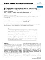
Left parotid lesions and all the other cervical lymphadenopathies previously reported were also reduced (Fig. 1).
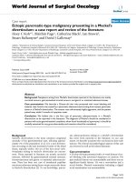
During follow-up, we witnessed a progressive dimensional decrease of the lesions. The right mediastinal enlargement shown on the first chest radiograph had
already disappeared at first checkup visit (Fig. 2). Right
parotid swelling disappeared, with US at 4 months after
therapy interruption showing residual lymphadenopathies of 16 mm in the right parotid and 23 × 12 mm in
the right part of the neck. We therefore decided to continue with a wait-and-see approach, with periodic clinical and US controls. Twelve months after the therapy
discontinuation, the child was completely asymptomatic,
with minimal residual lymph nodes within both right
di Dio et al. BMC Pediatrics (2016) 16:62
Page 3 of 7
Fig. 1 MRI of the face and neck findings. Evidence of a solid mass attributable to lymph nodes, entirely occupying right parotid cavity (a), with
marked improvement on second control after prednisone course (b)
parotid and cervical area. The “CARE” checklist is available as Additional file 1. This child’s clinical course is
summarized in the accompanying supplemental timeline file (Additional file 2).
Discussion
Systemic form of RDD is associated with various diseases
or factors involving the immune system, such as cases
discovered at the time or after the diagnosis of various
forms of leukemia or lymphoma, or even following bone
marrow transplant for precursor-B acute lymphoblastic
leukemia [6]. Lu et al. reported 4 cases in which RDD and
malignant lymphoma were identified in the same lymph
node biopsy specimen [7].
As in our case, the presence of EBV or HHV-6 in
histological samples has also been reported [8]. Nevertheless, no strong association can be found between one
of these factors and the occurrence of RDD.
However, it can be hypothesized that the disease may
be triggered by a generic immunological stimulus, which
could result in the accumulation and activation of histiocytes [9].
In our case, considering the presence of EBV in the
specimen and the serological pattern indicating a recent
infection (even though EBNA-IgG can be negative in a
small percentage of cases), it is possible that the virus
was the immunological trigger for the disease.
Cervical lymphadenopathy is present in over 90 %
of patients affected by RDD. Typically, it is painless,
bilateral and frequently massive, with single nodal elements measuring up to 6 centimeters [5]. However,
practically any group of lymph nodes can be involved
in RDD. Axillary and inguinal lymph nodes are enlarged in 38 and 44 % of cases, respectively. Mediastinal and hilar nodes are involved in approximately
30 % of patients. In a minority of cases, retroperitoneal lymph node localizations have also been described
[5]. Except for cervical nodes, the dimension of adenopathy in the other sites is usually smaller. In our
case, a non-massive cervical adenopathy was present,
associated with enlargement of right hilar nodes and
bilateral intraparotid nodes.
Extranodal involvement has been documented in 43 %
of cases [10]. The most commonly affected sites are the
upper respiratory tract (nasal cavities and paranasal
sinuses), skin, eyes and retro-orbital tissue and bone
tissues. Salivary glands and central nervous system are
less frequently involved. Localizations of RDD in lungs,
urogenital and gastrointestinal tract, breast, thyroid, and
even heart have also been reported [9, 11–13].
Fig. 2 chest x-rays findings. Evidence of right mediastinal enlargement (figure on the left) on first control, with disappearance on second
evaluation after prednisone course (figure on the right)
di Dio et al. BMC Pediatrics (2016) 16:62
The onset of the disease is generally subtle, with an
average of 3–6 months between the beginning of signs
and symptoms and the diagnosis. Non-specific systemic
symptoms can be present, including fever, malaise, weight
loss, and night sweats. They are more frequent in case of
an important nodal involvement, but cases of patients
with systemic symptoms in which RDD was purely extranodal have been reported [10]. In case of extranodal localizations, the clinical picture will depend on the affected
organ or apparatus. In our case, the only systemic symptom was the low-grade fever, appearing approximately
40 days after the first observation of the parotid swelling.
Laboratory findings in RDD are non-specific. The most
frequent features include leukocytosis, elevated ESR and
CRP levels, and polyclonal gammopathy. Normochromic
normocytic anemia and elevated serum ferritin levels have
also been described. Less common laboratory abnormalities include positivity for rheumatoid factor and antinuclear antibodies, and a reversal of the CD4/CD8 ratio in
peripheral lymphocytes [13]. There are also reports on
RDD complicated by the development of autoimmune
hemolytic anemia, especially in children, for which the
pathogenetic mechanism is not known [14].
Imaging exams may be helpful in differential diagnosis,
but they are not pathognomonic. In MRI studies of patients affected by RDD the areas involved were generally
T1 isointense, T2 isointense, diffusion isointense to gray
matter, and intensely enhancing with gadolinium,
whereas in CT images the lesions were hyperdense to
gray matter and intensely enhancing [15]. Ultrasound
can also be helpful, especially in neck involvement, to
see if lymphadenopathy is either focal or diffuse and in
follow up, being a noninvasive and relatively rapid exam.
Definitive diagnosis can only be made by histological
analysis of affected lymph nodes or tissues. The association between emperipolesis, defined as the presence of
phagocytized cells (mainly lymphocytes, but also plasma
cells, neutrophils or erythrocytes) in a histiocyte, and a
typical immunohistochemical pattern characterized by
positivity for S-100 protein and CD68 antigen and negativity for CD1a antigen, is diagnostic for RDD. Emperipolesis alone is highly suggestive of the disease, but it can
also be found in Langerhans cell histiocytosis (LCH), autoimmune hepatitis, lymphoma and rhinoscleroma, a
chronic granulomatous bacterial disease affecting the nose
and rarely the upper respiratory tract, caused by Klebsiella
rhinoscleromatis, a subspecies of Klebsiella pneumoniae
[14, 16]. Another characteristic is the absence of Birbeck
granules, which are instead typical of LCH. LCH and
RDD can be also distinguished by CD1a pattern, being
nearly always expressed in LCH. The clinical course of
RDD is generally benign, with spontaneous complete resolution in most cases, especially if the disease affects mainly
lymph nodes. However, locoregional recurrence of RDD
Page 4 of 7
and even dissemination can be possible, particularly in
forms with extranodal involvement. There have also been
reports of deaths directly caused by RDD, even in children, especially in severe extranodal forms, with involvement of CNS, kidneys or respiratory tract [17].
There is no consensus for the best therapeutic approach. An initial wait-and-see approach may be the best
option, given the fact that RDD is often self-limiting.
However, in case of significant extranodal disease and/
or compression of vital organs by massive lymphadenopathy, prompt therapy may be indicated.
When possible, surgery probably represents the best
solution, being curative if resection is complete. Recurrence after surgical therapy is very rare and limited to
incomplete debulking and multiorgan involvement [4].
Medical therapeutic options include corticosteroids, antibiotics, antiviral agents, chemotherapy, and radiotherapy.
However, no universally accepted treatment guidelines
exist. Several Authors reported successful treatments
using radiotherapy and/or corticosteroids in cases of
recurrences or incomplete resection, but this strategy was
not always effective. In our case, given the young age of
the patient and the apparently rapid progression of the
disease, we decided to treat the child using corticosteroids,
obtaining an important dimensional reduction of hilar,
cervical and parotid glands adenopathy, the resolution of
systemic symptoms, and normalization of blood tests.
Moreover, we chose to treat the patient with a relatively
short course of prednisone therapy at a standard dosage
of 1.5 mg/kg/day, obtaining a good clinical response and
no adverse effects.
In order to establish whether there could be a more
effective medical approach in the management of RDD,
in particular in children, we reviewed the literature
through both PubMed and Embase network using Rosai
Dorfman disease, sinus histiocytosis with massive lymphadenopathy and child as keywords and selecting all the
available pediatric manuscripts published between 2004
and 2014. In addition, we reviewed references cited in all
selected manuscripts to identify additional reports of
pediatric RDD. We did not include cases in which the
surgery was used as the only therapeutic approach (mainly
isolated intracranial forms of RDD). We selected also all
cases in which there was an apparent involvement of the
salivary glands at presentation, as in our patient, including
2 reports in which there was no information on clinical
outcome (Table 1) [18, 19]. In one of these two cases, only
surgery was performed, but we decided to include it in the
Review, given the involvement of the salivary glands as in
our case. In Table 2 we report a summary of all other
clinical reports found. Correlation between the clinical
outcome and the most effective therapy used in single
cases was obtained with the chi-square test. P values of
less than 0.05 were considered statistically significant.
di Dio et al. BMC Pediatrics (2016) 16:62
Page 5 of 7
Table 1 Pediatric cases of RDD with involvement of salivary glands
Age/ Clinical picture
sex at presentation
Main lesion location
Systemic symptoms and/or Nodal and
abnormal blood tests at
extranodal
presentation
involvement
Therapy and
Outcome
clinical evolution
Ref.
10/
M
Painless masses
Parotid and submandibular
around parotid and
glands bilaterally.
submandibular glands.
None
Apparently
None
both nodal
and extranodal
Symptom-free
[21]
9/M
Masses around
Submandibular glands
submandibular glands bilaterally.
None
Apparently
None
both nodal
and extranodal
Symptom-free
[21]
11/
M
1 year history of
painless bilateral
neck swelling.
Submandibular and parotid
glands bilaterally.
None
Both nodal
Surgery
and extranodal
Not reported
[18]
Mass at left common carotid
artery, descending aorta down
to the renal artery; MRI finding
of bilateral lesions in knee and
ankle.
High CRP and ESR,
Both nodal
None
hypergammaglobulinemia. and extranodal
Not reported
[19]
Recurrent fever 2 months
before presentation;
high ESR.
No recurrence [28]
after 28 month
of follow-up.
17/F Bilateral parotid
enlargement and
cervical lumps
localized in the
submandibular region.
12/F 1-month history of
Parotid and submandibular
enlarging and painless glands.
submandibular
lymphadenopathy.
Nodal
None
clear prevalence in males (68.6 % vs 31.4 %), as confirmed by other studies. During the period we considered, our case was the youngest reported. Almost half of
the patients had both nodal and extranodal involvement
(17). Eleven children had purely nodal RDD, whereas the
remaining 8 patients had only extranodal localizations.
There was no correlation between the type of RDD and
clinical outcome. However, we found a significant difference between the mean age of children and the type of
RDD (Table 3), with younger children being those most
frequently affected by purely nodal RDD. To our
Complete regression of RDD, improvement and clinical
stability of the disease were considered as clinical outcomes in our analysis. The only case in which the disease
led to death was not included in the statistical analysis regarding the connection between outcome and therapy, as
well as 3 cases in which outcome was not precise [17–20].
Overall, we found 35 pediatric cases of RDD in literature (36 including our case, which was considered in the
analysis). Mean age of the children described in the
manuscripts was 8.79 years [standard deviation (SD)
4.26, minimum 14 months, maximum 17 years], with a
Table 2 Summary of all other pediatric cases of RDD described between 2004 and 2014 (our case and cases in which only surgery
was used were not included; i.e. 33 cases)
Systemic
symptoms
Fever
Anemia
Fatigue
None
Not mentioned
# of cases
3
5
1
10
9
Ref.
[25, 29, 30]
[3, 24, 27, 29, 31]
[32]
[17, 20, 24, 28, 30, 32–35]
[6, 17, 23, 24, 26, 36–38]
Lesion location
Lymph nodes
Bone
Brain
Other
# of cases
18
8
5
6
Ref.
[17, 22, 25, 27–36, 39, 40]
[6, 22, 24, 34, 37, 40]
[20, 24, 26,
38, 39]
[3, 17, 23, 30, 32, 41]
Successful of main
treatment
Corticosteroids
Chemotherapy
Corticosteroids +
chemotherapy
Others
None
# of cases
6
7
5
5
10
Ref.
[3, 23, 30, 33, 36]
[6, 22–24, 32, 37, 40]
[17, 24, 34, 39]
[20, 25–27, 38]
[17, 24, 29, 31, 28,
35, 41]
Outcome
Complete regression
Partial regression
Clinical stability
Symptoms free
(no precise information
on disease outcome)
Death
# of cases
11
12
6
3
1
Ref.
[6, 22, 25–27, 28, 33,
35, 36, 38, 39]
[3, 23, 24, 29–32,
34, 39, 40]
[17, 24, 37]
[17, 29, 41]
[20]
di Dio et al. BMC Pediatrics (2016) 16:62
Page 6 of 7
Table 3 Chronological age at diagnosis of different forms of
RDD (nodal, extranodal, both nodal and extranodal). Data are
reported ad mean ± SD; Kruskal-Wallis test
Age (yrs)
Nodal
RDD
Extranodal
RDD
Nodal and
extranodal RDD
5.83 ± 4.89
10.0 ± 2.68
9.43 ± 3.54
p
0.018
knowledge, this epidemiological finding was not previously reported. In the analysis, we considered the age at
the time of first diagnosis, excluding 2 cases in which
this element was not reported [21].
Considering the medical therapeutic approaches, there
was a statistically significant difference correlating the
most effective strategy used in single cases and the clinical outcome (p 0.033). Prednisone and prednisolone
were the most used single drugs. In 6 cases they were
the only treatment used, with complete regression in 2
children and clinical improvement in the other 4 cases.
Chemotherapy was shown to be an effective strategy,
with complete regression in 4 patients, clinical improvement in 5 and clinical stability in 1 subject. However, it
has to be said that the chemotherapic agents reported
were various, giving different results. For example, 2chlorodeoxyadenosine (2-CDA) was found to be very effective in a case in which prednisone, 6-mercaptopurine
and vinblastine were used before, without clinical response [22]. Nevertheless, this drug was ineffective in
other reports [23, 24]. In 3 children, the successful treatment consisted of another medical approach (radiotherapy, rituximab in association with prednisone, αinterferon) [25–27]. However, in our opinion these strategies cannot be recommended as first-line treatment, as
they have been cited only in single case reports. Finally,
no treatment was used in 9 cases, with complete regression in 2 children, clinical improvement in 3 and clinical
stability in 4 subjects. This means that, even if RDD was
reported to have a benign course in most of the cases,
medical therapy may hasten remission, given the fact
that treated children had a significant higher percentage
of improvement (Table 4). Moreover, complications of
the disease are not so infrequent. That is why, in our
opinion, it would be safer, if complete surgical debulking
is not possible, to proceed with a therapeutic medical
approach, with the goal of hastening regression of the
Table 4 Type of approach and clinical outcome (p = 0.033,
chi-squared test). Cases in which only surgery was used
and/or outcome was death or not precise were not included;
i.e. 32 cases
Outcome/Therapy
None
Steroid
Chemo
Others
Complete regression
2
3
4
3
Clinical improvement
3
4
5
1
Clinical stability
5
0
2
0
disease and avoiding complications. In light of the data
presented, with all the limits related to a review of case
reports, in our opinion it would be safe and worthwhile
to start with a course of oral steroid therapy as a firstline approach. In case of no clinical response, there is
still no agreement on what might be the best treatment.
Chemotherapy was shown to be effective; however, clinical response to the same protocols did not appear to be
constant. Alternative therapies were effective in some
cases, but they should be tested in more subjects.
Conclusions
To our best knowledge, this is the first RDD case that
involves both parotid and hilar lymph nodes in a toddler,
apparently without extranodal localization.
Hilar adenopathy biopsy was not performed, but the
unilateral pattern, the absence of any other clinical
reason that could explain the mediastinal enlargement
and the prompt resolution after oral steroid therapy are
in our opinion 3 strong factors that confirm the adenopathy as another nodal localization of RDD.
Another interesting factor is that progression of the
disease was quite rapid, differently from other case reports
in which the development of parotid swelling was more
gradual.
A short course of prednisone was shown to be an
effective treatment in our case.
RDD should be included in differential diagnosis of
parotid swelling even in children.
A medical approach should be considered in all
pediatric cases of RDD.
Consent
Written informed consent was obtained from the
patient’s parents for publication of this Case report and
any accompanying images. A copy of the written consent
is available for review by the Editor of this journal.
Additional files
Additional file 1: CARE Checklist of information to include when
writing a case report. (DOC 1546 kb)
Additional file 2: Timeline. (PPT 138 kb)
Abbreviations
2-CDA: 2-Chlorodeoxyadenosine; CMV: cytomegalovirus; CNS: central nervous
system; CRP: C-reactive protein; CT: computed tomography; EBV: Epstein Barr virus; ESR: elevated erythrocyte sedimentation rate; HHV-6: herpesvirus;
LCH: Langerhans cell histiocytosis; MRI: magnetic resonance imaging;
MTX: methotrexate; RDD: Rosai-Dorfman disease; SD: standard deviation;
US: ultrasonography.
Competing interests
The authors declare that they have no competing interests.
di Dio et al. BMC Pediatrics (2016) 16:62
Authors’ contributions
FD was responsible for the conception and design of the study, data
collection and interpretation, and manuscript writing. IM cared for the
patient, collected samples, and drafted the manuscript EC participated in
data collection and analysis. PB participated in data collection and
interpretation, and critically revised the manuscript. BP analyzed the literature
and critically revised the manuscript. LI was responsible for the conception
and design of the study, data collection and interpretation, and manuscript
writing. All authors read and approved the final manuscript.
Received: 6 February 2015 Accepted: 21 April 2016
References
1. Brenn T, Calonje E, Granter SR, Leonard N, Grayson W, Fletcher CDM, McKee
PH. Cutaneous Rosai-Dorfman disease is a distinct clinical entity.
Am J Dermatopathol. 2002;24(5):385–91.
2. Kutlubay Z, Bairamov O, Sevim A, Demirkesen C, Mat MC. Rosai–Dorfman
disease: a case report with nodal and cutaneous involvement and review of
the literature. Am J Dermatopathol. 2013;0:1–5.
3. Azoulay R, Brisse H, Fre’neaux P, Ferey S, Kalifa G, Adamsbaum C. Lacrimal
location of sinus histiocytosis (Destombes-Rosai-Dorfman disease).
Am J Med Sci. 2013;345(3):200–10.
4. Mahzoni P, Zavareh MHT, Bagheri M, Neda Hani N, Moqtader B. Intracranial
ROSAI-DORFMAN disease. J Res Med Sci. 2012;17(3):304–7.
5. Sodhi KS, Suri S,MD, Nijhawan R, Kang M, Gautam V. Rosai-Dorfman disease:
unusual cause of diffuse and massive retroperitoneal lymphadenopathy.
Br J Radiol. 2005;78:845–7.
6. Ambati S, Chamyan G, Restrepo R, Escalon E, Fort J, Pefkarou A, Khatib ZA,
Dehner LP. Rosai–Dorfman disease following bone marrow transplantation
for Pre-B cell acute lymphoblastic leukemia. Pediatr Blood Cancer.
2008;51(3):433–5.
7. Lu D, Estalilla OC, Manning Jr JT, Medeiros LJ. Sinus histiocytosis with
massive lymphadenopathy and malignant lymphoma involving the same
lymph node: a report of four cases and review of the literature.
Mod Pathol. 2000;13:414–9.
8. Levine PH, Jahan N, Murari P, Manak M, Jaffe ES. Detection of human
herpes virus 6 in tissue involved by sinus histiocytosis with massive
lymphadenopathy (Rosai-Dorfman disease). J Infect Dis. 1992;166:291–5.
9. Oussama A. Rosai-Dorfman disease. />document.doc?id=54. 2011. Accessed 21 Apr 2016.
10. Zhu F, Zhang JT, Xing XW, Wang DJ, Zhu RY, Zhang Q, Wang HT, Lang SY.
Rosai-Dorfman disease: a retrospective analysis of 13 cases. Am J Med Sci.
2013;345(3):200–10.
11. Rosai J, Dorfman RF. Sinus histiocytosis with massive lymphadenopathy: a
newly recognized benign clinicopathological entity. Arch Pathol.
1969;87:63–70.
12. Foucar E, Rosai J, Dorfman RF. Sinus histiocytosis with massive
lymphadenopathy. Arch Otolaryngol. 1978;104:687–93.
13. Foucar E, Rosai J, Dorfman R. Sinus histiocytosis with massive
lymphadenopathy (Rosai-Dorfman disease): review of the entity.
Semin Diagn Pathol. 1990;7:19–73.
14. Sachdeva M, Abdulhaq H. A rare case of Rosai-Dorfman disease in an adult
male associated with auto-immune hemolytic anemia. Mediterr J Hematol
Infect Dis. 2013;5(1):e2013022.
15. Raslan OA, Schellingerhout D, Fuller GN, Ketonen LM. Rosai-Dorfman
disease in neuroradiology: imaging findings in a series of 10 patients.
Am J Roentgenol. 2011;196(2):W187–93.
16. Chou TC, Tsai KB, Lee CH. Emperipolesis is not pathognomonic for
Rosai-Dorfman disease: Rhinoscleroma mimicking Rosai-Dorfman disease,
a clinical series. J Am Acad Dermatol. 2013;69(6):1066–7.
17. Warrier R, Chauhan A, Jewan Y, Bansal S, Craver R. Rosai-Dorfman disease
with central nervous system involvement. Clin Adv Hematol Oncol.
2012;10(3):196–202.
18. Sridhara SK, Shah RK. Rosai-Dorfman in the submandibular salivary glands of
a pediatric patient. Laryngoscope. 2010;120 Suppl 4:S228.
19. Kasapoglu Gunal E, Kamali S, Akdogan MF, Cimen AO, Ocal L, Agan M, Gul
A, Inanc M, Konice M, Aral O. Rosai–Dorfman disease with factor XII
deficiency. Clin Rheumatol. 2009;28:733–6.
Page 7 of 7
20. Antuña Ramos A, Alvarez Vega MA, Alles JV, Antuña Garcia MJ, Meilán
Martínez A. Multiple involvement of the central nervous system in
Rosai-Dorfman disease. Pediatr Neurol. 2012;46:54–6.
21. Güven G, Ilgan S, Altun C, Gerek M, Gunhan O. Rosai–Dorfman disease of
the parotid and submandibular glands: salivary gland scintigraphy and oral
findings in two siblings. Dentomaxillofac Radiol. 2007;36:428–33.
22. Tasso M, Esquembre C, Blanco E, Moscardó C, Niveiro M, Payá A. Sinus
histiocytosis with massive lymphadenopathy (Rosai–Dorfman disease)
treated with 2-Chlorodeoxyadenosine. Pediatr Blood Cancer. 2006;47:612–5.
23. Stine KC, Westfall C. Sinus histiocytosis with massive lymphadenopathy
(SHML) prednisone resistant but dexamethasone. Pediatr Blood Cancer.
2005;44:92–4.
24. Rodriguez-Galindo C, Helton KJ, Sánchez ND, Rieman M, Jeng M, Wang W.
Extranodal Rosai-Dorfman disease in children. J Pediatr Hematol Oncol.
2004;26:19–24.
25. Kismet E, Köseoglu V, Atay AA, Deveci S, Demirkaya E, Tuncer K. Sinus
histiocytosis with massive lymphadenopathy in three brothers. Pediatr Int.
2005;47:473–6.
26. El Majdoub F, Brunn A, Berthold F, Sturm V, Maarouf M. Stereotactic
interstitial radiosurgery for intracranial rosai-dorfman disease. A novel
therapeutic approach. Strahlenther Onkol. 2009;185:109–12.
27. Alqanatish JT, Houghton K, Bond M, Senger C, Tucker LB. Rituximab
treatment in a child with Rosai-Dorfman disease and systemic lupus
erythematosus. J Rheumatol. 2010;37:1783–4.
28. Ruggiero A, Attinà G, Maurizi P, Mulè A, Tarquini E, Barone G, Lazzareschi I,
Riccardi R. Rosai-Dorfman disease: two case reports and diagnostic role of
fine-needle aspiration cytology. J Pediatr Hematol Oncol. 2006;28(2):103–6.
29. Lima FB, Barcelos PS, Constâncio AP, Nogueira CD, Melo-Filho AA.
Rosai-Dorfman disease with spontaneous resolution: case report of a child.
Rev Bras Hematol Hemoter. 2011;33(4):312–4.
30. Nandi M, Mondal RK, Datta S, Karmakar BC, Mukherjee K, Dhibar TK.
Rosai-Dorfman disease. Indian J Pediatr. 2008;75:290–4.
31. Maheshwari A, Seth A, Choudhury M, Aggarwal V, Patra B, Aggarwal S,
Mukherjee SB, Aneja S. Rosai-Dorfman disease: a case with
lymphadenopathy and liver involvement. J Pediatr Hematol Oncol.
2009;31:200–2.
32. Inoue S, Onwuzurike N. Venorelbine and Methotrexate for the treatment of
Rosai-Dorfman disease. Pediatr Blood Cancer. 2005;45:84–5.
33. Warpe BM, More SV. Rosai-Dorfman disease: a rare clinico-pathological
presentation. Austr Med J. 2014;7(2):68–72.
34. Yetiser S, Cekin E, Tosun F, Yildirim A. Rosai—Dorfman disease associated
with neurosensorial hearing loss in two siblings. Int J Pediatr
Otorhinolaryngol. 2004;68:1095–100.
35. Sardana D, Goyal A, Gauba K. Sinus histiocytosis with massive
lymphadenopathy: a “massive” misnomer. Diagn Cytopathol. 2014;00:1–5.
36. Folia M, Martin L, Duvillard C, Romanet P. A laryngeal Rosai-Dorfman
disease. Int J Pediatr Otorhinolaryngol Extra. 2011;6:285–7.
37. Tubbs RS, Kelly DR, Mroczek-Musulman EC, Hammers YA, Berkow RL,
Oakes WJ, Grabb PA. Spinal cord compression as a result of Rosai–Dorfman
disease of the upper cervical spine in a child. Childs Nerv Syst.
2005;21:951–4.
38. Miletic H, Röhling R, Stenzel W, Deckert M, Benz-Bohm G, Berthold F,
Voges J. 8-year-old child with a lesion in the left nucleus lentiformis.
Brain Pathol. 2008;18:598–601.
39. Jabali Y, Smrcka V, Pradna J. Rosai-Dorfman disease: successful long-term
results by combination chemotherapy with prednisone, 6-mercaptopurine,
Methotrexate, and vinblastine: a case report. Int J Surg Pathol.
2005;13(3):285–9.
40. Geara AR, Ayoubi MA, Achram MC, Chamseddine NM. Rosai-Dorfman
disease mimicking neurofibromatosis: case presentation and review of the
literature. Clin Radiol. 2004;59:625–30.
41. Yontz L, Franco A, Sharma S, Lewis K, McDonough C. A case of
Rosai-Dorfman disease in a pediatric patient with cardiac involvement.
Radiol Case. 2012;6(1):1–8.