Recurrent Streptococcus Pneumoniae 23 F meningitis due to cerebrospinal fluid leakage from the ear cannel: A case report
Bạn đang xem bản rút gọn của tài liệu. Xem và tải ngay bản đầy đủ của tài liệu tại đây (2.56 MB, 5 trang )
Li et al. BMC Pediatrics (2015) 15:195
DOI 10.1186/s12887-015-0509-2
CASE REPORT
Open Access
Recurrent Streptococcus Pneumoniae 23 F
meningitis due to cerebrospinal fluid
leakage from the ear cannel: a case report
Yu-Cheng Li1, Chun-Yu Chen2,3, Kang-Hsi Wu4,5, Huang-Tsung Kuo6,7* and Han-Ping Wu8,9*
Abstract
Background: Bacterial meningitis is a medical emergency, and immediate diagnostic steps must be taken to
establish the specific cause. Recurrence of bacterial meningitis in children is not only potentially life-threatening, but
also involves or induces psychological trauma to the patients through repeated hospitalization with many invasive
investigations.
Case presentation: A 6-year-old boy was diagnosed with recurrent bacterial meningitis caused by Streptococcus
Pneumonia 23 F. He had received serial imaging studies for identifying the cause. The initial sinus computed
tomography (CT) also showed sinusitis without bony defect of sinus. However, after performing nuclear scan, the
results showed cerebrospinal fluid (CSF) leaked originating from the right petrooccpital region into the middle ear.
Subsequent high resolution CT (HRCT) reports showed focal enlargement of the right facial nerve canal,
erosion of the bony canal at geniculate ganglion and tympanic segment with tiny high-density spots. The
reconstruction HRCT showed multiple bony defects at temporal bone. The magnetic resonance imaging
revealed multifocal bony destruction with CSF collection in the right petrous ridge, carotid canal and jugular
foramen. Eventually, CSF leakage to the right middle ear was confirmed and this could be the cause of the
recurrent bacteria meningitis in this patient.
Conclusion: Although recurrent bacterial meningitis in childhood is not common, this case report illustrates
that recurrence of meningitis within a short period should be considered as cause of underline immunologic
or anatomic defect.
Keywords: Streptococcus pneumoniae, Recurrent, Meningitis, Cerebrospinal fluid
Background
Bacterial meningitis is a medical emergency, and immediate diagnostic steps must be taken to establish the specific cause so that appropriate antimicrobial therapy can
be initiated [1, 2]. The mortality rate of untreated bacterial meningitis approaches 100 % and, even with optimal
therapy, morbidity and mortality may occur [2, 3]. Recurrence of bacterial meningitis in children may be
caused by many reasons from cranial or dural anatomic
defect and immumity deficiency [4]. Bacteria migration,
along congenital or acquired pathways from the skull or
spinal dural defects should be taken into consideration
when children had recurrent bacteria meningitis [5].
However, symptoms and signs of cerebrospinal fluid
(CSF) rhinorrhea or otorrhea are difficult to find in such
patients [6]. The CSF leakage caused by traumatic injury
is common, while leakage caused by congenital bony
abnormality is rarely reported. Here we present the case
of a 6-year-old boy with repeated bacterial meningitis
within 6 months and further imaging exanimations finally proved the cause of CSF leakage originating from
the right petrooccpital region into the middle ear.
* Correspondence: ;
6
School of Medicine, China Medical University, Taichung, Taiwan, R.O.C.
8
Division of Pediatric General Medicine, Department of Pediatrics, Chang
Gung Memorial Hospital at Linko, Kweishan, Taoyuan, Taiwan, R.O.C
Full list of author information is available at the end of the article
Case presentation
The 6-year-old boy complained of nausea, vomiting and
headache for one week. He received medical treatment
at local medical clinics initially, but his condition still
© 2015 Li et al. Open Access This article is distributed under the terms of the Creative Commons Attribution 4.0 International
License ( which permits unrestricted use, distribution, and reproduction in any
medium, provided you give appropriate credit to the original author(s) and the source, provide a link to the Creative
Commons license, and indicate if changes were made. The Creative Commons Public Domain Dedication waiver (http://
creativecommons.org/publicdomain/zero/1.0/) applies to the data made available in this article, unless otherwise stated.
Li et al. BMC Pediatrics (2015) 15:195
persisted without improvement. Progressed symptoms
and fever were also noted after initial medical treatment,
and, he was transferred to our emergency department
(ED) for further evaluation. At the ED, the previous history of the patient was obtained from his family. This boy
had experienced one earlier episode of AOM in his
young-infant stage and experienced a single episode of
acute sinusitis about 2 months prior to admission. Moreover, no any history of skull trauma was noted before
admission. However, the physical examinations revealed
general appearance as lethargy and neck stiffness with
positive meningitis signs (Brudzinski’s sign and Kerning
sign). After admission, blood was sampled for complete
blood count (CBC) with differential count (DC) analysis,
biochemistry, glucose levels, and blood culture. Immediately lumbar puncture with CSF survey (CSF analysis, bacterial culture, virus culture and CSF biochemistry test)
was also performed. The blood laboratory tests showed
leukocytosis with shift to the left (white blood cell (WBC)
count: 29190/mm3, and bands: 4 %), and the results of
CSF analysis revealed WBC count as 3240/uL with predominant neutrophils as 91 %, glucose levels as 55 mg/dL,
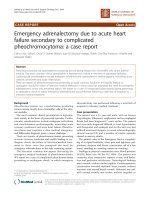
and total protein levels as 160.5 mg/dL. Moreover, the
gram stain of CSF showed Sptreptococcus Pneumoniae
(Fig. 1), and antibiotics with vancomycin and cefotaxime
were given immediately. The cultures of CSF and blood
both showed Sptreptococcus Pneumoniae 23 F. Based on
the report of the sensitivity to antibiotics in the strain of
23 F, vancomycin was useful and given continuously for
14 days. To trace back his past history, about 6 months
ago, this pediatric patient suffered from bacterial meningitis, and was admitted for survey and treatments. The CSF
gram stain showed Sptreptococcus Pneumoniae. Both CSF
and blood cultures also showed Sptreptococcus Pneumoniae 23 F. After complete antimicrobial treatment with
Fig. 1 Gram stain of the CSF showed Streptococcus Pneumoniae
(black arrow) in the patient
Page 2 of 5
vancomycin for 14 days, he was discharged home without
complication.
To further survey the cause of recurrent bacteria meningitis in such short period, we analyzed immunological
functions of this boy, including complements and various immunoglobulins. However, the results showed normal immunity. According to the previous history of
recurrent sinusitis for several weeks, we suspected that
recurrent meningitis may be due to a bony defect caused
by chronic sinusitis. Sinus computed tomography (CT)
was performed but only right side maxillary sinusitis was
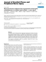
noted without any bony defect. Moreover, nuclear scan
was arranged and performed for studying CSF leakage.
Notably, the results showed CSF leaked originating from
the right petrooccpital region into the middle ear (Fig. 2).
Subsequent high resolution CT (HRCT) and magnetic
resonance imaging (MRI) of bilateral ears were both carried out. The HRCT reports showed focal enlargement
of the right facial nerve canal, erosion of the bony canal
at geniculate ganglion and tympanic segment with tiny
high-density spots (Fig. 3) and the reconstruction HRCT
showed multiple bony defect at petrous part of temporal
bone (Fig. 4). The MRI reports revealed multifocal bony
destruction with CSF collection in the right petrous
ridge (near the Meckel cave and facial nerve canal at
geniculate body ganglion region), carotid canal and jugular foramen (Fig. 5). Eventually, CSF leakage to the right
middle ear was confirmed and this may explain the
cause of the recurrent bacteria meningitis in this boy.
Further surgical approach for bony defect was suggested,
but his family refused and asked for medical treatments.
Therefore, after complete antimicrobial treatments with
vancomycin for 14 days, this patient was discharged
home, and received conjugated streptococcus pneumoniae vaccination (Prevenar 7) by self-payment, which is
not included in the program of our national schedule
vaccination at that time.
Discussion
Recurrence of bacterial meningitis in children is not only
potentially life-threatening, but also involves or induces
psychological trauma to the patients through repeated
hospitalization with many invasive investigations. In our
case report, this patient had suffered from bacteria meningitis twice, and required repeated hospitalization for
invasive CSF survey and for managements of infectious
emergency. This situation did suffer very much for the
patient and his family. Therefore, to avoid repeated meningitis again is essential for this patient and to understand why recurrence of bacterial meningitis occurred is
also important for primary clinicians. Clinically, it is reasonable for primary clinicians to survey for immune deficiency or CSF leakage caused by defect of anatomy [6–11].
In addition, the bacteria specificity could provide some
Li et al. BMC Pediatrics (2015) 15:195
Page 3 of 5
Fig. 2 Radioisotope cisternography showed CSF leak into right side middle ear area (red arrow)
Fig. 3 HRCT of the right side ear showed enlargement to facial
nerve cannel (red arrow)
informative clues: Based on some investigations, pneumoccocus or hemophilus may suggest cranial dural defects, E. coli or other gram negative bacillus may suggest
spinal dural defects, and meningococcus may suggest immunologic deficiency of the patient [3–5].
Moreover, spontaneous cerebrospinal fistula could be
difficult for clinicians to make the diagnosis and only revealed recurrent attacks of meningitis. Recurrent meningitis may occur in 92 % of such fistulas which indicates
the importance of accurate diagnosis and appropriate
treatments for CSF leakage [7]. Recurrent meningitis,
clear otorrhea, or rhinorrhea are signs requiring several
investigations of the temporal bone. When the ear drum
is intact, CSF passes down the eustachian tube and may
result in rhinorrhea. If the tympanic membrane is perforated, either spontaneously or after myringotomy, otorrhea may occur. Some case reports have reported that
congenital CSF leakage may present as serous otitis
media and be revealed at the time of myringotomy [12].
Also, CT scan involving 1-mm sections in coronal and
axial planes of the temporal bone is certainly the most
precise and reliable method available [13, 14]. In our
Li et al. BMC Pediatrics (2015) 15:195
Page 4 of 5
Fig. 4 Reconstruction in brain HRCT showed multiple bony destructions at the right side (black arrow) compared to the left side (red arrow)
case report, the initial CT scan could not find out the leakage This may be due to the difficult in identifying the right
location of CSF leakage by routine head or brain CT scan
which is too broad to image the otic capsule, ossicles, and
facial nerve accurately. Furthermore, the coronal images
Fig. 5 MRI showed CSF accumulation at right middle ear
are usually reconstructions, which provide significantly
less detail than the directly-obtained coronal image.
To test for CSF leakage, clinician may test the ear or
nose drainage for beta-2 transferrin, a desialylated form
of the protein transferring, which is almost exclusively
found only in CSF [15, 16]. Therefore, to localization of
the fistula may require diagnostic imaging studies [17].
Nuclear medicine examination (Radioisotope cisternography) or fluorescein dye study via lumbar puncture
should be considered to identify the location of leakage
[18]. In our case report, radioisotope cisternography
combined with HRCT (1-mm section) and MRI appeared helpful to identify the location. From this case report, we found that recurrent bacteria meningitis is
critical and should be prompt a search for an underline
immunologic or anatomic defect. CSF leakage is common to cause misdiagnosis or failure to make a timely
early diagnosis, which means that suitable treatment
may be delayed. Better knowledge of the possible sites
and pathways of fistulas (even rare ones) is necessary.
The different pathways of spontaneous CSF leakage
should be clearly understood and carefully examined by
the radiologists and primary clinicians. Congenital inner
ear malformation is an uncommon fistula route, which
can be misdiagnosed even regular CT (usually cut every
5 mm) is performed without performed high resolution
CT (usually cut every 1 mm). The treatment for this
Li et al. BMC Pediatrics (2015) 15:195
Page 5 of 5
congenital fistula is based on filling of the bone pathway,
which can be repaired with biometerials.
7.
Conclusions
Although recurrent bacterial meningitis in childhood is
not common, this clinical condition remains a neurological emergency for primary care physicians. This case
illustrates that recurrence of meningitis within a short
period should be considered as cause of underline immunologic or anatomic defect.
9.
Consent
Written informed consent was obtained from the patient’s parents for publication of this Case report and
any accompanying images. A copy of the written consents is available for review by the Editor of this journal.
Abbreviations
ED: Emergency department; CSF: Cerebrospinal fluid; WBC: White blood cell;
CT: Computed tomography; HRCT: High resolution computed tomography.
Competing interests
There is no conflict of interest related to this study.
Authors’ contributions
YCL, CYC and KHW reviewed the medical records, and drafted the
manuscript; HPW designed and oversaw the case report. HPW and HTK
revised the manuscript. All authors have read and approved the final
manuscript for publication.
8.
10.
11.
12.
13.
14.
15.
16.
17.
18.
Gacek RR, Gacek MR, Tart R. Adult spontaneous cerebrospinal fluid otorrhea:
diagnosis and management. Am J Otol. 1999;20:770–6.
Wetmore SJ, Herrmann P, Fisch U. Spontaneous cerebrospinal fluid otorrhea.
Am J Otol. 1987;8:96–102.
Sun HL, Wu KH, Chen SM, Chao YH, Ku MS, Hung TW, et al. Role of procalcitonin
in predicting dilating vesicoureteral reflux in young children hospitalized with a
first febrile urinary tract infection. Pediatr Infect Dis J. 2013;32:e348–54.
Wu KH, Tsai C, Wu HP, Sieber M, Peng CT, Chao YH. Human Application
of Ex Vivo Expanded Umbilical Cord-Derived Mesenchymal Stem Cells:
Enhance Hematopoiesis After Cord Blood Transplantation. Cell Transplant.
2013;22:2041–51.
Wu KH, Wu HP, Chan CK, Hwang SM, Peng CT, Chao YH. The role of
mesenchymal stem cells in hematopoietic stem cell transplantation: from
bench to bedsides. Cell Transplant. 2013;22:723–9.
Kuhweide R, Casselman JW. Spontaneous cerebrospinal fluid otorrhea from
a tegmen defect: transmastoid repair with minicraniotomy. Ann Otol Rhinol
Laryngol. 1999;108:653–8.
Steele RW, McConnell JR, Jacobs RF, Mawk JR. Recurrent bacterial meningitis:
coronal thin-section cranial computed tomography to delineate anatomic
defects. Pediatrics. 1985;76:950–3.
Lloyd MN, Kimber PM, Burrows EH. Post-traumatic cerebrospinal fluid
rhinorrhoea: modern high-definition computed tomography is all that is
required for the effective demonstration of the site of leakage. Clin Radiol.
1994;49:100–3.
Oberascher G. Cerebrospinal fluid otorrhea: new trends in diagnosis. Am J Otol.
1988;9:102–8.
Skedros DG, Cass SP, Hirsch BE, Kelly RH. Beta-2 transferrin assay in clinical
management of cerebral spinal fluid and perilymphatic fluid leaks.
J Otolaryngol. 1993;22:341–4.
Johnson DB, Brennan P, Toland J, O’Dwyer AJ. Magnetic resonance imaging
in the evaluation of cerebrospinal fluid fistulae. Clin Radiol. 1996;51:837–41.
Eljamel MS, Pidgeon CN, Toland J, Phillips JB, O’Dwyer AJ. MRI cisternography
and the localization of CSF fistulae. Br J Neurosurg. 1994;8:433–7.
Acknowledgements
We thank the Department of Radiology and Nuclear Medicine for his
assistance with the interpretation of the imaging studies of this patient.
Author details
1
Department of Pediatrics, Taichung Tzuchi Hospital, the Buddhist Medical
Foundation, Taichung, Taiwan, R.O.C. 2Division of Emergency Medicine,
Department of Pediatrics, Changhua Christian Hospital, Changhua, Taiwan,
R.O.C. 3School of medicine, Chung Shan Medical University, Taichung,
Taiwan, ROC. 4School of Chinese Medicine, China Medical University,
Taichung, Taiwan, ROC. 5Department of Hemato-oncology, Children’s
Hospital, China Medical University Hospital, China Medical University,
Taichung, Taiwan, ROC. 6School of Medicine, China Medical University,
Taichung, Taiwan, R.O.C.. 7Division of Developmental and Behavioral
Pediatrics, Children’s Hospital, China Medical University, Taichung, Taiwan,
ROC. 8Division of Pediatric General Medicine, Department of Pediatrics,
Chang Gung Memorial Hospital at Linko, Kweishan, Taoyuan, Taiwan, R.O.C.
9
College of Medicine, Chang Gung University, Taoyuan, Taiwan, R.O.C.
Received: 16 March 2015 Accepted: 14 November 2015
References
1. Kline MW. Review of recurrent bacterial meningitis. Pediatr Infect Dis J.
1989;8:630–4.
2. Lieb G, Krauss J, Collman H, Schrod L, Sorensen N. Recurrent bacterial
meningitis. Eur J Pediatr. 1996;155:26–30.
3. Bell BE. Bacterial meningitis in children. Pediatr Neurol. 1992;39:651–68.
4. Wen HY, Chou ML, Lin KL, Kao PF, Chen JF. Recurrence of pneumococcal
meningitis due to primary spontaneous cerebrospinal fluid fistulas. Chang
Gung Med J. 2001;24:724–8.
5. Schaad UB, Nelson JD, McCracken Jr GH. Recrudescence and relapse in
bacterial meningitis of childhood. Pediatrics. 1981;67:188–95.
6. Drummond DS, de Jong AL, Giannoni C, Sulek M, Friedman EM. Recurrent
meningitis in the pediatric patients - the Otolaryngologist’s role. Intl J Pediatr
Otorhinolaryngol. 1999;48:199–208.
Submit your next manuscript to BioMed Central
and we will help you at every step:
• We accept pre-submission inquiries
• Our selector tool helps you to find the most relevant journal
• We provide round the clock customer support
• Convenient online submission
• Thorough peer review
• Inclusion in PubMed and all major indexing services
• Maximum visibility for your research
Submit your manuscript at
www.biomedcentral.com/submit