Atypical X-linked agammaglobulinaemia caused by a novel BTK mutation in a selective immunoglobulin M deficiency patient
Bạn đang xem bản rút gọn của tài liệu. Xem và tải ngay bản đầy đủ của tài liệu tại đây (2.55 MB, 6 trang )
Lim et al. BMC Pediatrics 2013, 13:150
/>
CASE REPORT
Open Access
Atypical X-linked agammaglobulinaemia caused
by a novel BTK mutation in a selective
immunoglobulin M deficiency patient
Lee-Moay Lim1, Jer-Ming Chang2,6, I-Fang Wang3, Wei-Chiao Chang4,5, Daw-Yang Hwang1*
and Hung-Chun Chen1,6
Abstract
Background: X-linked agammaglobulinaemia (XLA) is the most common inherited humoural immunodeficiency
disorder. Mutations in the gene coding for Bruton’s tyrosine kinase (BTK) have been identified as the cause of XLA.
Most affected patients exhibit a marked reduction of serum immunoglobulins, mature B cells, and an increased
susceptibility to recurrent bacterial infections. However, the diagnosis of XLA can be a challenge in certain
patients who have near-normal levels of serum immunoglobulin. Furthermore, reports on XLA with renal
involvement are scant.
Case presentation: We report an atypical XLA patient who presented with selective immunoglobulin M (IgM)
immunodeficiency and nephropathy. He was diagnosed with selective IgM immunodeficiency, based on his
normal serum immunoglobulin G (IgG) and immunoglobulin A (IgA) levels but undetectable serum IgM level.
Intravenous immunoglobulin was initiated due to increased infections and persistent proteinuria but no
improvement in proteinuria was found. A lupus-like nephritis was detected in his kidney biopsy and the
proteinuria subsided after receiving a mycophenolate mofetil regimen. Although he had a history of recurrent
bacterial infections since childhood, XLA was not diagnosed until B-lymphocyte surface antigen studies and a
genetic analysis were conducted.
Conclusions: We suggest that B-lymphocyte surface antigen studies and a BTK mutation analysis should be
performed in familial patients with selective IgM deficiency to rule out atypical XLA.
Keywords: X-linked agammaglobulinaemia, Bruton’s tyrosine kinase, Proteinuria, Haematuria, Immunoglobulin
Background
X-linked agammaglobulinaemia (XLA) (OMIM # 300755)
is a humoural immunodeficiency disease characterised by
severe hypogammaglobulinaemia, defective B cell development, and extremely decreased numbers of mature B cells
[1]. In 1952, Colonel Ogden Bruton described the first
case of XLA in a boy with a history of recurrent bacterial
infections [2]. The gene responsible for XLA was identified in 1993 and named Bruton’s tyrosin kinase (BTK) [3].
The BTK gene is localised at Xq21.3-Xq22 and contains
19 exons spanning 37.5 kb [4]. A member of the Tec
* Correspondence:
1
Division of Nephrology, Department of Internal Medicine, Kaohsiung Medical
University Hospital, 100 Tze-You First Road, Kaohsiung City 807, Taiwan
Full list of author information is available at the end of the article
family, the BTK gene is a cytoplasmic tyrosine kinase
that plays a critical role in the development of B cells
[5]. Five domains of BTK, comprising pleckstrin homology (PH), Tec homology (TH), Src homology 3 (SH3),
Src homology 2 (SH2), and the kinase domain TK, have
been identified, with each having a distinctive function
[5]. The lack of functional BTK results in defective B
cell development at the pro-B and pre-B cell stages [6],
leading to a reduction of mature B cells in the peripheral
blood. The clinical diagnosis of XLA depends on a positive
family history of immunodeficiency, recurrent bacterial
infections before the age of 5 years, life-threatening bacterial infections in early childhood, and considerably low
levels of all isotypes of serum immunoglobulins [7]. These
indications are necessary for a definite diagnosis of XLA:
© 2013 Lim et al.; licensee BioMed Central Ltd. This is an Open Access article distributed under the terms of the Creative
Commons Attribution License ( which permits unrestricted use, distribution, and
reproduction in any medium, provided the original work is properly cited.
Lim et al. BMC Pediatrics 2013, 13:150
/>
Page 2 of 6
the patient must be male and have less than 2% CD19+ B
cells with mutations in the BTK gene, absent BTK mRNA
on a northern blot analysis of neutrophils or monocytes, absent BTK proteins in monocytes or platelets,
as well as maternal cousins, uncles, or nephews who
have mutations [8].
Most XLA-afflicted boys were diagnosed with repeated
or protracted bacterial infections during early childhood
after their maternal immunoglobulins had been lost [9],
and before the era of the intravenous immunoglobulin
(IVIG) and antibiotics, the disease could be life threatening. Currently, only 2 XLA cases associated with nephropathies can be found in the literature [10,11]. Here,
we report an atypical XLA case occurring with a novel
BTK mutation in a Chinese boy presenting with nephritis and selective IgM deficiency.
Table 1 Clinical characteristics of our patients with X-linked
agammaglobulinemia
Case presentation
A 6-year-old Chinese boy with a 2-year history of persistent haematuria and proteinuria found by routine screen
was referred to our department. He had suffered several
episodes of otitis media and maxillary sinusitis since
the age of 3 years without requiring hospitalisation. He
was diagnosed with selective IgM deficiency at the age
of 5 years. Clinical examinations revealed a normal gross
appearance and growth percentile, and there was no
pitting edema or skin rash. His family history was unremarkable except that his elder brother, who had experienced recurrent sinusitis and atopic dermatitis, had been
diagnosed with selective IgM deficiency at the age of
3 years. His brother had received intravenous immunoglobulin (IVIG) treatments and has normal renal function without proteinuria and haematuria. Examining our
patient’s kidneys by using ultrasound revealed that his
kidneys and urinary tract system were grossly normal.
Performing a dipstick urinalysis revealed that the urine
contained occult blood 3+ and protein 2+. His daily protein loss was 1.4 g/d. Other blood and urine biochemistry data, including titres of the antinuclear antibodies,
antistreptolysin-O, and autoantibodies related to systemic
lupus erythematosus were all negative (Table 1).
At the age of 6 years, the patient received 20 mg/d of
prednisolone orally for 3 months, which was later combined with 2 mg/d of chlorambucil for a further 6 months.
Neither treatment improved his proteinuria and haematuria. He suffered from more frequent episodes of sinusitis
during this treatment. Because of increased episode
of infections and persistent proteinuria, the treatment
regimen was followed by an IVIG of 400 mg/kg/4 wk
for a total of 16 weeks with no change in his proteinuria.
Three months after the first IVIG therapy, he was referred to us because of the proteinuria, and a renal biopsy was performed. Under light microscopy, only a
mild increase in the glomerular cellularity was noted.
C4 mg/dl
12.5
16.5
7-40
CH50 CAE unit
46.5
71.97
63-145
ANA
1:40
<1:40
Anti-dsDNA IU/ml
0.7
<10, negative
Anti-Ro U/ml
3.2
<7, negative
Anti-La U/ml
0.3
<7, negative
Anti-Sm U/ml
0.1
<5, negative
Anti-nRNP U/ml
0.8
<5, negative
Index Brother
case
Age years
Reference range
6
8
WBC × 1000/ul
6.5
6.7
6.0-10.4
Seg %
58
38
27.8-57.6
Lymph %
36
47
34.4-62.8
Mono %
3
8
2.0-7.6
Eosinophil %
IgG mg/dl
1
5
0-6.8
823
963
608-1572
IgA mg/dl
129
267
33-236
IgM mg/dl
<4.17
11.1
48-242
IgE IU/ml
184
2160
Child (3–9 y/o) : < 53 IU/mL
C3 mg/dl
48.2
66.9
77-195
Antistreptolysin-O IU/ml
Urine protein
<25.0
2+
<100 IU
-
Negative
Urine OB
3+
-
Negative
Serum Creatinine mg/dl
0.4
0.42
0.2-1.0
CD3 %
82
94.8
Child (>2 y/o) : 58-87
DR+/CD3 + %
12
15.5
CD19 %
1
0.8
Child (>2 y/o) : 5-23
CD4 %
44
35.9
Child (>2 y/o) : 32-62
T-cell subsets CD8 %
37
49.3
Child (>2 y/o) : 12-45
HLA-DR positive %
18
16.8
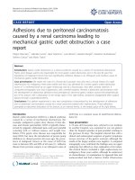
Immunofluorescence microscopy demonstrated a strong
staining of IgG, IgA, C3, IgG κ, and λ in the mesangium
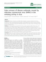
and glomerular basement membrane with equivocal patterns of IgM and C1q (Figure 1A-E). Electron microscopy
revealed diffuse foot process effacement and electronic
dense deposits over the subendothelial, subepithelial,
and paramesangial areas, where focal proliferative lupus
nephritis was suspected (WHO Class III) (Figure 2). These
lupus-like pathology results were inconsistent with his
clinical and autoimmune profile, whereby the diagnosis
of systemic lupus erythematosus cannot be made. His
following treatment regimen for nephritis consisted of
10 mg/d of prednisolone orally, in addition to a 6-month
regimen of 500 mg/m2/d of mycophenolate mofetil. His
proteinuria and haematuria gradually improved. Further
Lim et al. BMC Pediatrics 2013, 13:150
/>
Page 3 of 6
Figure 1 Immunofluorescence microscopy showed strong staining of (A) IgG, (B) IgA, (C) C3, (D) IgG kappa, and (E) IgG lambda over
mesangium and glomerular basement membrane (original magnification, × 400).
analysis of the surface antigens of T cells and B cells
showed considerably low CD19+ (Table 1), where XLA
was one of the potential diagnoses. An analysis of the
BTK gene revealed that the patient and his brother both
exhibited a c.347C > T (p.P116L) mutation inherited from
their mother (Figure 3). After a 2-year follow up, our
patient remains proteinuria-free with normal kidney function and no infections. Written informed consent was
obtained for the subjects included in this study and was
approved by the Kaohsiung Medical University Hospital
Institutional Review Board. The reference sequences for
the BTK gene are NG_009616.1 and NM_000061.2.
Discussion and conclusions
Eighty-five percent of patients with an early onset of
infections, pan-hypogammaglobulinaemia, and markedly
reduced B cells have BTK mutations [12]. Besides BTK,
mutations in the genes involved in pre-B cell receptor
and B cell receptor signalling, including IGHM (μ heavy
chain), IGLL1 (λ5), CD79A (Igα), CD79B (Igβ), BLNK,
and LRRC8A, were reported to block B cell development
and cause a similar clinical phenotype [13-18]. Recently,
a patient with an absence of p85α, resulting in an early
and severe defect in B cell development accompanied
with agammaglobulinaemia, but with minimal findings
in other organ systems was reported [19].
Amino acid P116 is well conserved among different
species. The variant c.347C > T was not observed in the
BTK base ( HGMD Biobase
( />1000 Genomes Project ( or
Figure 2 Electron microscopy revealed diffuse foot process
effacement and electronic dense deposition (arrows) over
subendothelial, subepithelial, and paramesangial areas
(original magnification, × 4,000).
Lim et al. BMC Pediatrics 2013, 13:150
/>
Figure 3 Chromatogram sequence of index patient and
family members.
the NHLBI Exome Sequencing Project (ESP) (http://evs.
gs.washington.edu/EVS/). The direct sequencing of 100
people of Chinese origin from Taiwan was performed and
the c.347C > T variant was not detected (data not shown).
Computer estimations of the function of p.P116L are
labelled “disease-causing” according to MutationTaster
(), and “probably damaging”
(a HumDiv score of 0.998 and a HumVar score of 0.949)
according to PolyPhen-2 ( />pph2/). Further analyses, including the expression of BTK
in mononuclear cells from the patients and the alteration
in the tyrosine 223 phosphorylation in monocytes after
activation through Toll-like receptors [20], should provide the degree of functional defect of this mutation.
Up to 10% of XLA patients have atypical presentation
and exhibit normal or near normal serum immunoglobulin levels or show significant levels of serum IgG
(approximately 800 mg/dL) with variable clinical findings
[21]. They are typically diagnosed at an older age with
less severe phenotypes. The CD19+ B cell remains less
than 1%, but certain “leaky” B-cells mature with higher
immunoglobulin levels in these atypical XLA cases [21].
The mechanism of this “leaky” phenomenon remains
unknown. Mutations that cause atypical XLA are similar
to those that cause classic XLA and include single amino
acid substitutions, splicing defects, premature stop codons,
promoter defects, and gene rearrangement [22]. Normal
levels of IgG accompanied with decreased IgM have been
reported in other cases involving BTK mutation in XLA
[23], but no autoimmune diseases have been reported. The
genotype-phenotype association in XLA has been studied,
but a strong correlation has not been established [24,25].
It is believed that other genetic and environmental factors
might affect the diverse phenotypes of XLA. The delay in
diagnosis is typically because of the significant levels of
Page 4 of 6
serum immunoglobulins regardless of the severity of
clinical phenotypes [26], as in our patient. The low
serum IgM led to the diagnosis of IgM deficiency in our
patient and his brother before further analyses could be
performed. The accurate diagnosis of XLA occasionally
requires BTK mutational analysis that identifies mutations, which may reside in any domain of the gene [27].
Up to 15% of XLA patients may present different
autoimmune manifestations including arthritis, diabetes,
hemolytic anaemia, scleroderma, and alopecia [28,29].
Autoimmunity accompanied by kidney disease is rarely
reported in XLA cases. There are 2 cases in the literature describing the IVIG-related nephropathies in XLA
[10,11]. Yoshino et al. reported a 3-year-old boy with
XLA who suffered from nephrotic syndrome with the
diagnosis of membranoproliferative glomerulonephritis
[10]. In this case, various IVIG preparations did not
improve the proteinuria and haematuria until methylprednisolone pulse therapy was introduced. Endo et al.
described a 23-year-old European man who had considerably mild proteinuria with a diagnosis of XLA [11].
The patient received several IVIG courses and developed
idiopathic membranous glomerulopathy during follow-up.
IVIG treatment was considered the cause of immune
depositions in the kidneys of these 2 patients. Our
patient developed proteinuria and haematuria before
IVIG treatments, and the renal biopsy performed shortly
after IVIG treatment showed diffuse immune complex
depositions. Compared with the patients in the previous
2 cases who received several years of IVIG supplements,
our patient received a relatively small amount of IVIG.
The kidney immune complex depositions either developed quickly after IVIG without additional kidney damage
(regarding proteinuria level), or these depositions actually
occurred before IVIG was administered. Although the
origin of the immune complex depositions remained unknown without a kidney biopsy before IVIG supplements,
our case raised the possibility that nephropathy can be
an entity of autoimmune diseases in XLA.
The goal of IVIG therapy in XLA is to maintain serum
IgG levels at 500–800 mg/dL and prevent recurrent bacterial infections that could be life threatening [30,31].
However, the treatment of autoimmunity in XLA is less
documented. In many cases, increasing the IVIG dosage
may ameliorate the autoimmune phenomenon [32]. The
nephropathy in our patient showed a resistance to steroids, chlorambucil, and IVIG treatment (although the
dose is less than the typical dose for autoimmune disease), but it responded well to mycophenolate and the
patient remained disease free.
Selective IgM deficiency is a rare form of dysgammaglobulinaemia with an incidence of less than 0.003% in the
general population [33,34]. The clinical and laboratory
criteria are poorly defined, either by an IgM level less
Lim et al. BMC Pediatrics 2013, 13:150
/>
than 2 standard deviations below standard, less than
10% of age-adjusted normal controls, or absolute levels
less than 10–20 mg/dL [35]. Immunoglobulin M deficiency can be presented as a primary or secondary disease. Secondary diseases are more commonly seen and
are often reported to be associated with bacterial and
viral infections, autoimmune diseases, and malignancies
[36,37]. Currently, no molecular defect has been determined to be responsible for IgM deficiency, and IVIG
may be instituted in cases of recurrent, debilitating, or
life-threatening infection, and/or in patients with concomitant functional IgG deficiencies [36]. Two patients
in an IgM deficiency study exhibited equal to or less than
2% CD19+ B cells [38], indicating that XLA should be a
differential diagnosis in familial selective IgM deficiency
despite normal IgG levels, as in our patient.
In summary, we reported a family with 2 siblings whose
immunoglobulin profiles, except their low CD19+ B cell
levels, were similar to selective IgM immunodeficiency.
A genetic analysis of this family revealed a novel BTK
variant, p.P116L, in both siblings, which makes XLA the
probable diagnosis. We therefore suggest that an analysis of the B-lymphocyte surface markers and BTK gene
should be performed in familial patients diagnosed with
selective IgM deficiency, which can be an atypical presentation of XLA. Furthermore, we reported another rare
glomerulonephritis case and demonstrated the successful
treatment of nephropathy in XLA-related autoimmunity
by using mycophenolate.
Consent
Written informed consent was obtained from the mother
of the patient for publication of this case report and any
accompanying images. A copy of the written consent is
available for review by the Series Editor of this journal.
Abbreviations
XLA: X-linked agammaglobulinemia; BTK: Bruton’s tyrosine kinase, PH,
Pleckstrin homology; TH: Tec homology; SH2: Src homology 2, SH3, Src
homology 3; IVIG: Intravenous immunoglobulin.
Competing interests
The authors declare that they have no competing interests.
Authors’ contribution
DYH and LML designed and performed research, analysed and interpreted
data, and wrote the manuscript; HCC analysed and interpreted data, wrote the
manuscript. JMC, WCC and IFW analysed and interpreted data. All authors read
and approved the final manuscript.
Acknowledgments
We thank the family for their willingness to participate in this study.
Author details
1
Division of Nephrology, Department of Internal Medicine, Kaohsiung Medical
University Hospital, 100 Tze-You First Road, Kaohsiung City 807, Taiwan.
2
Department of Internal Medicine, Kaohsiung Municipal Hsiao-Kang Hospital,
Kaohsiung, Taiwan. 3Department of Pediatrics, Kaohsiung Medical University
Hospital, Kaohsiung Medical University, Kaohsiung, Taiwan. 4Department of
Clinical Pharmacy, School of Pharmacy Taipei Medical University, Taipei, Taiwan.
Page 5 of 6
5
Master Program for Clinical Pharmacogenomics and Pharmacoproteomics,
School of Pharmacy, Taipei Medical University, Taipei, Taiwan. 6Faculty of Renal
Care, College of Medicine, Kaohsiung Medical University, Kaohsiung, Taiwan.
Received: 18 March 2013 Accepted: 24 September 2013
Published: 27 September 2013
References
1. Conley ME, Dobbs AK, Farmer DM, Kilic S, Paris K, Grigoriadou S,
Coustan-Smith E, Howard V, Campana D: Primary B cell immunodeficiencies:
comparisons and contrasts. Annu Rev Immunol 2009, 27:199–227.
2. Bruton OC: Agammaglobulinemia. Pediatrics 1952, 9(6):722–728.
3. Tsukada S, Saffran DC, Rawlings DJ, Parolini O, Allen RC, Klisak I, Sparkes RS,
Kubagawa H, Mohandas T, Quan S, et al: Deficient expression of a B cell
cytoplasmic tyrosine kinase in human X-linked agammaglobulinemia.
Cell 1993, 72(2):279–290.
4. Kwan SP, Kunkel L, Bruns G, Wedgwood RJ, Latt S, Rosen FS: Mapping of
the X-linked agammaglobulinemia locus by use of restriction
fragment-length polymorphism. J Clin Invest 1986, 77(2):649–652.
5. Vetrie D, Vorechovsky I, Sideras P, Holland J, Davies A, Flinter F,
Hammarstrom L, Kinnon C, Levinsky R, Bobrow M, et al: The gene involved
in X-linked agammaglobulinaemia is a member of the src family of
protein-tyrosine kinases. Nature 1993, 361(6409):226–233.
6. Nonoyama S, Tsukada S, Yamadori T, Miyawaki T, Jin YZ, Watanabe C,
Morio T, Yata J, Ochs HD: Functional analysis of peripheral blood B cells
in patients with X-linked agammaglobulinemia. J Immunol 1998,
161(8):3925–3929.
7. Shinomiya N, Kanegane H, Watanabe A, Yamaguchi Y, Futatani T, Miyawaki T:
Point mutation in intron 11 of Bruton’s tyrosine kinase in atypical X-linked
agammaglobulinemia. Pediatr Int 2000, 42(6):689–692.
8. Conley ME, Howard VC, et al: X-linked agammaglobulinemia. In
GeneReviews. Seattle: University of Washington, Seattle; 1993.
9. Ochs HD, Smith CI: X-linked agammaglobulinemia. A clinical and
molecular analysis. Medicine (Baltimore) 1996, 75(6):287–299.
10. Yoshino A, Honda M, Kanegane H, Obata K, Matsukura H, Sakazume S,
Katada Y, Miyawaki T, Ueda Y, Nagai T: Membranoproliferative
glomerulonephritis in a patient with X-linked agammaglobulinemia.
Pediatr Nephrol 2006, 21(1):36–38.
11. Endo LM, Giannobile JV, Dobbs AK, Foote JB, Szymanska E, Warnock DG,
Cook WJ, Conley ME, Schroeder HW: Membranous glomerulopathy in an
adult patient with X-linked agammaglobulinemia receiving intravenous
gammaglobulin. J Investig Allergol Clin Immunol 2011, 21(5):405–409.
12. Conley ME, Mathias D, Treadaway J, Minegishi Y, Rohrer J: Mutations in btk
in patients with presumed X-linked agammaglobulinemia. Am J Hum
Genet 1998, 62(5):1034–1043.
13. Yel L, Minegishi Y, Coustan-Smith E, Buckley RH, Trubel H, Pachman LM,
Kitchingman GR, Campana D, Rohrer J, Conley ME: Mutations in the mu
heavy-chain gene in patients with agammaglobulinemia. N Engl J Med
1996, 335(20):1486–1493.
14. Minegishi Y, Coustan-Smith E, Wang YH, Cooper MD, Campana D, Conley ME:
Mutations in the human lambda5/14.1 gene result in B cell deficiency and
agammaglobulinemia. J Exp Med 1998, 187(1):71–77.
15. Minegishi Y, Coustan-Smith E, Rapalus L, Ersoy F, Campana D, Conley ME:
Mutations in Igalpha (CD79a) result in a complete block in B-cell
development. J Clin Invest 1999, 104(8):1115–1121.
16. Minegishi Y, Rohrer J, Coustan-Smith E, Lederman HM, Pappu R, Campana D,
Chan AC, Conley ME: An essential role for BLNK in human B cell
development. Science 1999, 286(5446):1954–1957.
17. Sawada A, Takihara Y, Kim JY, Matsuda-Hashii Y, Tokimasa S, Fujisaki H,
Kubota K, Endo H, Onodera T, Ohta H, et al: A congenital mutation of the
novel gene LRRC8 causes agammaglobulinemia in humans. J Clin Invest
2003, 112(11):1707–1713.
18. Dobbs AK, Yang T, Farmer D, Kager L, Parolini O, Conley ME: Cutting edge:
a hypomorphic mutation in Igbeta (CD79b) in a patient with
immunodeficiency and a leaky defect in B cell development.
J Immunol 2007, 179(4):2055–2059.
19. Conley ME, Dobbs AK, Quintana AM, Bosompem A, Wang YD,
Coustan-Smith E, Smith AM, Perez EE, Murray PJ: Agammaglobulinemia
and absent B lineage cells in a patient lacking the p85alpha subunit
of PI3K. J Exp Med 2012, 209(3):463–470.
Lim et al. BMC Pediatrics 2013, 13:150
/>
20. Vargas-Hernandez A, Lopez-Herrera G, Maravillas-Montero JL, Vences-Catalan F,
Mogica-Martinez D, Rojo-Dominguez A, Espinosa-Rosales FJ, Santos-Argumedo L:
Consequences of two naturally occurring missense mutations in the structure
and function of Bruton agammaglobulinemia tyrosine kinase. IUBMB life 2012,
64(4):346–353.
21. Plebani A, Soresina A, Rondelli R, Amato GM, Azzari C, Cardinale F, Cazzola G,
Consolini R, De Mattia D, Dell’Erba G, et al: Clinical, immunological, and
molecular analysis in a large cohort of patients with X-linked
agammaglobulinemia: an Italian multicenter study. Clin Immunol 2002,
104(3):221–230.
22. Stewart DM, Tian L, Nelson DL: A case of X-linked agammaglobulinemia
diagnosed in adulthood. Clin Immunol 2001, 99(1):94–99.
23. Basile N, Danielian S, Oleastro M, Rosenzweig S, Prieto E, Rossi J, Roy A,
Zelazko M: Clinical and molecular analysis of 49 patients with X-linked
agammaglobulinemia from a single center in Argentina. J Clin Immunol
2009, 29(1):123–129.
24. Conley ME, Howard V: Clinical findings leading to the diagnosis of X-linked
agammaglobulinemia. J Pediatr 2002, 141(4):566–571.
25. Lopez-Granados E, de PerezDiego R, Ferreira Cerdan A, Fontan Casariego G,
Garcia Rodriguez MC: A genotype-phenotype correlation study in a group
of 54 patients with X-linked agammaglobulinemia. J Allergy Clin Immunol
2005, 116(3):690–697.
26. Maekawa K, Yamada M, Okura Y, Sato Y, Yamada Y, Kawamura N, Ariga T:
X-linked agammaglobulinemia in a 10-year-old boy with a novel
non-invariant splice-site mutation in Btk gene. Blood Cells Mol Dis 2010,
44(4):300–304.
27. Väliaho J, Smith CI, Vihinen M: BTKbase: the mutation database for X-linked
agammaglobulinemia. Hum Mutat 2006, 27(12):1209–1217.
28. Pessach IM: The relationship of x-linked primary immune deficiencies
and autoimmunity. Curr Allergy Asthma Rep 2010, 10(5):311–319.
29. Pessach IM, Notarangelo LD: X-linked primary immunodeficiencies as a
bridge to better understanding X-chromosome related autoimmunity.
J Autoimmun 2009, 33(1):17–24.
30. Eijkhout HW, van Der Meer JW, Kallenberg CG, Weening RS, van Dissel JT,
Sanders LA, Strengers PF, Nienhuis H, Schellekens PT: The effect of two
different dosages of intravenous immunoglobulin on the incidence of
recurrent infections in patients with primary hypogammaglobulinemia.
A randomized, double-blind, multicenter crossover trial. Ann Intern Med
2001, 135(3):165–174.
31. Moise A, Nedelcu FD, Toader MA, Sora SM, Tica A, Ferastraoaru DE,
Constantinescu I: Primary immunodeficiencies of the B lymphocyte. J Med
Life 2010, 3(1):60–63.
32. Etzioni A: Immune deficiency and autoimmunity. Autoimmun Rev 2003,
2(6):364–369.
33. Ohno T, Inaba M, Kuribayashi K, Masuda T, Kanoh T, Uchino H: Selective IgM
deficiency in adults: phenotypically and functionally altered profiles of
peripheral blood lymphocytes. Clin Exp Immunol 1987, 68(3):630–637.
34. Guill MF, Brown DA, Ochs HD, Pyun KH, Moffitt JE: IgM deficiency: clinical
spectrum and immunologic assessment. Ann Allergy 1989, 62(6):547–552.
35. Stiehm E, Ochs HD, Wikelstein JA: Immunologic Disorders in Infants and
children. 5th edition. PHiladelphia, Pa, USA: Elsevier Saunders; 2004.
36. Goldstein MF, Goldstein AL, Dunsky EH, Dvorin DJ, Belecanech GA, Shamir K:
Pediatric selective IgM immunodeficiency. Clin Dev Immunol 2008,
2008:624850.
37. Antar M, Lamarche J, Peguero A, Reiss A, Cole S: A case of selective
immunoglobulin M deficiency and autoimmune glomerulonephritis.
Clin Exp Nephrol 2008, 12(4):300–304.
38. Yel L, Ramanuja S, Gupta S: Clinical and immunological features in IgM
deficiency. Int Arch Allergy Immunol 2009, 150(3):291–298.
doi:10.1186/1471-2431-13-150
Cite this article as: Lim et al.: Atypical X-linked agammaglobulinaemia
caused by a novel BTK mutation in a selective immunoglobulin M
deficiency patient. BMC Pediatrics 2013 13:150.
Page 6 of 6
Submit your next manuscript to BioMed Central
and take full advantage of:
• Convenient online submission
• Thorough peer review
• No space constraints or color figure charges
• Immediate publication on acceptance
• Inclusion in PubMed, CAS, Scopus and Google Scholar
• Research which is freely available for redistribution
Submit your manuscript at
www.biomedcentral.com/submit