Báo cáo y học: "Optimization of 5-fluorouracil solid-lipid nanoparticles: a preliminary study to treat colon cancer"
Bạn đang xem bản rút gọn của tài liệu. Xem và tải ngay bản đầy đủ của tài liệu tại đây (1.62 MB, 11 trang )
Int. J. Med. Sci. 2010, 7
398
I
I
n
n
t
t
e
e
r
r
n
n
a
a
t
t
i
i
o
o
n
n
a
a
l
l
J
J
o
o
u
u
r
r
n
n
a
a
l
l
o
o
f
f
M
M
e
e
d
d
i
i
c
c
a
a
l
l
S
S
c
c
i
i
e
e
n
n
c
c
e
e
s
s
2010; 7(6):398-408
© Ivyspring International Publisher. All rights reserved
Research Paper
Optimization of 5-fluorouracil solid-lipid nanoparticles: a preliminary
study to treat colon cancer
Alaa Eldeen B. Yassin
1,2
, Md. Khalid Anwer
3
, Hammam A. Mowafy
1
, Ibrahim M. El-Bagory
1
, Mohsen A.
Bayomi
1,2
and Ibrahim A. Alsarra
1,2,
1. Department of Pharmaceutics, College of Pharmacy, King Saud University, P.O. Box 2457, Riyadh 11451, Saudi Arabia
2. Center of Excellence in Biotechnology Research, King Saud University, P.O. Box 2460, Riyadh 11451, Saudi Arabia
3. College of Pharmacy, Al-Kharj University, Al-Kharj, Saudi Arabia
Corresponding author: Professor Ibrahim A. Alsarra, Phone: +(966)-1-4677504, Fax: +(966)-1-4676363, E-mail: ialsar-
Received: 2010.07.10; Accepted: 2010.11.16; Published: 2010.11.22
Abstract
Solid lipid nanoparticle (SLNs) formulae were utilized for the release of 5-fluorouracil (5-FU)
i n s i d e t h e c o l o n i c m e d i u m f o r l o c a l t r e a t m e n t o f c o l o n c a n c e r . S L N s w e r e p r e p a r e d b y d o u b l e
emulsion-solvent evaporation technique (w/o/w) using triglyceride esters, Dynasan 114 or
Dynasan™ 118 along with soyalecithin as the lipid parts. Different formulation parameters;
including type of Dynasan, soyalicithin:Dynasan ratio, drug:total lipid ratio, and polyvinyl al-
cohol (PVA) concentration were studied with respect to particle size and drug entrapment
e f f i c i e n c y . R e s u l t s s h o w e d t h a t f o r m u l a 8 ( F 8 ) w i t h c o m p o s i t i o n o f 2 0 % 5 -FU, 27% Dynasan™
114, and 53% soyalithicin and F14 (20% 5-FU, 27% Dynasan™ 118, and 53% soyalithicin),
w h i c h w e r e s t a b i l i z e d b y 0 . 5 % P V A , a s w e l l a s F 1 0 w i t h s i m i l a r composition as F8 but stabilized
b y 2 % P V A w e r e c o n s i d e r e d t h e o p t i m u m f o r m u l a e a s t h e y c o m b i n e d s m a l l p a r t i c l e s i z e s a n d
relatively high encapsulation efficiencies. F8 had a particle size of 402.5 nm ± 34.5 with a
polydispersity value of 0.005 and an encap s u l a t i o n e f f i c i e n c y o f 5 1 % , F 1 0 h a d a 6 1 7 . 3 n m ± 5 4 . 3
particle size with 0.005 polydispersity value and 49.1% encapsulation efficiency, whereas
formula F14 showed a particle size of 343 nm ± 29 with 0.005 polydispersity, and an en-
capsulation efficiency of 5 9 . 0 9 % . D S C a n d F T I R r e s u l t s s u g g e s t e d t h e e x i s t e n c e o f t h e l i p i d s i n
the solid crystalline state. Incomplete biphasic prolonged release profile of the drug from The
t h r e e f o r m u l a e w a s o b s e r v e d i n p h o s p h a t e b u f f e r p H 6 . 8 a s w e l l a s s i m u l a t e d c o l o n i c m e d i u m
containing rat caecal contents. A burst release with magnitudes of 26%, 32% and 28.8% cu-
mulative drug released were noticed in the first hour samples incubated in phosphate buffer
p H 6 . 8 f o r b o t h F 8 , F 1 0 a n d F 1 4 , r e s p e c t i v e l y , f o l l o w e d b y a s l o w r e l e a s e profile reaching 50%,
46.3% and 52% after 48 hours.
Key words: Solid lipid nanoparticles, double emulsion, colon cancer; Dynasan, 5-fluorouracil, po-
lyvinyl alcohol
Introduction
Colorectal cancer is the second leading cause of
cancer-related death for men and women worldwide.
Each year there are about one million new cases of
colorectal cancer. Its incidence has increased over the
last 25 years [1]. Colorectal cancer is a disease that is
manifested by the formation of adenomatous polyps
and malignant cells in the colon [2]. These abnormal
cells creating tumors are characterized by unregulated
replication and the capability of spreading to other
sites [2]. Colon-specific delivery systems would allow
Int. J. Med. Sci. 2010, 7
399
for the local delivery of a high concentration of active
agents in the colon to improve pharmacotherapy and
reduce its potential systemic toxicity and side effects
[3]. The early detection, diagnosis, and the use of
more effective and less toxic systems would tre-
mendously improve the efficacy of therapy [1-3].
Nanotechnology has become a rapidly growing
field with potential applications in health and drug
therapy [4-6]. Nanoparticles have extraordinary
physical and chemical properties resulting from the
nanosize effect [7]. They play an important role in
cancer therapy to detect or to deliver the drug to the
cancerous cell without attacking the normal cells and
have good ability to form a complex with a variety of
drugs through chemical bindings [8]. The phenome-
non of nanoparticles has been applied in a number of
research approaches for the improvement of cancer
therapeutics including liposomes [9], polymeric na-
noparticles [10], dendrimers [11], and metallic nano-
particles [12]. Generally, lipid-based nanoparticles are
considered the least toxic among all the other nano-
particles for in vivo applications, in addition to the
significant progress that has been achieved in the de-
livery of DNA/RNA using lipid-based na-
no-assemblies [13].
Solid lipid nanparticles (SLNs) have been pro-
posed as an alternative drug carrier system to other
novel delivery approaches such as emulsions, lipo-
somes, and polymeric nanoparticles due to various
advantages, including feasibility of incorporation of
lipophilic and hydrophilic drugs, improved physical
stability, low cost, ease of scale-up, and manufactur-
ing [14,15]. In contrast to emulsions and liposomes,
the particle matrix of SLNs is composed of solid li-
pids. The majority of lipids commonly used are trig-
lyceride esters of hydrogenated fatty acids. Hydro-
genated cottonseed oil (Lubritab™ or Sterotex™),
hydrogenated palm oil (Dynasan™ P60 or Softisan™
154), hydrogenated castor oil (Cutina™ HR), and hy-
drogenated soybean oil (Sterotex™ HM, or Lipo™)
are typical examples [16].
General features of SLNs are their composition
of physiological compounds, possible routes of ad-
ministration by intravenous, oral and topical, large
scale production by high pressure homogenization,
and the relatively low costs of excipients [16-18]. A
number of studies have recently been published about
their production [19], physicochemical characteriza-
tion of particles [20], and drug incorporation and re-
lease [21]. SLNs carrying anticancer drugs such as
doxorubicin and paclitaxel had previously been d e-
v e l o p e d a n d t h e a n t i p r o l i f i r a t i v e e f f e c t o f S L N s v e r s u s
conventional drug formulations was also evaluated
on HT-29 cells. In vitro cytotoxicity of SLNs carrying
anticancer drugs was higher than that of conventional
drug formulations [22].
5-FU is an anticancer agent and the most widely
used drug in the treatment of malignancies arising
from breast, gastrointestinal tract, head, and neck re-
gions of the body for several decades [23]. It is consi-
dered the major chemotherapeutic agent with clinical
activity against colorectal cancer [24-27]. Localizing
5-FU directly to the colon is expected to reduce sys-
temic side effects allowing more effective and safe
therapy with higher tumor diffusivity [28]. 5-F U wa s
used in this study as a model drug. Improvement of
tissue distribution and targeting of drugs by using
SLNs have been reported for some drugs including
anticancers [29].
The aim of this work was to prepare and cha-
racterize 5-FU solid lipid nanparticles. A double
emulsion-solvent evaporation (w/o/w) method was
chosen and was optimized to obtain SLNs with low
particle size and a relatively high encapsulation effi-
ciency as well as a consistent release profile in simu-
lated colonic medium.
Materials and Methods
5-FU was obtained from Sigma-Aldrich Chemi-
cal Company (St. Louis, MO, USA). Dynasan 114
and 118 were acquired from Sasol Germany GmbH
(Witten, Germany). Soya lecithin 30% was purchased
from AppliChem (Darmstadt, Germany). Polyvinyl
alcohol (PVA), M.W. 22,000 was obtained from BDH
Laboratories (Poole, England). All other reagents and
chemicals were of analytical grade.
Preparation of solid lipid nanoparticles
Weighed amounts of soyalecithin and Dynasan
were dissolved in 10 ml of dichloromethane. Certain
am oun ts of 5-FU was dissolved in 4 ml of 2.5% w / v
lactose monohydrate in distilled water to avoid par-
ticle aggregation after freeze drying of SLNs. Both
lipid and aqueous solutions were mixed and emulsi-
fied by probe-sonication (Bandelin, Berlin, Germany)
for an optimized period of time (3×1 minutes) at 40%
voltage efficiency in an ice bath. The formed w/o
primary emulsion was immediately poured onto 40
ml aqueous solution of PVA continuously stirred at
1000 rpm over ice bath for 30 minutes. Then, the
temperature was increased gradually (15-18 °C) dur-
ing stirring and subjected to solvent evaporation for
another 30 minutes. Lipid nanoparticles were sepa-
rated from bulk aqueous phase by centrifugation at
14000 rpm for 30 min (Hettich, MIKRO-120, Tuttlin-
gen, Germany). After subsequent washing with cold
distilled water, the residue was dispersed in tris-HCl
b u f f e r p H 7 a n d f r e e z e -dried (Martin Christ Alpha-1-4
Int. J. Med. Sci. 2010, 7
400
LD freeze-drier, Osterode, Germany). Table 1,
represents the exact composition of each of the pre-
pared formula. The effect of different formulation
parameters, such as type of Dynasan, soyalici-
t h i n : D y n a s a n r a t i o , d r u g : t o t a l l i p i d r a t i o , a n d t h e P V A
concentration on the particle size and drug entrap-
ment efficiency were investigated.
Table 1. Composition of each of the prepared formulae
Formulations % w/w % PVA
Soyalicithin Dynasan 5-FU
F1 26 52
a
22 1
F2 52 26 22 1
F3 78 0 22 1
F4 52 27 20 1
F5 26 52 22 0.5
F6 52 26 22 0.5
F7 39 39 22 0.5
F8 53 27 20 0.5
F9 26 52 22 2
F10 53 27 20 2
F11 53 27 20 1
F12 53 27 20 3
F13 53 27
b
20 0.5
F14 53 27 20 0.5
F15 53 27 20 0.5
a
D114 is Dynasan™ 114
b
D118 is Dynasan™ 118
Measurement of particle size
The mean size and polydispersity index of the
size distribution for each formula were determined by
photon correlation spectroscopy using 90 Plus particle
size analyzer, Brookhaven Instruments Corporation
(Holtsville, New York, USA). The SLNs dispersions
were diluted 1:1000 with distilled water. Analysis was
performed at 25 °C with an angle of detection of 90°.
Each reported value is the average of three measure-
ments. The polydispersity index measures the size
distribution of the nanoparticles population.
Differential scanning calorimetry
The thermal behavior of some selected SLN
formulae was investigated by differential scanning
calorimetry (DSC) using a Shimadzu DSC-60 (Shi-
m a d z u C o r p o r a t i o n , T o k y o , J a p a n ) . S a m p l e s o f 4 -7 m g
were weighed and a heating rate of 10 ºC/min was
employed in the range of 25 ºC to 350 ºC.
Fourier transform infrared spectroscopy (FTIR)
The FTIR spectra of samples were recorded on
the on a PerkinElmer spectrum BX FTIR (PerkinEl-
mer, Waltham, MA, USA) using the potassium bro-
mide (KBr) disc technique. Samples equivalent to 2
mg of 5-FU were mixed with potassium bromide
(about 100 mg) in a clean glass pestle and mortar and
were compressed to obtain a pellet. Baseline was cor-
rected and the samples were scanned against a blank
KBr pellet background at a wave number ranging
from 4000-400 cm
-1
with a resolution of 1.0 cm
-1
.
Determination of % entrapment efficiency and
drug loading
The percentage drug entrapment efficiency
(%EE) and % drug loading (%DL) of 5-FU in SLNs
formulations were determined by centrifugation of
the colloidal samples at 14000 rpm at 25 °C for 30 min.
The non-entrapped 5-FU amounts in the supernatant
obtained after centrifugation of nanoparticles were
determined by UV spectroscopy at 266 nm.
T h e % EE of 5-FU entrapped within nanoparticles
was calculated by dividing the difference between the
total amount used (W
total 5-FU
) and the free amount
presented in the aqueous phase of supernatant (W
fre e
5-FU
) by the total amount used of 5-FU. The %DL w a s
obtained by dividing that difference by the total
weight of SLNs according to the following formulae:
Scanning el e c t r o n microscopic (SEM) analysis
The morphology characteristics of the prepared
SLNs were examined under the scanning electron
microscope (JSM-6360LV Scanning Microscope; Jeol,
Tokyo, Japan). Before microscopy, SLNs produced
from F14 were suspended in a phosphate buffer (pH
7) by vortex for 1 minute, and then one drop was
spread on a small clean slide cover and left to dry
overnight in a desicator. In the next day, they were
mounted on carbon tape and sputter-coated using a
thin gold palladium layer under an argon atmosphere
using a gold sputter module in a high-vacuum eva-
porator (JFC-1100 fine coat ion sputter; Jeol, Tokyo,
Japan). The coated samples were then scanned and
photomicrographs were taken at an acceleration vol-
tage of 20 kV.
In vitro release study
Certain weights from each of the selected for-
m u l a e q u i v a l e n t t o 1 m g 5 -FU were immersed in 10 ml
of phosphate buffer pH 6.8 in biological shaker at 37
°C and 80 rpm speed. Aliquots of 1 ml were with-
drawn at certain time intervals and replaced with an
Int. J. Med. Sci. 2010, 7
401
equal volume of fresh buffer. After centrifugation, the
amount of drug released was determined spectro-
photometrically by measuring the absorbance of each
aliquot supernatant at 266 nm.
Drug release in medium containing rat caecal
contents
The drug release was assessed using a procedure
introduced by Yassin et al. (2010) [30]. Briefly, male
rats of mixed breeds weighing 200-300 g were used
throughout this study. The rats were euthanized
while under ether anesthesia and the caecum was
exteriorized, legated at the two-ends and was
cut-l o o s e . T h e c o n t e n t s o f t h e f o r m e d c a e c a l b a g s w e r e
individually weighed, pooled, and suspended in a
chilled phosphate buffer saline (pH 6.8) to give a final
dilution of 3% (w/v). Weights equivalent to 1 mg
5-FU from F8, F10 and F14 were incubated in 20 ml of
the suspension at 37 °C ± 0.5 and shaken at 80 rpm
using a thermostatic shaking water bath. The experi-
ments were performed under nitrogen atmosphere to
simulate anaerobic conditions. Aliquots (1 ml) sam -
ples were withdrawn, filtered, diluted, and analyzed
usi n g H PLC at specified time intervals for 24 h. Re-
placement of samples were made by the medium
stored at the same temperature.
Assay method
A simple and sensitive stability-indicating HPLC
method with a UV detection using thymine as an in-
ternal standard was adopted [31]. The method was
utilized for the assessment of stability of 5-FU in rat
caecal content as simulated colon medium under
anaerobic conditions. Briefly, the HPLC system con-
sisted of a Waters Model 1515 HPLC pump, a Waters
autosampler, Model 717 plus (Waters Inc., Bedford,
MA, USA), a Waters 2487 dual absorbance UV detec-
tor (Waters Inc., Bedford, MA, USA) governed by a
microcomputer running Empower
®
software (version
1154). The detector wavelength was set at 260 nm.
Separation was achieved by isocratic elution with a
mobile phase of 40 mM phosphate buffer adjusted to
pH 7.0 using 10% w/v potassium hydroxide, deli-
vered at a flow-rate of 1.0 ml/min at ambient tem-
perature through a C
18
analytical, µ-Bondapack col-
umn (150 mm length × 4. 6 mm i.d., 10 µm part ic le
size).
Statistical analysis
The significance of difference among the differ-
ent formulae was tested by applying the one way
analysis of variance ANOVA test, while Paired t-test
was employed to determine the difference between
any two formulations using a statistical software
package (Statistical Analysis System, SAS Institute,
Inc., Cary, NC, USA). Differences between related
parameters were considered statistically significant
for p-value equal to or less than 0.05.
Results and Discussion
Particle size, entrapment efficiency and drug
loading
Table 2 presents the mean particle size, poly-
dispersity and entrapment efficiency for all the pre-
pared formulae. The polydispersity index is a meas-
ure of the width of the dispersion of particles. Narrow
dispersions comprise polydispersity index values
b e t w e e n 0 . 1 a n d 0 . 2 . H e n c e , a c c o r d i n g t o T a b l e 2 , m o s t
o f t h e d i s p e r s i o n s c a n b e labeled as a narrow disperse;
except F2 and F3 polydispersity index which was
slightly higher. Generally, the particle size s h o w e d a
wide range of variability ranging from 258 to 2743.7
nm depending on the lipid composition, drug to lipid
ratio, and the concentration of the stabilizer (PVA). It
is clear that a ratio of 2:1 soyalicithin: Dynasan and a
high lipid ratio in the nanoparticles are important
factors for getting smaller particle size. The PVA
concentration (0.5%) was found to be optimum for
both types of Dynasan.
Table 2. Entrapment efficiency, particle size and polydis-
persity for each of the prepared formulae
Formulation % EE %DL Particle size
(nm)
Polydispersity
F1 24.75 17.51
794.2 ± 113 0.046
F2 28.70 16.75
712.9 ± 138 0.297
F3 6.32 20.90 258 ± 49 0.277
F4 45.46 12.13 606.1 ± 63 0.006
F5 69.09 8.03 943.2 ± 97 0.041
F6 34.92 15.51 766.3 ± 104 0.064
F7 32.94 15.91 924.8 ± 68 0.005
F8 51.08 10.91 402.5 ± 34.5 0.005
F9 35.91 15.31 1216.7 ± 107 0.005
F10 49.10 11.29 617.3 ± 54.3 0.005
F11 26.17 15.59 651.6 ± 51.8 0.005
F12 53.03 10.51 2743.7 ± 183 0.005
F13 40.00 13.04 461.9 ± 52.1 0.130
F14 59.09 9.29 343.0 ± 29 0.005
F15 35.52 13.89 471.3 ± 38 0.005
The lipid core material and drug composition
were found to affect the extent of 5-FU entrapment in
SLNs. Loading efficiency of 5-FU ranging from
6.32-69.09% were also observed at different lipids and
drug ratios. As shown in Table 2, formulae F5, F8 and
F10 exhibited the highest entrapment efficiencies
among all the prepared formulae containing Dynasan
114 with values equal to 69.09%, 51.08% and 49.10%,
respectively. The Dynasan 118 containing formulae
F14 and F15 had entrapment efficiency values of
59.09% and 35.52%, respectively. Increasing the
Int. J. Med. Sci. 2010, 7
402
drug:total lipid ratio from 1:4 (F15) to 1:8 (F14 formu-
la) was found to have a positive effect on both particle
size and entrapment efficiency (i.e. smaller particle
size and higher entrapment efficiency).
The type of Dynasan showed a little effect on the
size and entrapment efficiency; however, comparing
F8 with both F13 and F15 revealed that Dynasan 114
was superior to Dynasan 118 when used with the
same composition. Briefly, F8 composed of drug to
lipid at 1:4 ratio and soyalithicin to Dynasan114 at 2:1
ratio (stabilized by 0.5% PVA) is considered the op-
timum formula for the Dynasan 114 group with re-
gards to the relatively small particle size (402.5 nm),
polydispersity value of (0.005), and high encapsula-
tion efficiency 51%. For the Dynasan 118 group, the
b e s t f o r m u l a w a s F 1 4 ( c o m p o s e d o f d r u g t o l i p i d a t 1 : 8
ratio and soyalithicin to Dynasan118 at 2:1 ratio),
which has particle size of 343 nm with 0.005 polydis-
persity and an encapsulation efficiency of 59.09%.
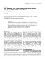
Particle morphology
SEM images of SLNs F14 were presented in Fig.
1 ( A , B , C ) . I t w a s c l e a r f r o m i m a g e A t h a t 5-FU loaded
SLNs were spherical in shape with rough or irregular
surfaces with the presence of some particle aggre-
gates. The presence of aggregates might be attributed
to a short redispersion time after centrifugation and
drying at room temperature. The sizes observed from
SEM micrographs were slightly higher than those
obtained from particle size analyzer. Micrographs B
and C showed irregular surfaces of single particles
und e r hi g h m a gni f ica t i ons .
Differential scanning Calorimetry (DSC)
Thermal behavior of the pure drug, Dynasan 114
and 118 compared with the thermograms of different
l y o p h i l i z e d S L N s f o r m u l a e i n t h e r a n g e o f 2 5 t o 3 5 0 º C
is shown in Fig. 2. The thermogram of the pure 5-FU
showed a sharp melting endotherm at approximately
282 ºC followed by decomposition, which was in
agreement with those reported previously [32, 33]. A
slight shift to the melting peaks of 5-FU to 240 °C was
only observed in the case of F9 and F15. Same obser-
vation was reported with 5-F U i n P L G A m i c r o s p h e r e s
[34]. The pure Dynasan 114 thermogram showed a
characteristic sharp peak at 58 ºC corresponding to the
melting of the lipid. This peak appeared in all ther-
mograms of the prepared SLNs formulae confirming
the solid crystalline state of the lipid inside the pre-
pared formulae. No change in the shape of the Dyna-
san peak was observed in the SLNs formulae [35-36].
Fig. 1. Scanning electron microscopy photomicrographs
for F14 SLNs: A, a field containing different particle sizes
using 3,300 X magnification power, B, a field showing two
single particles using 45,000 X magnification power, and C, a
field containing single particle using 50,000 X magnification
power.