Nghiên cứu chế tạo hệ vi cảm biến điện hoá trên cơ sở polyme dẫn biến tính để ứng dụng trong y sinh và môi trường tt tiếng anh
Bạn đang xem bản rút gọn của tài liệu. Xem và tải ngay bản đầy đủ của tài liệu tại đây (618.05 KB, 27 trang )
MINISTRY OF
VIETNAM ACADEMY OF
EDUCATION AND TRAINING
SCIENCE AND TECHNOLOGY
GRADATE UNIVERSIY OF SCIENCE AND TECHNOLOGY
Nguyen Hai Binh
RESEARCH ON FABRICATION OF THE ELECTROCHEMICAL
MIOCROSENSOR BASED ON MODIFIED CONDUCTIVE
POLYMER FOR APPLICATION IN BIOMEDICAL AND
ENVIRONMENTAL FIELDS
Major: Electronic materials
Code: 9.44.01.23
SUMMARY OF DOCTORAL THESIS IN MATERIALS SCIENCE
Ha Noi - 2018
This thesis was done at:
Laboratory of Biomedical Nanomaterials, Institute of Materials and Sciene,
Vietnam Academy of Science and Technology.
Supervisor: Prof. Ph.D. Tran Dai Lam
Reviewer 1: .....................................................
Reviewer 2: .....................................................
Reviewer 3: .....................................................
The dissertation will be defended at Graduate University of Science and Technology, 18
Hoang Quoc Viet street, Hanoi.
Time: .............,.............., 2018
This thesis could be found at National Library of Vietnam, Library of Graduate
University of Science and Technology, Library of Institute of Materials and Science,
Library of Vietnam Academy of Science and Technology.
INTRODUCTION
Currently, biosensors are considered as a potential device for application in many
fields such as biology, pharmaceuticals, agriculture, food safety and hygiene,
enviromental protection and industrial safety, etc... Biosensor is a device that uses
specific biological components in combination with a signal converter to detect,
measure or analyze chemical agents.
Electrochemical microsensor has a simple structure, easy to design and develop
structure, easy to integrate with micro-elements of the system, bactch fabrication. The
working electrodes, counter electrodes and reference electrodes are integrated on one
chip, which reduces the volume and mass of the sample to be analyzed due to reduces
electrode size. The elements of the electrochemical sensor are all employed on planar
technology so it is easy to pack, increase stability and repeatability.
Around the world, many research groups have developed micro-biosensor based on
microelectromechanical components with different physical-chemical effects such as
mass, presure, electrochemical... Comparison with micro-sensor using mass/pressure
effect, the electrochemical micro-sensor has more advantages such as designing and
manufacturing on the MEMS technology so small size, easy to batch fabricate to reduce
the price, more simple structure, easier integrate with microchannel- microvalve –
micropump system, easier package, easy to use the electrochemical methods to testing
the properties of the device. In Vietnam, some initial results on fabrication and
development of biosensor has been published by domestic research groups. The
research on the develop electrochemical microsystem applied in biomedical diagnosis
and environmental monitoring is being paid attetion and strongly invested in many
countries around the world. Vietnam is a country with a strong developing economy
with o population of nearly 90 million people, prospects for develeping electrochemical
microsystems and devices based on nanostructured materials would push science and
technology and has profound socio-ecomonic signification. Based on the science and
practical requirements, I choose to carry out the thesis “Research on fabrication of the
electrochemical miocrosensor based on modified conductive polymer for application
in biomedical and environmental”.
1
The issue of this thesis is to fabricate, develop and test the electrochemical
microbiosensor (as platform devices) with simply operation mode, fast response time,
high accuracy, easy to customize the structure, easy to integrate with other components.
With the aim of manufacturing some electrochemical microbiosenor based on
conductive polymers in which are modified by nanostrutured materials in existing
technological conditions in Vietnam, the thesis sets out the necessary problem have to
solve: designing an electrochemical miocrosensor suitable to the existing technological
conditions, conducting experiment to employe sensors, surveying the properties of the
fabricated microsensor, applying to analyze some indicators in biomedical,
environmental pollutants and food safety substances. On the obtained results, we would
concluse about the ability to fabricate, develop and apply the microsensor sytem in the
current technological conditions in the country.
Objectives of the thesis:
The electrochemical microbiosensors based on conductive polymer (PANi và P(1,5DAN)) are modified/functionalized with nanostructured materials (CNTs, Fe3O4
nanoparticles and Graphene).
Goals of the thesis:
Research on fabrication of the electrochemical microbiosensors based on conductive
polymer (PANi và P(1,5-DAN)) are modified/functionalized with nanostructured
materials (CNTs, hạt nano Fe3O4 nanoparticles and Graphene).
Applying the fabricated electrochemical micro-biosensor in biomedical and
environmental analysis.
Scientific and application of the thesis:
Study
on
modification/functionalization
of
conductive
polymers
using
nanostructured materials (CNTs, Fe3O4 nanoparticles and Graphene) to develop the
electrochemical biosensor and apply this biosensor in biomedical and environmental
analysis.
Research methods:
The thesis is conducted by experimental method. The integrated electrochemical
microelectrodes was fabricated by CMOS/MEMS technology. The surface
morphology of composite membrane based on modified/functionalized conductive
2
polymer with nanostructured materials was investigated by some techniques: FTIR,
Raman spectrum, FESEM, AFM. The electrochemical properties of the composite film
was evaluated by electrochemical analysis techniques: CV, Square Wave Voltammetry
and Electrochemical Impendance spectra. The biomedical and environmental testing
of electrochemical biosensor was performed by electrochemical techniques: CV,
Chronoamperometric and Square Wave Voltammetry on the Autolab PGS/TAT 30A
system (EcoChimie, Netherlands).
Contents of the thesis:
Research on electropolymerize the composite films based on conductive polymer
(PANi, P(1,5-DAN)) modified/functionalized by nanostructured materials (CNTs,
Fe3O4 nanoparticles, Graphene).
Study the surface morphology and electrochemical properties of composite films on
the surface of the integrated electrochemical microelectrodes.
Evaluate the characteristics of electrochemical biosensor based on composite
membrane (PANi, P(1,5-DAN)) and apply on the biomedical and environmental
analysis.
Structure of the thesis:
The main content of thesis is presented in 4 chapters. Chapter 1 is an overview of
electrochemical biosensors, conductive polymer materials (PANi, P(1,5-DAN)),
nanostructured materials and applications of electrochemical biosensors. Chapter 2
presents the technological and experimental processes to manufacture an integrated
electrochemical miocroelectrode system, electrochemical polymerization of composite
film, analytical techniques. Chapter 3 gives the results of the properties of employed
composite films based on conductive polymers (PANi và P(1,5-DAN)). Chapter 4
describes the results of apply the electrochemical microbiosensor in biomedical and
environmental analysis.
The research results of thesis was published in 10 scientific paper, including 04
articles published in ISI journal, 02 articles published in International Scopus journal
and 04 articles published in national journal.
Main results of the thesis:
Successfully fabricated the integrated electrochemical microelectrodes with
CMOS/MEMS technology.
Successfully electropolymerized the composite film based on conductive polymer
in which has been modified/functionalized by nanostructured materials. The structural
3
and electrochemical properties of composite films on the surface of electrochemical
microelectrodes have been studied.
Successfully developed the electrochemical biosensor based on conductive polymer
(PANi, P(1,5-DAN)) and applied in biomedical and environmental analysis.
Chapter I: OVERVIEW
I. Introduction to electrochemical biosensor
An electrochemical biosensor is a type of biosensor in which the working principle
based on electrochemical phenomenons that occur when an electric current through
electrolyte flask or by oxidation – reduction on the electrodes, the above phenomena
depend on the properties of the electrode, the nature and concentration of the solutions.
Electrochemical microsensor is an electrochemical sensor system with working
electrode has dimension smaller than 1mm (similar to the definition of Micro
ElectroMechanical System – MEMS). Electrochemical micro-biosensor allows
directly converting the biochemical signals as a results of interaction of protein-protein,
antigen-antibody, DNA-DNA, enzyme-subtrate into electrical signals.
II. Conducting polymer in electrochemical biosensor
Two types of electronic conductive polymers (PANi and PDAN) have been
polymerized and modified to develop the electrochemical biosensors thanks to their
advantages: good conductivity, easy processing, low cost, functional group – NH2 in
the polymer structure to create bonding with biological element, good stability and
durability. In addition, to enhance their conductivity, electrochemical activity, some
nanostructure materials (such as Carbon nanotubes, Graphene, Fe3O4 magnetic
nanopartices) will also be used for doping/denaturing with conductive polymers in
manufacturing – development the elctrochemical micro-biosensors.
III. Applications of electrochemical biosensor
The electrochemical biosesnsor has many applications in different fields such as: in
the field of health care (monitoring of blood glucose/cholesterol levels, determination
of DNA of HPV virus), in environmental monitoring (determination of the residues of
Atrazine), in food safety control (detection of mycotoxin Aflatoxin M1 in milk,
determination of concentration of lactose in milk).
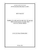
Chpater 2. THE FABRICATION OF ELECTROCHEMICAL BIOSENSOR
4
In this chapter, the experimental processes in fabrication - development and testing
of electrochemical biochemical sensors based on doped/modified conductive polymer
with nanostructured materials (Fe3O4 nanoparticles, carbon nanotubes, graphene
materials ...) are presented in detail. The diagram of experimental steps is shown in
Figure II.1 below.
CHẾ TẠO
HỆ
VI
ĐIỆN CỰC
TÍCH HỢP
CỐ ĐỊNH
PHÂN TỬ
ĐẦU DÒ
SINH
HỌC
TỔNG HỢP
MÀNG
POLYME
DẪN CHỨC
NĂNG HÓA
ĐO ĐẠC,
PHÂN
TÍCH,
THỬ
NGHIỆM
Figure II.1. Diagram of experimental steps for manufacturing - testing electrochemical
biosensor based on conductive polymer
I. Fabrication of the electrochemical microelectrodes
In the experimental framework of this thesis, we implement integrated
electrochemical microelectrode system on 1 chip including: working electrode (Pt),
counter electrode (Pt) and reference electrode (Ag/AgCl) on Si /SiO2 wafer (purchased
from Wafernet Inc, USA) (where Si p <100> wafer has a thickness of ~ 50 m and a
thickness of 1m SiO2) with thin Chromium (Cr) layer to increase the adhesion of
layers on the substrate.
Integrated electrochemical microelectrodes are fabricated based on microelectronic
technology by UV-photolithography, PVD-Physical Vapor Deposition, lift-off .. at the
Institute of Materials Science (IMS), Vietnam Academy of Science and Technology
(VAST) and at some abroad laboratories (Institute of Fundamental Electronics,
University of Paris 11, France and Department of Engineering and Science Systems,
National Tsinghua University, Taiwan). Integrated electrochemical microelectrodes
have dimensions: diameter of working electrode is 100m/200m or 500m, the width
of counter electrode/reference electrode is 100m/200m, the distance between the
electrodes is 100m/200m with the contact pad designed according to the USB
configuration.
II. Electropolymerization of the conductive polymer membrane
II.1. Electropolymerization of the polyaniline membrane
5
Electrolytic conducting solution consists of ANi 0.1M monomer in 0.5M H2SO4
containing MWCNTs-COOH (or Fe3O4-COOH) 1% w.t (compared to Aniline). The
polymerization process uses the Cyclic Voltammetry (CV) method in the potential
range of 0.0 - 0.9V (vs. Ag/AgCl), the scan rate of 50mV/s with a step of 10mV in 20
cycles. The synthesis process of pure PANi films in the same condition is also
conducted for comparison.
II.2. Electropolymerization of the polydiaminonaphthalen membrane
The P(1,5-DAN)-doped Fe3O4 films coated on working electrode (Pt) were
polymerized in 1,5-diaminonapthalene (DAN) solution of 5mM in 1M HClO4 and
Fe3O4 solution (10mg/ml) 0.5% w.t (compared to DAN) by electrochemical
polymerization CV method in the range of -0.02V to + 0.95V, scan rete of 50mV/s,
step of 10mV in 10 cycles. Pure PDAN films are also synthesized in the same
conditions to compare properties.
III. Immobilization of the biorecognition on the electrochemical miocroelectrodes
After the composite films on the basis of a multifunctional conductive polymer
membrane (denatured by nanostructured materials) was electropolymerized on the
surface of the working electrode (of the integrated microelectrode system), the
biological elements (biological probes such as enzymes, aptamers, DNA chains or
monoclonal antibodies ...) should be immobilized to the surface of the composite
membrane to develop electrochemical biosensors. Biological probes are immobilized
on the surface of composite membrane through chemical linkage (-NH-COO-) by
biological engineering. The biorecognition elements used in this thesis are biological
probes with high specificity such as enzymes (Glucose oxidase, Cholesterol oxidase
...), monoclonal antibodies, DNA sequences, aptamer sequences
IV. Electrochemical analytical methods
In this thesis, we have used many different electrochemical analysis methods to
investigate the properties of composite films (based on PANi and PDAN) and
determine the concentration of analytes in solutions such as: CV, SWV,
Chronoamperometric, EIS. Electrochemical experiments were performed on the
multifunction electrochemical device Autolab PGS/TAT 30 (EcoChimie, Netherlands)
at the Institute of Materials Science (VAST), Institute for Tropical Technology
(VAST), CETASD (Hanoi University of Science, Hanoi - Vietnam National
University).
6
V. The analytical methods for surface and structure of thin films
The surface and strutural analysis techniques such as FESEM, HRTEM, AFM,
FTIR, Raman spectrum are used in the study of the surface morphology of employed
membanes in the elctrochemical microbiosensors.
Chapter III. DEVELOPMENT OF THE ELECTROCHEMICAL MICROBIOSENSOR BASED ON CONDUCTING POLYMER
I. Development of the electrochemical micro-biosensor based on polyaniline
I.1. Functionalization the PANi film by using CNTs
CV spectra obtained in both cases are presented in Figure III.1 with similar shape,
this is the typical CV spectrum of PANi membrane electropolymerization. However, it
is very interesting that the intensity of electric current obtained in the case of composite
is about 10 times larger than the case of PANi. Thus with CNT doping in the membrane
may have increased: (i) the conductivity of the film and / or (ii) the contact surface
between the membrane and the solution containing the monomer.
800
PANi/CNTs
PANi
600
I (A)
400
200
0
-200
-400
-600
0.0
0.2
0.4
0.6
0.8
1.0
E (V)
Figure III.1. Spectrum polymerization by CV method of PANi film (a) and PANi / CNTs
membrane (b) at the 20th cycle on integrated microelectrodes
I.2. Functionalization the PANi film by using Fe3O4 nanoparticles
The electrochemical synthesis spectra of Fe3O4 doped PANi films are shown in
Figure III.2. We observed an increase in the electrochemical current density of the
7
Fe3O4-doped PANi membrane (solid line) when compared to the PANi membrane
(dashed line) (as shown in Figure III.3); This means that Fe3O4 nanoparticles may have
increased the current density of PANi films in the same experimental conditions
(design of electrode and PANi membrane properties equally), demonstrating the
doping of
Fe3O4
nanoparticles into PANi membrane increase the electrochemical
activity or the contact surface between the membrane and the monomer solution; that
leads to an increase in the ability of electron transfer in the configuration of
electrochemical sensors.
1000
1000
800
800
600
600
400
400
I /A
I /A
Fe3O4/PANi
200
0
0
-200
-200
-400
-400
-600
-600
-800
-0,2
0,0
0,2
0,4
0,6
0,8
PANi
200
-0,2
1,0
0,0
0,2
0,4
0,6
0,8
1,0
E /V vs. Ag/AgCl
E /V vs. Ag/AgCl
Figure III.2: Electropolymerization
Figure III.3. Comparison of
spectrum of Fe3O4 doping PANi films
electrochemical polymerization spectra of
PANi / Fe3O4 and PANi films
I.3 Development of the electrochemical micro-biosensor based on PANi/Grpahene
layer-by-layer structure
The thickness and structure and the functional group of PANi/Graphene films are
evaluated by Raman spectra (as shown in Figure III.4). The structural variation of
Graphene films before and after transferring to the working electrode surface Pt/PANi
is clearly observed in the Raman spectrum through comparison with Raman spectra of
PANi films and Graphene films. Raman spectrum of PANi/Graphene films (black lines)
shows the bands attributed to the PANi and Graphene (Gr), confirming the occurrence
of both of these components in the film. The question here is if the Gr has firmly bonded
by chemical bonding to PANi film or the Gr has only been mounted on this film
temporarily. In the thesis, it was found that the band situated at 1507 cm-1 (N-H bonding,
bipolaron) was collapsed, and in the same time, the band located at 1612 cm-1 (C-C
8
bonding, benzenoid) red shifts to 1597 cm-1. These results clearly demonstrated the
increase in concentration of benzenoid units; or on other hand, the chemical bonding
between PANi and Gr occurred. It was believed that those bondings are π-π bonding
between quinoid rings of PANi and Gr. Such bondings can facilitate charge transfer
between Gr and PANi, therefore influence the charge-carrier transport properties of the
material.
Figure III.4. Raman spectra of the films: Graphen, PANi và PANi/Graphen
The influence of glutaraldehyde (GA) on electrochemical behavior of PANi/Graphene
films is shown in Fig. III.5.
40
20
I /A
0
-20
-40
-60
-80
PANi/Graphen
PANi/Graphen/Glutaraldehyde
-100
-0,2
0,0
0,2
0,4
0,6
0,8
E /V vs. Ag/AgCl
Figure III.5. Electrochemical behavior of PANi/Gr film before and after GA imomobilization
9
The shape of CV curves did not change but the current intensity was decreased
slightly, suggesting the assembly of non-conductive organic compounds on the
membrane. The fact is that the GA was successfully immobilized on the surface of
micorosensor and influence on the electrochemical behavior of biosensor.
I.4 Development of the electrochemical micro-biosensor based on PANiFe3O4/Graphene structure
The surface morphology of composite PANi-Fe3O4/Graphen was examined by
FESEM (S-4800, Hitachi) at Institute of Materials Science (as show in Fig. III.6)
Graphene
Fe3O4 NPs
Figure III.6. FESEM image of PANi-Fe3O4/Graphen film
Some observations can be made from FE-SEM image of graphene/Fe3O4/PANi films
(Figure III.6). First, it shows a spongy and porous structure of PANi, which in turn can
be very helpful for enzyme entrapment. Second, doped core-shell Fe3O4 NPs (with the
diameter core of ca. 30 nm) could also contribute to further immobilization of
biomolecule, owing to their carboxylated shell. Furthermore, a thin and opaque
graphene layer was distinguishably seen on the top of the electrode surface.
The electrochemical activity of PANi/Fe3O4/graphene film increased about 8 times
compared with PANi film (Figure III.7) on the CV spectrum. The Fe3O4 nanoparticle
plays the role of electrolyte in the composite films. From Fig.III.7 4 it is clear that the
conductivity of composite was strongly enhanced with the presence of graphene film.
10
400 (1) PANi/Fe3O4/Graphene films
(1)
300 (2) PANi films
I / A
200
100
(2)
0
-100
-200
-300
-0,8
-0,6
-0,4
-0,2
0,0
0,2
0,4
0,6
E /V vs. Ag/AgCl
Figure III.7. The electrochemical behavior of composite film PANi-Fe3O4/Graphen
II. DEVELOPMENT OF THE ELECTROCHEMICAL MICRO-BIOSENSOR
BASED ON P(1,5-DAN) MEMBRANCE
II.1 Electropolymerization of the Fe3O4 nanoparticles-dopped P(1,5-DAN) membrance
When
doping
Fe3O4
nanoparticles
into
PDAN
films
during
in-situ
electropolymerization process, the Fe3O4 magnetic nanoparticles were linked to DAN
monomers via the bonding [Fe3O4]-COO-NH-[DAN] and increasing the electroactivity
of the membrane material. After 20 cycles, the current intensity of the PDAN/Fe3O4
film reaches ~ 120 A while the current intensity of the PDAN film is only ~ 8A, so
the current intensity of the PDAN/Fe3O4 film has increased greatly compared to the
with conventional PDAN film.
The electrochemical activity of PDAN/Fe3O4 films was investigated and compared
with PDAN films by CV spectrum (Figure III.8). Electrochemical spectrum of
PDAN/Fe3O4 composite has no change in shape but the signal strength increases
clearly, the spectral area is also increased (expressing the increase in electrochemical
conductivity of the film) about 10 times. Due to the electrical conductivity of
PDAN/Fe3O4 film increase, the output of electrochemical sensor also increased
accordingly, so which the sensitivity of sensor also increased.
11
60
40
I /A
20
0
-20
-40
P1,5-DAN
P1,5-DAN/Fe3O4
-60
0,0
0,2
0,4
0,6
0,8
1,0
E /V vs. Ag/AgCl
Hình III.8. Electrochemical behavior of fimls: PDAN and PDAN/Fe3O4
II.2 Fabrication of the electrochemical micro-biosensor based on Graphen/PDAN
membrance
Electrochemical behavior of Graphen/PDAN was studied and compared with PDAN
membrane by CV spectrum (Fig. III.9 below).
150
100
I / A
50
0
-50
Pt/Gr/P(1,5-DAN)
Pt/P(1,5-DAN)
-100
-150
-0.2
0.0
0.2
0.4
0.6
0.8
1.0
E /V vs. Ag/AgCl
Hình III.9. Electrochemical behavior of fimls: Pt/PDAN và Pt/Graphen/PDAN
Compared to the pure PDAN membrane, the electrochemical spectrum of the
Graphen / PDAN polymer film has no change in shape but the signal strength increases
markedly, the spectral area is also increased (demonstrating the enhancement of
electrochemical conductivity). of membrane) about 15 times. Due to the
12
electrochemical conductivity of the Gr/PDAN membrane, the output current of the
electrochemical sensor also increased accordingly, from which the sensor's sensitivity
increased. The increase of electrochemical conductivity of PDAN film on Graphene
material may be due to the interaction of NH2-Graphene group, which has changed the
band gap of the material, leading to an increase in the electronic conductivity of the
material.
Chương 4. APPLYING THE ELECTROCHEMICAL MICROBIOSENSOR ON THE ANALYTICAL
I. APPLYING ON THE BIOMEDICAL DIAGNOSTICS
I.1 Determination of the concentration of glucose
I.1.1 Determination of the concentration of glucose by using PANi/CNTs microbiosensor
The real-time response current of PANi/CNTs/GOx microsensor (with the
percentage of doping CNTs doped is 1.0% by weight) is shown in Figure IV.1 below.
Đường chuẩn của vi cảm biến trên cơ sở
màng composite PANi/CNTs có pha tạp
1,0%CNTs
0.7
9mM
8mM
0,4
7mM
0.6
6mM
0,3
4mM
0.5
3mM
∆i (μA)
I (A)
5mM
2mM
0,2
1mM
0.4
y = 0,0371x + 0,0074
R² = 0,9962
0,1
0.3
150
200
250
300
350
400
t (s)
0
0
2
4
6
Nồng độ (mM)
8
10
Figure IV.1. The real-time response
Figure IV.2. The response curve of
current of PANi/CNTs/GOx microsensor
PANi/CNTs/GOx microsensor in range 1-9
mM
It can be seen that the current intensity when measured in PBS solution (10mM, pH
= 7) is stable after about 200 seconds. When adding glucose solution, the current
intensity increases rapidly and reaches stability after about 30-40 seconds. However,
when the concentration of glucose exceeds 9mM, the increase in flow intensity is very
weak, even reduced. This may be due to the immense amount of GOx enzymes on the
electrode and the low activity (20kU).
13
The calibration curve describes the relationship between the difference in the
response current intensity ΔI (A) and the glucose concentration C (mM) added to the
electrolyte as shown in Figure IV.2. The regression equation has the form ΔI (A) =
0.0074 + 0.0371 * C (mM). The correlation coefficient of the regression equation
reaches 0.9962.
I.1.2 Determination of the concentration of glucose by using PANi-Fe3O4 microbiosensor
The current intensity of the oxidation process of glucose on the PANi/Fe3O4/GOx
sensor increases with the concentration of glucose in the solution shown in Figure
IV.3..
1.2
3.5mM
1,4
PANi with Fe3O4
3.0mM
PANi
1,2
1.0
2.0mM
0,8
0.8
I (A)
Current (A)
2.5mM
1,0
1.5mM
1.0mM
0,6
0.6
0.5mM
0,4
0.4
0,2
0.2
0,0
200
400
600
800
1000
0.5
Time (s)
1.0
1.5
2.0
2.5
3.0
3.5
Concentration (mM)
Figure IV.3. The current response of the
Figure IV.4. The calibaration curve of
PANi/Fe3O4/GOx microbiosensor
PANi/Fe3O4/GOx sensor
From the results in Figure IV.3, we determine the sensitivity of the micro sensor to
10 A.mM-1.cm-2 and the response time is less than 10s. From the calibration curve of
the sensor (Figure IV.4), the linear range of the PANi/Fe3O4/GOx micro-biosensor is
determined to be 0.5 to 3.5mM with R2 = 0.9992, LOD = 0.25mM. The regression
equation has the form: I (A) = 0.33021 * C (mM) + 0.04503.
I.1.3 Determination of the concentration of glucose by using PANiFe3O4/Graphen/Gox micro-biosensor
Figure IV.5 shows a typical current–time plot for the sensor at +0.7 V during
successive injections of glucose (3 mM increased injection, at room temperature,
without stirring, air saturated, in 50 mM PBS solution).
14
45
21,26
13,04
8,26
5,66
10,71
Iresponse /A
I /A
25
20
15
2,91mM
10
5
400
40
I (A) = 1,484*Cglucose + 6,764
35
R = 99,69
2
17,36
15,25
35
30
45
23,08
40
30
25
20
15
10
PA-Fe-Gr Glucose sensor
PANi-Fe3O4/co(St-AA)-Graphene films
5
600
800
1000
1200
1400
1600
0
Time /s
5
10
15
20
25
Glucose concentration C/mM
Hình IV.5. Amperometric responses of
Hình IV.6. Glucose calibration line and
PANi-Fe3O4/Graphen/GOx microsensor to
respective regression equation of PANi-
different added glucose concentrations
Fe3O4/Graphen/GOx microsensor
We found that the sensor has a short response time for changing glucose
concentrations in the solution, tresponse ~ 10-15s, the response current intenstity has good
stability at the various concentrations of glucose. The calibration plot indicates a good
and linear amperometric response to glucose within the concentration range from 2.9
to 23 mM (with regression equation of I (A) = 1.484 * C (mM) + 6.764, R2= 0.9969)
(as Fig. IV.6).
Thus, with a miniaturized dimension (500 µm) the above graphene patterned sensor
has shown much improved sensitivity to glucose, as high as 47 AmM-1cm-2 compared
to non-graphene one (10-30 AmM-1cm-2).
I.2. Determination of the concentration of cholesterol
I.2.1. Determination of the concentration of cholesterol by using PANi/CNTs microbiosensor
The current response curve of PANi/CNTs/ChOx micro-biosensors with the
presence of mediator K3[Fe(CN)6] at voltage E = -0.3V given in Figure IV.7. The
concentration of cholesterol is the diluted concentration (considering the change in
volume is negligible).
15
0.6
3.4
0,12mM
0.5
0,10mM
3.3
0,08mM
3.2
0.4
I (A)
I (A)
0,06mM
3.1
0,04mM
0.3
Y=A+B*X
Parameter
Value
Error
-----------------------------------------------------------A
0.01740
0.00926
B
4.30143
0.11885
------------------------------------------------------------
3.0
0.2
0,02mM
2.9
R
Sy
N
P
-----------------------------------------------------------0.99848
0.00994
6
<0.0001
------------------------------------------------------------
0.1
2.8
0.02
400
500
600
700
0.04
0.06
0.08
800
0.10
0.12
Nång ®é (mM)
Thêi gian (gi©y)
Figure IV.7. The real-time response curve
Figure IV.8. The calibration curve of
of PANi/CNTs/ChOx microsensor
PANi/CNTs/ChOx microsensor
The PANi/CNTs/ChOx microsensor reachs a stable current (-2,8A) in PBS buffer
solution of 50mM (pH = 7) after about 400 seconds. Based on the difference in the
response current intensity of the PANi/CNTs/ChOx microsensor and the total added
amount of cholesterol, a calibration curve for determining cholesterol at -0.3V
(compared with Ag/AgCl ) in the presence of K3[Fe(CN)6]. The regression equation
has the form ΔI (A) = 0.0174 + 4,3014*C (mM). Correlation coefficient of the
regression equation: R2 = 0.9985.
I.2.2 Determination of the concentration of cholesterol by using PANi/Fe3O4
The response current spectra of PANi/Fe3O4/ChOx-Fe3O4 microsensors is shown in
Figure IV.9 below. The results showed that the sensor gave good response (linear) in
the range of cholesterol concentrations from 0.196mM ÷ 1,803mM. At higher
cholesterol concentrations, when added to the electrolyte, the signal is noisy, the
current is poor. This is due to ChOx when catalyzing hydrolytic reactions of choleterol
that do not keep up with the added rate of substrate. For the results in the calibration
graph of the sensor (Figure IV.10), the regression equation of the calibration curve will
take the form: I (µA) = (21.45±1,271)×C (mM) + (-0,8352±1,1474), the correlation
coefficient of the regression equation reached R2 = 0.9929. The average sensitivity of
PANi/Fe3O4/ChOx-Fe3O4 micro sensors is S = 21.44 A.mM-1.cm-2.
16
45
40
40
35
30
I (A)
I (A)
30
20
25
20
15
10
Equation
10
0.99297
Value
5
0
y = a + b*x
Adj. R-Square
cholesterol
-- Intercept
-- Slope
0
200
400
600
800
0
0.0
1000
t (s)
0.3
0.6
0.9
1.2
Standard Error
-0.8352
0.66165
21.44897
0.57052
1.5
1.8
2.1
cholesterol (mM)
Figure IV.10. The calibration curve of
Figure IV.9. The real-time response curve
of PANi/Fe3O4/ChOx-Fe3O4 microsensor
PANi/Fe3O4/ChOx-Fe3O4 microsensor
I.2.3 Determination of the concentration of cholesterol by using PANiFe3O4/Graphen micro-biosensor
Figure IV.11 shows a typical current–time plot for the sensor at +0.7 V during
successive injections of cholesterol (2 mM increased injection, at room temperature,
without stirring, air saturated, in 50 mM phosphate buffered solution). The response
time of cholesterol sensor was smaller than 5s with cholesterol concentration change.
The calibration plot indicates a good and linear amperometric response to cholesterol
within the concentration range from 2 to 20 mM (with regression equation of I (µA)
= (2.15 ± 0.13) * C (mM), R2= 0.9986) (the inset in Figure IV.11). Thus, with a
miniaturized dimension (250 µm) the above graphene patterned sensor has shown
much improved sensitivity to cholesterol, as high as 1095.54 AmM-1cm-2. The
sensitivity of graphene cholesterol sensor was 2 times higher than that of CNTcholesterol sensor.
17
40
35
30
Current (mA)
Current /A
40
30
25
20
15
10
5
0
0
20
2
4
6
8
10
12
14
16
18
20
Concentration (mM)
10
0
0
200
400
600
800
1000
Time (s)
Hình IV.11. Amperometric responses of PANi-Fe3O4/Graphen/ChOx microsensor to
different added cholesterol concentrations (inset: the calibration curve of fabricated
cholesterol sensor)
I.3. Testing of DNA of HPV virus
The SWV graph is recorded after each process (activated with EDC/NHS, before and
3,5x10
-4
3,0x10
-4
2,5x10
-4
2,0x10
-4
1,5x10
-4
1,0x10
-4
5,0x10
-5
(1) + EDC/NHS
(2) + HPV-16-L1
(3) + 10nM anti-HPV
(4) + 20nM anti-HPV
(5) + 30nM anti-HPV
(6) + 40nM anti-HPV
(7) + 50nM anti-HPV
(1)
(2)
(3)
(4)
(5)
(6)
(7)
I /A
I /A
after immobilized aptamer, HPV antigen), as shown in Figure IV.12.
2.6x10
-4
2.4x10
-4
2.2x10
-4
2.0x10
-4
1.8x10
-4
1.6x10
-4
1.4x10
-4
0,0
-5,0x10
-5
-1,0x10
-4
-0,4
-0,2
0,0
0,2
0,4
0,6
0
E /V vs. Ag/AgCl
20
40
60
80
Anti-HPV-16 concentration /nM
Figure IV.12. SWV of PANi/CNTs
Figure IV.13. The response curve of
microsensor recorded after treatment with
PANi/CNTs microsensor with anti-HPV-16
EDC/NHS (curve 1), after grafting HPV-
concentration range from 10-80 nM
16-L1 (curve 2) and after complexation
with anti-HPV-16 (curve 3-7)
The spectrum of SWV analysis proved very clearly the formation of the complex of
aptamer HPV-16-L1 and its specific HPV antibody, through the linearly attenuating of
SWV peak current intensity. The calibration curve was constructed with a range of
18
different HPV concentrations in the range of 10-80 nM (shown in Figure IV.13). The
PANi/CNTs biosensor has a sensitivity response of 1.75 ± 0.2 (A.nM-1) (R2 = 0.997)
in a concentration range of 10–50 nM with limit of detection (LOD ) is 490pM. It can
be seen that the signal tends to be saturated with a concentration value above 80nM
II. APPLYING IN FOOD SAFETY CONTROL
II.1. Determination of the concentration of Aflatoxin M1 in milk
The ability to recognize the concentration of AFM1 of microsensors is determined
by a calibration curve with a range of different concentrations (from 6ng/L to 78ng/L
relative to a concentration of 18-240pM of AFM1) of AFM1 (molecular weight is ~
328Da). The analytical results of AFM1 concentration of microsensor by SWV method
shown in Figure IV.14 is quite similar to the electrochemical CV signal of micro
sensor.
(1) Fe3O4/PANi
7
(2) Fe3O4/PANi/Glu
(3) Fe3O4/PANi/Glu/APT
6
-1
(4) + AFM1 06ngL
-1
(5) + AFM1 18ngL
-1
(6) + AFM1 30ngL
-1
(7) + AFM1 60ngL
4
3
I / A
I /A
5
2
5,0
I (A) = -4,77*CAFM1 + 5,17 (A)
4,5
R = 0,9986
2
4,0
3,5
3,0
-1
2,5 LOD = 1,98 ngL
-1
LOQ = 6,62 ngL
2,0
1
0 10 20 30 40 50 60 70 80
SIGNAL OFF
0
-0,6
-0,4
-0,2
0,0
-1
AFM1 concentration /ngL
0,2
0,4
0,6
0,8
E /V vs. Ag/AgCl
Figure IV.14. SWV response of PANi/Fe3O4 microsensor with various AFM1 concentration
The results of microsensors are: sensitivity of 4.77 ± 0.2 (A/ ngL-1) (R2 = 0.9986)
in the range of 6 - 60 ngL-1 (approximate 18 to 240 pM) with LOD reaching 1.98ng/L
(the inset of Figure IV.14: the calibration line of the sensor).
II.2 Determination of the concentration of lactose in milk
II.2.1 Determination of the concentration of lactose by using P(1,5-DAN)/Fe3O4
micro-biosensor
The current response of the sensor under investigation at a voltage of 0.4V is shown
in Figure IV.15 below.
19
12
12
Lactose determination by PDAN/Fe3O4 biosensor
11
Iresponse = 5,88 + 0,38*Clactose (A/(mgmL ))
11,9
10
11
10,2
8,5
9
6,8
5,1
8
3,4
7
2
R = 99,65
-1
LOD = 0,19 mgmL
10
Iresponse /A
Iresponse /A
-1
13,6
1,7mg/ml
9
8
7
6
6
100
200
300
400
500
600
0
Time /s
2
4
6
8
10
12
Concentration of lactose /mgmL
14
-1
Figure IV.15 The real-time response curve
Figure IV.16. The calibration curve of
of PDAN/Fe3O4 microsensor
PDAN/Fe3O4 microsensor
From the real-time response, we found that the PDAN/Fe3O4 electrochemical
biosensor has a linear response to the concentration of the lactose in ~ 12 A range.
The response time of the microsensor is small (<10s) and the output current is very
stable at the survey sample concentrations. The stabilization time of micro-sensors in
buffer environments is short (<200s). These are good micro-sensor parameters that
require the connection of an electronic processing circuit to develop a device for rapid
analysis of lactose content in the sample.
Based on the real-time current response characteristics, a sensor of the sensor's
lactose-out signal concentration curve is determined (Figure IV.16). Based on the
calibration curve of the micro sensor, the sensitivity of the micro sensor is 0.38
A/(mg.mL-1), R2 = 0.9965 with LOD = 0.19mg/mL. Micro sensors have a linear
response in the concentration range of 0 - 14 mg/mL.
II.2.2 Determination of the concentration of lactose by using Graphen/P(1,5-DAN)
micro-biosensor
The calibration curve of the electrochemical biosensor Graphene/PDAN (shown in
Figure IV.17).
20
130
120
110
-1
Iresponse = 47,94 + 1,33*Clactose (A/(gmL ))
2
R = 99,5
-1
LOD = 1,3 gmL
Iresponse /A
100
90
80
70
60
50
40
0
10
20
30
40
50
-1
Concentration of lactose /gmL
60
Figure IV.17. The calibration curve of Graphene / PDAN micro sensor with variouslactose
concentration in solution
Based on the calibration curve of the micro-sensor, we determined the sensitivity of
the sensor to reach 1.33 A/(g.mL-1), R2 = 0.995 with LOD detection limit = 1.3 g
.mL-1 in the concentration range 0 ÷ 60g.mL-1..
III. APPLYING IN ENVIROMENTAL MONITORING
III.1 Detection of the trace concentration of herbicide Atrazine by using PANi/Fe3O4
micro-biosensor
We used SWV method and SIGNAL-OFF models to apply to electrochemical
sensors to determine very small levels of ATZ in solution (as shown in Figure IV.19).
The attenuation of electrochemical signal from lines (1) (7) shows good
performance of the sensor.
The calibration curve of the sensor is determined based on the SWV measurement
results of the sensor in the concentration range from 10-11 to 10-8M of Atrazine (shown
in Figure IV.19 below). The calibration curve of the sensor shows the linear
dependence between the output current and the concentration of Atrazine (in log) in
the range of 10-11M to 10-8M. Regression equation is: I (A) = (-306,02 ± 6,71) - (64,78
± 0,62) (logCATZ). Sensitivity of the sensor reached (64.78 ± 0.62) (A/logCATZ) with
R2 = 0.9915, LOD = 2.1x10⁻⁹M.
.
21
600
550
(1) PANi\Fe3O4
(2) PANi\Fe3O4 \Glu
500
(1)
(3) PANi\Fe3O4 \Glu-ATZ
-11
500
I /A
400
(4) [ATZ] = 10 M
-10
(5) [ATZ] = 10 M
-9
(6) [ATZ] = 10 M
-8
(7) [ATZ] = 10 M
450
(2)
(3)
400
(4)
I / A
700
(5)
300
(6)
200
300
250
S = 64,78 ± 0,62 (A/logCATZ)
200
R =0,9915
-9
LOD = 2,1 x 10 M
(7)
100
0
-0,8
350
2
150
-0,6
-0,4
-0,2
0,0
0,2
0,4
-13
0,6
-12
-11
-10
-9
-8
Concentration of Atrazine (logCATZ)
E /V vs. Ag/AgCl
Hình IV.18. The SWV response of the
Hình IV.19. The linear output of the
PANi/Fe3O4 immunosensor with ATZ
PANi/Fe3O4immunosensor with ATZ
concentration from 10-11 to 10-7 M
concentration from 10-11 to 10-7
III.2 Detection of the trace concentration of herbicide Atrazine by using
PANi/Graphen micro-biosensor
Fig. IV.20 shows the SWV curves of the immunosensor incubated at different
concentrations of ATZ. It was found that the current response (at +0.57V) decreased
with ATZ concentration. This is probably due to more ATZ binding to the immobilized
antibodies at higher ATZ concentrations, which acts as a definite kinetic barrier for the
electron transfer. The deposition of non-electronic materials like ATZ on
microelectrodes hindered the electroactive species to get onto the electrode and
reduced the electron exchange between the electrode and solution. The variation of
output current (intensity of SWV peak) with ATZ concentration was plotted in Fig.
IV.21. This curve shows a linear immune response (current change at +0.57V) against
logarithm of ATZ concentration with a regression equation: I=13.33logCATZ + 202 A
(R2 = 0.9786). As seen here, the range of detection over which we still have linear
immune response of the immunosensor is relatively large; meaning that high precision
can be easily achieved in a wide range of detection. The detection limit for the
immunosensor with the PANi/Gr layer was 43 pg.L-1, far below the maximum residue
level (100μgL-1) established by European Union.
22
550
500
450
360
340
350
I ()
I ()
400
Pt/PANi/Gr-GA
Pt/PANi/Gr-GA-ATZ
-11
Pt/PANi/Gr-GA-ATZ-ATZ 10 M
-10
Pt/PANi/Gr-GA-ATZ-ATZ 10 M
-9
Pt/PANi/Gr-GA-ATZ-ATZ 10 M
-8
Pt/PANi/Gr-GA-ATZ-ATZ 10 M
-7
Pt/PANi/Gr-GA-ATZ-ATZ 10 M
300
250
CATZ
320
300
200
Ipa=202,6 + 13,33.log (CATZ) ()
280
150
100
0,3
0,4
0,5
2
R = 0,9786
0,6
-11
E (V) vs. Ag/AgCl
-10
-9
-8
-7
Log (CATZ) (M)
Hình IV.20. The SWV response of the
Hình IV.21. The linear output of the
PANi/Gr immunosensor with ATZ
PANi/Gr immunosensor with ATZ
concentration from 10-11 to 10-7 M
concentration from 10-11 to 10-7
23