Detection of bovine rotavirus from Diarrheic bovine calves in Gujarat region, India
Bạn đang xem bản rút gọn của tài liệu. Xem và tải ngay bản đầy đủ của tài liệu tại đây (706.92 KB, 12 trang )
Int.J.Curr.Microbiol.App.Sci (2019) 8(9): 1282-1293
International Journal of Current Microbiology and Applied Sciences
ISSN: 2319-7706 Volume 8 Number 09 (2019)
Journal homepage:
Original Research Article
/>
Detection of Bovine Rotavirus from Diarrheic Bovine calves
in Gujarat region, India
Jayesh Patel1*, Rafiyuddin Mathakiya2 and Akash Golaviya3
1
Department of Veterinary Microbiology, College of Veterinary Science & Animal
Husbandry, AAU, Anand-388 001, Gujarat, India
2
Department of Veterinary Microbiology, College of Veterinary Science and A.H., AAU,
Anand, India
3
Department of Veterinary Microbiology, College of Veterinary Science & A.H., AAU, Anand,
India
*Corresponding author
ABSTRACT
Keywords
Neonatal Calf
Diarrhea, Calf
Scour,
Rotavirus,LAT,
ELISA, RT-PCR
Article Info
Accepted:
15 August 2019
Available Online:
10 September 2019
The aim of the current study was to identify the prevalence of bovine
rotavirus infection in bovine calves aged from 1 day to 2 months old in
Gujarat region. Studied samples were divided into 4 age groups, age group
I (1-10 days) age group II 911-20 days), age group III (21-30 days) and age
group IV (31-60 days). All the samples were screened by latex
agglutination test (LAT), ELISA and RT-PCR technique. Among the 117
fecal sample screened, 23.93%, 15.38% and 22.22%found positive for
Rotavirus by LAT, ELISA and RT-PCR respectively. Age wise, rotavirus
infection was highly prevalent in age group-I (1-10 days) i.e. 38.46%. Sex
wise, there was no clear-cut establishment of correlation for Rotavirus. The
present study showed that rotaviruses are involved in the neonatal calves’
diarrhea in Gujarat area
Introduction
Bovine rotavirus is a primary and most
common pathogen responsible for the NCD.
Rotavirus diarrhea in calves, piglets, foals and
lambs is often referred as “white scour” or
“milk scour”. Bovine rotavirus usually causes
diarrhea in calves at 1 to 2 weeks of age
(Dhama et al., 2009). It has been reported that
diarrhea in calves from 5-10 days of age is
commonly due to rotavirus and infected calves
excrete rotavirus in their feces upto the age of
6 to 8 weeks. Rotavirus was first isolated from
calves with diarrhea and since then has been
isolated from cattle, sheep, horses, dogs, cats,
chickens and turkeys (Mebus et al., 1969).
1282
Int.J.Curr.Microbiol.App.Sci (2019) 8(9): 1282-1293
The disease is usually seen in young animals
and the susceptibility to disease decreases as
the age progress, most likely due to changes in
animal physiology and/or acquired immunity
due to previous exposures (Estes and
Kapikian, 2007).
(ELISA), PAGE, Latex agglutination test
(LAT),
Electron
microscopy,
Passive
hemagglutination
assay
(PHA),
Immunoblotting and Immunofluorescence test.
The virus belongs to the genus Rotavirus of
subfamily Sedoreovirinae in the family
Reoviridae. Rotavirus is dsRNA, nonenveloped, 65-75 nm in diameter, icosahedron
symmetry having 11 double-stranded RNA
segments (16~21 kb) and is very stable over a
wide pH range with heat stability
(MacLachlan et al., 2016). The fully
infectious rotavirus particle consists of three
protein layers (i.e. core, inner capsid and outer
capsid) and is also termed triple-layered
particle (TLP). By electron microscopy, TLPs
resemble wheels (latin. rota), and this
appearance has led to the name of Rotavirus
for the genus (Flewett et al., 1974).
The present study was undertaken to ascertain
detection of bovine rotavirus from fecal
samples of diarrheal calves of cattle and
buffalo by Enzyme Linked Immunosorbent
Assay (ELISA) and Reverse TranscriptasePolymerase Chain Reaction (RT-PCR)
techniques.
The dsRNA segments of Rotavirus code for
six structural proteins (SPS) viz. VP7VP4VP6-VP1-VP2-VP3 and six non-structural
proteins (NSPs) viz. NSP1–NSP2–NSP3–
NSP4–NSP5/6. The VP6 protein comprises
the middle layer of the capsid and is the
protein, encoded by gene segment 6, to which
common immunodiagnostics are directed.
Rotavirus has 11 segments of the dsRNA as
the genome and that could be detected by
RNA-Polyacrylamide Gel Electrophoresis
(RNA-PAGE). The eleven segments of the
dsRNA are separated from each other based
on their electrophoretic mobility and it is very
sensitive method for determination of the
presence of Rotavirus in fecal samples. The
detection of 11 segments of dsRNA of RVA in
RNA-PAGE exhibited the 4-2-3-2 pattern of
migration.RVA are excreted in large numbers
in the feces and can be detected by the various
methods
viz.
Reverse
TranscriptasePolymerase Chain Reaction (RT-PCR),
Enzyme Linked Immunosorbent Assay
Materials and Methods
Collection of specimens
All the 117 fecal samples were collected from
calves of cattle and buffalo of 1 day to 2
months of age. Samples were collected from
farms of various region viz. Anand (n=31),
Surat (n= 68) and Junagadh(n=18). The fecal
samples were collected as per the method of
Yilmaz (2016). The samples were transferred
to sterile disposable plastic specimen vial. The
specimen vial was labelled properly according
to the species, age, sex and name of owner etc.
The sample was stored in deep freeze (-400C)
for detection of viral antigen until use.
Screening by Latex Agglutination Test
(LAT)
All the fecal samples were screened for
rotavirus antigen by LAT(Figure 1). LAT was
performed to detect rotavirus antigen in
fecal supernatants as per instruction by the kit
manufacturer (HiRotavirus Latex Test Kit,
LK08,HimediaPvt. Ltd.).
Sample Preparation for ELISA
The fecal sample was diluted in dilution buffer
(provided by kit manufacturer) volume per
volume (v/v). This was a qualitative dilution
only, which must allow the pipetting the fecal
1283
Int.J.Curr.Microbiol.App.Sci (2019) 8(9): 1282-1293
suspensions. The gruds were discarded by
natural sedimentation for about 10 minutes
without centrifugation.
was quantified and the ratio of purity was
evaluated following the method of Sambrook
and Russel (2012).
Screening by ELISA
Molecular Detection of Rotavirus by RTPCR
All the samples again screened rotavirus
antigen by ELISA. ELISA was performed to
detect bovine rotavirus in the fecal samples by
the kit manufacture (Bio X Diagnostic
Multiscreen Ag ELISA kit (BIO K 315/1,
Belgique). This 96 well plate ELISA kit also
used for diagnosis of Coronavirus and E. coli
F5 attachment factor from fecal sample of
calves along with Rotavirus(Figure 2). The
ELISA plate is sensitised with a mixture of
antibodies that are specific for the above 3
pathogens. These antibodies capture the
corresponding pathogens in the faecal
samples. The net optical density of each
sample was calculated by following formula:
Val(ue)=
The optical density of ≥6.0 at 450nm
wavelength were considered as positive for
Rotavirus by antigen detection sandwich
ELISA
Extraction of Viral RNA
Viral RNA extraction was done from fecal
suspensions (10% v/v) by using a QIAamp
Viral RNA Mini Kit, QIAGEN (Cat. No.
52904) following the manufactures protocol.
Quality and Quantity Check of RNA
For quantification of viral RNA, absorbance
was read in Nanodrop Spectrophotometer at
260nm.
An optical density (OD) 1 corresponds to
40µg/ml for dsRNA. A ratio of 260:280
provides an estimate of purity of the nucleic
acid. Pure preparations of RNA have 260:280
ratio of 2.0. The viral RNA from fecal samples
The RT-PCR assay for specific VP6 gene was
standardized using the viral genomic RNA
extracted by QIAamp RNA Mini Kit,
QIAGEN (Cat. No. 52904)(Table 1).
The genomic RNA was quantitated by
Nanodrop and used as a template for cDNA
synthesis
by
using
components
of
PrimeScriptTM RT-PCR kit, TaKaRa (Cat.
No. RR014A). The reaction conditions for
RT-PCR using PrimeScriptTM RT-PCR kit,
TaKaRa were standardized to get the desired
specific product. The reaction mixture of
polymerase chain reaction (PCR)as per Table
2 and thermo cycling conditions used for PCR
to amplify full-length VP6 gene as per
Table.3.
Visualization of PCR products by agarose
gel electrophoresis
To confirm the targeted PCR amplification,
5µl of the PCR products from each tube was
mixed with 1µl of 6X gel loading buffer and
electrophoresed along with DNA molecular
weight marker (HiMediaPvt. Ltd) on 2.0 %
agarose (Low EEO, SeaKem) gels containing
0.5μg/ml ethidium bromide (Sigma-Aldrich,
USA) at 80V in 0.5X Tris Borate EDTA
(TBE) buffer. The amplified product was
visualized as single compact band of expected
size under UV light and documented by gel
documentation system (Genetix Biotech Pvt.
Ltd, Delhi).
Results and Discussion
A total 117 fecal samples were collected from
diarrheic calves from farms of various region
1284
Int.J.Curr.Microbiol.App.Sci (2019) 8(9): 1282-1293
viz. Anand, Surat and Junagadh, and screened
for the presence of Rotavirus using latex
agglutination test (LAT), ELISA and RTPCR.
Out of 117 diarrheic samples screened for
Rotavirus, 28 (23.93%), 18 (15.38%) and 26
(22.22%) samples were found positive for
Rotavirus by LAT, ELISA and RT-PCR
method, respectively as mentioned in Table 4.
The results of RT-PCR were taken in to
consideration to show the prevalence of
Rotavirus. The RT-PCR technique is widely
used for the detection of the RNA viruses. It
helps in the detection of viral nucleic acid
during initial stages of infection without
waiting for higher virus titre and development
of immune response in the affected host
species. The amplification of fragment of VP6
gene of Bovine rotavirus in RT-PCR indicated
that calves were suffering from rotaviral
infection (Figure 3).In the present study,
overall prevalence of Rotavirus recorded was
almost similar to Suresh et al., (2012),
Abdulazeez et al., (2017), and Pardo-Mora
(2018) they observed24.10%, 25.60% and
19.70%, respectively. In contrast to present
study, Mondal et al., (2013), Yilmaz (2016)
andGill et al., (2017) observed low prevalence
of Rotavirus i.e. 11.23%, 7.57% and 10%,
respectively.
associated with infectious agents, and
probably due to nutritional or other
management factors, or because other noninvestigated pathogens might be involved as
reported by Ammar et al., (2014).
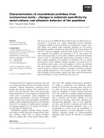
Out of 117 fecal samples, 28 (23.93%)
samples were found positive for Rotavirus by
latex agglutination test as indicated in the
form of clump/agglutination of latex particles
in the test sample and positive control, and
absence of agglutination in negative control
(Figure 4). AlYousif et al., (2001) and Singh
(2011) reported higher 52% and 32.07%
prevalence of rotavirus infection whereas
Sukura et al., (1990) reported lower (12%)
prevalence of Rotavirus by LAT.
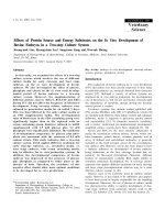
In the present investigation, 18 (15.38%)
samples were found positive by ELISA(Figure
5). The result is in agreement with Al-Robaiee
and Al-Farwachi (2013), Ammar et al., (2014)
and Singh et al., (2015), as they reported
15.50%, 14.63 and 12% prevalence of
Rotavirus.
In contrast to present study, Wei et al.,
(2013), Khamees et al., (2015) and
Abdulazeez et al., (2017) reported42.05%,
43.50% and 36% prevalence of Rotavirus.
Region wise Prevalence of Rotavirus
The difference in Rotavirus prevalence in
diarrheic calves which established in the
present study and by above mentioned
investigators is in agreement due to scientific
fact that rotaviruses area worldwide
distributed and able to infect humans and
domesticated animals. Various factors like
hygienic measures, proper access to maternal
colostrum, nutrition season and climatic
factors such as rainfall, temperature, relative
humidity etc. might be responsible for
rotaviral diarrhea as reported by Abdulazeez et
al., (2017). Negative results may occur
because some cases of diarrhea might not be
Rotavirus was highly prevalent in Junagadh
(38.88%) as compared to Anand (22.58%) and
Surat (17.64%) region. This high and low
prevalence in various regions might be due to
the difference in climatic conditions, sample
size, management practices, personal hygiene,
the age at which sample was collected and
farm size (Table 5).
Age and Sex wise Prevalence of Rotavirus
In the present study, the association between
the different age groups and sex with the
1285
Int.J.Curr.Microbiol.App.Sci (2019) 8(9): 1282-1293
occurrence of diarrhea due to Rotavirus was
studied (Table 6).
The rotavirus infection (38.46%) highly
prevalent in 1-10 days of age group of calves
followed by 30.76%, 19.23% and 11.53% for
age group of 11-20 days, 21-30 days and 3160 days.
Table.1 Oligonucleotide primer (VP6) sequence used in RT-PCR of BRV
Primers
Sequences (5’-3’)
VP6 F
VP6 R
GACGGVGCRACTACATGGT (19)
GTCCAATTCATNCCTGGTGG (20)
Expected
Reference
product
size (bp)
379
Yilmaz, 2016
Table.2 Reaction of polymerase chain reaction (PCR)
Reagents
Volume (µl)
5
10 X PCR Buffer II
2
dNTP Mixture (10 Mm each)
10
VP6 F (20 µM)
10
VP6 R (20 µM)
0.5
TaKaRa Ex Taq HS (5 U/ µl)
5
cDNA
17.5
Sterile water
50
Total
Table.3 Thermo cycling conditions used for PCR to amplify full-length VP6 gene
Sr. No.
Step
Temperature
Time
Denaturation
94oC
30 seconds
1
Annealing
55oC
30 seconds
2
o
Extension
72 C
90 seconds
3
40
Cycles
Table.4 Detail information of Rotavirus positive samples by various methods
Sr. No.
Sample code
1
2
3
4
5
6
7
8
A-8
A-9
A-10
A-11
A-17
A-18
A-30
A-31
Bovine Calf
Age
Sex
(Days)
15
Female
3
Female
27
Female
45
Male
20
Male
7
Female
30
Male
7
Male
1286
LAT
ELISA
RT-PCR
++++
++
++++
+
++
+
++
++++
++
++++
++
++
Positive
Negative
Positive
Positive
Positive
Positive
Positive
Positive
Int.J.Curr.Microbiol.App.Sci (2019) 8(9): 1282-1293
9
10
11
12
13
14
15
16
17
18
19
20
21
22
23
24
25
26
27
28
29
S-34
S-35
S-43
S-49
S-50
S-55
S-56
S-75
S-76
S-84
S-86
S-88
S-90
S-92
J-101
J-102
J-103
J-104
J-105
J-108
J-115
45
30
8
11
7
6
11
2
25
1
1
2
14
35
7
15
5
25
14
20
30
Female
Female
Male
Male
Male
Male
Male
Male
Male
Female
Female
Female
Male
Female
Male
Female
Male
Female
Male
Male
Female
Total
+++
+
+++
++++
++
++++
++++
+++
++
+++
+++
+++
++
+++
+
+
+++
++
+++
+++
+++
28/117
(23.93%)
++
++
+++
+++
+++
+++
+++
+++
+++
+++
+++
+++
+++
18/117
(15.38%)
Positive
Positive
Positive
Positive
Negative
Positive
Positive
Positive
Negative
Positive
Positive
Positive
Positive
Positive
Positive
Positive
Positive
Positive
Positive
Positive
Positive
26/117
(22.22%)
Table.5 Rotavirus prevalence in various regions
Regions
Total no. of sample
Junagadh
Anand
Surat
Total
18
31
68
117
No. of positive
samples
07
07
12
26
Prevalence %
38.88
22.58
17.64
22.22
Table.6 Rotavirus prevalence by RT-PCR in various age groups and sex
Age groups(Days)
No. of positive samples
I
(1-10)
II
(11-20)
III
(21-30)
IV
(31-60)
Total
10 (38.40%)
Sex wise Prevalence of Rotavirus
No. of male calves
No. of female calves
Infected
infected
6
4
(60%)
(40%)
6
2
(75%)
(25%)
1
4
(20%)
(80%)
1 (33.33%)
2
(66.66)
14
12
(53.85%)
(46.15%)
8 (30.76%)
5 (19.23%)
3 (11.53%)
26
1287
Int.J.Curr.Microbiol.App.Sci (2019) 8(9): 1282-1293
Fig.1Latex agglutination test kit
Fig.2 Components of Sandwich ELISA kit
1288
Int.J.Curr.Microbiol.App.Sci (2019) 8(9): 1282-1293
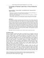
Fig.3 Agarose gel showing amplified product for VP6- gene ofRotavirus isolates (approximately 379bp)
L: DNA Molecular weight ladder – 1000bp,
1-4: Isolates (Samples)
P: Positive control
N: Negative control
L
1
2
3
379bp
100bp
1289
4
P
N
Int.J.Curr.Microbiol.App.Sci
(2019) 8(9): 1282-1293
Fig. 4 Screening for Rotavirus
using Latex Agglutination
Test kit (1-4: Samples, 5:
Positive control, 6: Negative control
Fig.5 Screening for Rotavirus using Sandwich ELISA (C1-H9: Samples, A1: Positive
control, B1: Negative control
The prevalence of Rotavirus is higher in 1-10
days age group, this finding is supported by
Ammar et al., (2014) and Khamees (2015)
they also reported (60.80% and 19.23%) and
higher prevalence of Rotavirus infection in 110 days age group. A higher infection rate in
the first week of life suggests widespread
rotavirus in this group of younger calves
reported by Selim et al., (1991) and Majeed et
al., (2011). As numerous factors interplay in
precipitating clinical diarrhoea, it is difficult to
make absolute conclusions based on the
limited information. However, rotavirus
infection has been shown to be more
1290
Int.J.Curr.Microbiol.App.Sci (2019) 8(9): 1282-1293
important than other agents in diarrhoea in
young calves of around 1 week of age (4-14
days) (Selim et al., 1991). In the present
investigation, 22 (18.80%) diarrheic calves
were infected with both E. coli and Rotavirus.
management and veterinary care as well as
animal suffering.
The resultant of effectiveness of sex on
infection rate in present study which slightly
higher in male (53.84%) than female (46.15%)
but not much difference similar finding to
those of Dash et al., (2011) and Hassan et al.,
(2014), they recorded infection rate (20.37%)
in male and (12.76%) in female and 37.5% in
males and 40% in females, respectively. In
contrast to the present study, Ammar et al.,
(2014) recorded higher frequency of infection
of rotavirus in female (60.86%) as compared
to the male (39.13%). The anatomical,
functional and hormonal similarities of body
systems of male and female calves in early
ages lead to non-specific resistance against
infection, but degree of contamination with
virus, dose of viruses, exposing to stress
factors, consumption of colostrum or not and
other many environmental and management
factors, all effect on infection rate and severity
in both sexes of calves in same or different
periodic age as reported by Hassan et al.,
(2014). Calves within their first month of life
are highly susceptible to viral diarrhea
probably due to suckling milk, which
neutralizes the acidic pH of their digestive
tract and in turn allows several pathogens to
survive (Dhama et al., 2009: Cho and Yoon,
2014). Several improvements in vaccination,
medication and management have been
implemented to reduce the incidence of NCD.
However, NCD is still persistent because it is
complex and multifactorial and can be
triggered by several different infectious and
non-infectious causes (Cho and Yoon, 2014).
Direct losses are due to dehydration, reduced
growth rate, and high morbidity and mortality.
In the present study, the difference in the
results of nine samples was recorded for LAT
and ELISA i.e. LAT showed nine samples
positive for Rotavirus while the same samples
were negative by ELISA. The LAT results
(23.93%) were almost similar to that of the
RT-PCR (22.22%). These results indicated
that LAT was more sensitive than ELISA and
thus, former technique should be used for
screening of Rotavirus detection. However,
two fecal samples were weak positive by LAT
and negative by ELISA and RT-PCR. This
variation in result of LAT and RT-PCR might
be due to cross agglutination with another
organism present in the fecal sample.
Indirect losses are due to the imposition of
trade restrictions and increased costs of
Comparison of LAT, ELISA and RT-PCR
for Rotavirus detection
Acknowledgement
I would like to acknowledge my major guide
Dr. R. A. Mathakiya, Assistant Professor,
Dept. of Veterinary Microbiology, AAU,
Anand and Veterinary College of Veterinary
Science & Animal Husbandry for their moral
and financial support to my research work.
References
Abdulazeez, A. A., and Abed, M. N. 2017.
Genotyping of Rotavirus in neonatal
calves with acute gastroenteritis in
Iraq. Advances in Microbiology, 7(12),
863.
Al-Robaiee, I. A., and Al-Farwachi, M. I.
2013. Prevalence of rotaviral infection
in diarrheic neonatal calves in Mosul
city, Iraq. Veterinary World, 6, 538540.
Al-Yousif, Y., Anderson, J., ChardBergstrom, C., Bustamante, A.,
Muenzenberger, M., Austin, K., and
Kapil, S. 2001. Evaluation of a latex
1291
Int.J.Curr.Microbiol.App.Sci (2019) 8(9): 1282-1293
agglutination kit (VirogenRotatest) for
detection of Bovine Rotavirus in fecal
samples. Clinical and Diagnostic
Ammar, S. S. M., et al., 2014. Prevalence of
Rotavirus (GARV) and Coronavirus
(BCoV) associated with neonatal
diarrhea in calves in western Algeria.
Asian Pacific Journal of Tropical
Biomedicine, 4, S318-S322.
Cho, Y. I., and Yoon, K. J. 2014. An overview
of calf diarrhea-infectious etiology,
diagnosis, and intervention. Journal of
Veterinary Science, 15(1), 1-17.
Dash, S. K., Tewari, A., Kumar, K., Goel, A.,
and Bhatia, A. K. 2011. Detection of
Rotavirus from diarrhoeic cow calves
in Mathura, India. Veterinary World,
4(12), 554-556.
Dhama, K., Chauhan, R. S., Mahendran, M.,
and Malik, S. V. S. 2009. Rotavirus
diarrhea in bovines and other domestic
animals.
Veterinary
Research
Communications, 33(1), 123.
Estes, M. K., and Kapikian, A. Z. 2007.
Fields’ Virology, 5th edition. Kluwer,
Philadelphia, 1971-1974.
Flewett, T. H., Bryden, A. S., Davies, H.,
Woode, G. N., Bridger, J., and Derrick,
J. 1974. Relation between viruses from
acute gastroenteritis of children and
newborn
calves.
The
Lancet,
304(7872), 61-63.
genotype from diarrhoeic calves in northern
and southern states of India. Veterinary
Gill, G. S., Kaur, S., Dwivedi, P. N., and Gill,
J. P. S. 2017. Comparative prevalence
and molecular characterization of
group A Rotavirus in cow calves of
Punjab, India. Journal of Animal
Research, 7(5), 927-933.
Hasaan, H. A. H. Q. H., and Mansur, K. A.
2014. Detection of Bovine Rotavirus in
diarrheic calves by using rapid test in
some Mid-Euphrates provinces.
Khamees A. K. 2015. Detection of Rota- and
Corona viral antigens in diarrheic
newly born calves. Benha Veterinary
Medical Journal, 29(1), 9-16.
Laboratory Immunology, 8(3), 496-498.
Maclachlan, N. J., Dubovi, E. J., Barthold, S.
W., Swayne, D. F., and Winton, J. R.
2016. Fenner's Veterinary Virology.
5th.
Ed.
Amsterdam,
Boston,
Elsevier/Ap. ISBN – 9780128009468.
Majeed, Q. A., Al-Batel, M. K., Abdou, N. E.
M., El-Azazy, O. M., Sami, A. M., and
El-Said, H. 2011. Infectious causes of
neonatal diarrhea in cattle in Kuwait
with special
Mebus, C. A., Underdahl, N. R., Rhodes, M.
B., andTwiehaus, M. J. 1969. Calf
diarrhea(scours): reproduced with a
virus from a field outbreak. Research
Bulletin, 233.
Methods, 189(1), 36-40.
Mondal, A., Sharma, K., Malik, Y. S.,
andJoardar, S. N. 2013. Detection of
group A Rotavirus in faeces of
diarrhoeic bovine porcine and human
population from eastern India by
reverse transcriptase–polymerase chain
reaction. Population, 09-16.
Pardo-Mora, D., Vargas-Bermúdez, D. S.,
Oliver-Espinosa, O., and JaimeCorrea,
J.
2018.
Molecular
characterization
of
Rotaviruses
isolated from calves with bovine
neonatal diarrhea (BND) in Colombia.
Infection, 22(2), 99-104.
Practitioners, 13(1), 1-8.
reference to Cryptosporidiosis. Journal of
Animal and Veterinary Advances,
10(17),
Sambrook, J. R., & Russel, D. W. 2012. DW
2001. RNA: A Laboratory Manual.
Selim, S. A., Aziz, K. M. S., Sarker, A. J., and
Rahman, H.1991. Rotavirus infection
in calves in Bangladesh. Veterinary
Research Communications, 15(4), 327333.
Singh, T. C., andJhala, M. K. 2011.
Comparing relative sensitivity and
1292
Int.J.Curr.Microbiol.App.Sci (2019) 8(9): 1282-1293
specificity of LA and RNA-PAGE in
detecting bovine rotaviruses. Buffalo
BulleitnIbic, Kasetsart University, Po
Box 1084 Bangkok 10903, Thailand,
30(1), 36.
Singh, T., Singh, A. P., Malik, Y. P. S., and
Prasad, M. 2015. G and P types of
bovine group A Rotavirus in northern
India. Indian Journal of Animal
Research, 49(6).
Sukura, A., andNeuvonen, E. 1990. Latex test
for rapid Rotavirus diagnosis in calves.
Acta Veterinaria Scandinavica, 31(1),
1-4.
Suresh, T., Rai, R. B., Dhama, K., Sawant, P.
M., Kumar, D., and Bhatt, P. 2012.
Determination of G and P type
diversity of group A Rotaviruses and
detection of a new
Wei, S., Gong, Z., Che, T., Guli, A., and Tian,
F. 2013. Genotyping of calves’
Rotavirus in China by reverse
transcription
polymerase
chain
reaction. Journal of Virological
Yilmaz, V. 2016. Investigation of Rotavirus
Infection in Calves with Diarrhea in
Northeast Turkey. Animal and
Veterinary Science, 4, 1, 1-4.
How to cite this article:
Jayesh Patel, Rafiyuddin Mathakiya and Akash Golaviya 2019. Detection of Bovine Rotavirus
from Diarrheic Bovine calves in Gujarat region. Int.J.Curr.Microbiol.App.Sci. 8(09): 12821293. doi: />
1293