Isolation and characterization of exopolysaccharide producing bacillus cereus from brown seaweed- sargassum wightii
Bạn đang xem bản rút gọn của tài liệu. Xem và tải ngay bản đầy đủ của tài liệu tại đây (396.08 KB, 10 trang )
Int.J.Curr.Microbiol.App.Sci (2019) 8(9): 1302-1311
International Journal of Current Microbiology and Applied Sciences
ISSN: 2319-7706 Volume 8 Number 09 (2019)
Journal homepage:
Original Research Article
/>
Isolation and Characterization of Exopolysaccharide producing
Bacillus cereus from Brown Seaweed- Sargassum wightii
V. A. Minimol*, Pankaj Kishore, Ranjit K. Nadella, K. R. Sreelakshmi, S. S. Greeshma,
M. M. Prasad and Suseela Mathew
ICAR-Central Institute of Fisheries Technology, Matsyapuri,
P. O., Willingdon Island, Cochin, India
*Corresponding author
ABSTRACT
Keywords
Exopolysaccharides,
brown seaweeds,
Bacillus cereus,
FTIR
Article Info
Accepted:
15 August 2019
Available Online:
10 September 2019
The bacterial extracellular polymeric substances (EPS) have huge
applications in biotechnological and industrial sector. Biochemical
composition determines its role in biofilm formation and pathogenicity as
well as beneficial applications in various industries as thickeners,
stabilizers, gelling agents etc. In the present study, aimed at screening the
EPS producing bacteria from three species of brown seaweed and the EPS
producing bacteria was isolated from Sargassium wightii. The EPS
extraction was optimized and maximum production was recorded in BHI
broth (1.65 mg ml-1) and media supplemented with glucose (1.56 mg ml-1).
The extracted EPS contained 69.9% carbohydrate. Structural analysis by
FTIR revealed the presence of carbohydrate (peak at 3292.93 cm−1, &
1200-1000 cm−1) and S-S stretch (peak at 600 cm−1 & 492 cm−1). The
isolate was identified as Bacillus cereus by 16S rRNA sequencing and
species specific PCR targeting bla gene. Complete study on biochemical
composition and structure of EPS will facilitate the employment of this
novel biopolymer in food and pharmaceutical industry.
Introduction
Biopolymers are the polymers obtained from
various biological organisms containing
covalently bonded monomers and are
classified into polysaccharides, polypeptides
and polynucleotides (Ates, 2013). The main
source of biopolymers from marine
environment includes macro algae, micro
algae, bacteria, and fungi. Among the
microorganisms, bacteria are widely accepted
as the source of exopolysaccharide with
different functional properties and can be
exploited
for
novel
industrial
and
biotechnological
applications.
Exopolysacchrides (EPS) are high molecular
weight polymers secreted by bacteria,
consisting of different functional groups such
1302
Int.J.Curr.Microbiol.App.Sci (2019) 8(9): 1302-1311
as acetyl, succinyl or pyruvyl, sulfate etc
(Tang et al., 2012). Traditional polysaccharide
sources include both plant and algae which
includes functional starch, glactomanan,
pectin, carrageenan, pectin, and alginate
(Vroman, and Tighzert, 2009). Microbial
sources include xanthan gum, gellan, alginate,
glucans, cellulose, hyaluronan, succinoglycan
and levan (Zhao et al., 2017). Biodegradation
ability of EPS from bacterial origin, can
replace the traditional polysaccharides sources
to a larger extent. However, the high cost of
production and low yield from bacterial
sources may limit the use in industrial scale.
Microbial cells generally contain extracellular
biopolymeric structures which aids in rigidity
as well as functional attributes to overcome
adverse conditions. In general microbial cells
produces two types of EPS, namely capsule
EPS which produces during the log phase of
bacterial growth and slime EPS which are
produced mainly during the stationary phase
(Plante
and
Shriver,
1998).
The
polysaccharide fraction of bacterial EPS
mainly
comprises
of
either
homo
polysaccharides or heteropolysaccharides
(Kumar et al., 2007). The studies have been
shown that the extracted extracellular
materials
(polysaccharides,
lipids,
glycoproteins and lipopolysaccharides) have
wide applications in textile and food industries
as stabilizers, gelling agents, adhesives,
thickening agents, emulsifying agents,
flocculants and flushing agents (Becker et al.,
1998).
In the recent years, researchers are focusing on
the bacterial communities and its interaction
with seaweeds. Among all the microbes
associated with seaweeds, bacteria can be seen
either on seaweed surface or in cell cytosol
and even determine the life cycle of
eukaryotic organisms (Delbridge et al., 2004).
Nicholas et al., (2005) reported better seaweed
survival because of nutrient enhancement by
bacterial EPS. In marine environment, EPS
helps to increase the elemental uptake and
dissolution of organic compounds which
inturn make them available for microbial
growth and other surrounding communities
(Logan and Hunt, 1987). The bacterial
associates with Laminaria japonica was
reported to have plantlet growth promoting
effect (Dimitrieva et al., 2006).
The exploitation of biopolymers with novel
functionalities from marine environments has
been considerably increased. In this study, an
attempt was made to isolate and extract EPS
produced from seaweed associated bacteria.
Further, the chemical and structural properties
of EPS produced from seaweed associated
bacterium in pure cultures lead to unravel the
constituents which will help to increase our
understanding of the seaweed-bacterial
association in the marine environment.
Materials and Methods
Isolation and purification of bacteria
producing exopolysaccharide (EPS)
Three species of seaweed viz Sargassum
wightii, Turbinaria connoides, Padina
gymnocephalus were collected from Gulf of
Mannar, Mandapam coast, Tamil Nadu (India)
and screened for exopolysaccharide producing
bacteria. A total of 25g was dried seaweed
aseptically suspended in phosphate buffered
saline (1X) and serial dilutions were made.
From each dilution, one ml was then
inoculated into tubes of 9 ml EPS culture
medium containing 0.2 g KH2PO4; 1.5 g
K2HPO4; 0.2 g MgSO4.7H2O; 0.1 g
CaSO4.2H2O; 2.0 mg FeCl3; 0.5 g yeast
extract and 20 g sucrose (per liter). Then the
samples were spread plated in duplicate onto
trypticase soy agar (BD, Mumbai, India) and
incubated at 37oC for 48 hrs. Bacteria that
produce EPS characterized by colonies of
bacteria that form thick slime (mucoid) was
1303
Int.J.Curr.Microbiol.App.Sci (2019) 8(9): 1302-1311
subsequently selected (Tallgren et al., 1999)
and purified by streaking the four quadrants to
obtain single colonies.
(Blumenkrantz, and Asboe-Hansen, 1973) and
sulphate content (Cha et al., 1999).
Structural characterization of crude EPS
Extraction
bacteria
of
exopolysaccharide
from
EPS was extracted following the method of
Berekaa and Ezzeldin (2018). The bacterial
isolates were inoculated to the EPS culture
medium and incubated at a temperature of
37oC for 10 days in the shaker incubator
(MaxQ 6000,Thermoscientific,USA) with 200
rpm rotation. At the end of incubation, the
cultures were centrifuged (Centrifuge 5804R,
Eppendorf, India) at 9000 rpm for 20 min. The
supernatant was collected and mixed with cold
alcohol in 1:2 ratio. Deposition of biomass in
the form of exopolysaccharide was washed
with distilled water and dried at 60o C for 2-4
hours. The isolate which yielded maximum
production of EPS was selected for further
studies.
Effect of different culture media and
carbon sources on Exopolysaccharide
production
The liquid medium showing maximum yield
was optimized by inoculating the bacterial
culture in to three different liquid media i.e,
luria bertani broth, trypticase soy broth, brain
heart infusion broth supplemented with 2 %
glucose and incubated at a temperature of 37
o
C for 10 days in shaker incubator at 200 rpm.
Similarly, the effect of different carbon
sources (2%) viz., glucose, maltose, fructose,
cellobiose and trehalose on EPS production
were also studied as per Sonawdekar and
Gupte (2016).
Biochemical characterization of crude EPS
Chemical analysis of EPS was performed by
determining the carbohydrate (Dubois et al.,
1956), protein (Lowry et al., 1951), uronic
Structural analysis of crude EPS was carried
out using FTIR (Fourier-transform infrared
spectroscopy). Infrared spectra of the purified
EPSs fractions were recorded in the 4000–400
cm−1 region using a FT-IR system (Nicolet iS
10, Thermo Fisher Scientific, USA)
Biochemical and molecular identification of
EPS producing bacteria
Biochemical identification of the bacterial
isolate was carried by tests such as Gram
staining, oxidase, catalase, citrate utilization
and starch hydrolysis (Tallent et al., 2012).
The biochemically confirmed isolate was
further confirmed by 16S rRNA sequencing
employing universal primers 27F and 1492R
(27F 5'-AGAGTTTGA TCCTGGCTCAG-3'
and 1492R (5'-GGTTACCTTGTTACGAC
TT-3') as well as Bacillus cereus specific PCR
targeting bla gene (Das et al., 2009). Briefly,
the crude DNA was extracted from the
overnight culture by boiling method (Oliwa
Stasiak et al., 2010). For 16S rRNA
sequencing, amplification was performed
using PCR mixture made to a final volume of
30μl containing 1X PCR buffer, 1.5 mM
MgCl2, 2.5 μl of each forward and reverse
primer, 100μM of dNTPs mixed with 1U Taq
DNA polymerase, 2 μl of DNA template and
rest by adding sterile Millipore water (Sanchez
et al., 2011). The reaction was carried out in a
thermocycler (Applied biosystem, USA) with
following steps-an initial denaturation at 94oC
for 2min, followed by 30 cycles of
denaturation at 94o C for 30 sec, annealing at
52oC for 60 sec, extension at 68o C for 90 sec
and a final extension at 68o C for 7 min. PCR
reaction condition for the primer bla consisted
of 30 cycles of 94°C for 45 sec (denaturation),
55°C for 45 sec (annealing) and 72°C for 45
1304
Int.J.Curr.Microbiol.App.Sci (2019) 8(9): 1302-1311
sec (extension). After amplification, the PCR
products were analyzed by electrophoresis
with 1.5% agarose gel. The partial bacterial
16S rRNA gene sequences were submitted to
NCBI (National centre for biotechnology
Information) BLAST search on the gene bank
nucleotide database to identify the sequences
with higher similarity.
Results and Discussion
Screening of bacteria from seaweed for
production of exopolysaccharides
The exploitation of bacterial metabolites with
bioactive potential from marine environment
become a major area of research due to its
biocompatible, non toxic properties which opt
out the role of synthetic polymers to a large
extent (Mano et al., 2007). Seaweeds are rich
in bioactive polymers having commercial
significance
and
major
source
of
hydrocolloids such as alginate, agar and
carrageenan. In the present study, brown
seaweeds viz, Sargassum wightii, Padina
gymnocephalus, and Turbinaria connoides
were screened for EPS producing bacteria.
Initial screening for EPS production by the
bacterial isolates was carried out on the basis
of appearance of colony on the TSA plate. A
total of five distinct morphological isolates
(B1, B2, C1, W1 and W2) from Sargassum
wightii and Padina gymnocephalus were
selected. A comparative study on the potential
of these isolates for maximal production of
exopolysaccharide was carried out by using
liquid broth media. The colony characteristics
of the selected isolates and EPS yield in EPS
culture media are presented in Table 1. Out of
five isolates, the isolate B1 from Sargassum
wightii showed maximum EPS production
(1.06 mg ml-1) and was selected for further
studies (Fig 1). The EPS production is often
accompanied with aging of the culture and
exhaustion of available nutrients in the media.
The appearance of thick slime/mucoid
colonies on the solid media was taken to be
the initial screening criteria for polysaccharide
producing bacteria (Hereher et al., 2018).
According to Mostefaoui et al., (2014) the
probable easiest method for screening of EPS
producing bacterial colonies is visual
inspection even though it is insensitive.
The results of biochemical characterization of
the isolates are depicted in Table 2. The
isolate was identified as Bacillus cereus by
16S rRNA sequencing and the sequences was
submitted in the public domain with accession
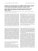
number MK595701.1. Polymerase chain
reaction targeting species specific bla gene
showed amplification of 533bp (Fig 2). Li et
al., (2016) reported an extracellular matrix
containing an unusual polysaccharide, in the
dormant spores of B. cereus and investigated
its key role in the adaptation and fitness to the
environment. Sonawdekar and Gupte (2016)
have isolated EPS producing B. cereus from
oil contaminated sites from in and around
Navi Mumbai and Thane districts of
Maharashtra. Similarly, EPS producing
Bacillus cereus GU 812900 was isolated from
the stainless steel test panel and it contained
54%
sugar
and
1.85%
protein
(Bragadeeswaran et al., 2011). Singh and
others (2011) identified the major bacteria
associated with seaweeds Ulva and Gracilaria
a Marinomonas spp. and Bacillus spp.
Production of
Bacillus cereus
exopolysaccharides
by
Effect of media
The bacterial production of EPS depends
highly on composition of substrate and
environmental conditions (Rabha et al., 2012).
Among different media tested for EPS
production, the dry weight of EPS was
recorded maximum in BHI (1.65 mg ml-1)
followed by LB broth (1.12 mg ml -1) and TSB
(0.62 mg ml-1), which indicate BHI medium
1305
Int.J.Curr.Microbiol.App.Sci (2019) 8(9): 1302-1311
provides suitable nutrients for the production
of EPS. BHI is more nutritious than LB and
TSB in terms of nitrogenous source and hence
the bacteria get sufficient time to produce the
polysaccharide to protect the deleterious
effects of slow exhaustion of nutrients from
the media. Pal and Paul (2013) reported that
EPS production in Cupriavidus pauculus KPS
201 is positively influenced with the increase
of nitrogen and phosphate in the growth
medium.
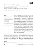
Effect of carbon source
The effect of carbon source on EPS
production by B. cereus was studied with both
monosaccharides (glucose and fructose) and
disaccharides (maltose, cellobiose and
trehalose). Among the different sugars
supplemented in media, glucose (1.56 mg ml1
) was most efficient for EPS production (Fig
3). It has been reported that the EPS
production is directly correlated with the
amount of carbohydrate present in the medium
and its optimum concentration varies
depending upon the individual microorganism
(Ergene and Avcı, 2017). Glucose was the
most efficient carbon source for Lactobacillus
delbrueckii
subsp.
bulgaricus
and
Streptococcus thermophiles (Yuksekdag and
Aslim, 2008). Grobben et al., (1997) reported
that three times more production of EPS with
glucose than fructose as sugar source and the
type of EPS also varies with carbon source.
These results are in contrary to Hereher et al.,
(2018) who reported that due to the relative
ease of polymerization, disaccharides help in
EPS production than monosaccharides.
Further, Lee et al. (1997) observed that
sucrose was efficient for the production of
EPS
from
Bacillus
polymyxa.
The
polysaccharide, starch was more effective in
enhancing
the
EPS
production
by
Pseudomonas stutzeri AS22 than glucose
(Maalej et al., 2014).
Chemical and structural analysis of EPS
Fazio et al., (1982) have shown that EPS from
marine bacterium is rich in galacturonic acids.
During chemical analysis of the EPS from B.
cereus, it was observed that EPS contained
69.9% carbohydrate, 8.1% protein, 3.2% total
uronic content and 1.5% sulphate content.
This is in accordance with observation of
Singh et al. (2011) who reported that the
quantity of carbohydrates, protein and sulfate
was 343.14, 107.68 and 50.28 mg l-1,
respectively in EPS produced by Bacillus
licheniformis.
Generally, the bacterial EPSs have higher
carbohydrate content than sulphate and protein
(Zhenming and Yan, 2005).
Structural analysis of EPS was carried out by
FT-IR analysis (Fig. 4) which showed a
characteristic N-H and OH stretch at around
3292.93 cm−1 and a C-H stretching vibration
at around 2925 cm−1 (Deepika et al., 2016).
The absorption peaks within 1650-1540 cm−1
attributed to vibrations of a CO, NH and CN
bending of protein and peptides.
The absorption peaks within 1200-1000 cm−1
attributed to vibrations of a broad stretch of C
O and C O C glycosidic bands, which revealed
the presence of carbohydrates (Zhang et al.,
2013) that would be sugar monomers in the
EPS.
The absorption peak at 600 cm−1 and 492 cm−1
could be attributed to the S-S stretch. The
absorption observed at 1500-1600 cm−1 could
be attributed to the stretching vibration of
C=C and C–N groups. Peaks at 884 cm−1
ascertain the presence of glycosidic linkage
bonds. The composition and components of
exopolymeric substance of bacteria have large
implications in their bioactive properties.
1306
Int.J.Curr.Microbiol.App.Sci (2019) 8(9): 1302-1311
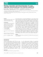
Fig.1A) colony morphology of exopolysaccharide producing bacterial isolates on Trypticase
soya agar and (B) crude extract of exopolysaccharide produced by Bacillus cereus in Luria
bertani broth
A
B
Fig.2 B cereus group specific PCR.
Lane 1: 1kb ladder, Lane 2: B. cereus isolates recovered from S. wightii. Lane 3: Positive
control, Lane 4: Negative control
Fig.3 Effect of different sugars on EPS production by Bacillus cereus
1307
Int.J.Curr.Microbiol.App.Sci (2019) 8(9): 1302-1311
Table.1 Morphological and biochemical characteristics of the isolates and the EPS yield
Isolates
Sample source
Colony morphology
Gram reaction
Oxidase reaction
Motilitiy
Indole
Catalase
Glucose fermentation
Lactose fermentation
Starch hydrolysis
Protease activity
Citrate utilization
EPS yield (mg ml-1)
BI
Sargassum
wightii
Light
brown,
mucoid,
large 2-3mm
size, smooth
Gram
positive,
spore
forming, rod
Positive
Motile
Negative
Positive
Positive
Negative
Positive
Positive
Positive
0.82
B2
Sargassum
wightii
Light
brown,
mucoid,
large 2-3mm
size, smooth
Gram
positive
spore
forming, rod
Positive
Motile
Negative
Positive
Positive
Negative
Positive
Positive
Positive
1.06
C1
Sargassum
wightii
Creamy,
mucoid, small
1-2mm
size,
smooth
WI
Padina
gymnocephalus
Whitish, Mucoid
large 2-3mm size,
irregular edge
Gram negative Gram negative,
non
spore non
spore
forming, cocci
forming,
rod
shaped
Negative,
Positive
Non motile
Non motile
Negative
Positive
Positive
Positive
Positive
Positive
Positive
Negative
Negative
Negative
Negative
Negative
Negative
Positive
Nil
0.3
Fig.4 FT-IR Spectrum of crude EPS from Bacillus cereus
1308
W2
Padina
gymnocephalus
Whitish, Mucoid
large 2-3mm size,
irregular edge
Gram negative
Positive
Non motile
Positive
Positive
Positive
Negative
Negative
Negative
Positive
Nil
Int.J.Curr.Microbiol.App.Sci (2019) 8(9): 1302-1311
The present study gives an insight to the
production of EPS by bacteria associated with
Sargassum wightii, Turbinaria connoides,
Padina gymnocephalus. Bacillus cereus was
the potential EPS producing bacterium
isolated and the EPS production was enhanced
in the presence of glucose. The extracted
exopolysaccharide from Bacillus cereus may
be applied in many fields such as textiles,
pharmaceuticals and food industry after
carrying out further studies pertaining to its
unique properties.
References
Ates, O., 2015. Systems biology of microbial
exopolysaccharides
production.
Frontiers in bioengineering and
biotechnology, 3, p.200.
Becker, A., Katzen, F., Pühler, A. and Ielpi,
L., 1998. Xanthan gum biosynthesis and
application:
a
biochemical/genetic
perspective. Applied microbiology and
biotechnology, 50(2), pp.145-152.
Berekaa, M.M. and Ezzeldin, M.F., 2018.
Exopolysaccharide
from
Bacillus
mojavensis DAS10-1; Production and
Characterization. Journal of PurE and
aPPliEd Microbiology, 12(2), pp.633640.
Berekaa, M.M. and Ezzeldin, M.F., 2018.
Exopolysaccharide
from
Bacillus
mojavensis DAS10-1; Production and
Characterization. Journal of PurE and
aPPliEd Microbiology, 12(2), pp.633640.
Blumenkrantz, N. and Asboe-Hansen, G.,
1973. New method for quantitative
determination of uronic acids. Analytical
biochemistry, 54(2), pp.484-489.
Blumenkrantz, N. and Asboe-Hansen, G.,
1973. New method for quantitative
determination of uronic acids. Analytical
biochemistry, 54(2), pp.484-489.
Bragadeeswaran, S., Jeevapriya, R., Prabhu,
K., Rani, S.S., Priyadharsini, S. and
Balasubramanian,
T.,
2011.
Exopolysaccharide
production
by
Bacillus cereus GU812900, a fouling
marine bacterium. African Journal of
Microbiology Research, 5(24), pp.41244132.
Cha, J.M., Cha, W.S. and Lee, J.H., 1999.
Removal of organo-sulphur odour
compounds by Thiobacillusnovellus
SRM,
sulphur-oxidizing
microorganisms. Process Biochemistry,
34(6-7), pp.659-665.
Cha, J.M., Cha, W.S. and Lee, J.H., 1999.
Removal of organo-sulphur odour
compounds by Thiobacillusnovellus
SRM,
sulphur-oxidizing
microorganisms. Process Biochemistry,
34(6-7), pp.659-665.
Dubois, M., Gilles, K.A., Hamilton, J.K.,
Rebers, P.T. and Smith, F., 1956.
Colorimetric method for determination
of sugars and related substances.
Analytical chemistry, 28(3), pp.350-356.
Dubois, M., Gilles, K.A., Hamilton, J.K.,
Rebers, P.T. and Smith, F., 1956.
Colorimetric method for determination
of sugars and related substances.
Analytical chemistry, 28(3), pp.350-356.
Ergene, E. and Avcı, A., 2017. Effects of
Cultural
Conditions
on
Exopolysaccharide
Production
by
Bacillus sp. ZBP4. Journal of
Agricultural Sciences, 24(3), pp.386393.
Kumar, A. S., Mody, K., and Jha, B. (2007).
Bacterial
exopolysaccharides-a
perception. J. Basic Microbiol. 47(2),
103–117.
Li, Z., Hwang, S. and Bar-Peled, M., 2016.
Discovery of a unique extracellular
polysaccharide in members of the
pathogenic Bacillus that can co-form
with spores. Journal of Biological
Chemistry, 291(36), pp.19051-19067.
Lowry, O.H., Rosebrough, N.J., Farr, A.L. and
Randall,
R.J.,
1951.
Protein
1309
Int.J.Curr.Microbiol.App.Sci (2019) 8(9): 1302-1311
measurement with the Folin phenol
reagent. Journal of biological chemistry,
193, pp.265-275.
Lowry, O.H., Rosebrough, N.J., Farr, A.L. and
Randall,
R.J.,
1951.
Protein
measurement with the Folin phenol
reagent. Journal of biological chemistry,
193, pp.265-275.
Mano, J.F., Silva, G.A., Azevedo, H.S.,
Malafaya, P.B., Sousa, R.A., Silva, S.S.,
Boesel, L.F., Oliveira, J.M., Santos,
T.C., Marques, A.P. and Neves, N.M.,
2007. Natural origin biodegradable
systems in tissue engineering and
regenerative medicine: present status
and some moving trends. Journal of the
Royal Society Interface, 4(17), pp.9991030.
Oliwa‐Stasiak, K., Molnar, C.I., Arshak, K.,
Bartoszcze, M. and Adley, C.C., 2010.
Development of a PCR assay for
identification of the Bacillus cereus
group species. Journal of applied
microbiology, 108(1), pp.266-273.
Pal, A. and Paul, A.K., 2013. Optimization of
cultural conditions for production of
extracellular
polymeric
substances
(EPS) by serpentine rhizobacterium
Cupriavidus pauculus KPS 201. Journal
of Polymers, 2013.
Plante, C.J. and Shriver, A.G., 1998.
Differential lysis of sedimentary bacteria
by Arenicola marina L.: examination of
cell wall structure and exopolymeric
capsules as correlates. Journal of
Experimental Marine Biology and
Ecology, 229(1), pp.35-52.
Sanchez, M.C., Llama‐Palacios, A., Blanc, V.,
Leon, R., Herrera, D. and Sanz, M.,
2011. Structure, viability and bacterial
kinetics of an in vitro biofilm model
using six bacteria from the subgingival
microbiota. Journal of periodontal
research, 46(2), pp.252-260.
Sheetal Sonawdekar and Arpita Gupte. 2016.
Production and Characterization of
Exopolysaccharide produced By Oil
Emulsifying
Bacteria.
Int.J.Curr.Microbiol.App.Sci. 5(2): 254262.
Singh, R.P., Shukla, M.K., Mishra, A.,
Kumari, P., Reddy, C.R.K. and Jha, B.,
2011. Isolation and characterization of
exopolysaccharides
from
seaweed
associated
bacteria
Bacillus
licheniformis. Carbohydrate polymers,
84(3), pp.1019-1026.
Sonawdekar, S. and Gupte, A., 2016.
Production and characterization of
exopolysaccharide produced by oil
emulsifying bacteria. Int J Curr
Microbiol App Sci, 5(2), pp.254-262.
Tallent, S.M., Kotewicz, K.M., Strain, E.A.
and Bennett, R.W., 2012. Efficient
isolation and identification of Bacillus
cereus group. Journal of AOAC
International, 95(2), pp.446-451.
Tallgren, A.H., Airaksinen, U., von
Weissenberg, R., Ojamo, H., Kuusisto,
J.
and
Leisola,
M.,
1999.
Exopolysaccharide-producing bacteria
from sugar beets. Appl. Environ.
Microbiol., 65(2), pp.862-864.
Tang, X. Z., Kumar, P., Alavi, S., and
Sandeep, K. P. (2012). Recent advances
in biopolymers and biopolymer-based
nanocomposites for food packaging
materials. Crit. Rev. Food Sci. Nutr. 52,
426–442.
doi:10.1080/10408398.2010.500508
Vroman, I., and Tighzert, L. (2009).
Biodegradable polymers. Materials 2,
307–344. doi:10.3390/ma2020307
Zhang, N., Liu, X., Yu, L., Shanks, R.,
Petinaks, E. and Liu, H., 2013. Phase
composition and interface of starch–
gelatin blends studied by synchrotron
FTIR micro-spectroscopy. Carbohydrate
polymers, 95(2), pp.649-653.
Zhang, N., Liu, X., Yu, L., Shanks, R.,
Petinaks, E. and Liu, H., 2013. Phase
composition and interface of starch–
1310
Int.J.Curr.Microbiol.App.Sci (2019) 8(9): 1302-1311
gelatin blends studied by synchrotron
FTIR micro-spectroscopy. Carbohydrate
polymers, 95(2), pp.649-653.
Zhao, M., Cui, N., Qu, F., Huang, X., Yang,
H., Nie, S., Zha, X., Cui, S.W.,
Nishinari, K., Phillips, G.O. and Fang,
Y., 2017. Novel nano-particulated
exopolysaccharide
produced
by
Klebsiella
sp.
PHRC1.
001.
Carbohydrate polymers, 171, pp.252258.
How to cite this article:
Minimol, V. A., Pankaj Kishore, Ranjit K. Nadella, K. R. Sreelakshmi, S. S. Greeshma, M. M.
Prasad and Suseela Mathew 2019. Isolation and Characterization of Exopolysaccharide
producing
Bacillus
cereus
from
Brown
Seaweed-Sargassum
wightii.
Int.J.Curr.Microbiol.App.Sci. 8(09): 1302-1311. doi: />
1311