N-terminal Domain of tlyA from mycobacterium tuberculosis displayed concentration dependent ordered structure
Bạn đang xem bản rút gọn của tài liệu. Xem và tải ngay bản đầy đủ của tài liệu tại đây (187.79 KB, 9 trang )
Int.J.Curr.Microbiol.App.Sci (2019) 8(10): 1917-1925
International Journal of Current Microbiology and Applied Sciences
ISSN: 2319-7706 Volume 8 Number 10 (2019)
Journal homepage:
Original Research Article
/>
N-terminal Domain of tlyA from Mycobacterium tuberculosis Displayed
Concentration Dependent Ordered Structure
V.B. Shivaleela1, Srihari Prathapaneni2, P. Sharada1* and K. Giri Gowda2
1
Department of Biotechnology, Basaveshwara Engineering College,
Bagalkot-587102, Karnataka, India
2
Sampoorna International Institute of Agri. Science & Horticulture Technology,
Mandya-571433, Karnataka, India
*Corresponding author
ABSTRACT
Keywords
Mycobacterium
tuberculosis, Nterminal domain,
Circular dichroism,
tlyA, Maltose
binding protein
Article Info
Accepted:
15 September 2019
Available Online:
10 October 2019
Mycobacterium tuberculosis (Mtb), the causative agent of the disease tuberculosis,
is an ancient pathogen and a major cause of death worldwide. Although various
virulence factors of M. tb have been identified, its pathogenesis remains
incompletely understood. TlyA is a virulence factor that is evolutionarily
conserved in many gram-positive bacteria, but its full length structure and function
in the pathogenesis of infection with Mtb has not been elucidated. In the present
study, we cloned, expressed and purified N-terminal domain of tlyA, which play a
crucial role in the binding of the co-substrate S-adenosyl-L-methionine. We
characterized the protein by SDS-PAGE and Circular Dichroism. TlyA model
generated using tlyA crystal structure, clearly indicates E59 separates between Nterminal domain (NTD) and C-terminal domain (CTD).
Introduction
Mycobacterium tuberculosis is the causative
agent of tuberculosis (TB), most successful
gram-positive bacterial pathogen, primarily
infects human lungsand is a major global
public health problem, with approximately 9
million new cases and nearly 2 million deaths
each year (WHO, 2018).
Efforts to search for virulence factors of M.
tuberculosis (Mtb) is unrelenting, many
researchers have identified genes that may
serve as potential targets for vaccine
development. Among the unexplored gene
products of Mtb, tlyA (Rv1694) was recently
identified as a possible virulence factor. TlyA
protein have a haemolysin activity and tlyA is
a 268 amino acid polypeptide (Martino MC et
al., 2001). The tlyA gene is also present in
several pathogenic mycobacterial species,
including Mycobacterium tuberculosis and
Mycobacterium
leprae.
Although,
M.
tuberculosis and M. leprae evolved from a
1917
Int.J.Curr.Microbiol.App.Sci (2019) 8(10): 1917-1925
common ancestor, M. leprae possesses fewer
genes. Genes conserved between the two
species are hence considered important for
pathogenicity and virulence.Almost all tlyA
homologues have K-D-K-E domain for 2hydroxy-ribose methylation in ribosomal RNA
(Wren et al., 1998).
When TlyA is introduced into non-haemolytic
M. smegmatis strains, and cloned into E.coli, it
showed contact dependent haemolytic activity
(Wren et al., 1998). It has been previously
shown that in H37Rv, the tlyA gene may be a
part of an operon containing at least three
other genes: tlyA (Rv1694), ppnk (Rv1695)
and RecN (Rv1696), homologous to E.coli
RecN (Wren et al., 1998). TlyA is also known
to function as a ribosomal RNA
methyltransferase. It is known to methylate
50S and 30S ribosomal RNA and makes Mtb
susceptible to the peptide antibiotic
capreomycin (Monshupee et al., 2012).
Despite intense research on Mtb pathogenesis,
detailed molecular mechanisms of the role of
distinct mycobacterial virulence factors
remain in completely understood. To
understand its mechanism of pathogenesis, the
functions of numerous M. tuberculosis gene
products are being characterized in animal
models. Recently, Rahman et al., (2010)
reported that tlyA (Rv1694) of M. tuberculosis
possesses haemolytic activity by binding with
and oligomerizing into host cell membranes.
Resistance to antibiotics in Mtb can aquire via
mutation of tlyA, protein belongs to a unique
group of methyltransferases for which the loss
of function confers bacterial antibiotic
resistance. Many bacterial genera lack tlyA,
the potent antibiotic activity of capreomycin is
specific against Mtb. (Kumar et al., 2011). In
this study, our aim was to understand the
structure and possible role of tlyA N-terminus
in the interaction of SAM binding inMtb.
Materials and Methods
Strains and plasmid
pCDF vector system was obtained from
Invitrogen (California, USA) and was used
according to the manufacturer’s instructions.
E.coli DH5α competent cells were obtained
from Invitrogen (California, USA).
Isolation of genomic DNA
Bacterial culture (50ml) was harvested at
optical density of A6000.5-0.6 at 37°C by
centrifugation at 4150 rpm for 7 mins. The
pellet was resuspended by adding 6ml of
freshly prepared chloroform-methanol (3:1)
solution and vortexed until the bacteria were
lysed as evident by a clear bottom layer. 6ml
of Tris-buffered phenol (pH 8) was added and
vortexed. 9ml of guanidinium thiocyanate
buffer (GTC) solution was added and
vortexed. The sample was centrifuged at
10000x g for 10-15 mins and a clear
supernatant was collected. DNA was
precipitated out by adding equal volumes of
isopropanol, mixed gently and centrifuged at
13-14,000 rpm for 10-15 mins. The pellet was
suspended in 4 ml TE buffer and transferred to
an eppendorf tube. The DNA was used for
PCR with primers for tlyA gene.
PCR amplification of tlyA N-terminal
domain (NTD)
Oligonucleotide
primers
used
for
amplification of Mtb-tlyA NTD were designed
based on the tlyA sequence from
mycobacterium tuberculosis strain H37Rv
deposited in genome database (NCBI
accession no.AQO55200.1). Primers were
designed based on its sequence for generating
a truncation of tlyA NTD. The sequence of the
forward primer was 5′-GCGGAATTCA
TGGCACGACGTGCCCGCGTT-3′ and the
reverse primer was 5′- TATGGTACCTTC
1918
Int.J.Curr.Microbiol.App.Sci (2019) 8(10): 1917-1925
ACTGTCGGTCACCAC-3′.
The
PCR
reaction mixture consisted of 5 µl of 5X
Phusion buffer supplied with the enzyme, 200
µM of each dNTPs, 0.5 µM of each primer,
500ng of DNA template, and of 0.02 U/µl
Phusion DNA polymerase (New England
Biolabs, Massachusetts, USA) and water to a
final volume of 50 µl. The gene was
successfully amplified using the following
PCR conditions: 98 °C for 30 sec followed by
30 cycles of denaturation at 98 °C for 10 sec,
annealing at 60 °C for 30sec, extension at 72
°C for 30sec and the final extension was
carried out at 72 °C for 10 min on a PTC-100
Thermocycler (M.J. Research, Watertown,
MA). The PCR product was analyzed on a 1
% agarose gel electrophoresis and DNA band
corresponding to the expected size was
purified using a gel extraction kit
(ThermoFisher
Scientific,
Waltham,
Massachusetts, USA).
Cloning and DNA sequencing
The PCR product was subcloned into plasmid
DNA using the modified MBP-His-pCDF
vector system (Novagen, Wisconsin, USA)and
TEV recognition site upstream of the MCSI
(multiple cloning site I). The PCR product and
MBP-His-pCDF vector was digested with
restriction enzymes EcoRI and KpnI for 2h at
37o C, product was purified. 2 µl of purified
PCR product was mixed with 0.5 µl linearized
MBP-His-pCDF cloning vector in presence of
0.5 µl T4 DNA ligase (ThermoFisher
Scientific, Waltham, Massachusetts, USA)
and incubated overnight at 16 °C. Then the
ligation mixture was directly used for the
transformation of CaCl2 competent DH5α cells
by heat shock method (Inoue et al., 1990).
Colony PCR was performed to screen positive
colonies. Positive colonies were picked,
grown overnight in 5 ml of LB broth at 37 °C
and plasmids were isolated using commercial
mini-prep kit (GCC Biotech, West Bengal,
India). Restriction digestion screening of the
isolated plasmids was done to select the
construct containing the correct size insert and
selected
constructs
were
sequenced.
Sequencing was performed in both the
directions using vector specific T7 promoter
primer.
Expression and purification
The MBP-His-pCDF vector containing MtbtlyANTD was transformed into E. coli BL21
(Star) competent cells. For protein expression,
transformed BL21 (Star) cells were grown at
37ºC to an optical density of 0.6 at 600 nm
(OD600) and induced with 200M isopropylß-thiogalactopyranoside (IPTG). Induced
cultures were transferred to 16 C and cells
were grown for 12-14 h. Cells were harvested
by centrifugation at 18,000 rpm at 4ºC and cell
pellets were stored at -20ºC until further use.
For protein purification, cell pellets from 1
litre culture were resuspended in 20 ml of ice
cold binding buffer containing 50 mM
TrisHCl (pH 7.5), 300 mM sodium chloride,
10% glycerol (v/v) and 5 mMβmercaptoethanol.
PMSF
was
added
immediately after the lysis(0.2 mM). Cells
were disrupted by sonication on ice with 50%
amplitude and a pulse of 20 sec on and 60 sec
off for 15 min. The lysate was centrifuged at
18,000 rpm for 1h at 4ºC to separate
supernatant from cell debris. The supernatant
was loaded onto 5 ml Ni-NTA affinity column
pre-equilibrated with the binding buffer.
Protein was eluted by running a linear gradient
of 0–1000 mM imidazole in 60 ml of buffer A
(50 mM TrisHCl (pH 7.5), 1 M imidazole, 300
mM sodium chloride and 10% glycerol (v/v)]
at a flow rate of 1 ml/min. Eluted fractions
were analyzed on sodium dodecyl sulfatepolyacrylamide gel electrophoresis (SDSPAGE) and fractions containing tlyA NTD
were pooled and dialyzed against the buffer A
(50 mM TrisHCl (pH 7.5), 300 mM sodium
chloride and 10% glycerol(v/v).
1919
Int.J.Curr.Microbiol.App.Sci (2019) 8(10): 1917-1925
TEVprotease cleavage of MBP-His tag
Dialyzed tlyA protein was transferred to 50 ml
falcon tube and subjected to TEV proteolysis
(Yarden et al., 2003), TEV to protein ration
used was (1:100) and incubated at 18ºC for
overnight and the sample was loaded onto 5
ml Ni-NTA affinity column pre-equilibrated
with the binding buffer. TEV cleaved protein
was eluted by 30 ml of buffer A [50 mM
TrisHCl (pH 7.5), 300 mM sodium chloride
and 10% glycerol (v/v)] at a flow rate of 1
ml/min. MBP-His tag bound to Ni-NTA
column, whereas unbound protein without tag
was eluted out.
Gel filtration chromatography
Size
exclusion
chromatography
was
performed using Hi-Load 16/60 prep grade
Superdex75 column pre-equilibrated with
buffer containing 20mMTris-HCl (pH 7.5),
1M NaCl, 10% (v/v) glycerol and 5mM βmercaptoethanol using AKTA purification
system (GE Healthcare). Protein was
concentrated up to 5 ml and injected using 5
ml injector, flow rate of the column was fixed
at 0.8 ml/min. Fractions collected were
analyzed on 15% SDS-PAGE and fractions
containing tlyA NTD were pooled and
concentrated. Protein concentration and yield
were determined using the Bio-Rad protein
assay kit with bovine serum albumin (BSA) as
a standard.
SDS-PAGE
SDS-PAGE was performed according to the
method of Laemmli (1970). The expressed
soluble fractions were diluted with the sample
buffer 1:5 ratio and boiled for 3min before
loading. Standard protein marker was used as
a broad range protein standard to estimate the
molecular weight of the proteins (Thermo
Fisher Scientific, Waltham, Massachusetts,
USA). The protein sample was isolated at
room temperature with a current of 20mA.
The proteins were stained with Coomassie
brilliant blue G-250 (Bio-Rad, Hercules,
California, United States)
Circular Dichroism studies
Measurements were performed using a
Chirascan
CD
spectrometer
(Applied
Photophysics) according to the method of
Whitmore et al., (2008). Cuvette path length
used was 1 mm, and sample concentration was
0.30 mg/ml. Protein was dialyzed with the
buffer contained 10 mM sodium phosphate,
pH 8.0, 200 mMNaCl. The purity of samples
was checked by SDS-PAGE and sizeexclusion chromatography. Each spectrum
was averaged from four repeated scans
ranging between 180 and300 nm at a scan rate
of 1.25 nm/s. Raw data were corrected by
subtracting the contribution of the buffer to
the signal.
Results and Discussion
Cloning tlyA NTD in pCDF vector
The tlyA NTD (residues 1 to 59) was
subcloned into pCDF vector (Invitrogen,
California, USA) containing a TEV
recognition site upstream of the MCS. The
expression vectors encode a Maltose binding
protein and hexahistidine tag on the Nterminus, followed by a TEV protease site and
ensuing desired coding sequence. It was than
expressed as a MBP-His fusion protein in E.
coli (star) strain as described.
Expression and purification
Modified MBP-His-TEV-pCDF vector system
was used as the expression vector which
harbors a strong promoter, T7. Maltose
binding protein (MBP) tag helps in protein
folding and pCDF-Mtb-tlyA was transformed
to E.coli strains BL21 (star). The recombinant
protein expression level was high when over
produced. Soluble form of the protein was
1920
Int.J.Curr.Microbiol.App.Sci (2019) 8(10): 1917-1925
detected with the BL21 (star) strain, MBP and
His-tagged tlyA was confirmed by analyzing
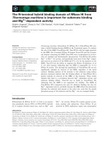
the protein on 15% SDS-PAGE (Figure 1A).
Temperature and IPTG concentration for
protein production were optimized and
optimum temperature for obtaining maximum
protein production was 16C, whereas
optimum concentration of IPTG was found to
be 200 M. The MBP fusion protein was
purified
using
standard
affinity
chromatography with Ni-NTA beads. Both
maltose binding protein and histidine tag were
removed by cleavage with TEV protease. The
TEV protease is highly specific and does not
cleave other sites on the protein. Gel filtration
profile showed single predominant peak
indicating the Mtb-tlyA NTD, eluted protein
is homogenous and protein eluted after 85 ml
on Superdex 75 column, which was further
confirmed by SDS-PAGE (Data not shown).
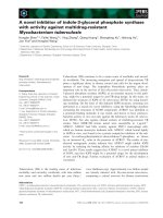
Mass of the protein was further confirmed by
MALDI-TOF
(Figure
1B).
Fractions
corresponding to protein on SDS-PAGE were
pooled, concentrated and stored in -80C.
Figure.1(A) Purification of Mtb-tly ACTD (B) MALDI-TOF studies of purified Mtb-tly ACTD
MBP-His cleaved tlyA NTD
Purified tlyA NTD
Intens. [a.u.]
Figure.1B
L 0:E8 MS, BaselineSubtracted, Smoothed
8000
6000
7640.0
7835.9
4000
3909.8
2263.8
2000
0
2000
3000
4000
5000
6000
7000
1921
8000
9000
10000
m/z
Int.J.Curr.Microbiol.App.Sci (2019) 8(10): 1917-1925
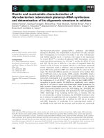
Figure.2 (A)CD spectroscopic analysis Mtb-tlyACTD at 0.4 mg/ml concentration. (B) CD
spectra of Mtb-tlyACTD at concentration of 2 mg/ml.
Figure.2A
Figure.2B
1922
Int.J.Curr.Microbiol.App.Sci (2019) 8(10): 1917-1925
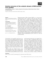
Figure.3 Mtb-TlyA model generated using I-Tasser server using CTD crystal structure as a
template showing NTD and CTD separates at E59.
Figure.3
NTD
E59
CTD
Secondary structure studies using circular
Dichroism
To explain more precisely about the SAM
binding site, we purified the tlyA NTD,
residues 1–59, and measured the CD spectrum
in the concentration of 0.4 to 2.0 mg/ml.
Disordered structure of the truncated Nterminus tlyA was observed at 0.4 mg/ml
(Figure
2A),
whereas
increasing
concentrations indicated increasing fractions
of helical secondary structure (Figure 2B).
Such behaviour is consistent with intrinsically
disordered protein that upon association with
protein undergoes a structured transition,
facilitating binding with its target, tlyA CTD
containing methyltransferase domain (Witek
et al., 2017). Amino acid identity shared
among bacterial tlyA NTD is not so high. The
full-length tlyA protein comprises a
1923
Int.J.Curr.Microbiol.App.Sci (2019) 8(10): 1917-1925
methyltransferase domain, extending from
residue64 to 268, and NTD, from residues 1
to 59. What might be the structure of the Nterminal domain? The CD spectrum strongly
suggests that the tlyA NTD is predominantly a
disordered structure, although it may have
small fractions of helical and extended
conformation (Figure 3). Bioinformatics
analysis of the tlyANTD is equivocal, with
one algorithm predicting some secondary
structure and others predicting disorder. The
structural attributes of the tlyA NTD suggest
that while alone it is largely disordered, it
nevertheless may provide a target for protein
interactions that would perforce induce
structure upon binding.
Based on the previous studies by Witek et al.,
(2017), homology model prediction studies
indicated, Glu59surface exposed amino acid
separates between NTD and CTD of MtbtlyA.
Studies also indicated that NTD and CTD
were intact even after cleavage under the
solution conditions.
Acknowledgement
One of the authors Shivaleela V B
acknowledge TEQIP-III to Basavehwar
Engineering College (A), Bagalkot for the
financial support.
Conflicts of interest
The authors declare no conflict of interest.
References
Colovos C and Yeates TO (1993) Verification
of protein structures: patterns of
nonbonded
atomic
interactions.
Protein Sci. 2:1511–1519.
Gasteiger E, Gattiker A, Hoogland C, Ivanyi
I, Appel RD and Bairoch A (2003)
ExPASy: the proteomics server for indepth protein knowledge and analysis.
Nucl Acids Res 31:3784–3788.
Inoue H, Nojima H and Okayama H. (1990)
High efficiency transformation of
Escherichia coli with plasmids, 30: 96,
23-28.
Kumar A, Saigal K, Malhotra K, Sinha KM,
and Taneja B (2011) Structural and
Functional
Characterization
of
Rv2966c Protein Reveals an RsmDlike
Methyltransferase
from
mycobacterium tuberculosis and the
Role of Its N-terminal domain in
target recognition. The Journal of
Biological Chemistry, 286:22, 19652–
19661.
Laemmli U (1970) Denaturing (SDS)
discontinuous gel electrophoresis,
Nature. 277: 680–685.
Martino, M. C., et al., (2001). "Helicobacter
pylori
pore-forming
cytolysinorthologue TlyA possesses in
vitro hemolytic activity and has a role
in colonization of the gastric mucosa."
Infect Immun, 69(3): 1697-1703.
Maus CE, Plikaytis BB and Shinnick TM
(2005) Mutation of tlyA confers
capreomycin
Resistance
in
mycobacterium
tuberculosis.
Antimicrobial
agents
and
Chemotherapy, Feb. 2005:571–577.
Monshupanee,
T.,
et
al.,
(2012).
"Capreomycin
susceptibility
is
increased by TlyA-directed 2'-Omethylation on both ribosomal
subunits." Mol. Microbiol., 85(6):
1194-1203.
Opatowsky Y, Chomsky-Hecht O, Kang M,
Campbell KP and Hirsch JA (2003)
The
Voltage-dependent
Calcium
Channel Subunit Contains Two Stable
Interacting Domains, The Journal of
Biological Chemistry, 278 (52),
52323–52332.
Pruitt KD, Tatusova T and Maglott DR
(2005). NCBI Reference Sequence
(RefSeq): a curated non-redundant
1924
Int.J.Curr.Microbiol.App.Sci (2019) 8(10): 1917-1925
sequence database of genomes,
transcripts and proteins. Nucleic Acids
Res. 33 (Database issue):501–4.
Rahman A, Srivastava SS, Sneh A, Ahmed N,
and Krishnasastry MV (2010)
Molecular characterization of tlyA
gene
product,
Rv1694
of
mycobacterium tuberculosis: a nonconventional hemolysin and a
ribosomal RNA methyl transferase.
BMC Biochem. 11:35.
Turoverov KK, Kuznetsova IM and Uversky
VN (2010) The protein kingdom
extended. Ordered and intrinsically
disordered proteins, their folding,
supramolecular complex formation,
and aggregation. Prog. Biophys. Mol.
Biol. 102, 73–84.
Whitmore L and Wallace BA (2008) Protein
secondary structure analyses from
circular
dichroism
spectroscopy:
methods and reference databases.
Biopolymers 89, 392–400.
Witek MA, Kuiper EG, Minten E, Crispell
EK and Conn GL (2017) A Novel
Motif for S-Adenosyl-L-methionine
binding by the ribosomal RNA
methyltransfer
asetlyA
from
mycobacterium tuberculosis. The
Journal of Biological Chemistry. 292:
5, 1977–1987.
World Health Organization (2018) Global
Tuberculosis Report 2018, World
Health
Organization,
Geneva,
Switzerland.
Wren, B. W., et al., (1998). "Characterization
of a haemolysin from Mycobacterium
tuberculosis with homology to a
virulence
factor
of
Serpulina
hyodysenteriae." Microbiology 144 (Pt
5): 1205-1211.
How to cite this article:
Shivaleela, V.B., Srihari Prathapaneni, P. Sharada and Giri Gowda, K. 2019. N-terminal
Domain of tlyA from Mycobacterium tuberculosis Displayed Concentration Dependent Ordered
Structure. Int.J.Curr.Microbiol.App.Sci. 8(10): 1917-1925.
doi: />
1925