Regulator of G protein signaling 17 as a negative modulator of GPCR signaling in multiple human cancers
Bạn đang xem bản rút gọn của tài liệu. Xem và tải ngay bản đầy đủ của tài liệu tại đây (485.92 KB, 10 trang )
The AAPS Journal, Vol. 18, No. 3, May 2016 ( # 2016)
DOI: 10.1208/s12248-016-9894-1
Review Article
Theme: Heterotrimeric G Protein-based Drug Development: Beyond Simple Receptor Ligands
Guest Editor: Shelley Hooks
Regulator of G Protein Signaling 17 as a Negative Modulator of GPCR Signaling
in Multiple Human Cancers
Michael P. Hayes1 and David L. Roman1,2,3,4
Received 21 September 2015; accepted 15 February 2016; published online 29 February 2016
Abstract. Regulators of G protein signaling (RGS) proteins modulate G protein-coupled receptor
(GPCR) signaling networks by terminating signals produced by active Gα subunits. RGS17, a member of
the RZ subfamily of RGS proteins, is typically only expressed in appreciable amounts in the human
central nervous system, but previous works have shown that RGS17 expression is selectively upregulated
in a number of malignancies, including lung, breast, prostate, and hepatocellular carcinoma. In addition,
this upregulation of RGS17 is associated with a more aggressive cancer phenotype, as increased
proliferation, migration, and invasion are observed. Conversely, decreased RGS17 expression diminishes
the response of ovarian cancer cells to agents commonly used during chemotherapy. These somewhat
contradictory roles of RGS17 in cancer highlight the need for selective, high-affinity inhibitors of RGS17
to use as chemical probes to further the understanding of RGS17 biology. Based on current evidence,
these compounds could potentially have clinical utility as novel chemotherapeutics in the treatment of
lung, prostate, breast, and liver cancers. Recent advances in screening technologies to identify potential
inhibitors coupled with increasing knowledge of the structural requirements of RGS-Gα protein-protein
interaction inhibitors make the future of drug discovery efforts targeting RGS17 promising. This review
highlights recent findings related to RGS17 as both a canonical and atypical RGS protein, its role in
various human disease states, and offers insights on small molecule inhibition of RGS17.
KEYWORDS: cancer; drug discovery; GPCR; G protein; regulator of G protein signaling.
INTRODUCTION
G protein-coupled receptors (GPCRs) are the largest
class of proteins in the human genome and regulate various
physiological processes, ranging from chemosensation to
neurotransmission (1). Due to their evolutionarily conserved
function as small molecule binding proteins, GPCRs have
proved to be useful targets for the development of therapeutic agents. Currently, one third to one half of drugs marketed
in the USA act on a GPCR, targeting diseases like
hypertension, asthma, schizophrenia, and prostate cancer.
Interestingly, over 30% of these drugs elicit their effects by
binding to one of only 50 receptors, which represents only
∼13% of the non-olfactory GPCR-ome, leaving ample room
for future GPCR-targeted drug discovery efforts (2). Furthermore, as G protein-mediated signaling events have been
1
Department of Pharmaceutical Sciences and Experimental Therapeutics, University of Iowa, Iowa City, Iowa, USA.
2
Cancer Signaling and Experimental Therapeutics Program, Holden
Comprehensive Cancer Center, University of Iowa Hospitals and
Clinics, Iowa City, Iowa, USA.
3
115 S. Grand Avenue, S327 PHAR, Iowa City, Iowa 52242, USA.
4
To whom correspondence should be addressed. (e-mail: )
1550-7416/16/0300-0550/0 # 2016 American Association of Pharmaceutical Scientists
clinically validated for therapeutic use, proteins downstream
of these receptors have gained attention as potential sites of
chemical intervention, such as the regulator of G protein
signaling (RGS) protein family. Inhibition of RGS proteins by
small molecules represents a means by which to enhance
GPCR signals by increasing the lifetimes of GTP-bound,
active Gα subunits. One member of the RGS family that has
recently emerged as a potential drug target is RGS17, as it
has been implicated in a number of the most common forms
of cancer, including lung, breast, prostate, and liver cancers
(3–5).
Guanine Nucleotide-Binding Protein (G Protein) Signaling
GPCRs exert their effects by acting as guanine nucleotide exchange factors (GEFs) on G protein α subunits,
thereby translating extracellular stimuli into intracellular
signaling cascades. Gα subunits can be grouped together
based on primary sequence identity, their downstream
signaling partners, and their sensitivity to RGS protein
activity. The inhibitory Gα subunits Gαi, Gαo, and Gαz result
in inhibition of adenylyl cyclases (AC), decreased cellular
cAMP levels, and are sensitive to RGS-mediated GAP
activity, whereas the stimulatory Gαs family activates AC,
increasing intracellular cAMP, and are insensitive to RGS
proteins (6). The activation of the Gαq/11 family, which is also
550
RGS17 in Multiple Forms of Cancer
sensitive to regulation by RGS family members, results in
increased phospholipase C (PLC) activity, ultimately resulting
in calcium mobilization (7). Finally, the Gα12/13 family
activates RhoGEF, which acts as a GAP for Gα12/13 subunits
and a GEF for the small GTPase Rho, linking GPCR
signaling to Rho-mediated cellular events, such as cytoskeletal rearrangements and cell division (8). Upon stimulation
with ligand, a ternary complex is formed between the ligand,
GPCR, and Gαβγ heterotrimer, where GDP is exchanged for
GTP in the Gα subunit, which then dissociates from the
obligate Gβγ dimer (9). Both the Gβγ and GTP-bound Gα
are then able to initiate signaling cascades through interaction
with downstream effectors, such as AC, PLC, ion channels,
and RhoGEF. In order to terminate signaling, Gα hydrolyzes
GTP to GDP via its intrinsic GTPase activity, and Gα-GDP
then associates with βγ, reforming the inactive Gαβγ
heterotrimer, thus terminating signaling (Fig. 1).
Regulators of G Protein Signaling
RGS proteins, as GTPase acceleration proteins (GAPs),
function to expedite signal termination by increasing the rate
of GTP hydrolysis and decreasing the lifetime of Gα-GTP by
orders of magnitude (10). The defining feature of the RGS
family, which is composed of 20 canonical members, is the
presence of a highly conserved, approximately 120 amino acid
region that binds activated Gα subunits, termed the RGS
Homology (RH) domain. This domain is composed of nine α
helices, α1-9, that form a two-lobed structure composed of
the bundle and terminal subdomains (Fig. 2a) (11). Aside
from the RH domain, RGS proteins can contain a number of
accessory domains, leading to their subdivision into four
distinct families based on sequence similarity and the
inclusion of these additional domains, as shown in Fig. 2b.
Additionally, there are approximately 11 noncanonical RGSlike proteins, including GPCR kinases (GRKs), RhoGEFs,
and sorting nexins, that contain RH domains but ostensibly
perform important functions other than or in addition to
acting as Gα GAPs.
The RZ Family
The RZ family is composed of four members, each of
which was shown to be highly homologous to RGSZ1 upon
their initial discovery. The members of this family, RGS17
(RGSZ2), 19 (GAIP), 20 (RGSZ1), and Ret-RGS, are
encoded by three genes Rgs17, Rgs19, and Rgs20. Rgs20
undergoes alternative splicing, giving rise to RGS20 and RetRGS (12,13). As compared to other RGS families, the RZ
family proteins are small and relatively simple. Each member
contains a short N-terminal poly-cysteine (pCys) string, an
RH domain, and a very short C-terminus (13). The pCys
string serves as a substrate for palmitoylation in RGS19,
anchoring the protein in the membrane (14), and this
mechanism is likely conserved in all members of the family,
based on conservation of this sequence and their identification as membrane-bound proteins (15,16). Additionally, all
members of the RZ family can bind to Gαz, though some
family members are capable of binding additional Gα
subtypes (13,17,18).
551
RGS19, the first identified member of the RZ family, was
discovered in 1995 via yeast-two hybrid (Y2H) screening that
employed Gαi3 as bait, and its discovery was notable because
it was the first time a mammalian RGS-Gα protein-protein
interaction had been observed (19). RGS19 and RSG17 share
50% amino acid identity and 75% similarity with the bulk of
the divergence occurring at the extreme N-termini and the
region between the pCys string and the RH domain.
Additionally, unique to RGS19 is a C-terminal PDZ binding
motif that enables GIPC binding, which may act as a scaffold
to regulate RGS19 recruitment (20,21). Functionally, recent
work has begun to show possible connections between
RGS19 and nociception and pain due to its ability to regulate
serotonergic and opiate signals (22,23).
RGS20 was first identified due its GAP activity toward
Gαz, and subsequent efforts determined that it, in fact, had
higher affinity for Gαz than other Gαi/o proteins, leading to its
initial description as RGSZ1 (16,24). Of all the RZ family
members, RGS20 most closely resembles RGS17, as these
two proteins have 53% amino acid identity and 72%
similarity. Notably, the pCys string is perfectly conserved
between RGS20 and 17, though RGS20 harbors a 31 residue
N-terminal extension that RGS17 lacks. A significant body of
evidence exists relating RGS20 function to the regulation of
opioid signaling through the μ-opioid receptor (μOR) (25–
27). As noted above, Ret-RGS is a splice variant of the gene
that also encodes for RGS20, resulting Ret-RGS being 147
residues longer than RGS20. Though Ret-RGS contains the
pCys string common to RZ members, it also contains a
putative membrane spanning domain, potentially further
tethering it to cellular membranes (15). Ret-RGS is the RZ
family member most distinct from RGS17, as the proteins’
primary sequences are only 33% identical and 44% similar,
though the lower degree of similarity can be almost
completely attributed to Ret-RGS’s extended N-terminus.
REGULATOR OF G PROTEIN SIGNALING 17
Gene Structure
Like other RGS proteins, RGS17 was first identified
during Y2H screening for its ability to interact with an
activated Gα subunit, namely constitutively active mutants
of Gαo (13,28). Rgs17 is located on murine chromosome 10
and at position 6q25.3 in humans (29). Subsequent work
identified that in humans Rgs17 can be transcribed into
mRNAs varying in length from 2 to 8 kb, but as only a single
cDNA for RGS17 has been detected, it is presumed that
these differences occur in untranslated regions (10).
Normal Tissue Distribution
The endogenous tissue distribution of Rgs17 is largely
variable depending on the animal species and methodology
employed, but the overall consensus is that RGS17 is found in
the central nervous system. In humans, Rgs17 mRNA can be
detected in the nucleus accumbens (NAc), parahippocampal
gyrus, and putamen, but the highest levels of expression are
observed in the cerebellum, though overall Rgs17 is
expressed to a much lower degree than other RGS family
members (30). Low levels of human Rgs17 is also observed in
552
Hayes and Roman
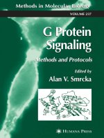
Fig. 1. GPCR-G protein activation cycle. Upon ligand binding to a
GPCR, Gαβγ binds the receptor, where GDP on the Gα subunit is
exchanged for GTP, leading to dissociation of this complex. Gα and
βγ are then free to activate downstream signaling pathways. Signaling
is terminated when an RGS protein binds the Gα-GTP, leading to
GTP hydrolysis to GDP. RGS then dissociates from Gα-GDP, which
is sequestered by βγ, reforming the heterotrimer and priming the
cycle for reactivation upon future GPCR-ligand binding events.
Adopted from PDB Structures: 1AGR (Gα, RGS), 3SN6 (GPCR,
Gα, βγ) (11,73)
the testis (13,30). In mice, Rgs17 exists in the cerebral cortex
and to a higher extent in the striatum and NAc (31). In rats,
Rgs17 can be detected in the frontal cortex, striatum, NAc,
and, interestingly, atrial myocytes (32,33). Moreover, Rgs17
expression can be induced in cultured rat smooth muscle cells
by platelet-derived growth factor DD (PDGF-DD), indicating
a link between GPCR and receptor tyrosine kinase signaling
(34). Additionally, Rgs17 levels are subject to regulation by
neurotransmitter signaling through dopamine receptors. Genetic knockout of the D1 dopamine receptor (D1R) leads to
decreased Rgs17 expression in the medial frontal cortex of
mice; however, when D1R signaling is reduced via prenatal
cocaine exposure in rabbits, increased Rgs17 expression is
observed (31). In rats, prenatal exposure to the D2R agonist
quinpirole results in increased Rgs17 expression in the frontal
cortex, striatum, and NAc (33). Taken together, the tissue
expression discrepancies exhibited between species highlight
the importance of working with human tissue, preferably
primary, whenever possible and that findings from rodent
models may not always be directly translatable to human
health.
GTPase Accelerating Protein Activity
After RGS17 was discovered and identified as being a
member of the RZ family, it was proposed that RGS17 would
be specific for Gαz, similar to RGS20. Early work demonstrated that RGS17 can, in fact, bind and accelerate the
GTPase activity of Gαz, but unlike RGS20, it is not
necessarily specific for this subtype. RGS17 is capable of
binding Gαi1-3, Gαo, and Gαz and displays a preference for
Gαz and Gαo subunits in GAP assays involving purified
proteins. Oddly, in assays using membrane preparations,
RGS17 displays preferential binding to Gαi and Gαo rather
than Gαz, implying that these interactions may be more
relevant in a cellular context. At equimolar concentrations,
RGS17 shows faster GTPase acceleration than RGS20 on all
inhibitory Gα, though neither acts as quickly as RGS4 (13).
Additionally, RGS17 has been shown to bind Gαq using both
immunoprecipitation and surface plasmon resonance, though
in vitro GAP assays have been unable to detect RGS17mediated Gαq GTPase acceleration (13,35). Interestingly,
RGS17 is capable of reducing calcium flux elicited by the
thyrotropin-releasing hormone receptor, which couples to
Gαq/11. This has lead to the hypothesis that RGS17 may
physically occlude interactions between Gαq/11-GTP and its
downstream effectors, thereby acting as an effector antagonist
(13). RGS17 has also been shown to regulate signals
generated by other GPCRs coupled to inhibitory G proteins,
most notably the D2R, M2 acetylcholine receptor, and μΟR
(13,36). In fact, in vivo at the μOR, RGS17 has been shown to
regulate signaling through Gαz in murine periaqueductal grey
matter (PAG), and mice lacking RGS17 show increased
antinociception and faster tolerance development in response
to opioids (36).
Noncanonical Functions and Interactions
Aside from its canonical role as a GAP toward activated
Gα subunits, a number of unique or atypical functions of
RGS17 have been described, some of which seem to be
mediated by the pCys string as opposed to the RH domain.
RGS17 in Multiple Forms of Cancer
553
relevant in vivo as RGS17 and RGS20 both co-precipitate
with the μOR in mouse PAG synaptosomal preparations (36).
RGS17 also contains two PDZ binding domains at residues
61–64 and 75–79 that bind to the N-terminal PDZ domain of
neural nitric oxide synthase, which functions to couple
NMDA glutamate receptor signals to μOR (39). In addition
to binding HINT1, the pCys string of RGS17, 19, and 20
mediates interaction with GAIP-interacting protein Nterminus (GIPN), an E3 ubiquitin ligase that degrades Gαi3.
This suggests that RZ RGS proteins can serve as a scaffold to
link activated Gα subunits to ubiquitin-dependent
proteasomal degradation in vitro (40). This function is notable
because it compliments the overall role of RGS proteins as
negative regulators of Gα signaling using a GAP-independent
mechanism.
Post-translational Modification
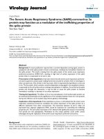
Fig. 2. RGS homology domain and the RGS protein family. a The
RH domain is composed of nine α-helices, forming a structure of two
distinct lobes: the terminal lobe containing both the N- and C-termini
(α1-3, 8, 9) and the bundle domain containing a four-helix, antiparallel bundle (α4-7). Gα subunits engage the bottom of the
structure, largely through contacts made with the bundle domain
PDB: 1ZV4 (RGS17). b Domain composition and identified members
of the different families of RGS proteins. RZ and R4 proteins are the
simplest RGS proteins, composed of an RH domain with short Nterminal regions and are approximately 190–240 residues long. The
R7 family contains a few accessory domains and is much longer than
RZ/R4 members at 470–675 residues. The R12 family is the largest
and most complex set of RGS proteins at 500–1000+ residues, except
for RGS10, which is closer to the R4 family in length but is grouped
in the R12 family based on RH sequence identity. pCys poly-Cysteine
string, RH RGS homology, AH amphipathic helix, DEP disheveled/
Egl-10/pleckstrin domain, GGL G protein γ- like, PDZ Psd-95/DlgA/
ZO1 domain, PTB phosphotyrosine-binding domain, RBD Raf-like
Ras binding domain, GOLoco Gαi/o loco
The most well-established noncanonical function of RGS17 is
its ability to act as a scaffold in a complex surrounding the
μOR. RGS17, as well as RGS19 and 20, interacts with
histidine triad nucleotide binding protein 1(HINT1) through
its pCys string, as first identified via Y2H screening for
proteins that directly bind to RGS20 (25). The formation of
this complex is dependent on the presence of Zn2+. RGS17’s
pCys string coordinates two Zn 2+ , each of which is
coordinated by four cysteine residues, forming a structure
known as a zinc ribbon (37). The HINT1-RGS17 complex
then engages the μOR and recruits protein kinase C (PKC) γ
to the plasma membrane, where PKCγ phosphorylates the
receptor, preventing further activation as a means of desensitization (38). The HINT1-RGS17 association with the
receptor appears to be mediated by RGS17 rather than
HINT1, as RGS17 is able to interact directly with μΟR
intracellular regions, namely the C-terminus and intracellular
loop 3. Moreover, the formation of this RGS-receptor
complex is not specific to the μΟR, as RGS17 is capable of
binding peptides derived from intracellular portions of
serotonin (1A and 2A), dopamine (D2), and cannabinoid
(CB1) receptors, as determined using surface plasmon
resonance (37). Furthermore, this interaction seems to be
Though the RZ family consists of little more than a
pCys string and an RH domain, RGS17 is subject to
modification and regulation through a number of posttranslational modifications. The first post-translational
modification of an RZ family member identified was the
palmitoylation of RGS19 on its pCys string, which largely
serves to regulate intracellular trafficking and localization.
Palmitoylation involves a reversible reaction between Cys
residues on the RGS protein and the carboxylic acid
moiety of the 16-carbon fatty acid palmitate, the addition
of which tethers the RGS protein to membranes. This
serves to concentrate RGS proteins to the same subcellular compartments as Gα subunits, which also exist as
lipid-modified proteins within cells, though unmodified
RGS proteins are able to exist in the cytosolic fraction
of cells (14). It is assumed that this mechanism holds true
for other members of the RZ family, considering that the
pCys string is perfectly conserved between RGS19 and
RGS17.
In addition to covalent modification by lipids, RGS17 is
also a substrate for phosphorylation. When it was first
identified, RGS17 was noted for containing a number of
putative sites for phosphorylation, as its primary protein
sequence contains six potential casein kinase sites and three
PKC sites (13). RGS17 was also identified in a large-scale
search for proteins containing phosphotyrosine residues in
murine brain samples. RGS17 can be phosphorylated on
Y137 at the base of α5, though the kinase responsible for this
modification and its functional consequence have yet to be
determined (41). Additionally, RGS19 is phosphorylated on
Ser151 by mitogen-activated protein kinase 1, increasing its
GAP activity toward Gαi3. This residue lies between in loop
between α5 and α6 in the RH domain and is conserved across
the RZ family, indicating that all members of the family are
likely substrates (42).
RGS17 can also be covalently linked to sugars. In the
mouse brain, RGS17 exists as a glycoprotein that purifies with
the fraction containing glycosylated proteins. Furthermore,
when immunoblotted, RGS17 is observed as a series of bands
of varying molecular weights, and the higher molecular
weight species are sensitive to glycosidase treatment (36).
The location and functional implications of these modifications have yet to be explored.
554
In addition to lipidation, phosphorylation, and glycosylation, RGS17 is also a substrate for sumoylation by SUMO1,
2, and 3 and is detected in mouse synaptosomes in its
sumoylated form. K90 in α3 and K121 in α4 are two potential
sumoylation sites in RGS17. The sumoylated forms preferentially coimmunoprecipitate with Gα and μOR, meaning that
this modification possibly changes function of RGS17 from a
GAP to a scaffold or effector antagonist (43). Additionally,
RGS17 contains two SUMO interaction motifs, one of which
(residues 64–67) is able to noncovalently associate with
SUMO and other sumoylated proteins, leaving open the
possibility of RGS17 forming even higher order SUMOdependent scaffolding complexes (44).
RGS17 also serves as a substrate for ubiquitination at
K147, located between α5 and α6, as found during a largescale proteomic effort. Ubiquitinated RGS17 could be
detected in murine brain and kidney tissues, but not liver,
heart, or muscle (45). The exact function of RGS17
ubiquitination is unknown, but this modification likely marks
RGS17 for degradation through the proteosome.
RGS17 AND DISEASE
Lung and Prostate Cancer
RGS17’s first link to cancer was its identification as a
potential marker for familial lung cancer, as a susceptibility
locus was tracked to chromosome 6q23-25, the genomic
location of Rgs17. Further work showed that RGS17 is often
overexpressed in both lung and prostate cancers by 8.3- and
7.5-fold, respectively (3,46). Furthermore, it has been shown
that knockdown of RGS17 in lung cancer-derived cultured
cells decreases tumor volume by 59–75% in a mouse
xenograft model of cancer. Moreover, RGS17 overexpression
causes increased expression of proteins with cAMP response
elements (CRE) in their promoter region. These results
indicate that the proliferative effect observed in RGS17dependent cancers is likely due to RGS17’s GAP activity
toward inhibitory Gα subunits, resulting in increased activity
of the PKA-CREB pathway. Increased RGS17 would lead to
decreased Gαi/o signaling, decreased AC inhibition, increased
formation of cAMP, increased PKA activity, and CREB
activation, ultimately altering the transcription of CREregulated genes (3). In some lung cancer cell lines, it has
been shown that RGS17 protein levels can be regulated by
microRNAs (miRNA, miR), which are short, non-coding
RNA sequences that regulate translation of their target
mRNA sequences. In lung cancer, there is evidence that the
specific miRNA that regulates expression of RGS17 is Hsamir-182, expression of which drastically reduces the amount
of endogenous RGS17. In fact, expression ectopic of Hsa-mir182 recapitulates what is observed when RGS17 is specifically
knocked down using synthetic shRNA, and increased Hsamir-182 is sufficient to reduce the growth and proliferation of
lung cancer in vitro (47).
Hepatocellular Carcinoma (HCC)
Similar to what has been observed in prostate and lung
cancers, RGS17 mRNA is detectable in rat HCC tissue, but
not normal whole liver tissue or hepatocytes. Likewise, in 5 of
Hayes and Roman
7 human HCC samples analyzed, RGS17 mRNA was
significantly overexpressed as compared to patient-matched
control tissue (p = 0.011), though when all seven samples were
analyzed together, no statistical significance was observed
(p = 0.061). Again similar to previous reports of RGS17 in
cancer, increased expression correlates to increased cellular
proliferation in HepG2 cells, and knockdown of RGS17 via
RNA interference results in decreased cellular proliferation.
Additionally, decreased RGS17 is correlated with decreased
intracellular cAMP levels, presumably through increased Gαi/
o-mediated inhibition of AC. Interestingly, the work performed in the HCC cancer model could not detect changes in
protein expression levels in the presence of Hsa-mir-182
overexpression. In fact, in HCC, it seems that RGS17 protein
stability might be regulated by proteosomal degradation, as
the presence of proteosome inhibitor MG132 results in
increased RGS17 in vitro (4). The presence of proteosomal
degradation of RGS17 further validates reports that RGS17 is
a substrate for ubiquitination in vivo (45). The fact that Hsamir-182 did not regulate RGS17 protein levels in HCC could
be due to a cell line or tissue type-dependent phenomenon,
though a thorough examination of this hypothesis has yet to
be realized (4). In addition to proteosomal degradation, it is
possible that RGS17 levels are epigenetically regulated. In
HCC tissues that show copy number losses on chromosome
6q, decreased methylation of CpG sites in Rgs17 is observed,
likely leading to increased RGS17 expression (48).
Breast Cancer
Recently, a number of findings relating RGS17 to breast
cancer have begun to emerge. Similar to prostate, lung, and
liver cancers described above, RGS17 can be upregulated in
cancerous versus noncancerous tissue. Using
immunohistological staining, RGS17 protein was found in
96% of cancerous samples, whereas it was only detectable in
57% of normal samples. Furthermore, RGS17 expression was
absent or very low in 12 of 28 normal samples, and low in the
remaining 16, but 85% (74 of 87) of cancerous samples had
moderate to high expression (5). Additionally, in breast
cancer, RGS17 expression is positively correlated with p63
expression, a protein that can be over expressed in a number
of cancers, including breast, lung, and prostate cancers
(5,49,50). RGS17 knockdown via RNA interference inhibited
cancer cell migration in a wound healing assay and invasion in
a Boyden chamber assay, recapitulating results seen in HCC
and lung cancers (3–5). In breast cancer tissue, a novel
miRNA, miR-32, capable of modulating RGS17 expression
was identified, and it was also shown that this miRNA is
specifically downregulated in cancerous breast tissue as
compared to surrounding normal tissue. Overexpression of
miR-32 causes decreased RGS17 expression and reductions
in cancer cell proliferation, migration, and invasion (5). In
breast cancer cells, the mechanism by which RGS17 is initially
upregulated remains unknown, but in vitro work has shown
that one possible mechanism is by chromosomal rearrangements. In MCF7 cells, chromosomal instability can result in a
chromosomes 3 and 6 rearrangement, placing the IRA1
promoter upstream of the RGS17 coding sequence, though
the consequence of this on transcript level has yet to be
identified (51). Additionally, RGS17 is upregulated in MCF-7
RGS17 in Multiple Forms of Cancer
cells after treatment with ionizing radiation, though the
ultimate consequence of this increase remains unknown (52).
Ovarian Cancer
In ovarian cancer, it appears that RGS17 is capable of
mediating chemoresistance, the ability of malignancies to
grow in the presence of chemotherapeutic drugs. When
cancerous cell lines are exposed to chemotherapeutic agents
(cisplatin [cis-diamminedichloroplatinum (II)], vincristine, or
paclitaxel), a loss of RGS17 expression is observed in cells
that become chemoresistant. Moreover, knockdown of
RGS17 expression via RNA interference is sufficient to
increase cell survival and decrease the growth inhibition
response following challenge with these compounds. Conversely, overexpression of RGS17 leads to increased sensitivity to drug treatment, though the effect is less pronounced
(53). Mechanistically, RGS17 in ovarian cancer cells appears
to modulate the PI3K/AKT survival pathway, rather than the
cAMP-PKA-CREB pathway like in HCC, lung, and prostate
cancers (3,53). Lysophosphatidic acid (LPA) can act in an
autocrine manner, such that binding to one of its receptors
activates Gαi proteins, resulting in the phosphorylation and
activation of protein kinase B (Akt) and the promotion of cell
survival. Increased RGS17 results in decreased Akt activation
following treatment with LPA, thus representing a mechanism for growth arrest. Therefore, the loss of RGS17
promotes increased growth and survival through increased
Gαi-mediated activation of the Akt signaling axis (53).
Acute Myeloid Leukemia (AML)
Recent work has implicated a possible role for RGS17 in
AML chemoresistance that could prove similar to that
identified in ovarian cancer. The expression of miR-363 is
inversely related to response to chemotherapy, and increased
miR-363 is evident in bone marrow samples from patients
with chemoresistant AML. Most importantly, RGS17 has
been identified as a target gene of miR-363 (54). It is
tempting to speculate that increased miR-363 would correlate
to decreased RGS17 levels, increased Akt activation, and
ultimately, diminished response to chemotherapeutic agents,
though this hypothesis has yet to be tested. Alternatively,
analysis of miR-363 levels in chemosensitive and resistant
ovarian cancer cells could prove to be of merit.
Neurological Disorders
As RGS17 is expressed to the highest degree in the brain
in healthy individuals, it comes as no surprise that RGS17 has
also been indicated in various neurological conditions.
Unfortunately, many of its potential roles have been identified via large-scale screening efforts, and there is little to no
mechanistic insight into its exact role. For example, RGS17
expression is decreased by nearly an order of magnitude in
clinical depression, as determined via RNA microarray
analysis of postmortem brain samples from patients with
and without a history of major depressive disorder (55).
There also has been an association of singe nucleotide
polymorphisms (SNPs) at chromosome 6q25, the location of
Rgs17, with bipolar disorder, though a definite role of RGS17
555
has yet to be established (56). RGS17 may also be involved in
addiction and drug abuse. Differences in RGS17 expression
levels have been correlated to morphine preference differences observed between C57BL/6J and DBA/2J mice (57).
DBA/2J mice exhibit higher levels of RGS17 protein and
mRNA expression in the NAc, midbrain, and brainstem,
possibly explaining the decreased reward and, therefore,
decreased preference for morphine as compared to C57BL/
6J mice in a two-bottle test (58). In humans, Rgs17 SNPs are
associated with substance abuse, most notably one SNP that
results in lowered RGS17 expression is correlated with
increased alcohol, marijuana, and opioid dependence in both
African and European Americans (59). Additionally, one
study found that Rgs17 SNPs have been associated with
smoking initiation in an Asian population (60).
RGS17 and Metastatic Disease
As noted above, reduction of RGS17 activity via RNA
interference is able to reduce the migratory and invasive
phenotypes of cells derived from HCC, lung, and breast
cancers, implying that RGS17 could be involved in metastatic
processes (3–5). It is very likely that these observations are
due aberrant signaling, as RGS17’s canonical role is to
negatively regulate inhibitory Gα signaling. An abundance
of RGS17 could lead to persistent inhibition Gαi/o, leading to
an imbalance in Gαi/o/Gαs signaling and ultimately excessive
AC-mediated cAMP production, as has been shown in both
lung cancer and HCC cells (3,4). Excessive cAMP would then
lead to CREB activation through PKA, resulting in excessive
transcription of CREB target genes, which has also been
observed in lung cancer cells (3). This could lead to increased
levels of CREB target genes that are directly involved in
metastasis and anchorage-independent cell growth, such as
vascular endothelial growth factor (VEGF), type IV collagenases, or cyclin D1, though this is somewhat speculative as
only cyclin D1 expression as been experimentally shown to
decrease in response RGS17 knockdown (3,61–63).
CHEMICAL INHIBITION OF RGS PROTEINS
RGS-Gα Druggability
Since their discovery in the mid 1990s, RGS proteins
have remained of great interest for drug discovery and
development due to their ability to modulate GPCR
signaling cascades. Traditionally, protein-protein interactions (PPIs) have been categorized as undruggable, but
recent successes in the field challenge this assumption
(64). In fact, recently PPI inhibitors have even begun to
enter clinical trials, such as SAR1118 for dry eye and
navitoclax for cancer (65,66). As RGS proteins have no
intrinsic catalytic activity and exert their function by
binding activated Gα subunits, previous drug discovery
efforts have primarily focused on identifying molecules
capable of inhibiting the Gα-RGS PPI (67). The most
apparent means to achieve this would be by identifying
molecules capable of binding directly to the residues that
form the interaction surface of the Gα or RGS. This
interface, also referred to as the A site, has been the
subject of numerous previous efforts to design inhibitors
556
Hayes and Roman
targeting RGS4, a member of the R4 family. Using the
previously solved structure of the RGS4-Gα complex, Jin
and coworkers designed cyclic peptides that mimicked the
Gα switch I region, inhibiting the RGS4-Gα interaction
with micromolar potency (67). This work proved that
inhibition of the interaction was possible, but as peptides
generally tend to make poor drugs, alternative methods to
identify inhibitors were sought (68).
Ultimately, high-throughput screening against RGS
proteins has proved the most fruitful in identifying lead
compounds with inhibitory activity toward these PPIs,
with methodologies ranging from bead-based flow cytometry and luminescence to colorimetric monitoring of Gα
GTPase activity (68–71). Interestingly, screening against
RGS4 has often identified cysteine-reactive compounds
that bind covalently to a site distinct from the A site
(72,73). This site is closer to a region that has been
termed the B site that binds endogenous phospholipids to
regulate GAP activity, establishing the hypothesis that
inhibition of the RGS-Gα PPI can be achieved through
molecules that act allosterically to the actual interaction
interface (74,75).
RGS17 Inhibition
Due to its role in lung, liver, breast, and prostate cancers,
our research group has interest in the development of small
molecules capable of inhibiting the RGS17-Gα interaction.
We hypothesize that chemical inhibition of RGS17 would
recapitulate the reduction in invasion, migration, and tumor
size in cancer that is observed when RGS17 expression is
reduced via RNA interference (3–5). Additionally, specific
chemical inhibitors of RGS17 could serve as tool compounds
to help unravel the cancer type-specific functions of RGS17
that have been previously reported (3,53). RGS17 merits
further evaluation as a potential drug target due to its
relatively narrow pattern of expression in normal human
tissue and its specific upregulation in the cancers of interest.
As RGS17 is generally relegated to CNS tissues (30), we
hypothesize that potential side effects of an inhibitor could be
mitigated if the compound is large (>400 Da) and/or
sufficiently hydrophilic, and thus incapable of crossing the
blood-brain barrier.
To this end, we have pursued high-throughput screening,
as in the past, it has been successful in identifying inhibitors of
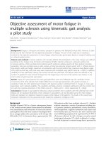
Fig. 3. Chemical inhibitors of RGS17 and potential sites for inhibitor
selectivity. a Chemical structures of previously identified RGS17
inhibitors. The RL-series of compounds was discovered using a
luminescent bead-based screen of 1300 compounds against RGS17Gαo PPI (66). The UI inhibitors were identified using a colorimetric
assay of RGS4-induced Gα GTPase activity and further work
identified their activity toward RGS17 (65). b Residues unique to
RGS17 as opposed to other RZ family members could facilitate
identification of binding contacts that confer specificity for RGS17.
Residues unique to RGS17’s primary sequence are shown in green
sticks. Residues that are shared or are extremely similar (Asp v. Glu,
for example) with one RZ family member are indicated as yellow
sticks. Residues that are completely conserved across the RZ family
are indicated in grey, and the side chains are not shown
RGS17 in Multiple Forms of Cancer
other RGS proteins (68). Initial efforts in the screening of
∼3500 compounds have identified six compounds capable of
inhibiting RGS17-Gα formation in vitro with micromolar
affinity, though issues with RGS protein specificity or the
presence of potentially reactive chemical moieties have
lessened the promise of these compounds (Fig. 3a) (69,70).
In order to increase the chances of success of identifying
specific RGS17 inhibitors that lack reactive functional groups,
larger chemical libraries need to be tested against RGS17 and
ongoing efforts in our lab are aimed at doing exactly that. As
other members of the RGS family are involved in important
physiological processes, such as heart rate regulation and
vision, pan-RGS inhibition could be deleterious. Thus,
identification of molecules that specifically inhibit RGS17 is
of the utmost importance. As noted before, the RH domain
of RGS17, 19, and 20 is highly conserved, but there are a
number of residues unique to RGS17. As shown in Fig. 3b,
many of these divergent residues are located in the terminal
subdomain, especially α9. Additionally, there are a few
RGS17-specific residues in the bundle subdomain, distal to
the Gα interface and near the region identified as the B site in
RGS4, which makes the discovery of RGS17-specific compounds more promising (Fig. 3b) (75). Future efforts will
focus on exploring the druggability of this site in RGS17,
potentially using fragment-based screening and structurebased methods, as this paradigm is beginning to gain traction
in PPI inhibition drug discovery programs (76).
CONCLUSION
RGS17 is able to negatively regulate GPCR signaling
through a variety of mechanisms, from its activity as Gα
GAP to targeting Gα subunits for proteosomal degradation to promoting receptor desensitization. It has been
implicated in regulating proliferation, migration, and
invasion in some of the most common forms of human
cancer, including lung, breast, prostate, and liver cancers.
This information coupled with RGS17’s expression in only
a limited number of human tissues makes it a potential
target for the development of a new class chemotherapeutic agents. Specific RGS17 inhibitors incapable of
permeating the blood-brain barrier would have few
predicted on-target adverse effects, though the identification of such molecules is needed for pre-clinical validation
of this hypothesis. As all previously identified RGS17
inhibitors lack specificity and/or contain potentially reactive moieties, future work remains to be done in the area
of RGS17 inhibition with small molecules. Though preliminary work has been performed to meet this goal,
future efforts must focus on the screening of larger, more
diverse compound libraries, as increasing the area of
chemical space interrogated will increase the likelihood
of success. Additionally, alternative drug development
methodologies employing a priori knowledge and
structure-based screening paradigms may be fruitful in
accelerating the identification of RGS17 inhibitors.
ACKNOWLEDGMENTS
This work was supported by NIH 5R01CA160470
(DLR), NIH T32GM067795 (MPH), and American Foundation for Pharmaceutical Education Predoctoral Fellowship
(MPH).
557
REFERENCES
1. Takeda S, Kadowaki S, Haga T, Takaesu H, Mitaku S.
Identification of G protein-coupled receptor genes from the
human genome sequence. FEBS Lett. 2002;520(1–3):97–101.
2. Esbenshade TA. G protein-coupled receptors as targets for drug
discovery. In: Lundstrom KH, Chiu ML, editors. G proteincoupled receptors in drug discovery. Boca Raton: Taylor &
Francis; 2006. p. 15–36.
3. James MA, Lu Y, Liu Y, Vikis HG, You M. RGS17, an
overexpressed gene in human lung and prostate cancer, induces
tumor cell proliferation through the cyclic AMPPKA-CREB
pathway. Cancer Res. 2009;69(5):2108–16.
4. Sokolov E, Iannitti DA, Schrum LW, McKillop IH. Altered
expression and function of regulator of G-protein signaling-17
(RGS17) in hepatocellular carcinoma. Cell Signal.
2011;23(10):1603–10.
5. Li Y, Li L, Lin J, Hu X, Li B, Xue A, et al. Deregulation of
RGS17 expression promotes breast cancer progression. J Cancer
Educ. 2015;6(8):767–75.
6. Simonds WF. G protein regulation of adenylate cyclase. Trends
Pharmacol Sci. 1999;20(2):66–73.
7. Smrcka AV, Hepler JR, Brown KO, Sternweis PC. Regulation of
polyphosphoinositide-specific phospholipase C activity by purified Gq. Science. 1991;251(4995):804–7.
8. Siehler S. Regulation of RhoGEF proteins by G12/13-coupled
receptors. Br J Pharmacol. 2009;158(1):41–9.
9. Gilman AG. G proteins: transducers of receptor-generated
signals. Annu Rev Biochem. 1987;56:615–49.
10. Berman DM, Wilkie TM, Gilman AG. GAIP and RGS4 are
GTPase-activating proteins for the Gi subfamily of G protein
alpha subunits. Cell. 1996;86(3):445–52.
11. Tesmer JJ, Berman DM, Gilman AG, Sprang SR. Structure
of RGS4 bound to AlF4-activated G(i alpha1): stabilization
of the transition state for GTP hydrolysis. Cell.
1997;89(2):251–61.
12. Barker SA, Wang J, Sierra DA, Ross EM. RGSZ1 and Ret RGS:
two of several splice variants from the gene RGS20. Genomics.
2001;78(3):223–9.
13. Mao H, Zhao Q, Daigle M, Ghahremani MH, Chidiac P. Albert
461 PR. RGS17/RGSZ2, a novel regulator of Gi/o, Gz, and Gq
s i g n a l i n g . T h e J o u r n a l o f b i o l o g i c a l c h e m i s t r y.
2004;279(25):26314–22.
14. De Vries L, Elenko E, Hubler L, Jones TL, Farquhar MG. GAIP
is membrane-anchored by palmitoylation and interacts with the
activated (GTP-bound) form of G alpha i subunits. Proc Natl
Acad Sci U S A. 1996;93(26):15203–8.
15. Faurobert E, Hurley JB. The core domain of a new retina
specific RGS protein stimulates the GTPase activity of
transducin in vitro. Proc Natl Acad Sci U S A. 1997;94(7):2945–
50.
16. Wang J, Ducret A, Tu Y, Kozasa T, Aebersold R, Ross EM.
RGSZ1, a Gz-selective RGS protein in brain. Structure, membrane association, regulation by Galphaz phosphorylation, and
relationship to a Gz gtpase-activating protein subfamily. The
Journal of biological chemistry. 1998;273(40):26014–25.
17. Glick JL, Meigs TE, Miron A, Casey PJ. RGSZ1, a Gz-selective
regulator of G protein signaling whose action is sensitive to the
phosphorylation state of Gzalpha. The Journal of biological
chemistry. 1998;273(40):26008–13.
18. Tu Y, Wang J, Ross EM. Inhibition of brain Gz GAP and other
RGS proteins by palmitoylation of G protein alpha subunits.
Science. 1997;278(5340):1132–5.
19. De Vries L, Mousli M, Wurmser A, Farquhar MG. GAIP, a
protein that specifically interacts with the trimeric G protein G
alpha i3, is a member of a protein family with a highly conserved
core domain. Proc Natl Acad Sci U S A. 1995;92(25):11916–20.
20. De Vries L, Lou X, Zhao G, Zheng B, Farquhar MG. GIPC, a
PDZ domain containing protein, interacts specifically with the C
terminus of RGS-GAIP. Proc Natl Acad Sci U S A.
1998;95(21):12340–5.
21. Jeanneteau F, Guillin O, Diaz J, Griffon N, Sokoloff P. GIPC
recruits GAIP (RGS19) to attenuate dopamine D2 receptor
signaling. Mol Biol Cell. 2004;15(11):4926–37.
558
22. Wang Q, Traynor JR. Modulation of mu-opioid receptor
signaling by RGS19 in SH491 SY5Y cells. Mol Pharmacol.
2013;83(2):512–20.
23. Wang Q, Terauchi A, Yee CH, Umemori H, Traynor JR. 5HT1A receptor-mediated phosphorylation of extracellular
signal-regulated kinases (ERK1/2) is modulated by regulator of
G protein signaling protein 19. Cell Signal. 2014;26(9):1846–52.
24. Wang J, Tu Y, Woodson J, Song X, Ross EM. A GTPaseactivating protein for the G protein Galphaz. Identification,
purification, and mechanism of action. The Journal of biological
chemistry. 1997;272(9):5732–40.
25. Ajit SK, Ramineni S, Edris W, Hunt RA, Hum WT, Hepler JR,
et al. RGSZ1 interacts with protein kinase C interacting protein
PKCI-1 and modulates mu opioid receptor signaling. Cell Signal.
2007;19(4):723–30.
26. Garzon J, Rodriguez-Munoz M, Lopez-Fando A, Garcia-Espana
A, Sanchez-Blazquez P. RGSZ1 and GAIP regulate mu- but not
delta-opioid receptors in mouse CNS: role in tachyphylaxis and
acute tolerance. Neuropsychopharmacology. 2004;29(6):1091–
104.
27. Sanchez-Blazquez P, Rodriguez-Munoz M, Montero C, Garzon
J. RGS-Rz and RGS9-2 proteins control mu-opioid receptor
desensitisation in CNS: the role of activated Galphaz subunits.
Neuropharmacology. 2005;48(1):134–50.
28. Jordan JD, Carey KD, Stork PJ, Iyengar R. Modulation of rap
activity by direct interaction of Galpha(o) with Rap1 GTPaseactivating protein. The Journal of biological chemistry.
1999;274(31):21507–10.
29. Sierra DA, Gilbert DJ, Householder D, Grishin NV, Yu K,
Ukidwe P, et al. Evolution of the regulators of G-protein
signaling multigene family in mouse and human. Genomics.
2002;79(2):177–85.
30. Larminie C, Murdock P, Walhin JP, Duckworth M, Blumer KJ,
Scheideler MA, et al. Selective expression of regulators of Gprotein signaling (RGS) in the human central nervous system.
Brain Res Mol Brain Res. 2004;122(1):24–34.
31. Stanwood GD, Parlaman JP, Levitt P. Genetic or pharmacological inactivation of the dopamine D1 receptor differentially alters
the expression of regulator of G-protein signalling (Rgs)
transcripts. Eur J Neurosci. 2006;24(3):806–18.
32. Doupnik CA, Xu T, Shinaman JM. Profile of RGS expression in
single rat atrial myocytes. Biochim Biophys Acta.
2001;1522(2):97–107.
33. Maple AM, Perna MK, Parlaman JP, Stanwood GD, Brown RW.
Ontogenetic quinpirole treatment produces long-lasting decreases in the expression of Rgs9, but increases Rgs17 in the
striatum, nucleus accumbens and frontal cortex. Eur J Neurosci.
2007;26(9):2532–8.
34. Alexander MR, Murgai M, Moehle CW, Owens GK. Interleukin1beta modulates smooth muscle cell phenotype to a distinct
inflammatory state relative to PDGF-DD via NF-kappaBdependent mechanisms. Physiol Genomics. 2012;44(7):417–29.
35. Soundararajan M, Willard FS, Kimple AJ, Turnbull AP, Ball LJ,
Schoch GA, et al. Structural diversity in the RGS domain and its
interaction with heterotrimeric G protein alpha-subunits. Proc
Natl Acad Sci U S A. 2008;105(17):6457–62.
36. Garzon J, Rodriguez-Munoz M, Lopez-Fando A, SanchezBlazquez P. The RGSZ2 protein exists in a complex with muopioid receptors and regulates the desensitizing capacity of Gz
proteins. Neuropsychopharmacology. 2005;30(9):1632–48.
37. Sanchez-Blazquez P, Rodriguez-Munoz M, Bailon C, Garzon J.
GPCRs promote the release of zinc ions mediated by nNOS/NO
and the redox transducer RGSZ2 protein. Antioxid Redox
Signal. 2012;17(9):1163–77.
38. Rodriguez-Munoz M, de la Torre-Madrid E, Sanchez-Blazquez
P, Wang JB, Garzon J. NMDAR-nNOS generated zinc recruits
PKCgamma to the HINT1-RGS17 complex bound to the C
terminus of Mu-opioid receptors. Cell Signal. 2008;20(10):1855–
64.
39. Garzon J, Rodriguez-Munoz M, Vicente-Sanchez A, Bailon C,
Martinez-Murillo R, Sanchez-Blazquez P. RGSZ2 binds to the
neural nitric oxide synthase PDZ domain to regulate mu-opioid
receptor-mediated potentiation of the N-methyl-D-aspartate
receptor-calmodulin-dependent protein kinase II pathway.
Antioxid Redox Signal. 2011;15(4):873–87.
Hayes and Roman
40. Fischer T, De Vries L, Meerloo T, Farquhar MG. Promotion of
G alpha i3 subunit down-regulation by GIPN, a putative E3
ubiquitin ligase that interacts with RGS-GAIP. Proc Natl Acad
Sci U S A. 2003;100(14):8270–5.
41. Ballif BA, Carey GR, Sunyaev SR, Gygi SP. Large-scale
identification and evolution indexing of tyrosine phosphorylation
sites from murine brain. J Proteome Res. 2008;7(1):311–8.
42. Ogier-Denis E, Pattingre S, El Benna J, Codogno P. Erk1/2dependent phosphorylation of Galpha-interacting protein stimulates its GTPase accelerating activity and autophagy in human
colon cancer cells. The Journal of biological chemistry.
2000;275(50):39090–5.
43. Rodriguez-Munoz M, Bermudez D, Sanchez-Blazquez P, Garzon
J. Sumoylated RGS-Rz proteins act as scaffolds for Mu-opioid
receptors and G-protein complexes in mouse brain.
Neuropsychopharmacology. 2007;32(4):842–50.
44. Garzon J, Rodriguez-Munoz M, Vicente-Sanchez A, GarciaLopez MA, Martinez-Murillo R, Fischer T, et al. SUMO-SIM
interactions regulate the activity of RGSZ2 proteins. PLoS One.
2011;6(12):e28557.
45. Wagner SA, Beli P, Weinert BT, Scholz C, Kelstrup CD, Young
C, et al. Proteomic analyses reveal divergent ubiquitylation site
patterns in murine tissues. Mol Cell Proteomics.
2012;11(12):1578–85.
46. You M, Wang D, Liu P, Vikis H, James M, Lu Y, et al. Fine
mapping of chromosome 6q23-25 region in familial lung cancer
families reveals RGS17 as a likely candidate gene. Clin Cancer
Res. 2009;15(8):2666–74.
47. Sun Y, Fang R, Li C, Li L, Li F, Ye X, et al. Hsa-mir-182
suppresses lung tumorigenesis through down regulation of
RGS17 expression in vitro. Biochem Biophys Res Commun.
2010;396(2):501–7.
48. Shen J, LeFave C, Sirosh I, Siegel AB, Tycko B, Santella RM.
Integrative epigenomic and genomic filtering for methylation
markers in hepatocellular carcinomas. BMC Med Genomics.
2015;8:28.
49. Di Como CJ, Urist MJ, Babayan I, Drobnjak M, Hedvat CV,
Teruya-Feldstein J, et al. p63 expression profiles in human
normal and tumor tissues. Clin Cancer Res. 2002;8(2):494–
501.
50. Au NH, Gown AM, Cheang M, Huntsman D, Yorida E, Elliott
WM, et al. P63 expression in lung carcinoma: a tissue microarray
study of 408 cases. Appl Immunohistochem Mol Morphol.
2004;12(3):240–7.
51. Hahn Y, Bera TK, Gehlhaus K, Kirsch IR, Pastan IH, Lee B.
Finding fusion genes resulting from chromosome rearrangement
by analyzing the expressed sequence databases. Proc Natl Acad
Sci U S A. 2004;101(36):13257–61.
52. Jung S, Lee S, Lee J, Li C, Ohk JY, Jeong HK, et al. Protein
expression pattern in response to ionizing radiation in MCF-7
human breast cancer cells. Oncol Lett. 2012;3(1):147–54.
53. Hooks SB, Callihan P, Altman MK, Hurst JH, Ali MW, Murph
MM. Regulators of G-Protein signaling RGS10 and RGS17
regulate chemoresistance in ovarian cancer cells. Mol Cancer.
2010;9:289.
54. Mosakhani N, Raty R, Tyybakinoja A, Karjalainen-Lindsberg
ML, Elonen E, Knuutila S. MicroRNA profiling in
chemoresistant and chemosensitive acute myeloid leukemia.
Cytogenet Genome Res. 2013;141(4):272–6.
55. Shelton RC, Claiborne J, Sidoryk-Wegrzynowicz M, Reddy R,
Aschner M, Lewis DA, et al. Altered expression of genes
involved in inflammation and apoptosis in frontal cortex in
major depression. Mol Psychiatry. 2011;16(7):751–62.
56. Ferreira MA, O’Donovan MC, Meng YA, Jones IR, Ruderfer
DM, Jones L, et al. Collaborative genome-wide association
analysis supports a role for ANK3 and CACNA1C in bipolar
disorder. Nat Genet. 2008;40(9):1056–8.
57. Doyle GA, Furlong PJ, Schwebel CL, Smith GG, Lohoff FW,
Buono RJ, et al. Fine mapping of a major QTL influencing
morphine preference in C57BL/6 and DBA/2 mice using
congenic strains. Neuropsychopharmacology. 2008;33(12):2801–
9.
58. Doyle GA, Schwebel CL, Ruiz SE, Chou AD, Lai AT, Wang MJ,
et al. Analysis of candidate genes for morphine preference
quantitative trait locus Mop2. Neuroscience. 2014;277:403–16.
RGS17 in Multiple Forms of Cancer
59. Zhang H, Wang F, Kranzler HR, Anton RF, Gelernter J.
Variation in regulator of G-protein signaling 17 gene (RGS17)
is associated with multiple substance dependence diagnoses.
Behav Brain Funct. 2012;8:23.
60. Yoon D, Kim YJ, Cui WY, Van der Vaart A, Cho YS, Lee JY, et
al. Large-scale genome-wide association study of Asian population reveals genetic factors in FRMD4A and other loci influencing smoking initiation and nicotine dependence. Hum Genet.
2012;131(6):1009–21.
61. Wu D, Zhau HE, Huang WC, Iqbal S, Habib FK, Sartor O, et al.
cAMP-responsive element-binding protein regulates vascular
endothelial growth factor expression: implication in human
prostate cancer bone metastasis. Oncogene. 2007;26(35):5070–7.
62. Xie S, Price JE, Luca M, Jean D, Ronai Z, Bar-Eli M. Dominantnegative CREB inhibits tumor growth and metastasis of human
melanoma cells. Oncogene. 1997;15(17):2069–75.
63. Kumar AP, Bhaskaran S, Ganapathy M, Crosby K, Davis MD,
Kochunov P, et al. Akt/cAMP-responsive element binding
protein/cyclin D1 network: a novel target for prostate cancer
inhibition in transgenic adenocarcinoma of mouse prostate
model mediated by Nexrutine, a Phellodendron amurense bark
extract. Clin Cancer Res. 2007;13(9):2784–94.
64. Arkin MR, Tang Y, Wells JA. Small-molecule inhibitors of
protein-protein interactions: progressing toward the reality.
Chem Biol. 2014;21(9):1102–14.
65. Rudin CM, Hann CL, Garon EB, Ribeiro de Oliveira M,
Bonomi PD, Camidge DR, et al. Phase II study of single agent
navitoclax (ABT-263) and biomarker correlates in patients with
relapsed small cell lung cancer. Clin Cancer Res.
2012;18(11):3163–9.
66. Zhong M, Gadek TR, Bui M, Shen W, Burnier J, Barr KJ, et al.
Discovery and development of potent LFA-1/ICAM-1 antagonist
SAR 1118 as an ophthalmic solution for treating dry eye. ACS
Med Chem Lett. 2012;3(3):203–6.
67. Jin Y, Zhong H, Omnaas JR, Neubig RR, Mosberg HI.
Structure-based design, synthesis, and pharmacologic evaluation
of peptide RGS4 inhibitors. J Pept Res. 2004;63(2):141–6.
559
68. Roman DL, Talbot JN, Roof RA, Sunahara RK, Traynor JR,
Neubig RR. Identification of small-molecule inhibitors of RGS4
using a high-throughput flow cytometry protein interaction assay.
Mol Pharmacol. 2007;71(1):169–75.
69. Monroy CA, Mackie DI, Roman DL. A high throughput screen
for RGS proteins using steady state monitoring of free phosphate
formation. PLoS One. 2013;8(4):e62247.
70. Mackie DI, Roman DL. Development of a novel highthroughput screen and identification of small-molecule inhibitors
of the Galpha-RGS17 protein-protein interaction using
AlphaScreen. J Biomol Screen. 2011;16(8):869–77.
71. Roman DL, Ota S, Neubig RR. Polyplexed flow cytometry
protein interaction assay: a novel high-throughput screening
paradigm for RGS protein inhibitors. J Biomol Screen.
2009;14(6):610–9.
72. Roman DL, Blazer LL, Monroy CA, Neubig RR. Allosteric
inhibition of the regulator of G protein signaling-Galpha proteinprotein i nt eract i on by CCG-49 86. Mol P harm acol.
2010;78(3):360–5.
73. Blazer LL, Roman DL, Chung A, Larsen MJ, Greedy BM,
Husbands SM, et al. Reversible, allosteric small-molecule inhibitors of regulator of G protein signaling proteins. Mol Pharmacol.
2010;78(3):524–33.
74. Ishii M, Fujita S, Yamada M, Hosaka Y, Kurachi Y. Phosphatidylinositol 3,4,5-trisphosphate and Ca2+/calmodulin competitively bind to the regulators of G-protein-signalling (RGS)
domain of RGS4 and reciprocally regulate its action. Biochem J.
2005;385(Pt 1):65–73.
75. Popov SG, Krishna UM, Falck JR, Wilkie TM. 686 Ca2+/
Calmodulin reverses phosphatidylinositol 3,4, 5-trisphosphatedependent inhibition of regulators of G protein-signaling
G T P a s e - a c t i v a t i n g p r o t e i n a c t i v i t y. J B i o l C h e m .
2000;275(25):18962–8.
76. Dias DM, Van Molle I, Baud MG, Galdeano C, Geraldes CF,
Ciulli A. Is NMR fragment screening fine-tuned to assess
druggability of protein-protein interactions? ACS Med Chem
Lett. 2014;5(1):23–8.