Design and evaluation of a novel felbinac transdermal patch: Combining ion-pair and chemical enhancer strategy
Bạn đang xem bản rút gọn của tài liệu. Xem và tải ngay bản đầy đủ của tài liệu tại đây (457.47 KB, 10 trang )
AAPS PharmSciTech, Vol. 17, No. 2, April 2016 ( # 2015)
DOI: 10.1208/s12249-015-0342-9
Research Article
Design and Evaluation of a Novel Felbinac Transdermal Patch: Combining Ion-Pair
and Chemical Enhancer Strategy
Nannan Liu,1 Wenting Song,1 Tian Song,1 and Liang Fang1,2
Received 14 February 2015; accepted 23 May 2015; published online 13 June 2015
Abstract. The aim of this study was to design a novel felbinac (FEL) patch with significantly higher
(P<0.05) skin permeation amount than the commercial product SELTOUCH® using ion-pair and chemical enhancer strategy, overcoming the disadvantage of the large application area of SELTOUCH®.
Six complexes of FEL with organic amines diethylamine (DEA), triethylamine (TEA), N-(2′-hydroxyethanol)-piperdine (HEPP), monoethanolamine (MEtA), diethanolamine (DEtA), and triethanolamine
(TEtA) were prepared by ion-pair interaction, and their formation were confirmed by differential
scanning calorimetry (DSC), powder X-ray diffraction (pXRD), infared spectroscopy (IR), and proton
nuclear magnetic resonance spectroscopy (1H-NMR). Subsequently, the effect of ion-pair complexes and
chemical enhancers were investigated through in vitro and in vivo experiments using rabbit abdominal
skin. Results showed that FEL-TEA was the most potential candidate both in isopropyl palmitate (IPP)
solution and transdermal patches. Combining use of 10% N-dodecylazepan-2-one (Azone), the optimized
FEL-TEA patch achieved a flux of 18.29±2.59 μg/cm2/h, which was twice the amount of the product
SELTOUCH® (J=9.18±1.26 μg/cm2/h). Similarly, the area under the concentration curve from time 0 to
time t (AUC0-t) in FEL-TEA patch group (15.94±3.58 h.μg/mL) was also twice as that in SELTOUCH®
group (7.31±1.16 h.μg/mL). Furthermore, the in vitro skin permeation results of FEL-TEA patch was
found to have a good correlation with the in vivo absorption results in rabbit. These findings indicated that
a combination of ion-pair and chemical enhancer strategy could be useful in developing a novel transdermal patch of FEL.
KEY WORDS: chemical enhancer; felbinac-triethylamine (FEL-TEA); in vitro/in vivo correlation
(IVIVC); ion-pair; transdermal patch.
INTRODUCTION
Nowadays, nonsteroidal anti-inflammatory drugs
(NSAIDs) remain the most commonly used drugs for
treatment of osteoarthritis, rheumatoid arthritis, and
acute pain [1]. However, gastrointestinal side effects
resulting from repeated oral administration limit their
use [2]. As a result, topical products of these drugs,
which reduce the risk of gastrointestinal disorders and
enhance patients’ compliance, have become more and
more popular [3].
As a potent NSAID, felbinac (FEL) has been widely
used for treatment of osteoarthritis, rheumatoid arthritis,
muscle inflammation, and acute soft tissue injuries in topical preparations [4–7]. Currently, FEL patches have been
available in Japan and Korea, but the product has a large
application area of 70 cm2, which is far beyond the desired
size (that is, a surface area of ≤40 cm2) and decreased
patients’ compliance [8]. To decrease the large area of the
product, the permeation of FEL needs to be further
1
Department of Pharmaceutical Sciences, Shenyang Pharmaceutical
University, 103 Wenhua Road, Shenyang, Liaoning 110016, China.
2
To whom correspondence should be addressed. (e-mail:
)
1530-9932/16/0200-0262/0 # 2015 American Association of Pharmaceutical Scientists
enhanced. Ultrasound therapy was ever used to enhance
the effectiveness of FEL gel [9]. However, this method was
not especially effective in improving the hydrophilicity of
FEL and then increasing the permeability of FEL.
Considering the lipophilic property of FEL (Log P=2.58),
the partition from lipophilic stratum corneum (SC) to hydrophilic epidermis (ED) may be a principal resistance [10].
Therefore, ion-pair complexation, an effective technique to
influence a drug’s Log P [11,12], was chosen to decrease
FEL’s lipophilicity and enhance its permeability. Additionally, chemical enhancer is also a widely used approach to
increase the skin permeation of drugs [13]. A combination
of chemical enhancer and ion-pair strategy was used to
maximize the permeability of ionized drugs [14,15].
In this work, six organic amines, diethylamine (DEA),
triethylamine (TEA), N-(2′-hydroxy-ethanol)-piperdine
(HEPP), monoethanolamine (MEtA), diethanolamine
(DEtA), and triethanolamine (TEtA) were selected to prepare ion-pair complexes with FEL, and the different permeation behaviors of these complexes through rabbit abdominal
skin were further discussed. On this basis, the skin permeation
amount of FEL was further enhanced with combined use of
chemical enhancers. Finally, the effect of the combination of
ion-pair and chemical enhancer strategy was evaluated both
in vitro and in vivo.
262
Combining Ion-Pair and Enhancer Strategy
MATERIALS AND METHODS
Chemicals and Animals
Felbinac (FEL) was provided by Hubei Xunda Pharmaceuticals Co., Ltd. (Hubei, China). Ethanolamine (MEtA), diethanolamine (DEtA), triethanolamine (TEtA),
diethylamine (DEA), triethylamine (TEA), and N-(2′-hydroxy-ethanol)-piperdine (HEPP) were purchased from
Tianjin Bodi Chemicals Co., Ltd. (Tianjin, China). Isopropyl palmitate (IPP), N-dodecylazepan-2-one (Azone), isopropyl myristate (IPM), Span80 (SP), propylene glycol
(PG), and l-menthol (MT) were obtained from Alfa Aesar
(MA, USA). Duro-Tak® 87-4098 (PSA) was purchased
from Henkel Corp. (NJ, USA). Methanol of HPLC grade
was supplied by the Hanbang Science and Technology
Co., Ltd. (Jiangsu, China). SELTOUCH® tape 70
(felbinac, 70 mg/140 cm 2) was obtained from Teikoku
Seiyaku Co., Ltd. (Osaka, Japan). All other chemicals
were of analytical grade.
Male rabbits weighing 1.8–2.2 kg were supplied by the
Experimental Animal Center of Shenyang Pharmaceutical
University (Shenyang, China). All animal experiments were
performed according to the NIH Guidelines for the Care and
Use of Laboratory Animals as well as the guidelines for
animal use published by the Life Science Research Center of
Shenyang Pharmaceutical University.
Preparation and Characterization
Preparation of Ion-Pair Complexes
Equimolar amount of FEL and organic amines were
dissolved in ethanol and stirred for 2 h. Then, the solvent
was removed using a rotary evaporator, and products were
obtained after drying in a vacuum for 24 h.
DSC and pXRD Characterization
Subsequently, FEL and its solid complexes were identified by differential scanning calorimetry (DSC) and powder
X-ray diffraction (pXRD). The pXRD patterns of samples
were measured with DX-2700 XRD diffractometer (Dandong,
China) using Cu Kα radiation (tube operated at 40 kV,
40 mA). Data were collected over the 2θ range of 3-50°.
IR and 1H-NMR Characterization
FEL and its complexes were also characterized by infrared spectra (IR) and 1H-NMR. For 1H-NMR study, samples
were dissolved into deuterated chloroform (CDCl3) and
analyzed with an Advance-400 MHz instrument (Bruker, Germany). Chemical shifts (δ) for CH groups were reported in
parts per million relative to tetramethylsilane.
263
onto a release linear followed by drying at 50°C for 20 min.
After removal of the solvent, the products were covered with
backing membranes.
In Vitro Studies
Apparent Partition Coefficient Experiments
The apparent partition coefficients of FEL and its complexes were measured by the classic shake-flask method [16].
Equal volumes of distilled water and n-octanol and an appropriate amount of drugs were added into a sealed glass vial and
agitated to achieve equilibrium at 32°C for 48 h. After centrifugation, the sample concentration in each phase was determined by HPLC.
Apparent Solubility Measurements
The solubilities of FEL and its complexes in IPP solutions
were determined at 32°C, by adding excessive drugs to the
vehicle in glass vials. All vials were shaken for 48 h until
equilibrium. After centrifugation and dilution, the concentration of each drug was determined by HPLC.
In Vitro Skin Permeation Experiments
Excised rabbit abdominal skin was used to evaluate
the skin permeation of FEL and its ion-pair complexes
and prepared according to a previous report [14]. In vitro
skin permeation experiments were performed using twochamber side-by-side glass diffusion cells. The excised
rabbit skin was mounted between the diffusion cells, with
dermal side facing the receptor compartment. The receptor cell was filled with 3 mL pH 7.4 phosphate buffer
(PBS), and the donor cell was suspensions composed of
FEL or its ion-pair complexes in IPP. For the skin permeation experiments from patches, donor compartments
were exchanged to patches stuck on the SC side of skin.
Solutions in both compartments were stirred at about
600 rpm and maintained at 32°C. At pre-determined time
intervals, 2 mL samples were withdrawn from the receptor
compartment for analysis, and then an equal volume of
fresh receptor medium was added to maintain the constant volume. The samples were analyzed by HPLC
method.
The cumulative amount of each drug permeating per unit
area (Q) versus time was plotted. The steady-state flux (J, μg/
cm2/h) was calculated from the slope of linear region of the
plot. The enhancement ratio (ER) was defined as Q for the
ion-pair group or enhancer-containing group divided by the
same parameter for the control group containing only FEL or
FEL-TEA.
In Vivo Studies
Preparation of Patches
Rabbit Skin Irritation Test
FEL or its ion-pair complexes equivalent to the amount
of FEL, penetration enhancers and pressure sensitive adhesive (PSA) were dissolved in ethanol and mixed thoroughly
with a magnetic bar. The resulting mixture was then coated
Four healthy rabbits were used to test the skin irritation
of FEL-TEA patch according to the Draize method [17]. One
day prior to the experiment, each rabbit abdominal skin was
Liu et al.
264
shaved and divided into four areas. Each area was grouped
and treated as follows:
In Vitro/In Vivo Correlation
& Control group—non-treated
& Positive group—standard irritant (10% aqueous solution of
The predicted in vitro skin permeation profiles of
FEL-TEA transdermal patches in rabbits were obtained
using the following formula as described in a previous
study [18].
lauryl sodium sulfate)
& Negative group—blank patch (6 cm2, without any drug)
& FEL-TEA group—the optimized FEL-TEA patch (6 cm2)
Then patches were removed after a period of 12 h, and
the resulting reactions (erythema and edema) after removing
patches at 24, 48, and 72 h were evaluated by a scale of scores
as follows:
0.0~0.4: negligible response;
0.5~1.9: slight response;
2.0~4.9: moderate response;
5.0~8.0: severe response.
Z
C ðt Þ ¼
t
I ðθÞW ðt−θÞdθ ¼ I ðθÞ*W ðθÞ
0
where C(t), I, and W are the plasma concentration in
rabbit as a function of time, the input into the system (i.e.,
in vitro skin permeation results), and the weighting function
(i.e., intravenous data), respectively. And * stands for the
convolution operator. Therefore, I(t), the in vitro permeation
results of FEL-TEA patch, can be predicted from the in vivo
absorption data and intravenous data by the deconvolution
method.
Administration and Sampling
I ðt Þ ¼ RðθÞ
..
W ðθÞ
Twelve male rabbits weighing 1.8–2.2 kg were randomly divided into two groups, and the day prior to the
experiments, an abdominal area of about 48 cm 2 was
shaved carefully without damaging the skin. For group
A, rabbits were treated with the commercial product
SELTOUCH® (FEL, 0.5 mg/cm2) on the abdominal area
for 12 h. For comparison, animals in group B were
applied with the optimized FEL-TEA patch (equals to
FEL 0.5 mg/cm2) on the same area. Blood samples were
collected at 0.083, 0.167, 0.5, 1, 2, 3, 4, 6, 8, 10, 12, and
14 h after transdermal administration. After a washout
period of 2 days, rabbits in group B were given an
intravenous administration of FEL-TEA (equals to FEL
4 mg/kg) via the marginal ear vein, and blood samples
were collected at 0.083, 0.167, 0.25, 0.5, 0.75, 1, 2, 3, 4, 5,
and 6 h after intravenous administration. Plasma were
obtained by centrifugation at 16,000 rpm for 5 min and
stored at −70°C until analysis.
The amounts of FEL and its complexes were determined by HPLC. The HPLC system consists of an L-2130
pump (Hitachi Ltd., Japan), an L-2420 variable wavelength ultraviolet absorption detector (Hitachi Ltd., Jap a n ) , a n d a n H T- 2 2 0 A c h r o m a t o g r a p h i c c o l u m n
incubator (Dalian Huida Scientific instruments, Ltd.).
Chromatographic separation was achieved on Diamonsil
C18 column, 200 mm×4.6 mm×5 μm, by using a mobile
phase containing methanol: 0.02 mol/L pH 4.5 NH4AcHAc buffer solution (75:25, v/v) at a flow rate of 1 mL/
min. Ethylparaben was used as the internal standard and
detection wavelength was set at 254 nm.
Treatment and Analysis of Plasma Samples
Data Analysis
A 100 μL aliquot of rabbit plasma was mixed with
10 μL ethylparaben solution and 10 μL 1 mol/L hydrochloric acid before extracted with 1 mL ethyl acetate by
vortex for 10 min. The mixture was centrifuged at
16,000 rpm for 5 min, and the organic layer was transferred to another tube and evaporated under nitrogen at
40°C. Then, the residue was reconstituted in 100 μL mobile phase and centrifuged at 16,000 rpm for 5 min. A
20 μL aliquot of supernatant was injected into the HPLC
system for analysis.
Each experimental value was an average of minimum
four measurements. Statistical analysis was conducted by
using Student’s t test and all data were presented as mean
±standard deviation (SD). A difference between data was
considered significant when P<0.05.
where the symbol // denotes the deconvolution operation.
Quantitative Analysis
RESULTS AND DISCUSSION
Characterization of FEL Ion-Pair Complexes
DSC and pXRD Characterization
Pharmacokinetic Analysis
The peak blood concentration (Cmax) was obtained
directly from the concentration-time profile. The area
under the concentration curve from time 0 to time t
(AUC 0-t ), and the mean residence time (MRT) were
obtained by noncompartmental analyses with the help
of WinNonlin®.
All FEL complexes were characterized by DSC and
pXRD, except for FEL-HEPP in the sticky liquid state.
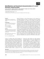
As presented in Fig. 1, FEL had a sharp endothermic
peak at 160°C, and all FEL complexes had lower melting temperatures. According to literatures [19], it was
probably due to the different arrangement of molecules
in the crystal lattice, and FEL complexes may have
Combining Ion-Pair and Enhancer Strategy
265
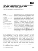
Fig. 2. Powder X-ray diffractograms of FEL and its ion-pair
complexes
Fig. 1. DSC curves of felbinac and its ion-pair complexes at a heating
rate of 10°C/min
lower crystalline lattice energy [20]. Figure 2 showed
the pXRD patterns of solid-state forms of FEL and its
complexes. The distinct differences in the diffraction
patterns of FEL and its complexes also demonstrated
the different arrangement of molecules in the crystal
lattice [21].
that of C=O acceptor groups in FEL, thus leading to the
electron redistribution of the corresponding carboxyl group
acted as a donor group and the blue shift of C=O stretching
vibration in this group [23,26]. This explanation also
conformed to the proton-transfer model of Huyskens and
Zeegers-Huyskens [27], which showed that the larger pKa
difference between the proton donor (FEL) and acceptor
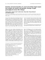
IR and 1H-NMR Characterization
Infrared spectroscopy (IR) plays an important role in
studying the formation of ion pairs [22,23]. In IR spectrum
of FEL (Fig. 3), the absorption at 1687 cm−1 was assigned to
the stretching vibration of C=O group. In the case of FEL
complexes with MEtA, DEtA, TEtA, and HEPP, the
absorption at 1687 cm−1 was red shifted to 1581, 1634, 1588,
and 1580 cm−1, respectively, and that red shift was reckoned as
a criterion for hydrogen bonding [22]. Contrary to the above
complexes, the C=O stretching bands in FEL complexes with
DEA and TEA got blue shifted to 1700 and 1693 cm−1,
separately. This phenomenon was not contradictive to the
aforementioned redshift criterion. As the carboxylic acid
groups in FEL can form dimers by the intermolecular
hydrogen bonding [24], the R3-N acceptor groups in DEA
and TEA might disrupt the original intermolecular hydrogen
bond due to the formation of new intermolecular hydrogen
bond with the carboxyl donor groups in FEL. Based on
literatures [25], it could be inferred that the electronegativity
of R3-N acceptor groups in DEA and TEA were weaker than
Fig. 3. IR spectra of felbinac and its ion-pair complexes
Liu et al.
266
Table I. 1H NMR Chemical Shifts of FEL and Its Ion-Pair Complexes for Proton on Carbon
Permeants
FEL
FEL-DEA
FEL-TEA
FEL-MEtA
FEL-DEtA
FEL-TEtA
FEL-HEPP
δ (ppm)
Δδ (ppm)
3.72
0
3.63
−0.09
3.64
−0.08
3.60
−0.12
3.58
−0.14
3.66
−0.06
3.62
−0.10
(DEA and TEA) indicated stronger hydrogen bond
interaction.
NMR spectroscopy also offered a good evidence for
hydrogen bonding and was therefore used to analyze the
interaction between FEL and organic amines in IPP, based
on the chemical shift change of the methenyl proton near
the carboxyl group. However, the complicated structure of
IPP interfered the spectra of samples, deuterated chloroform (ε r =4.81) was chosen as substitutions of IPP
(ε r =3.18) based on its comparable dielectric constant
[23]. As illustrated in Table I, the signal of the methenyl
proton in all complexes brought out upfield shifts compared with that in FEL. It could be elucidated that there
existed hydrogen interactions between FEL and organic
amines. In detail, the carboxyl group of FEL had an
electrophilic effect on methenyl, which decreased the electron atmosphere density and caused a downfield shift of
the methenyl proton. After the introduction of organic
amines, hydrogen bond was formed between the carboxyl
group of FEL and the basic organic amine, which impaired the deshielding effect and brought out an upfield
shift of the methenyl proton [28]. In a word, all characterization results demonstrated the formation of FEL ionpair complexes.
In Vitro Evaluation
The Effect of Organic Amines on the Skin Permeation of FEL
As FEL is a weak acid, six organic amines were
chosen to prepare ion-pair complexes with FEL and the
permeation of these complexes from both IPP and transdermal patches were investigated. IPP is a frequently used
cosmetic ingredient with low dielectric constant (εr=3.18),
which can contribute to the formation of ion pairs and
simulate the highly lipophilic matrix such as pressuresensitive adhesives [23,29]. Different from the permeation
experiments from patches, the permeation experiment
from IPP ignores the influence of patch matrix; thus, the
flux from IPP can represent the skin permeability of drugs
to some extent. The permeation profiles from IPP and
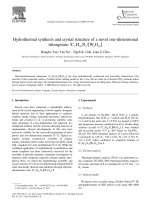
relevant parameters are presented in Fig. 4 and Table II.
As depicted in Fig. 4, TEA, DEA, and HEPP had a
positive effect on the permeation of FEL, and among
them, TEA had the greatest enhancing effect, while other
amines, i.e., TEtA, MEtA, and DEtA, exerted negative
effects. The different effects of amines can be explained
by the altered physicochemical properties of a drug due to
the formation of ion-pair complexes [30].
As illustrated in Fig. 5a, the flux of FEL ion-pair
complexes increased with the increasing solubility
(r=0.9929), which indicated that solubility was an important factor affecting their permeation rate [31]. However,
for FEL-DEA and FEL-HEPP, the introduction of amines
did not increase their solubility, but their flux was increased. This suggested that the flux increase of these
FEL complexes could be attributed in part to their different solubility in the donor phase and there existed other
factors affecting their flux [32]. In Fig. 5b, the flux also
increased with the increasing n-octanol/water partition coefficient Log P of FEL ion-pair complexes (r=0.9498).
This suggested Log P might be another important factor.
According to the two-layer skin model [10], the simplified
skin consists of a lipophilic SC and an underlying hydrophilic ED. For hydrophilic drugs, the lipophilic SC layer
provides a main barrier. While for lipophilic drugs, the
partition from SC to hydrophilic ED becomes a ratelimiting step. Thus, to achieve enhanced skin permeability,
drugs should possess balanced lipid and water solubility.
As a lipophilic drug, FEL is almost insoluble in water and
the distribution from SC to ED may be a principal resistance. With the help of organic amines like TEA, DEA,
and HEPP, the lipophilicity of FEL decreased to a suitable level, making it easier to partition into the ED and
thereby brought about an enhanced permeability. In contrast, FEL complexes with MEtA, DEtA, and TEtA exhibited lower permeation than FEL. It may also be due to
the altered solubility and Log P of FEL complexes. As
can be seen from Table II, the flux of FEL complexes
with MEtA, DEtA, and TEtA decreased as their decreasing solubility in donor phase. This indicated that solubility
was an important factor affecting the flux of FEL complexes. Meanwhile, the lipophilicity of these complexes
also influenced their permeability. As MEtA, DEtA, and
TEtA had strong hydrophilicity, the introduction of these
amines greatly reduced the lipophilicity of FEL even to
Fig. 4. Effect of ion-pair complexes on the permeation of felbinac
from IPP (n=4)
Combining Ion-Pair and Enhancer Strategy
267
Table II. Permeation Parameters of FEL and Its Ion-Pair Complexes from IPP Through Rabbit Abdominal Skin (n=4) and Corresponding
Physicochemical Properties
Permeants
Log Pamines
Log Pion pairs
Sa (mg/mL)
Sb (mg/mL)
pKa
J (μg/cm2/h)
FEL-TEA
FEL-DEA
FEL-HEPP
FEL
FEL-TEtA
FEL-MEtA
FEL-DEtA
1.65
0.66
0.50
–
−0.99
−1.76
−1.48
0.81
0.36
0.30
2.58
−0.02
−0.38
−0.52
3.50±0.28
2.40±0.07
2.37±0.07
3.05±0.03
0.29±0.02
0.19±0.01
0.02±0.01
6.50±0.43
9.27±0.48
178.24±2.71
5.26±0.06
21.25±2.32
19.66±2.31
35.68±1.61
10.62
10.76
8.96
–
7.77
8.71
9.16
177.60±32.21
111.86±22.54
94.03±15.45
74.35±6.13
8.45±0.48
6.32±0.60
4.72±0.16
a
b
Solubility in isopropyl palmitate (IPP)
Solubility in phosphate buffer (pH 7.4)
become hydrophilic. That hydrophilic character hindered
their partition into the lipophilic SC layer, thus presenting
a negative effect. Therefore, both solubility and Log P
had a major influence on the flux of FEL ion-pair complexes, and those organic amines which could alter the
Log P of a drug to a proper level would have a positive
effect on the drug’s permeability.
In addition, the pKa of counter ions was reckoned as
another factor affecting the permeability of ion pairs in
previous reports. The fluxes of flurbiprofen ion pairs were
found to increase with the increasing pKa values of
amines and this was attributed to the stronger attractive
force between flurbiprofen and amines [28]. Xi et al. also
demonstrated that pKa of counter ions could affect the
stability of their ion pairs, thus influencing the permeability of ion pairs [23]. Although amines with relatively high
pKa exhibited enhancing effect on FEL, the correlation
between the flux of FEL complexes and pKa of amines
was not quite so successful (r=0.7998), probably because
the different fluxes of ion-pairs were influenced by several
factors together including both parent drugs and counterions. But this pKa effect can still be seen in TEA and
DEA, with relatively higher pKa, DEA, and TEA also
exhibited significantly promoting effect on FEL, and this
may also be due to their stronger attractive force and
more stable formation of complexes with FEL [27]. This
explanation was also consistent with the IR results, in
which the red-shift phenomenon in TEA and DEA suggested their stronger interaction with FEL.
In transdermal patches, ion-pair strategy was also
used due to the promoting effect of TEA, DEA, and
HEPP in IPP solution system. PSA Duro-Tak® 87-4098
without functional groups was used to prepare transdermal patches, thus avoiding the polar functional groups’
damage to ion-pair structure. As shown in Figs. 6 and 7,
the order of the permeation amounts of FEL ion-pair
complexes from patches was almost the same as that from
IPP solution (r=0.9762). That means the lipophilic IPP
Fig. 5. a Relationship between the flux of FEL ion-pair complexes from IPP and their solubility in IPP. b Relationship between the flux of FEL
ion-pair complexes from IPP and Log P of these complexes
268
Liu et al.
Fig. 6. Effect of ion-pair complexes on the permeation of felbinac
from transdermal patches (n=4)
Fig. 8. Effect of chemical enhancers on the permeation of FEL-TEA
from transdermal patches (n=4)
solution system can predict the permeation of drugs from
patches prepared with lipophilic PSA Duro-Tak® 87-4098.
In PSA, FEL-TEA (5%, w/w, based on adhesive weight)
still had the highest flux (J=6.07±1.11 μg/cm2/h), which
was significantly higher than that of FEL (J=3.16
±0.36 μg/cm2/h). This indicated the feasibility of ion-pair
strategy used in transdermal patches, and therefore, FELTEA was used to substitute FEL for designing a more
effective transdermal patch.
used and the concentration of enhancers was initially
fixed at 5% (w/w).
As shown in Fig. 8, the relatively lipophilic enhancers Azone (Log P=6.02, obtained from SciFinder
database) and IPM (Log P=7.25, obtained from Hui M
et al. 2014) had greater enhancement effect on the permeation of FEL-TEA, and Azone had the greatest promoting effect (P<0.05). It has been widely accepted that
the predominant route of penetration is through the
intercellular lipid domains [34]; therefore, these results
suggested lipophilic enhancers could partition well into
the modified SC. Furthermore, Azone was reckoned to
exert its enhancing effect by partitioning into stratum
corneum and disrupting the packings of the bilayer lipids
[13,36]. Subsequently, the influence of Azone concentration was further studied. As illustrated in Fig. 8, the
permeation amount of FEL-TEA increased as the concentration of Azone increased from 5 to 10%, but when
it increased to 15%, the permeation of FEL-TEA was
Combined Effect of Chemical Enhancers
To further increase the cumulative amounts of
FEL-TEA patch, chemical enhancer was introduced
and combined with ion-pair strategy in this study [33].
N-Dodecylazepan-2-one (Azone), isopropyl myristate
(IPM), Span80 (SP), propylene glycol (PG), and l-menthol (MT), five commonly used penetration enhancers
known to be safe or used commercially [13,34,35], were
Fig. 7. Relationship between the flux from transdermal patches and
the flux from IPP of FEL and its ion-pair complexes
Fig. 9. The penetration profiles of patches containing different concentration of FEL-TEA and compared with the commercial FEL
patch (n=4)
Combining Ion-Pair and Enhancer Strategy
269
Table III. Results of Rabbit Skin Irritation Test (n=4)
Treatment
24 h
48 h
72 h
FEL-TEA patch
Standard irritant
0.25±0.10
6.17±0.19
0.0
6.56±0.19
0.0
7.67±0.50
not further increased. This could be explained by the
effect of Azone on the hydration of SC, which had a
negative influence on the partition of FEL-TEA [37,38].
Overall, 10% Azone had the greatest enhancement effect
on FEL-TEA and it was chosen for designing the formulation of FEL-TEA patch.
To make the skin permeation results comparable
with the commercial product, the concentration of
FEL-TEA in the optimized patch was increased to
7%, which equaled to the amount of FEL in the product SELTOUCH® (0.5 mg/cm 2). As shown in Fig. 9, the
flux of the optimized patch containing 7% FEL-TEA
was significantly higher than that of the commercial
product. The in vitro evaluation results indicated that
it was useful to maximize the flux of FEL by combining
ion-pair and chemical enhancer strategy. The optimized
patch contained the adhesive Duro-Tak® 87-4098, 7%
FEL-TEA, and 10% Azone, and it was used in further
study.
In Vivo Evaluation
Skin Irritation Test
As was showed in Table III, the optimized FEL-TEA
patch (containing 10% Azone) produced no irritation to
the rabbit skin compared with the standard irritant group.
Skin irritation response depends on the amount of Azone
released from the PSA layer. As a lipophilic enhancer,
Azone had a good compatibility with the PSA and it
appeared to have a lower release rate from the acrylic
PSA without influence from the type of adhesive [39]. In
this study, the acrylic type PSA Duro-Tak® 87-4098 was
used as matrix, and therefore, not all Azone could be
released from the optimal FEL-TEA patch in the administration period and the safety of using Azone could be
assured [40].
Pharmacokinetic Analysis
To further evaluate the enhancement effect of combing
ion-pair and chemical enhancer strategy, both the optimized
FEL-TEA patch and commercial FEL patch SELTOUCH®
were applied in rabbit to study their pharmacokinetics. Relevant profiles and parameters were presented in Fig. 10 and
Table IV.
Compared to injection group, the MRT in FELTEA patch group was prolonged to 4.80±0.28 h, which
was more than seven times higher than that in injection
group. This was believed to be due to the continuous
replenishment of drug into the systemic circulation by
constant drug delivery from transdermal patches. The
MRT in FEL-TEA patch group (4.80±0.28 h) and FEL
commercial patch group (5.20±0.15 h) showed no significant difference, but the FEL-TEA group achieved significantly higher C max (2.23±0.49 μg/mL) and AUC 0-t
(15.94±3.58 h.μg/mL) values than the commercial patch
group, which indicated the optimized FEL-TEA patch
had higher skin permeation amount than the commercial product in vivo. The in vivo results also indicated
Fig. 10. a Plasma concentration-time profiles of FEL after intravenous injection of 8 mg FEL (in the form of FEL-TEA) through ear marginal
vein of rabbit (n=4). b Plasma concentration-time profiles of FEL after transdermal administration of FEL-TEA patch and commercial FEL
patch at the abdominal site of rabbit (n=4)
Liu et al.
270
Table IV. Pharmacokinetic Parameters of FEL after Intravenous Injection and Transdermal Administration of Patches (n=4)
Intravenous
Transdermal
Parameters
FEL-TEA
FELTEA patch
Dose of FEL (mg)
Cmax (μg/mL)
AUC0–t (h.μg/mL)
MRT (h)
8
24.17±3.07
16.18±3.74
0.67±0.13
24
2.23±0.49
15.94±3.58
4.80±0.28
Commercial
patch
24
0.86±0.18
7.31±1.16
5.20±0.15
the feasibility of maximizing the flux of FEL by combining ion-pair and chemical enhancer strategy.
In Vitro/In Vivo Correlation
In vitro/in vivo correlation (IVIVC) is defined as a
predictive model about the relationship between in vitro
property of a dosage form and relevant in vivo performance [41]. For transdermal delivery, the in vitro property refers to the rate of skin permeation, and the
in vivo performance is the drug concentration in plasma. In previous reports, IVIVC has been established for
some drugs in topical preparations, and the
deconvolution method showed a good prediction performance [18,42,43].
Thus, based on the in vivo absorption data of FELTEA patch group and FEL-TEA injected group, in vitro
skin permeation results were predicted by the
deconvolution method with the help of WinNonlin®. As
was seen from Fig. 11, the predicted in vitro drug profiles
were consistent with the actual observed in vitro profiles
(r=0.9951), which demonstrated that in vitro skin permeation studies could be used to predict the in vivo performance of FEL-TEA transdermal patches.
Fig. 11. Observed and fitted permeation profiles of FEL-TEA from
transdermal patches through the rabbit abdominal skin in vitro
CONCLUSION
In this work, a novel transdermal patch of FEL was
achieved by combining ion-pair and chemical enhancer
strategy. The optimized patch containing FEL-TEA and
10% Azone had significantly higher skin permeation
amount (P<0.05) and AUC 0-t value than the product
SELTOUCH® in vitro and in vivo. And furthermore, the
in vitro skin permeation results of the optimized FELTEA patch were shown to be useful to predict the
in vivo drug absorption profiles. Therefore, a combination
of ion-pair and chemical enhancer strategy could be useful
in developing a novel transdermal patch of FEL.
REFERENCES
1. Zhang C, Wang L, Yang W, Wang XS, Fawcett JP, Sun YT, et al.
Validated LC–MS/MS assay for the determination of felbinac:
application to a preclinical pharmacokinetics study of felbinac
trometamol injection in rat. J Pharm Biomed Anal. 2009;50:41–5.
2. Heyneman CA, Lawless-Liday C, Wall GC. Oral versus topical
NSAIDs in rheumatic diseases: a comparison. Drugs.
2000;60:555–74.
3. Hadgraft J, du Plessis J, Goosen C. The selection of non-steroidal
anti-inflammatory agents for dermal delivery. Int J Pharm.
2000;207:31–7.
4. Shiba T, Shikata Y, Takagawa N, Sasaki M, Yanaka M, Sugiyama
Y, et al. Antiinflammatory and analgesic activities of felbinac
adhesive preparation (felbinac patch). Jpn Pharmacol Ther.
1992;20:81–94.
5. Shinkai N, Korenaga K, Takizawa H, Mizu H, Yamauchi H.
Percutaneous penetration of felbinac after application of transdermal patches: relationship with pharmacological effects in rats.
J Pharm Pharmacol. 2008;60:71–6.
6. Leeb B. Topical felbinac in therapy of athletic injuries. Fortschr
Med. 1994;112:77–80.
7. Dawson M, McGee CM, Vine JH, Nash P, Watson TR, Brooks
PM. The disposition of biphenylacetic acid following topical application. Eur J Clin Pharmacol. 1988;33:639–42.
8. Tan HS, Pfister WR. Pressure-sensitive adhesives for transdermal
drug delivery systems. Pharm Sci Technol Today. 1999;2:60–9.
9. Van der Windt DAWM, Van der Heijden GJMG, Van den Berg
SGM, Ter Riet G, De Winter AF, Bouter LM. Ultrasound therapy for musculoskeletal disorders: a systematic review. Pain.
1999;81:257–71.
10. Bronaugh RL, Stewart RF. Methods for in vitro percutaneous
absorption studies VI: preparation of the barrier layer. J Pharm
Sci. 1986;75:487–91.
11. Agharkar S, Lindenbaum S, Higuchi T. Enhancement of solubility of drug salts by hydrophilic counterions: properties of organic
salts of an antimalarial drug. J Pharm Sci. 1976;65:747–9.
12. Berge SM, Bighley LD, Monkhouse DC. Pharmaceutical salts. J
Pharm Sci. 1977;66:1–19.
13. Williams AC, Barry BW. Penetration enhancers. Adv Drug Deliver Rev. 2012;64:128–37.
14. Zhang YX, Cun DM, Kong X, Fang L. Design and evaluation of
a novel transdermal patch containing diclofenac and
teriflunomide for rheumatoid arthritis therapy. Asian J Pharm
Sci. 2014;9:251–9.
15. Cheong HA, Choi HK. Effect of ethanolamine salts and enhancers on the percutaneous absorption of piroxicam from a
pressure sensitive adhesive matrix. Eur J Pharm Sci.
2003;18:149–53.
16. Hui M, Quan P, Yang YY, Fang L. The effect of ion-pair formation combined with penetration enhancers on the skin permea t i o n o f lo x o p r o f e n . D r u g D e l i v. 2 0 1 4 . d oi :10 .31 09 /
10717544.2014.981768.
17. Draize JH, Woodard G, Calvery HO. Methods for the study of
irritation and toxicity of substances applied topically to the skin
and mucous membranes. J Pharmacol Exp Ther. 1944;82:377–90.
Combining Ion-Pair and Enhancer Strategy
18. Sun L, Cun DM, Yuan B, Cui HX, Xi HL, Fang L, et al. Formulation and in vitro/in vivo correlation of a drug-in-adhesive transdermal patch containing azasetron. J Pharm Sci. 2012;101:4540–8.
19. Katritzky AR, Jain R, Lomaka A, Petrukhin R, Maran U,
Karelson M. Perspective on the relationship between melting
points and chemical structure. Cryst Growth Des. 2001;1:261–5.
20. Mazzenga GC, Berner B. The transdermal delivery of zwitterionic drugs I: the solubility of zwitterions salts. J Control Release.
1991;16:77–88.
21. Chawla G, Gupta P, Thilagavathi R, Chakraborti AK, Bansal
AK. Characterization of solid-state forms of celecoxib. Eur J
Pharm Biopharm. 2003;20:305–17.
22. Arunan E, Desiraju GR, Klein RA, Sadlej J, Scheiner S, Alkorta
I, et al. Defining the hydrogen bond: an account (IUPAC Technical Report). Pure Appl Chem. 2011;83:1619–36.
23. Xi HL, Wang ZY, Chen Y, Li W, Sun L, Fang L. The relationship
between hydrogen-bonded ion-pair stability and transdermal
penetration of lornoxicam with organic amines. Eur J Pharm
Sci. 2012;47:325–30.
24. Van Eerdenbrugh B, Fanwick PE, Taylor LS. 2-(Biphenyl-4yl)acetic acid (felbinac). Acta Cryst. 2010;E66:O2609–U908.
25. Huheey JE. The electronegativity of groups. J Phys Chem.
1965;69:3284–91.
26. Liu NN, Zhang YQ, Cun DM, Quan P, Fang L. Effect of backing
films on the transdermal delivery of donepezil from patches.
AAPS PharmSciTech. 2014;15:1569–73.
27. Huyskens PL, Zeegers-Huyskens T. Molecular associations and
acid–base equilibriums. J Chim Phys Phys Chim Biol. 1964;61:81–
6.
28. Ma X, Fang L, Guo JP, Zhao NX, He ZG. Effect of counter-ions
and penetration enhancers on the skin permeation of
flurbiprofen. J Pharm Sci. 2010;99:1826–37.
29. Barrow GM. The nature of hydrogen bonded ion-pairs: the reaction of pyridine and carboxylic acid in chloroform. J Am Chem
Soc. 1956;78:5802–6.
30. Megwa SA, Cross SE, Whitehouse MW, Benson HAE, Roberts
MS. Effect of ion pairing with alkylamines on the in-vitro dermal
penetration and local tissue disposition of salicylates. J Pharm
Pharmacol. 2000;52:929–40.
31. Michaels AS, Chandrasekaran SK, Shaw JE. Drug permeation
through human skin: theory and in vitro experimental. AICHE J.
1975;21:985–96.
271
32. Minghetti P, Cilurzo F, Casiraghi A, Montanari L, Fini A.
Ex vivo study of transdermal permeation of four
diclofenac salts from different vehicles. J Pharm Sci.
2007;96:814–23.
33. Auner BG, Valenta C, Hadgraft J. Influence of lipophilic counterions in combination with phloretin and 6-ketocholestanol on the
skin permeation of 5-aminolevulinic acid. Int J Pharm.
2003;255:109–16.
34. Lane ME. Skin penetration enhancers. Int J Pharm. 2013;447:12–21.
35. Narishetty STK, Panchagnula R. Transdermal delivery of zidovudine: effect of terpenes and their mechanism of action. J Control Release. 2004;95:367–79.
36. Pilgram GSK, Van der Meulen J, Gooris GS, Koerten HK,
Bouwstra JA. The influence of two azones and sebaceous
lipids on the lateral organization of lipids isolated from human stratum corneum. Biochim Biophys Acta. 2001;1511:244–
54.
37. Sugibayashi K, Nakayama S, Seki T, Hosoya K, Morimoto Y.
M e c h a n i s m o f sk i n p e n e t r a t i o n - e n h a n c i n g e f f e c t b y
Laurocapram. J Pharm Sci. 1992;81:58–64.
38. Díez-Sales O, Watkinson AC, Herráez-Domínguez M, Javaloyes
C, Hadgraft J. A mechanistic investigation of the in vitro human
skin permeation enhancing effect of Azone®. Int J Pharm.
1996;129:33–40.
39. Qvist MH, Hoeck U, Kreilgaard B, Madsen F, Frokjaer S.
Release of chemical permeation enhancers from drug-inadhesive transdermal patches. Int J Pharm. 2002;231:253–
63.
40. Liu C, Fang L. Drug in adhesive patch of zolmitriptan: formulation and in vitro/in vivo correlation. AAPS Pharm Sci Tech. 2014.
doi:10.1208/s12249-015-0303-3.
41. Uppoor VRS. Regulatory perspectives on in vitro
(dissolution)/in vivo (bioavailability) correlations. J Control Release. 2001;72:127–32.
42. Qi X, Liu RR, Sun D, Ackermann C, Hou H. Convolution
method to predict drug concentration profiles of 2,3,5,6tetramethylpyrazine following transdermal application. Int J
Pharm. 2003;259:39–45.
43. Flynn GL, Shah VP, Tenjarla SN, Corbo M, DeMagistris D,
Feldman TG, et al. Assessment of value and applications of
in vitro testing of topical dermatological drug products. Pharm
Res. 1999;16:1325–30.