Screening for incidence of microsporidian parasite Enterocytozoon hepatopenaei (EHP) in litopenaeus vannamei from aquaculture ponds in SPSR Nellore district of Andhra Pradesh, India
Bạn đang xem bản rút gọn của tài liệu. Xem và tải ngay bản đầy đủ của tài liệu tại đây (654.28 KB, 12 trang )
Int.J.Curr.Microbiol.App.Sci (2018) 7(3): 1098-1109
International Journal of Current Microbiology and Applied Sciences
ISSN: 2319-7706 Volume 7 Number 03 (2018)
Journal homepage:
Original Research Article
/>
Screening for Incidence of Microsporidian Parasite
Enterocytozoon hepatopenaei (EHP) in Litopenaeus vannamei from
Aquaculture Ponds in SPSR Nellore District of Andhra Pradesh, India
M. Raveendra1*, P. Hari Babu1, T. Neeraja1, D. Pamanna1,
N. Madhavan1, A.S. Sahul Hameed2 and Ch. Srilatha3
1
2
College of Fishery Science Muthukur, Nellore, Andhra Pradesh, India
OIE Reference Laboratory for WTD, Department of Zoology, C. Abdul Hakeem College,
Melvisharam, Tamil Nadu, India
3
College of Veterinary Science, S.V.V.U Tirupati, India
*Corresponding author
ABSTRACT
Keywords
EHP, HPM,
Parasite, PCR,
Shrimp, WFS
Article Info
Accepted:
10 February 2018
Available Online:
10 March 2018
Hepaotopancreatic Microsporidiosis (HPM) caused by Enterocytozoon hepatopenaei
(EHP), a microsporidian parasite to be associated with slow (retarded or stunted) growth
and white feces syndrome (WFS) in cultured shrimp in many of the shrimp growing
countries in Asia, also in India. In the present study, shrimp samples from various shrimp
ponds from different Mandals of SPSR Nellore district, Andhra Pradesh, India, were
collected over a period of five months (February 2016 to June 2016). Important diagnosis
observed were histopathological studies, molecular technique (PCR). Histologically, the
infected animals showed severe degeneration of hepaotopancreatic tubule, basophilic
inclusions resembling the developmental stages of EHP were found in the epithelial cells
and large number of spore aggregations was observed in the tubular lumen. Enlargement
of haemal sinuses was also observed in some cases. From this study, out of 50 pond case
studies, 31 cases were showing EHP symptoms with 62 % prevalence by using tools of
detection like PCR and histology.
Introduction
Shrimp farming is one of the most profitable
and fastest-growing sectors of the aquaculture
industry. Over the past decade, global farmed
shrimp production has grown almost threefold
from 1.13 million tons in 1999 to over 3.43
million tons in 2009 (Jory, 2010). China
ranked first in shrimp aquaculture with 40% of
total cultured shrimp production followed by
Thailand (15%), Vietnam (12%) and
Indonesia (10%) (FAO, 2011). Shrimp
continues to be the largest single commodity
in value terms, accounting for about 15% of
the total value of internationally traded fishery
products in 2012. It is mainly produced in
developing countries, and much of this
production finds its way into the international
trade (FAO, 2014). Penaeid shrimps
(Litopenaeus
vannamei
and
Peneaus
monodon), which comprise around 80% of
total farmed shrimp production (FAO, 2009).
1098
Int.J.Curr.Microbiol.App.Sci (2018) 7(3): 1098-1109
According to the production statistics from
Marine Products Export Development
Authority (MPEDA), after the introduction of
the species, export production has grown from
1731 metric tonne (mt) to 3,53,413 mt in
2014-15.
Andhra Pradesh has the second largest
brackish water area in India after West
Bengal, covering an area of about 37,560 ha
besides the coastline of 972 km, spreading
over nine districts namely Srikakulam,
Vijayanagaram,
Visakhapatnam,
East
Godavari, West Godavari, Krishna, Guntur,
Prakasam and Sri Potti Sri Ramulu Nellore.
Development of coastal aquaculture in Andhra
Pradesh is centered on shrimp (L. vannamei)
farming. The culture area increased from 264
Ha to 37,560 Ha during the period from 200910 to 2014-15. Andhra Pradesh (2, 76,077 mt)
is the leading farmed shrimp producer with
78% and the rest of India production was 77,
336 mt (MPEDA, 2015). The shrimp (L.
vannamei) exports from Andhra Pradesh
increased to all time high and valued at more
than Rs. 14,000 Crores for the year 2014-15
(MPEDA, 2015).
High stocking density (maximum permissible
is 60 PLs/m2) and use of compounded pelleted
feed in order to achieve higher production
rates impose stress on the shrimps, making
them susceptible to diseases (Alavandi et al.,
1995). The diseases may be caused by various
etiological agents such as viruses, bacteria,
fungi, parasites, algal toxins, nutritional
deficiency or the adverse environment. In
India, the gross economic losses due to shrimp
(P. monodon) diseases were estimated at more
than Rs. 1,000 crores in 2006-2008 and loss
continues even now (Kalaimani et al., 2013).
Several shrimp diseases are new or newly
emerged in Asia that causes serious economic
losses in shrimp farms, including Acute
Hepatopancreatic Necrosis Disease (AHPND)
or Early Mortality Syndrome (EMS),
Hepatopancreatic Microsporidiosis (HPM),
Hepatopancreatic Haplosporidiosis (HPH),
Aggregated Transformed Microvilli (ATM)
and Covert Mortality Disease (CMD). In
addition to these, White Spot Disease (WSD),
Yellow Head Disease (YHD) and Infectious
Myonecrosis (IMN) continued their share of
losses (Thitamadee et al., 2016).
The Global Aquaculture Alliance (GAA,
2013) has estimated that losses to the Asian
shrimp culture sector amount to US$ 1.0
billion. World farmed shrimp production
volumes decreased in 2012 and particularly in
2013, mainly as a result of disease-related
problems, such as Early Mortality Syndrome
(EMS) (FAO, 2014).
In Nellore district the frequent outbreaks of
diseases such as White Spot Syndrome Virus
(WSSV), Black Gill Disease (BGD), Running
Mortality Syndrome (RMS), Loose Shell
Syndrome (LSS), White Faecal Syndrome
(WFS), White Muscle Disease (WMD) and
Infectious Hypodermal and Haematopoietic
Necrosis (IHHN) in shrimps causing
economic loss to the aquaculture industry
(Srinivas et al., 2016).
Recently, shrimp farms in Asia and other areas
have been reporting heavy infection with a
microsporidian
parasite,
Enterocytozoon
hepatopenaei (EHP) in cultured L. vannamei
impacting the production due to severe growth
retardation (Newman, 2015).
Hepatopancreatic microsporidiosis (HPM) is
caused by Enterocytozoon hepatopenaei
(EHP), it was first reported as an unnamed
microsporidian from growth retarded black
tiger shrimp Penaeus monodon from Thailand
in 2004 (Chayaburakul et al., 2004). It was
subsequently characterized in detail and
named in 2009 (Tourtip et al., 2009). During
2004, it was not statistically associated with
1099
Int.J.Curr.Microbiol.App.Sci (2018) 7(3): 1098-1109
slow growth. Although EHP does not appear
to cause mortality, recent information from
shrimp farmers in Southeast Asian countries
indicates that it is associated with severe
growth retardation in P. vannamei. EHP
outbreaks are occurring widely in China,
Indonesia, Malaysia, Vietnam and Thailand.
Very recently, EHP is also reported from slow
growing shrimp in India. Thus, EHP is an
emerging problem that is under urgent need of
control (Sritunyalucksana et al., 2014).
L. vannamei samples drawn from Andhra
Pradesh, Tamil Nadu and Puri (Odisha) were
tested positive for EHP by PCR, and some
samples
were
found
positive
by
histopathology (CIBA, 2015).
Stunting of L. vannamei in shrimp culture
ponds for various reasons including EHP has
created confusion among shrimp farmers and
farmers are unable to harvest the crop though
it is uneconomical to continue the crop with
stunted shrimp.
Materials and Methods
Sampling area
The present study was carried out for a period
of five months between February, 2016 to
June, 2016. Shrimp (L. vannamei) with the
sign of stunted growth were collected for this
study from different shrimp farms located in
Nidiguntapalem,
Krishnapatnam
village,
Pottempadu, Bodiswamikandriga, Pantapalem
of Muthukurmandal, Esuruwaka, Daruvukatta,
Uttama Nellore, Konduru of Kota mandal,
Dugarajapatnam, Mallam, Raghavavaripalem,
Kothagunta, Thuppaguntapalem, Tupilipalem
of Vakadumandal, Chintavaram, Eruru village
of Chillakurumandal, Ganagapatnam village
of Indukuripetmadal, Mudivarthi village of
Vidavalurumandal and Jadagogula village of
Bhogolumandal, SPSR Nellore district,
Andhra Pradesh, India.
Experimental shrimp
The experimental culture shrimp of the present
study was Litopenaeus vannamei cultured in
semi-intensive and intensive farms of the
above mentioned areas.
Primers
Published universal primers were used for the
amplification of ssu rRNA gene of
Enterocytozoon hepatopenaei isolates. The
names of the primers, sequence and
amplification size are given below:
Collection of samples
Fifty (50) ponds were selected for study which
were experiencing size variation/growth
retardation and white feces syndrome. On
each sampling day, a minimum of 60 shrimps
were examined for diseases of species as per
OIE guidelines (OIE, 2013). Information on
behavioral abnormalities, gross and clinical
signs were recorded on the sampling sheet.
From each pond 2-4 shrimps were taken for
diagnosis and the hepatopancreas of each
sample were dissected out and fixed in
Davidson’s fixative for histopathology and
along with Davidson’s fixative from the 50
samples, 15 were separately fixed in 95%
alcohol for molecular diagnosis (Bell and
Lightner, 1984). Whole infected shrimps were
also wrapped individually in sterile polythene
bags, placed in icebox and brought to the
laboratory. On reaching laboratory they were
transferred to refrigerator and analyzed /
processed.
Histopathology
Histopathology was conducted in the
Department of Pathology, College of
Veterinary Science, S.V.V.U. Tirupati. The
hepatopancreas of infected and normal
shrimps were fixed in alcoholic Davidson’s
1100
Int.J.Curr.Microbiol.App.Sci (2018) 7(3): 1098-1109
fixative for 48-72 h for comparative study.
After fixation the tissues were transferred to
70% ethyl alcohol and kept overnight.
Histopathological analysis was made as
described by Roberts (2001)
Molecular diagnosis
Molecular diagnosis has done at OIE
Reference Laboratory for WTD, Department
of Zoology, C. Abdul Hakeem College,
Melvisharam, Tamil Nadu.
read Sequencing kit (Applied Biosystems).
The nucleotide sequence of E. hepatopenaei
(small subunit rRNA gene) has been deposited
in (Gen-Bank accession no. KU198278). The
sequence was aligned using bioinformatics
tools such as standard nucleotide BLAST and
multiple
sequence
analysis
clustalW
(Thompson et al., 1994). Significant similarity
with sequences available in GenBank was
searched using BLAST at National Center for
Biotechnology Information (NCBI).
Results and Discussion
DNA extraction
Clinical signs of infected shrimp
Hepatopancreas were homogenized in NTE
buffer (0.2 M NaCl, 0.02 M Tris–HCl and
0.02 M EDTA, pH 7.4), and 10% tissue
suspension was made. The suspension was
centrifuged at 3000 g for 15 min at 4 °C, and
supernatant was collected. The tissue
suspension was mixed with an appropriate
amount of digestion buffer (100 mM NaCl, 10
mM Tris–HCl, pH 8.0, 50 mM EDTA, pH 8.0,
0.5% sodium dodecyl sulphate and 0.1 mg
mL-1 proteinase K) and incubated for 2 h at
65°C to extract the DNA. After incubation, the
digests were deproteinized by successive
phenol/chloroform/isoamyl alcohol extraction
and DNA was recovered by ethanol
precipitation and dried. The dried DNA pellet
was suspended in TE buffer and used as a
template for PCR amplification.
Agarose gel electrophoresis
Polymerase chain reaction products were
analyzed by electrophoresis in 0.8% agarose
gels stained with ethidium bromide and
visualized by ultraviolet transillumination.
DNA sequensing and analysis
The amplified PCR product was purified using
Qiagen plasmid minipreparation spin column.
Sequence analysis was performed on an Auto-
Shrimp (L. vannamei) with the sign of slow
growth were collected for this study from
different shrimp farms located in SPSR
Nellore district, Andhra Pradesh, India. All the
samples which we collected are slow growing
as well as normally growing. These animals
were apparently same except for reasons of
slow growth and white fecal matter.
From the selected 50 ponds a shrimp
population 4-10 animals with typical clinical
symptoms (white feces and slow growth) were
selected for diagnosis. Test results showed 31
pond shrimp samples (Table 1) were tested
positive for EHP both by histopathology and
PCR. Among the 31 pond samples, 15
samples were positive in slow growth with
white feces syndrome and 16 pond samples
were positive in slow growth without white
feces.
The shrimp samples collected from white
feces syndrome affected ponds were showing
floating strands of white feces and some time
the fecal strand was hanging from the anal
portion of the shrimp. When the problem was
severe, all the floating fecal strands were
coming to sides of the pond, and it become
easy for the pond manager to recognize the
abnormality. Associated with the white feces
1101
Int.J.Curr.Microbiol.App.Sci (2018) 7(3): 1098-1109
syndrome is drop in daily feed consumption,
slow growth and some shrimp mortality also.
The freshly dead shrimp also showed loose
shell condition. During the study period, the
white feces syndrome first appears 50–70 days
of culture (DOC). After the appearance of
white feces, shrimp health will deteriorate if
some management interventions are not
adopted. These include treatments with
medicated feed (garlic etc.) and reduction in
daily feed ration. In general, the shrimp in the
WFS ponds showed FCR of over 2.92–3.17
(can be considered as 3.0) as compared to the
range of normal growth ponds 1.83–1.94 (can
be considered as 2.0).
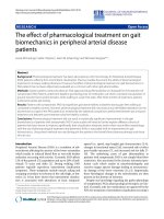
Histopathology
Histologically, severe necrotic changes were
noticed in the hepatopancreas (Fig. 1)
whereas, EHP spores were observed in
cytoplasm (Fig. 2). Developmental stages of
EHP (Fig. 3) and large eosinophilic to
basophilic inclusions indicating presumptive
developmental stages of the microsporidian
could be noticed in the tubular epithelium
(Fig. 4). These stages were predominantly
seen in the distal ends of hepatopancreatic
tubules and most of the tubular epithelium in
this region showed detachment from the basal
membrane (Fig. 5 and 6).
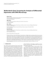
The basal part of the tubular epithelium
showed granular material and spore-like
structures. Abnormally enlarged haemal
sinuses were also noticed in the inter-tubular
spaces (Fig. 7). In some of the sections, the
spores were noticed in vacuolated structures.
Sloughing of the tubular epithelial cells was
pronounced in heavily infected HP and large
spore aggregations were noticed in the tubular
lumen (Fig. 8). In some of the hepatopancreas
sections, large number of rod-shaped bacterial
cells was also noticed in the tubular lumen
indicating secondary infection.
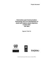
Molecular characterization
The results of the targeted surveillance of EHP
in Litopenaeus vannamei of SPSR Nellore
district, Andhra Pradesh from February 2016
to June 2016 are presented in Table 1. In 0.8%
agarose gel electrophoresis, samples with EHP
infection show a band of PCR (510 bp)
(Fig. 9).
Shrimp samples from the different villages
were tested for EHP infection. Out of 50 pond
shrimp samples, 31 samples were found to be
EHP positive with 62% prevalence both by
PCR and histologically (Table 1). Selected
microsporidian isolate of Enterocytozoon
hepatopenaei
KU198278
was
further
characterized and identified through ssu rRNA
analysis. The detailed information of the
bacterial strain used, host species, clinical
signs, site of infection, Gen Bank accession
numbers are presented in Table 2.
Shrimp farms in Asia and other areas have
been reporting heavy infection with a
microsporidian
parasite,
Enterocytozoon
hepatopenaei (EHP) in cultured L. vannamei
impacting the production due to severe growth
retardation (Newman, 2015). The parasite was
first recorded from growth retarded tiger
shrimp, Penaeus monodon from Thailand and
reported as an undesignated microsporidian
(Chayaburakul et al., 2004).
Later, this parasite was identified in P.
monodon and named as EHP by Tourtip et al.,
(2009). The occurrence of this parasite was
reported in pond-reared P. monodon from
Vietnam, China, Indonesia, Malaysia and
Thailand (Ha et al., 2010) and in P. stylirostris
from Brunei (Tang et al., 2015). EHP is also
reported from slow growing shrimp in India
(Sritunyalucksana el al., 2014) and very
recently, its occurrence was reported in farm
reared L. vannamei in India (Rajendran et al.,
2016).
1102
Int.J.Curr.Microbiol.App.Sci (2018) 7(3): 1098-1109
Fig.1 Degenerative and necrotic tubules
H&E,x100
Fig.2 EHP spores present in the cytoplasm
H&E,x400
Fig.3 Development stages of EHP
H&E,x400
Fig.4 Basophilic inclusion (arrow; H&E,
x400), EHP spores (star; H&E, x400)
Fig.5 EHP spores (star), basophilic inclusion
(arrow) H&E,x400
Fig.6 Developmental stages of EHP spores
(star) and detachment of tubular lumen
(arrow) H&E, x400)
Fig.7 Enlargement of haemal sinus (star) and
EHP spores (arrow) (H&E, x400)
Fig.8 EHP infected tissue section
(H&E,x400)
1103
Int.J.Curr.Microbiol.App.Sci (2018) 7(3): 1098-1109
Fig.9 0.8%Agarose gel showing PCR product of EHP of naturally infected Litopenaeus
vannamei
Primers
Primers
Sequence (5’-3’)
GCC TGA GAG ATG GCT CCC ACG T
GCG TAC TAT CCC CAG AGC CCG A
Amplification size
510-bp
Reference
Tang et
al., 2015
Table.1 Targeted surveillance of Enterocytozoon hepatopenaei in Litopenaeus vannamei of
SPSR Nellore district, Andhra Pradesh from February 2016 to June 2016
Case No
and Date
1)
20.02.2016
2)
20.02.216
3)
20.2.2016
4)
28.02.216
5)
28.02.2016
6)
28.02.216
Location of
Pond
Nidiguntapale
m, Muthukur
Nidiguntapale
m, Muthukur
Nidiguntapale
m, Muthukur
K.P.Village,
Muthukur
K.P.Village,
Muthukur
K.P.Village,
Muthukur
Sign of
Stunted
growth &
White
Feces
Syndrome
Yes
ABW
(g) on
Date of
Samplin
g
Pond
area
(Ha)
PL's
stocked
125
20
1.0
Yes
110
16
Yes
105
Yes
DOC
Confirmation of
EHP
Histol
ogy
PCR
400000
+ ve
+ ve
0.8
250000
- ve
16
0.4
100000
- ve
55
6
0.4
100000
- ve
Yes
55
7
0.4
100000
- ve
Yes
60
7
0.4
100000
- ve
Not
tested
Not
tested
Not
tested
Not
tested
Not
tested
1104
Int.J.Curr.Microbiol.App.Sci (2018) 7(3): 1098-1109
7)
28.02.216
8)
29.02.2016
9)
12.03.2016
10)
16.03.2016
11)
21.03.2016
12)
26.03.2016
13)
12.04.2016
14)
15.04.2016
15)
15.04.2016
16)
16.04.2016
17)
16.04.2016
18)
03.05.2016
19)
03.05.2016
20)
05.05.2016
21)
05.05.2016
22)
05.05.2016
23)
07.05.2016
24)
25.05.2016
25)
25.05.2016
K.P.Village,
Muthukur
Gangapatnam,
Indukuripeta
Mudivarthi,
Vidavaluriu
Ramudupalem,
Vidavaluru
Ramudupalem,
Vidavaluru
Gangapatnam,
Indukuripeta
Pottempadu,
Muthukur
Esuruwaka,
Chittamuru
Esuruwaka,
Chittamuru
Daruvkatta,
Kota
Uttama
Nellore, Kota
Utukuru,
Vidavaluru
Mudivarthi,
Vidavaluru
Pottempadu,
Muthukur
Jadagogula,
Bhogolu
Jadagogula,
Bhogolu
Kandriga,
Muthukur
Mallam,
Vakadu
Mallam,
Vakadu
Yes
60
7
0.4
100000
- ve
Yes
115
25
0.8
400000
- ve
Yes
95
16.6
0.8
400000
- ve
Yes
105
20
0.8
400000
- ve
Yes
92
16
0.8
400000
- ve
Yes
64
10
1.0
600000
- ve
Yes
50
8
0.8
400000
- ve
Yes
55
9
0.8
500000
- ve
Yes
50
8
0.8
500000
- ve
Yes
55
6
1.2
700000
- ve
Yes
60
5
0.8
500000
- ve
Yes
64
8
0.6
150000
- ve
Yes
77
18
1.0
600000
- ve
Yes
90
16
0.4
200000
- ve
Yes
44
4
0.48
400000
- ve
Yes
50
5
0.8
300000
- ve
Yes
60
10
0.4
150000
- ve
Yes
105
14
1.0
300000
- ve
Yes
95
10
1.0
500000
+ ve
26)
25.05.2016
27)
25.05.2016
28)
25.05.2016
29)
26.05.2016
30)
26.05.2016
31)
26.05.2016
32)
26.05.2016
Mallam,
Vakadu
Raghavavaripal
em, Vakadu
Kothagunta,
Vakadu
Chintavaram
Yes
78
9
0.4
200000
- ve
yes
85
10
1.0
400000
- ve
Yes
90
12.5
1.0
400000
- ve
Yes
90
12.5
1.0
500000
+ ve
Eruru
Yes
90
14
0.8
300000
- ve
Eruru
Yes
84
12.5
0.8
200000
- ve
Eruru
Yes
85
12
1.0
400000
- ve
1105
Not
tested
Not
tested
Not
tested
Not
tested
Not
tested
Not
tested
Not
tested
Not
tested
Not
tested
Not
tested
Not
tested
Not
tested
Not
tested
- ve
Not
tested
Not
tested
Not
tested
Not
tested
+ ve
Not
tested
Not
tested
Not
tested
+ ve
Not
tested
Not
tested
Not
tested
Int.J.Curr.Microbiol.App.Sci (2018) 7(3): 1098-1109
33)
27.05.2016
34)
27.05.2016
35)
27.05.2016
36)
06.06.2016
37)
07.06.2016
38)
08.06.2016
39)
14.06.2016
40)
27.06.2016
41)
27.06.2016
Tupilipalem
Yes
90
14
1.2
400000
- ve
Tupilipalem
Yes
80
11.1
1.2
400000
- ve
Not
tested
- ve
Tupilipalem
Yes
80
8
0.8
400000
+ ve
+ ve
Pantapalem,
Muthukur
B.S.Kandriga,
Muthukur
B.S.Kandriga,
Muthukur
Pantapalem
Yes
104
17
0.8
300000
- ve
- ve
Yes
58
9
1.0
200000
- ve
- ve
Yes
53
4
0.6
200000
- ve
- ve
Yes
104
17
0.8
300000
- ve
- ve
Dugarajapatna
m, Vakadu
Dugarajapatna
m, Vakadu
Yes
105
13
0.8
400000
- ve
- ve
Yes
105
15
0.8
400000
- ve
- ve
42)
27.06.2016
43)
27.06.2016
44)
27.06.2016
45)
27.06.2016
46)
27.06.2016
47)
27.06.2016
48)
27.06.2016
49)
27.06.2016
50)
28.06.2016
Dugarajapatna
m, Vakadu
Dugarajapatna
m, Vakadu
Konduru,
Vakadu
Konduru
Yes
100
16
0.8
400000
- ve
- ve
Yes
115
16.6
0.8
400000
+ ve
+ ve
Yes
125
30
1.2
400000
- ve
- ve
Yes
45
6
1.0
400000
- ve
Tupilipalem
Yes
85
16.6
0.8
500000
- ve
Tupilipalem
Yes
85
11.1
0.8
500000
+ ve
Not
tested
Not
tested
+ ve
Tupilipalem
Yes
90
10
0.8
500000
+ ve
+ ve
Tupilipalem
Yes
88
12.5
1.0
500000
- ve
Thuppaguntapa
lem
Yes
86
11
0.8
500000
+ ve
Not
tested
+ ve
Table.2 Molecular characterization of EHP strain isolated from infected/
diseased cultured shrimp
Shrimp species
Length of
Gen Bank
of consensus
Accession
Disease/Clinical
Site
sign
infection
Litopenaeus
Stunted
(bp)
Hepatopancreas 510
vannamei
growth/White
feces Syndrome
1106
sequence
Identification
number
KU198278
EHP
Int.J.Curr.Microbiol.App.Sci (2018) 7(3): 1098-1109
Histologically,
heavily
infected
hepatopancreas showed severe necrotic
changes as evidenced by sloughing of tubular
epithelial cells, degenerated cells and spore
accumulation in the tubular lumen. In the
present study histological features are
observed in accordance with the reports on
the hepatopancreatic microsporidian infection
in P. monodon and L. vannamei
(Chayaburakul et al., 2004 and Tourtip et al.,
2009). Further, the parasite affinity for the
epithelial cells has been reported for the genus
Enterocytozoon, including the humaninfecting species E. bieneusi (Desportes et al.,
1985). Some of the histological changes
observed in the infected hepatopancreas of P.
vannamei due to EHP also showed similarity
with Enterospora canceri reported from crab
(Stentiford et al., 2007).
PCR and Light microscopic observation of L.
vannamei from 50 ponds located in SPSR
Nellore district revealed the prevalence of
EHP infection (16%). However, PCR
screening from 15 ponds showed relatively
high prevalence of EHP infection (33%)
compared to histopathology results.
The prevalence of EHP was estimated to be
63.5% for 137 samples by Rajendran et al.,
(2016). In the present study the EHP
prevalence was observed as 16% for 50
samples. If we calculate these 50 samples as
137, the prevalence was 23%. Rajendran et
al., (2016) collected animals from white
faeces syndrome (WFS) affected pond
showed higher prevalence of EHP (96.4%)
compared to those from the unaffected pond
(39.7%). But in the present study, WFS
affected pond showed low prevalence (12%)
for 25 samples compared to those from only
stunted growth ponds (20%) for 25 samples.
We agree with the statement given by
Rajendran et al., (2016) that the EHP could be
detected from slow growing as well as WFSaffected animals.
Previously Ha et al., (2010) had been reported
that the association of microsporidian
Enterocytozoon hepatopenaei with white
faeces syndrome (WFS) and Felgel (2012)
has indicated that severe infection with a
microsporidian morphologically similar to E.
hepatopenaei was associated with WFS of P.
vannamei. In the present study WFS affected
ponds showed low prevalence. Ponds with
stunted growth only but no WFS showed
higher prevalence. These results are
comparable
to
the
statement
of
Tangprasittipap et al., (2013) that EHP is not
the cause of WFS.
References
Alavandi, S. V., Vijayan, K. K. and
Rajendran, K. V., 1995. Shrimp
diseases,
their
prevention
and
control. CIBA Bulletin, 3: 1-17.
Bell, T.A. and Lightner, D.V. 1984. IHHN
virus: infectivity and pathogenicity
studies in Penaeus stylirostris and
Penaeus vannamei. Aquaculture. 38:
185–194.
Chayaburakul, K., Nash, G., Pratanpipat, P.,
Sriurairatana,
S.
and
Withyachumnarkul, B., 2004. Multiple
pathogens found in growth-retarded
black tiger shrimp Penaeus monodon
cultivated in Thailand. Dis. Aquat. Org.
60: 89–96.
Chiyansuvata,
P.,
Chantangsi,
C.,
Chutmongkonkul, M., Tangtrongpiros,
J. and Chansue, N., 2015. Molecular
Biological Screening of Enterocytozoon
hepatopenaeiin Aquatic Macro Fauna in
Pacific White Shrimp (L. vannamei)
Pond. Proceedings of the 14th
Chulalongkorn University Veterinary
Conference (CUVC) 2015: Responsible
for Lives Bangkok, Thailand. April 2022.
1107
Int.J.Curr.Microbiol.App.Sci (2018) 7(3): 1098-1109
CIBA, 2015. Annual report 2014-15. Central
Institute
of
Brackish
Water
Aquaculture, Chennai.
Desportes. I., Le Charpentier, Y., Galian, A.,
Bernard, F., Coch and Priollet, B.,
Lavergne, A., Ravisse, P. and
Modigliani, R. 1985. Occurrence of a
new microsporidian: Enterocytozoon
bieneusi n. g., n. sp., in the enterocytes
of human patients with AIDS. J
Protozool 32:49–60.
FAO (Food and Agriculture Organization of
the United Nations), 2009. Fishmeal
market report—May 2009. Food and
Agriculture Organization of the United
Nations—Globefish. Online: http://
www.globefish.org.
FAO (Food and Agriculture Organization),
2014. The state of world fisheries and
aquaculture. Rome, Italy: Food and
Agriculture Organization of the United
Nation.
Flegel, T.W., 2012. Historic emergence,
impact and current status of shrimp
pathogens in Asia. J. Invertebr. Pathol.
110: 166-173.
Food and Agriculture Organization (FAO).
2011. The State of world fisheries and
aquaculture 2008. Food and Agriculture
Organization of the United Nations,
Rome, Italy, pp. 1-235.
Global Aquaculture Alliance (GAA). 2013.
Cause of EMS shrimp disease
identified. GAA News Releases.
Available:
/>Accessed 29 March 2014.
Ha, N.T.H., Ha, D.T., Thuy, N.T. and Lien,
V.T.K.,
2010.
Enterocytozoon
hepatopenaei has been detected
parasitizing tiger shrimp (Penaeus
monodon) cultured in Vietnam and
showing white feces syndrome (In
Vietnamese with English abstract).
Agric. Rural Dev.: Sci. Technol. 12: 45–
50 (translation from Vietnamese).
Jory, D. E. 2010. GOAL production data
projects
tempered
aquaculture
growth. Global aquaculture advocate.
1: 8–10.
Kalaimani, N., Ravisankar, T., Chakravarthy,
N., Raja, S., Santiago, T.C. and
Ponniah, A.G. 2013. Economic Losses
due to Disease Incidences in Shrimp
Farms of India. Fish. Techn. 50: 80-86.
MPEDA, 2015. Annual Report 2014-2015,
Marine Products Export Development
Authority, Cochin.
Newman, S.G. 2015. Microsporidian impacts
shrimp production-Industry efforts
address control, not eradication. Global
Aquaculture Advocate, March/April
2015, 16-17.
Rajendran, K.V., Shivam, S., Praveena, P.E.,
Sahayakajan, J.J., Kumar, T.S., Avunje,
S., Jagadeesan, V., Babu, P.S.V.A.N.V.,
Prande, A., Krishnan, A.N., Alavandi,
S.V. and Vijayan, K.K. 2016.
Emergence
of
Enterocytozoon
hepatopenaei (EHP) in farmed Penaeus
(Litopenaeus) vannamei in India.
Aquaculture. 454: 272–280.
Roberts, R.J. 2001. The parasitology of
teleosts, In: Fish pathology, Roberts,
R.J., (Ed.), W. B. Sauders, London, pp.
254 - 296.
Srinivas, D. and Venkatrayulu, Ch.
2016.Current Status and Prospects of
Pacific White Shrimp Litopenaeus
vannamei (Boone, 1931) Farming in
Coastal Districts of Andhra Pradesh in
India. International Journal of Science
and Research. 5(3): 1211-1214.
Sritunyalucksana, K., Sanguanrut, P.,
Salachan P. V., Thitamadee, S. and
Flegel, T.W. 2014. Urgent appeal to
control spread of the shrimp
microsporidian parasite Enterocytozoon
hepatopenaei (EHP).
Stentiford, G.D. and Bateman, K.S. 2007.
Enterospora canceri sp., an intranuclear
microsporidian
parasite
1108
Int.J.Curr.Microbiol.App.Sci (2018) 7(3): 1098-1109
infection of hermit crab Eupagurus.
Dis. Aquat. Org. 75: 73–78.
Tang, K.F.J., Pantoja, C.R., Redman, R.M.,
Han, J.E., Tran, L.H. and Lightner, D.V.
2015. Development of in situ
hybridization and PCR assays for the
detection
of
Enterocytozoon
hepatopenaei (EHP), a microsporidian
parasite infecting penaeid shrimp. J.
Invertebr. Pathol. 130: 37–41.
Tangprasittipap, A., Srisala, J., Chouwdee, S.,
Somboon, M., Chuchird, N., Limsuwan,
C., Srisuvan, T., Flegel, T.W. and
Sritunyalucksana, K., 2013. The
microsporidian
Enterocytozoon
hepatopenaei is not the cause of white
feces syndrome in white leg shrimp
Penaeus (Litopenaeus vannamei). BMC
Vet. Res. 9: 139.
Thitamadee, S., Prachumwat, A., Srisala, J.,
Jaroenlak,
P.,
Salachanb,
P.V.,
Sritunyalucksana, K., Flegel, T.W. and
Itsathitphaisrn, O., 2016. Review of
current disease threats for cultivated
penaeid shrimp in Asia. Aquaculture.
452: 69-87.
Tourtip, S., Wongtripop, S., Stentiford, G.D.,
Bateman, K.S., Sriurairatana, S.,
Chavadej, J., Sritunyalucksana, K. and
Withyachumnarnkul,
B.
2009.
Enterocytozoon hepatopenaei sp. nov.
(Microsporida: Enterocytozoonidae), a
parasite of the black tiger shrimp
Penaeus
monodon
(Decapoda:
Penaeidae):
fine
structure
and
phylogenetic relationships. J. Invertebr.
Pathol. 102: 21–29.
How to cite this article:
Raveendra, M., P. Hari Babu, T. Neeraja, D. Pamanna, N. Madhavan, A.S. Sahul Hameed and
Srilatha, Ch. 2018. Screening for Incidence of Microsporidian Parasite Enterocytozoon
hepatopenaei (EHP) in Litopenaeus vannamei from Aquaculture Ponds in SPSR Nellore
District of Andhra Pradesh, India. Int.J.Curr.Microbiol.App.Sci. 7(03): 1098-1109.
doi: />
1109