Quantitative and morphological evaluation of the effect of platelet rich plasma on collagen fibers in experimentally induced skeletal muscles injury in adult male albino rats
Bạn đang xem bản rút gọn của tài liệu. Xem và tải ngay bản đầy đủ của tài liệu tại đây (936.95 KB, 9 trang )
Int.J.Curr.Microbiol.App.Sci (2018) 7(7): 3808-3816
International Journal of Current Microbiology and Applied Sciences
ISSN: 2319-7706 Volume 7 Number 07 (2018)
Journal homepage:
Original Research Article
/>
Quantitative and Morphological Evaluation of the Effect of Platelet Rich
Plasma on Collagen Fibers in Experimentally Induced Skeletal
Muscles Injury in Adult Male Albino Rats
Shimaa M. Badr*, Reda H Elbakary, Essam M. Laag, Naglaa I. Sarhan
and Nafisa A. Elbakary
Histology Department, Faculty of Medicine, Tanta University, Egypt
*Corresponding author
ABSTRACT
Keywords
Platelet rich plasma,
Muscle injury,
Fibrosis, Mallory
trichrome
Article Info
Accepted:
26 June 2018
Available Online:
10 July 2018
Muscle injuries present frequently in sports medicine. Healing with conventional therapy
is often inadequate, leading to incomplete functional recovery due to fibrosis. Generating
substantial interest in the potential for emerging technologies such as platelet-rich plasma
(PRP) to enhance soft-tissue healing and decrease time of recovery became mandatory.
The aim of this work was to evaluate the role of PRP in promoting healing of
experimentally induced skeletal muscle injury in adult male albino rat model. 48 adult
male albino rats were used and were divided into three groups. Group I was served as
control for obtaining muscle specimens from their gastrocnemius muscle and PRP. Group
II was exposed to bilateral gastrocnemius muscles injury and was left without treatment.
Group III were subjected to bilateral gastrocnemius injury and were immediately treated
with PRP intramuscularly. Muscle specimens were excised after 1, 7 and 21 days from the
onset of injury to be processed for light microscopic study after staining with Mallory
trichrome stain. Assessments of the mean area percentage of collagen fibers were also
statistically analyzed. Treatment with PRP resulted in enhanced regeneration of skeletal
muscle injury without fibrosis. The PRP treated group demonstrated absence of fibrosis on
days 14 and 21 as compared to their associates in non-treated group. Local injection of
PRP into the injured gastrocnemius muscle resulted in enhanced muscle regeneration
without fibrosis.
Introduction
Muscle injuries are common and may be
associated with impaired functional capacity,
especially among athletes. The results of
healing with conventional therapy including
rest, ice, compression, and elevation are often
inadequate, generating substantial interest in
the potential for emerging technologies such
as platelet-rich plasma (PRP) to enhance the
process of soft-tissue healing and to decrease
time to recovery (Mosca and Rodeo, 2015).
There are abundant evidences suggesting that
growth factors (GFs) may play a key role in
the healing process, especially in the early
3808
Int.J.Curr.Microbiol.App.Sci (2018) 7(7): 3808-3816
stages of inflammation. These observations
constituted the basis for the use of platelet rich
plasma (PRP) as a new therapeutic tool in the
field of dentistry, acute trauma, chronic
tendinopathies and plastic surgery (Foster et
al., 2009; Kazakos et al., 2009; and Kaux and
Criclaard, 2013)
Nowadays there is increase of using PRP in
playground injury resides in the fact that it is a
simple, efficient and minimally invasive
method of obtaining a natural concentration of
GFs and cytokines from the α granules
(Menetrey et al., 2000), so this work was
carried out to evaluate the role of platelet rich
plasma in healing of experimentally induced
skeletal muscle injury in adult male albino
rats.
Materials and Methods
This study was carried out using 48 adult male
albino rats of average weight 200 grams. Rats
were housed in clean and properly ventilated
cages under the same environmental
conditions and fed on a standard laboratory
diet. They were allowed a two weeks preexperimentation period to be acclimatized to
the laboratory conditions. The experiment was
approved by the local ethical Committee of
Faculty of Medicine, Tanta University, Egypt.
The animals were divided into three groups
Group I (control group): 24 rats subdivided
into three subgroups: Subgroup IA (`12 rats):
from which blood was collected from retro
orbital plexus for platelet rich plasma
preparation. Subgroup IB(4 rats): from which
the gastrocnemius muscles were obtained
from both lower limbs without any maneuvers
after 1, 7, 14 and 21 days.
Subgroup IC (8 rats): from which the
gastrocnemius muscles were obtained from
both lower limbs after 1, 7, 14 and 21 days of
their injection with platelet rich plasma
without injury.
Group II (Muscle injury induced group) (12
rats): The rats were anesthetized the skin over
the gastrocnemius muscle of both lower limbs
was shaved and cleaned with betadine
solution. The gastrocnemius muscle was
palpated guided by tendon Achillis and was
crushed between the blades of the hemostat at
level 3 for 2 minutes (Allbrook, 1962). The
muscles of both lower limbs were injured and
left without treatment. This group was
subdivided into four equal subgroups: IIA, IIB
IIC and IID where muscle specimens were
obtained at day 1, 7, 14 and 21 after injury.
Group III (Muscle injury and PRP treated
group) (12 rats): the gastrocnemius muscle of
both lower limbs were injured as in group II
and
were
injected
intramuscularly
immediately with 100 µl (0.1 ml) of platelet
rich plasma by insulin syringe within the
gastrocnemius muscle. (9) This group was
further subdivided into four equal subgroups:
IIIA, IIIBIIIC and IIID. The muscle specimens
were obtained at day 1, 7, 14 and 21 after
injury.
Blood collection and preparation of platelet
rich plasma
Blood was collected from donor rats
(subgroup I A 3 rats for each time period)
from retro-orbital plexus and occasionally by
cardiac puncture after being anesthetized by
ether inhalation (Kim et al., 2011; and
Quarteiro et al., 2015). 2.5-3 ml were
collected from each rat by sterile syringe
containing 0.3 ml of 3.8% sodium citrate
(0.5ml complete blood was taken to count
number of platelets in complete blood sample
by adding 10 µl to 2.5 ml platelet counting
solution using hemocytometer, it ranged from
(355x103 – 470x103/µl). The collected citrated
blood was then put in sterile 15ml centrifuge
3809
Int.J.Curr.Microbiol.App.Sci (2018) 7(7): 3808-3816
(falcon) tube and was centrifuged at 3000 rpm
for 7 minutes. Subsequently the supernatant
(containing the buffy coat with platelets and
leucocytes) was aspirated by a micropipette
leaving heavy red blood cells. The supernatant
was then placed in another sterile tube and
centrifuged at 4000 rpm for 5 minutes. Cell
pellet (platelets pellet) appeared in the bottom
of the tube represented the plasma part rich in
platelets (platelet rich plasma, PRP). The
supernatant which represent the plasma poor
in platelets (platelets poor plasma, PPP) was
aspirated leaving only 1 ml to suspend the cell
pellet in it.
The resuspended cell pellet was aspirated (10
µl were taken to hemocytometer for manual
counting of platelets to be sure it was PRP as
it is nearly 5 times the number in complete
blood sample) (11).The number of platelets was
ranging from 1.4x10⁶ - 2.6x10⁶/µl.- Then PRP
was activated by adding calcium chloride at
ratio 10:1 (0.1 ml of calcium chloride to each
1 ml of PRP. Once PRP was activated, it
should be injected rapidly within 10 minutes
to avoid jellification of the plasma.
Results and Discussion
Group I (Control group)
1- Subgroup IB showed the same results for
all intervals (1, 7, 14 and 21 days) in the form
of normal few blue collagen fibers in
epimysium and perimysium and mainly
around blood vessels at intervals (Figure 1).
2- Subgroup IC (Gastrocnemius muscle was
injected with platelet rich plasma without
injury) (Figure 2).
After 1 and 7 days, gastrocnemius muscles
depicted blue fine collagen fibers inbetween
muscle fibers.
After 14 and 21 days revealed similar findings
like the control group IB.
Group II (Muscle injury induced group)
displayed blue collagen fibers scattered
inbetween inflammatory cells after 1 day
which increased after 7 days to be prominently
presented around muscle fibers after 14 and 21
days (Figure 3).
Tissue processing
For histological study muscle samples were
fixed in 10% formalin buffered saline and
processed to get paraffin sections for Mallory
Trichrome staining.
Morphometric study and statistical analysis
Area percentage of collagen fibers in Mallory
trichrome stained sections was assessed in 10
randomly different microscopic fields for each
specimen for each rat at a magnification
power of (x10). This was done using image J
software (V 1.48). The data were analyzed by
Kruskal-Wallis test (Non-parametric ANOVA
test). Differences were regarded as significant
if P> 0.05.
Group III (Muscle injury and PRP treated
group) denoted blue collagen fibers scattered
inbetween inflammatory cells after 1 day
which increased after 7 days to be distributed
inbetween newly formed muscle fibers. After
14 and 21 days collagen fibers decreased and
arranged in the endomysium and perimysium
to be more or less similar to control group
(Figure 4).
Statistical results
Morphometric study showed insignificant
difference (P- value = 0.4207) in the mean
area percentage of collagen between subgroup
(IB) (control group), subgroup (IC), subgroup
(IIA) and subgroup (IIIA)on the 1st day. This
difference became significant on the 7th day
and the 14th day with (P-value = 0.0009) and
3810
Int.J.Curr.Microbiol.App.Sci (2018) 7(7): 3808-3816
(P-value = 0.0008) in subgroups IIB and IIC
respectively when compared to other groups
on the same days.
Within the same group II, the mean area
percentage of collagen in subgroup IID was
significantly increased when compared with
subgroup
IIA
(P-value<0.01)
and
insignificantly in relation to subgroups IIB and
IIC (P- value >0.05 for both).
Statistical analysis also, showed significant
increase in the mean area percentage of
collagen on the 21th day in non-treated
subgroup (IID) as compared to control,
subgroup (IC), subgroup (IIID) (P-value =
0.0025). Remarkably, group III displayed
significant decrease in the mean area
percentage of collagen in subgroup (IIID)
when compared with subgroup IID (p=0.0025)
and subgroup IIIB (P-value <0.05). This
difference
became
insignificant
when
compared with subgroups IIIA, IIIC, control
and subgroup IC on the same day (P-value
>0.05 for all).
Furthermore, the mean area percentage of
collagen in subgroup IC at the four periods,
exhibited insignificant difference when
compared to control group (P-value = 0.6256).
Table 1 and Histogram (1).
Table.1 Comparison between the studied groups as regard mean ± SD of area percentage of
collagen fibers
Group IB
Group IC
1
day
Area
percentage of
collagen fibers
7
days
14
days
Group II
21
days
Group III
II A
II B
II C
II D
III A
III B
III C
III D
1
Day
7
days
14
days
21
days
1
day
7
days
14
days
21
days
Mean
3.07
2.88
2.21
2.43
2.38
4.12
16.37
21.27
22.04
4.07
10.25
8.40
4.93
SD
0.6666
1.738
1.073
0.4809
1.115
1.557
1.443
2.694
3.082
1.984
1.786
2.550
1.648
Kruskal-Wallis test (Non
parametric ANOVA test)
P1
0.6256
Not
significant
P1 comparison between
subgroups IB, IC (1 day), IC (7
days), IC (14 days) & IC (21
days)
P2
0.0019*
Significant
P2 comparison between
subgroups IIA, IIB, IIC & IID
P3
0.0036*
Significant
P4
0.4207
Not
significant
P3comparison between groups
IIIA, IIIB, IIIC & IIID
P4 comparison between
subgroups IB IC (1 day), II A &
III A
P5
0.0009*
Significant
P6
0.0008*
Significant
P7
0.0025*
Significant
3811
P5comparison between subgroups
IB IC (7 days), II B & III B
P6 comparison between
subgroups IB IC (14 days), II C &
III C
P7 comparison between
subgroups IB IC (21 days), II D &
III D
Int.J.Curr.Microbiol.App.Sci (2018) 7(7): 3808-3816
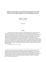
Figure.1 A photomicrograph of a transverse section in the gastrocnemius muscle of a control rat
(subgroup IB), showing blue collagen fibers in endomysium (arrow) and in perimysium around
blood vessels (arrow heads). (Mallory's trichrome, x200)
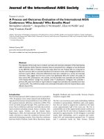
Figure.2 Sections in the gastrocnemius muscles of subgroup IC
A: after one dayshowing blue collagen fibers scattered between inflammatory cells (arrow
heads). B: after 7 days, denotes blue fine collagen fibers in endomysium (arrows) and
perimysium (arrow heads).
(Mallory's trichrome, A and B x200)
3812
Int.J.Curr.Microbiol.App.Sci (2018) 7(7): 3808-3816
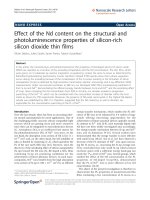
Figure.3 Sections in the gastrocnemius muscles of group II A: after 1 day showing blue collagen
fibers inbetween and around muscle fibers (arrow heads). B: after 7 days reveals more collagen
fibers (arrow heads). C and D: after 14 and 21 days respectively demonstrate prominentincrease
in collagen fibers (arrow heads). (Mallory trichrome, A, B, C and D x400)
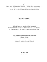
Figure.4 Sections in the gastrocnemius muscles of group III.
A: after 1 day shows blue collagen fibers in between muscle fibers (arrow heads). B: after 7 days
depicts more collagen fibers (arrow heads) inbetween newly formed muscle fibers. C and D:
after 14 and 21 days respectively demonstrate less collagen fibers in endomysium (arrow heads)
and in perimysium (arrows) to be more or less similar to control.
(Mallory trichrome, A, B, C and D x400)
3813
Int.J.Curr.Microbiol.App.Sci (2018) 7(7): 3808-3816
Histogram.1 Comparison between the studied groups as regard mean of area percentage of
collagen fibers
Muscle injuries are common and may be
associated with impaired functional capacity,
especially among athletes. Pain and restricted
range of motion due to these injuries can lead
to decreased performance and limited ability
to play. With the exception of muscle
complete ruptures/ avulsions, complications
like myositis ossificans and the persistence of
uncomfortable symptoms in chronic injuries,
almost all the acute muscle damages are
usually treated non-surgically. Conventional
therapy including rest, ice, compression,
elevation, is often considered the treatment of
choice. Experimental and clinical studies
demonstrated that myogenesis is not restricted
only to the prenatal period but may also
occurs during the healing period after muscle
tissue damage (Järvinen et al., 2005; Mosca
and Rodeo, 2015; and Benazzo et al., 2017).
`
Concentrated growth factors (GFs) within
platelet rich plasma, act synergistically during
the different phases of the healing processes
when compared with the use of isolated GFs.
Platelet rich plasma is simply obtained and
easily prepared with a little risk of developing
an immune response (Borrione et al., 2010).
Therefore, this study was carried out to
evaluate the role of platelet rich plasma in the
healing of experimentally induced skeletal
muscle injury in adult male albino rat model.
Remarkably, the muscle specimens of group
II (non-treated) that were obtained after 7, 14
and 21days depicted increased amount of
collagen fibers in endomysium and
perimysium which were measured and
statistically analyzed. This was evidenced at
day 21 when compared to days 7 and 14
within the same group and when compared to
control and treated group with evident
fibrosis. The same findings were observed by
Fisher and Rathgaber, (2006) who found that,
at 6 days post trauma, muscles appeared to
regenerate with focal interstitial fibrosis and
3814
Int.J.Curr.Microbiol.App.Sci (2018) 7(7): 3808-3816
multiple subsarcolemmal nuclei or central
located nuclei with prominent nucleoli. With
persistence of residual focal areas of fibrosis
after 14 days.
These changes were explained by Järvinen et
al., (2005); and Järvinen et al., (2007) who
reported that satellite cells can proliferate and
mature into myoblasts, which can form
multinucleated myotubes and ultimately
myofibers. The ends of the ruptured
myofibers are typically prevented from
reuniting completely by the scar tissue that
forms during healing. Järvinen et al., (2005)
also mentioned that the process of scar
formation begins almost immediately
following injury. Inflammatory cells degrade
the blood clot while fibrin cross-links form an
initial extracellular matrix that functions as an
initial scaffold to support a reparative
response.
In this study, morphometric and statistical
results also showed significant decrease of the
mean of area percentage of collagen fibers in
PRP treated group at day 21 when compared
with other subgroups within the same group
and when compared to the same period in
group II (Muscle injury induced group). But
this difference wasn't significant when
compared with the control group. This was
explained by Quarteiro et al., (2015) who
mentioned that, during repair and remodeling
phases, deposition of collagen in an organized
and gradual manner is the most important
characteristic for assuring balance between
lysis of the old cell matrix and synthesis of
the new matrix. This is an essential condition
for successful regeneration of the injured
muscle tissue. In addition, the initially
produced collagen is thinner than the collagen
from the healthy tissue; this initial collagen is
then reabsorbed and thicker collagen is
produced along the tension lines, and this is
positively correlated with increase in tensile
strength.
From the previous discussion, it was observed
that PRP had a significant effect on
enhancement of muscle regeneration after
injury without fibrosis as compared to nontreated group. The same finding was
documented by Sanchez et al., 2009 who
stated that full recovery of functional
capabilities was restored in half the expected
time, and images showed full regenerated
muscle tissue after PRP treatment. According
to Hamilton and Best, 2011Platelets are rich
in growth factors that can stimulate
myogenesis and mitigate inflammation.
In conclusion, treatment with PRP resulted in
enhanced regeneration of skeletal muscle
injury without fibrosis.
References
Allbrook, D. 1962. An electron microscopic
study of regenerating skeletal muscle.
J Anat, 96: 137–152.
Benazzo, F., Bargagliotti, M., Combi, A., and
Zanon, G. 2017.Surgical treatment of
acute and chronic muscle injuries. In:
Muscle and Tendon Injuries. By:
Canata, G., d'Hooghe, P., and Hunt, K.
(eds). Springer, Berlin, Heidelberg.
pp. 181-191.
Borrione, P., Gianfrancesco, A.D., Pereira,
M.T., and Pigozzi, F. 2010. Plateletrich plasma in muscle healing. Am J
Phys Med Rehabil, 89: 854-861.
Fisher, B.D., and Rathgaber, M. 2006.An
overview of muscle regeneration
following acute injury. J Phys Ther
Sci, 18: 57-66.
Foster, T.E., Puskas, B.L., Mandelbaum,
B.R., Gerhardt, M.B., and Rodeo, S.A.
2009. Platelet-rich plasma: from basic
science to clinical applications. Am J
Sports Med, 37(11): 2259-2272.
Hamilton, B.H., and Best, T.M. 2011.
Platelet-enriched plasma and muscle
strain injuries: challenges imposed by
3815
Int.J.Curr.Microbiol.App.Sci (2018) 7(7): 3808-3816
the burden of proof. Clin J Sports
Med, 21:31–36.
Järvinen, T.A., Järvinen, T.L., Kääriäinen,
M., Aärimaa, V., Vaittinen, S.,
Kalimo, H., and Järvinen, M.
2007.Muscle injuries: optimising
recovery. Best Pract Res Clin
Rheumatol, 21 (2): 317 - 331.
Järvinen, T.A., Järvinen, T.L., Kääriäinen,
M., Kalimo, H., and Järvinen, M.
2005.Muscle injuries: biology and
treatment. Am J Sports Med, 33 (5):
745 - 764.
Kaux, J.F., and Crielaard, J.M. 2013. Plateletrich plasma application in the
management
of
chronic
tendinopathies. Acta Orthop Belg,
79(1):10-15.
Kazakos, K., Lyras, D.N., Verettas, D.,
Tilkeridis, K., and Tryfonidis, M.
2009. The use of autologous PRP gel
as an aid in the management of acute
trauma wounds. Injury, 40(8):801805.
Kim, D.H., Je, Y.J., Kim, C.D., Lee, Y.H.,
Seo, Y.J., Lee, J.H., et al.2011. Can
platelet rich plasma be used for skin
rejuvenation. Evaluation of effects of
platelet rich plasma on human dermal
fibroblast. Ann Dermatol, 23: 424431.
Menetrey, J., Kasemkijwattana, C., Day, C.S.,
Bosch, P., Vogt, M., Fu, F.H., et
al.2000. Growth factors improve
muscle healing in vivo. J Bone Joint
Surg Br, 82(1):131–137.
Mosca, M.J., and Rodeo, S.A.2015. Plateletrich plasma for muscle injuries: game
over
or
time
out?Curr
Rev
Musculoskelet Med, 8(2):145–153.
Quarteiro, M.L., Tognini, J.R.F., Flores de
Oliveira, E.L., and Silveira, I.
2015.The effect of platelet-rich plasma
on the repair of muscle injuries in rats.
Rev Bras Ortop, 50(5): 586–895.
Sánchez, M., Anitua, E., Orive, G., Mujika, I.,
and Andia, I. 2009.Platelet-rich
therapies in the treatment of
orthopaedic sport injuries. Sports
Med, 39 (5):345–354.
How to cite this article:
Shimaa M. Badr, Reda H Elbakary, Essam M. Laag, Naglaa I. Sarhan and Nafisa A. Elbakary.
2018. Quantitative and Morphological Evaluation of the Effect of Platelet Rich Plasma on
Collagen Fibers in Experimentally Induced Skeletal Muscles Injury in Adult Male Albino Rats.
Int.J.Curr.Microbiol.App.Sci. 7(07): 3808-3816. doi: />
3816