Cell wall proteome of sugarcane stems: comparison of a destructive and a nondestructive extraction method showed differences in glycoside hydrolases and peroxidases
Bạn đang xem bản rút gọn của tài liệu. Xem và tải ngay bản đầy đủ của tài liệu tại đây (1.12 MB, 17 trang )
Calderan-Rodrigues et al. BMC Plant Biology (2016) 16:14
DOI 10.1186/s12870-015-0677-0
RESEARCH ARTICLE
Open Access
Cell wall proteome of sugarcane stems:
comparison of a destructive and a nondestructive extraction method showed
differences in glycoside hydrolases and
peroxidases
Maria Juliana Calderan-Rodrigues1, Elisabeth Jamet2,3, Thibaut Douché2,3, Maria Beatriz Rodrigues Bonassi1,
Thaís Regiani Cataldi1, Juliana Guimarães Fonseca1, Hélène San Clemente2,3, Rafael Pont-Lezica2,3ˆ
and Carlos Alberto Labate1*
Abstract
Background: Sugarcane has been used as the main crop for ethanol production for more than 40 years in Brazil.
Recently, the production of bioethanol from bagasse and straw, also called second generation (2G) ethanol, became a
reality with the first commercial plants started in the USA and Brazil. However, the industrial processes still need to be
improved to generate a low cost fuel. One possibility is the remodeling of cell walls, by means of genetic improvement
or transgenesis, in order to make the bagasse more accessible to hydrolytic enzymes. We aimed at characterizing the
cell wall proteome of young sugarcane culms, to identify proteins involved in cell wall biogenesis. Proteins were
extracted from the cell walls of 2-month-old culms using two protocols, non-destructive by vacuum infiltration vs
destructive. The proteins were identified by mass spectrometry and bioinformatics.
Results: A predicted signal peptide was found in 84 different proteins, called cell wall proteins (CWPs). As expected,
the non-destructive method showed a lower percentage of proteins predicted to be intracellular than the destructive
one (33 % vs 44 %). About 19 % of CWPs were identified with both methods, whilst the infiltration protocol could lead
to the identification of 75 % more CWPs. In both cases, the most populated protein functional classes were those of
proteins related to lipid metabolism and oxido-reductases. Curiously, a single glycoside hydrolase (GH) was identified
using the non-destructive method whereas 10 GHs were found with the destructive one. Quantitative data analysis
allowed the identification of the most abundant proteins.
Conclusions: The results highlighted the importance of using different protocols to extract proteins from cell walls to
expand the coverage of the cell wall proteome. Ten GHs were indicated as possible targets for further studies in order
to obtain cell walls less recalcitrant to deconstruction. Therefore, this work contributed to two goals: enlarge the
coverage of the sugarcane cell wall proteome, and provide target proteins that could be used in future research to
facilitate 2G ethanol production.
Keywords: Cell wall protein, Saccharum sp, Stem, Proteomics, Second generation ethanol
* Correspondence:
ˆDeceased
1
Departamento de Genética, Laboratório Max Feffer de Genética de Plantas,
Escola Superior de Agricultura “Luiz de Queiroz”, Universidade de São Paulo,
Av. Pádua Dias 11, CP 83, 13400-970 Piracicaba, SP, Brazil
Full list of author information is available at the end of the article
© 2016 Calderan-Rodrigues et al. Open Access This article is distributed under the terms of the Creative Commons
Attribution 4.0 International License ( which permits unrestricted use, distribution,
and reproduction in any medium, provided you give appropriate credit to the original author(s) and the source, provide a link
to the Creative Commons license, and indicate if changes were made. The Creative Commons Public Domain Dedication
waiver ( applies to the data made available in this article, unless otherwise
stated.
Calderan-Rodrigues et al. BMC Plant Biology (2016) 16:14
Background
The use of Saccharum sp. to produce second generation
(2G) ethanol can reduce waste and increase the yield without expanding the crop area, contributing to a cleaner,
more efficient and more sustainable production. However,
from the economic point of view, the costs of the process
need to be reduced, mostly those related to the enzymes
used to deconstruct plant cell walls. Therewith, research is
mainly focused on the identification of new enzymes that
could efficiently degrade cell walls [1]. Other studies have
been developed from the biomass perspective, describing
the plant cell wall components [2–5], and even altering
them attempting to achieve a higher ethanol 2G yield. Since
pre-treatments facilitate cell wall digestibility to increase
ethanol production, when altering plant cell wall components, focus should be either on lignin- carbohydrate complex cleavage and hemicellulose removal, or lignin
modification and even on redistribution and cellulose
decrystallization [6].
Plant cell walls are mainly composed of polysaccharides
and cell wall proteins (CWPs) [7]. Proteomics studies have
revealed the large diversity of CWPs [8–10]. They have
been grouped in different functional classes according to
predicted functional domains and experimental data: polysaccharide modifying proteins, oxido-reductases and proteases, have been found as major classes. Structural proteins
such as hydroxyproline-rich glycoproteins, namely extensins, arabinogalactan proteins and hydroxyproline/prolinerich proteins, have been estimated to account for about
10 % of the cell wall mass in dicots [11] and approximately
1 % in monocots [12]. However, only a few of them have
been identified in proteomics studies. CWPs are involved in
growth and development, signaling and defense against
pathogens. They virtually take part in most functions of the
cells [4, 11, 13]. They can affect cell fate, being able to sense
stress signals and transmitting them to the cell interior
[14]. They can also have tissue-specific functions , such as
playing roles in cuticle formation [15]. Due to this versatility, plant cell walls are the subject of many fields of
research.
In the case of grasses, type II-cell walls present specific
features [7]. The cellulose microfibrils are interlocked by
glucuronoarabinoxylans, instead of xyloglucans of type
I-cell walls. In addition, the grass cell walls contain a
substantial portion of non-cellulosic polymers ‘wired on’
the microfibrils by alkali-resistant phenolic linkages.
As mentioned above, plant cell walls contain enzymes
capable of modifying the cell wall matrix [16]: endoglucanases which cleave the polysaccharide backbones; glycosidases which remove side chains; transglycosylases which
cut the polysaccharides and link them together; esterases
which remove methyl groups of pectins, and cleave ester
bonds in polysaccharide chains; and class III peroxidases
(Prxs) which form or break phenolic bonds. Altogether,
Page 2 of 17
these enzymes offer many possibilities to modify the
structure and the mechanical properties of cell walls,
and thus biomass structure [3]. Besides, the addition
of plant glycosidases during the hydrolysis of corn stover could increase the ethanol yield [17]. These examples show that the repertoire of CWPs could provide
interesting tools to improve the deconstruction of cell
walls.
As commonly known, classical CWPs share common features. The first one is a signal peptide at the N-terminus of
the protein which is responsible for their targeting to the
endoplasmic reticulum (ER) [18], the first organelle of the
secretory pathway [19]. The signal peptide is not formed by
a consensus amino acid sequence. However, it has a positively charged n-region at its N-terminus and a central
hydrophobic h-region followed by a polar c-region at its Cterminus comprising the cleavage site [20]. In addition,
CWPs do not possess the canonical ER retention signal
KDEL or HDEL tetrapeptide at their C-terminus [19, 20].
The third feature is that they do not present a transmembrane domain. When passing through the secretory
pathway, proteins go from ER to the Golgi complex in
order to be packed into vesicles and directed to be secreted.
Plasma membrane proteins show the same features as
CWPs except that they have a trans-membrane domain
[20, 21].
Cell wall proteomics require challenging strategies
comprising several steps, from the extraction to the
identification of the proteins, compared to other subcellular proteomics works. Despite the technical hurdles,
a lot of studies have been successful [8, 9]. Several aerial
organs have been studied in different plant species, such
as alfalfa [22], Linum usitatissimum [23], Solanum tuberosum [24], and Arabidopsis thaliana [25]. In Brachypodium distachyon leaves and stems, different classes of
proteins have been identified and it was possible to address some of them to the mechanism of 2G biofuel production [26]. It is then possible to alter their expression
to improve cell walls deconstruction, such as the upregulation of a cell wall transcript in rice [27].
In a recent publication, 69 CWPs have been described from isolated cells obtained from cell suspension cultures of sugarcane [28]. However, the
description of the cell wall proteome from a differentiated organ is still missing. In this work, two different strategies were developed to extract the CWPs of
two month-old stems: either a destructive method
(DT Method) or a non-destructive one (ND Method),
i.e. vacuum infiltration [29]. Proteins were identified
by mass spectrometry (MS) and bioinformatics. The
results were compared regarding the number and the
type of CWPs. Quantitative MS data were used to
identify the most abundant CWPs in sugarcane
culms.
Calderan-Rodrigues et al. BMC Plant Biology (2016) 16:14
Page 3 of 17
Results
Extraction of proteins from cell walls
Two-month-old sugarcane culms were selected for presenting a soft and young material, at an early stage of development. The use of young organs could lead to the
identification of proteins involved in cell wall expansion,
thus clarifying the mechanisms that the plant itself uses to
allow growth.
Sugarcane features four stages of development: (i)
germination and emergence, (ii) tillering phase, (iii) grand
growth period and (iv) ripening phase, when sugar accumulates [30]. The tillering phase begins about 40 days after
planting and can last up to 120 days, being the early stage
of plant development [31, 32]. In this work, plants were
collected 60 days after planting, halfway from the maximum tillering, measuring around 40–50 cm in height from
the bottom to the upper leaf. This age was also chosen to
allow distinguishing leaves and culms visually.
The DT Method was a destructive one relying on the
grinding of the material and its centrifugation in solutions of increasing sucrose concentration. On the contrary, the ND Method was a non-destructive one, since
it maintained the cell structures intact while performing
the extraction of CWPs by vacuum infiltration of the
tissues. Thus, it was expected that the DT Method
would be able to extract more wall-bound proteins than
the ND one. In both protocols, protein extraction from
cell walls was performed using 0.2 M CaCl2 and 2 M
LiCl. The efficiency of CaCl2 to release CWPs could rely
on the fact that demethylesterified homogalacturonans
strongly chelate calcium [33], solubilizing weakly-bound
proteins by a competition mechanism [34]. On the other
hand, LiCl was used to extract mostly hydroxyproline-rich
glycoproteins [35] All the experiments were performed in
duplicates.
The DT Method produced around 518 μg of proteins
from 35 g of culms (fresh weight). Regarding the ND
Method, the yield was slightly lower: around 667 μg of proteins were recovered from about 50 g of culms (fresh
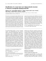
weight). Figure 1 shows the patterns of the proteins
extracted from sugarcane culms. The presence of thin resolved bands after staining showed the quality of the procedure with no degradation pattern. Each biological
replicate, using either method, showed a pattern very similar to that of its counterpart and each method gave rise to a
different pattern.
Identification of proteins by MSE and bioinformatics
analyses
Proteins were analyzed by shotgun LC-MS/MS, after tryptic
digestion. The identification of proteins was performed
using the translated-SUCEST database containing ESTs
[36]. Homologous genes in Sorghum bicolor, the closest related species with a fully sequenced genome, were
Fig. 1 1D-electrophoresis of proteins extracted form 2-month-old
sugarcane culm cell walls. Proteins have been extracted using either
the DT or the ND Method. The biological repeats corresponding to
each Methods are respectively numbered 1–2 and 3–4. The molecular
mass markers (MM) are indicated in kDa on the left
systematically searched for. Predictions of sub-cellular
localization and functional domains were done on translated ESTs when they were full-length, otherwise on homologous S. bicolor coding sequences. Because of the high
level of ploidy of the sugarcane genome [37], in some cases,
different ESTs matched the same S. bicolor gene.
More detailed results of MS analyses, such as protein score and number of matched peptides, can be
found in Additional files 1, 2, 3 and 4. About 65 %
and 82 % of the proteins identified were found in
both biological replicates, in the DT and ND
Methods, respectively. These Methods allowed the
identification of 70 and 103 different proteins from
the translated-SUCEST database, respectively. From
these, 39 (56 %) and 69 (67 %) proteins respectively
had a predicted signal peptide, no known intracellular
retention signal such as an endoplasmic retention signal and one trans-membrane domain at most
(Table 1). These proteins were considered as CWPs
(Additional file 5), and the others as intracellular proteins (Additional file 6). The DT and ND Methods
lead to the identification of different sets of proteins.
Calderan-Rodrigues et al. BMC Plant Biology (2016) 16:14
Page 4 of 17
Table 1 CWPs identified in sugarcane young culms
SUCEST accession
numbera
Number of
peptidesb
Number of Protein score
unique
peptidesb
Femtomole
average
S. bicolor
homologues
Functional annotation
Extraction
method
Proteins acting on polysaccharides
SCCCCL3001B10.b
16; 16
3; 6
4368.653; 1207.559
87.40605
Sb01g010840.1
GH1
ND
SCJFLR1017E03
7; 8
1; 1
627.7585; 389.633
4.65435
Sb01g010840.1
GH1
ND
SCEQLB1066E08
11; 5
9; 2
1258.872; 452.9845
6.72165
Sb01g010825.1
GH1
ND
SCEQHR1082B01
9; 7
8; 6
2256.574; 388.3896
28.182652
Sb02g028400.1
GH1
ND
SCEZLB1007A09
18; 12
11; 8
4982.336; 2259.83
41.55905
Sb01g008030.2
GH3
ND
SCEQLR1093F09*
12; 18–20; 14 5; 8–6; 5
523.6582; 1309.333 – 17.23065 –
3688.09; 10810.11
50.3928
Sb01g008040.3* GH3
DT - ND2
SCCCCL4009F05
20; 14
16; 12
10955.92; 8452.891
156.84746
Sb06g030270.1
GH3
ND
SCQSAM1030G04
3; 2
3; 2
8506.176; 6709.646
71.61725
Sb06g030270.1
GH3
ND
SCQSRT2031D12
10; 1
7; 9
1169.301; 2716.698
34.5685
Sb03g045490.1
GH17
ND
SCVPRZ3029G05
3; 2
2; 1
853.3968; 815.915
15.9117
Sb03g040600.1
GH18
ND
SCJLLB2076C12
9; 8
4; 6
3324.314; 1244.686
40.19725
Sb06g021220.1
GH19
ND
SCEZRZ3015E11
8; 5
6; 5
3106.044; 2091.409
55.74035
Sb01g048140.1
GH19
ND
SCCCCL5004G07
8; 8
5; 6
7937.636; 2173.338
76.40835
Sb10g000660.1
GH28
ND
SCJFRT1007G04
4; 2
1; 1
4752.977; 2164.125
31.240002
Sb10g000660.1
GH28
ND
SCCCCL6004H07
9; 8
7; 7
633.8843; 542.2719
19.80175
Sb01g040750.1
GH35
ND
SCVPRZ3029F03
6; 4
4; 4
1035.417; 246.8027
8.5248
Sb03g029700.1
Acyl esterase
(homologous to AtPMR5)
ND
SCSGLR1025E03
5; 2–5; 8
4; 2–4; 8
460.239; 887.95161045.544; 226,6407
19.8042 –
13.1516
Sb02g042780.1
Pectin methylesterase
(carbohydrate esterase
family 8, CE8)
DT – ND
12.9346
Oxido-reductases
SCCCRZ1002B03
8; 1
3; 2
708.6577; 447.837
Sb01g041770.1
Prx homologous to SbPrx20
DT
SCCCRT1001G12
9; 9–14; 9
5; 6–7; 5
2689.641; 2574.119 – 65.36725 –
10308.06; 9888.154
77.029495
Sb04g008590.1
Prx homologous to SbPrx71
DT - ND
SCCCLB1004B09*
9; 16
4; 4
690.8371; 1079.03
26.908451
Sb10g027490.1* Prx homologous to SbPrx139
DT
SCEQRT2030A04*
7; 12
1; 3
306.9711; 658.3382
7.35725
Sb10g027490.1
Prx homologous to SbPrx139
DT
SCCCLR1C03A09
12; 11
8; 7
845.2605; 759.2087
46.113102
Sb09g004650.1* Prx homologous to SbPrx115
DT
SCCCLR1C05G08*
11; 11
5; 8
1494.011; 1461.387
66.7759
Sb03g024460.1* Prx homologous to SbPrx65
DT
SCRLAD1042E05
6; 4–5; 2
1; 2–1; 1
2528.694; 933.4598 – 17.7972 –
873.3041; 1444.467
10.54785
Sb09g002740.1* Prx homologous to SbPrx108
DT – ND
SCVPRZ2035F03*
11; 8–9; 5
6; 6–4; 3
2417.947; 1151.436 – 42.28915 –
1401.302; 1033.822
17.264
Sb09g002740.1
Prx homologous to SbPrx108
DT - ND
SCVPLB1020D03
2; 8
2; 7
372.5895; 854.9581
Sb03g046760.1
Prx homologous to SbPrx68
SCEPRZ1011A06*
7; 11–12; 6
3; 5–4; 3
866.9835; 940.5853 – 17.7399 –
5970.014; 1026.785
45.46655
Sb03g010250.1* Prx homologous to SbPrx54
SCCCAD1001B08
3; 3
1; 1
9547.608; 3981.619
Identified
Sb03g010740.1
SCJFRZ2013F04
7; 1
1; 1
23916.97; 9892.667
7.78455
SCJLRT1019B02
9; 6
1; 1
16559.67; 6310.499
14.36725
SCEQRT1024D03
1; 1
3; 2
16709.58; 4972.862
60.093697
Sb03g010740.1
Prx homologous to SbPrx55
ND
SCCCAD1001C08
6; 5
3; 4
7855.956; 13571.95
28.358952
Sb02g042860.1
Prx homologous to SbPrx47
ND
SCQSST3114C09
5; 8
5; 4
2082.782; 583.5138
16.338501
Sb01g031740.1
Prx homologous to SbPrx14
ND
SCBFFL4112F05
2; 3
1; 2
1435.365; 4620.795
35.5025
Sb06g018350.1
Blue copper binding protein
(plastocyanin)
DT
23.364399
Prx homologous to SbPrx55
DT
DT - ND
ND
ND
ND
Calderan-Rodrigues et al. BMC Plant Biology (2016) 16:14
Page 5 of 17
Table 1 CWPs identified in sugarcane young culms (Continued)
SCRFHR1006G03
3; 2
2; 1
391.4637; 1712.47
1.22305
Sb01g010510.1
Blue copper binding protein
(plastocyanin)
DT
SCJLLR1104H07
3; 3
3; 3
912.4164; 417.4785
15.684
Sb07g011870.1
blue copper binding protein
(plastocyanin)
ND
SCEPAM1021H07
8; 3
3; 3
924.3445; 347.9209
12.423151
Sb10g027270.1
Multicopper oxidase
ND
Proteins related to lipid metabolism
SCCCAM2002F12
4; 5–4; 5
1; 1–1; 1
716.9828; 1069.318 – 19.3443 –
5961.475; 9251.332
115.4035
Sb03g038280.1
LTP
DT – ND
SCBFLR1046E09
5; 6–4; 5
1; 1–1; 1
816.9942; 1488.674 – 34.07175 –
14868.01; 16290.19
242.60735
Sb03g038280.1
LTP
DT - ND
SCVPRZ2039B03
5; 6–4; 5
1; 1–1; 1
816.9942; 2065.907 – Identified 14868.01; 16290.19
identified
DT - ND
SCVPRZ2041C11
5; 6–4; 5
1; 1–1; 1
902.0069; 1488.674 – 8.77335 18617.13; 24758.37
identified
DT - ND
SCCCLR1072C06
3; 2–6; 5
1; 1–1; 1
243.1648; 2495.928 – 6.5461 –
35001.54; 23317.92
132.9761
SCRFLR1012A10
3; 2–7; 5
1; 1–1; 1
345.6155; 2314.583 – Identified 35158.61; 23317.92
identified
DT – ND
SCEPRT2047G01
10; 5
1; 1
35204.81; 23919.76
250.87096
ND
SCEZLR1031G07
5; 4
1; 1
34256.59; 23296.33
Identified
ND
SCRUSB1064D08
9; 5
1; 1
35280.34; 23317.92
Identified
ND
SCEPLB1044H04
3; 4–3; 4
1; 1–1; 1
3924.14; 3159.724 –
1548.66; 926.7365
189.36455 – Sb01g049830.1
46.1548
SCEZLB1006F09
3; 4–3; 2
1; 1–1; 1
SCCCLR1048F06 SCCCLR1048F06
10; 13–5; 4
SCBGLR1114E07
Sb08g002700.1
LTP
DT - ND
LTP
DT – ND
6772.019; 11919.76 – 194.30121 – Sb08g002670.1
8845.361; 13343.73
146.9349
Protease inhibitor/seed
storage/LTP family
DT - ND
1; 1–1; 2
91432.66; 77846.23 – 318.6974 –
176534.4; 124470.6
352.27365
Protease inhibitor/seed
storage/LTP family
DT - ND
5; 5
2; 2
145690.4; 125905.7
SCCCCL3004H07.b 3; 3
2; 2
145392.5; 124459
Identified
ND
SCVPHR1092G06
4; 4
2; 2
145392.5; 124470.6
Identified
ND
SCUTST3131G03
3; 6–4; 3
2; 1–1; 1
6793.345; 3745.457 – 150.85635 – Sb08g002690.1
21630.89; 20854.45
109.76019
SCCCCL3001E03.b*
5; 7
2; 3
3263.44; 1945.298
SCJFRZ2033G07
4; 3
1; 1
SCRUFL4024B04
4; 3
1; 1
SCCCRZ1001H02
3; 3
1; 1
7741.618; 3045.282
70.59645
Sb03g039880.1
LTP
ND
SCCCRZ2002G09
5; 5
2; 2
20532.6; 6676.209
49.378
Sb06g016170.1
LTP
ND
Sb08g002660.1* Protease inhibitor/seed
storage/LTP family
ND
Sb08g002690.1
588.4425
ND
Protease inhibitor/seed
storage/LTP family
39.72605
Sb01g033830.1* LTP
26826.32; 13939.5
8.15625
Sb08g002700.1
26805.06; 13987.92
202.91615
LTP
DT – ND
ND
ND
ND
SCQSFL3039E08.b
5; 5
2; 2
22572.43; 7334.285
20.2533
SCCCLR1024C05*
6; 3
1; 1
11552.57; 5130.042
5.59605
ND
SCCCLR1076D05
6; 5
1; 1
16880.65; 8523.134
129.08115
SCEPLB1044H11*
7; 3
1; 1
11788.22; 5527.095
13.30225
SCCCLR2C03F01
3; 3
1; 1
9426.873; 6414.531
78.86415
Sb08g002670.1
Protease inhibitor/seed
storage/LTP family
ND
SCCCRT1003B03
6; 4
2; 3
722.451; 408.6039
26.09375
Sb10g003930.1
GDSL lipase
ND
SCBGLR1023G11
6; 8
5; 8
553.142; 705.4267
24.15155
Sb04g029670.1
Asp protease, peptidase A1
DT
SCBGLR1097G03
4; 6
3; 3
1475.072; 7045.55
168.19795
Sb05g027510.1
Asp protease, peptidase A1
DT
SCMCLR1123H12
6; 7–3; 2
3; 3–2; 1
1722.723; 4799.767 – 122.60135 – Sb05g027510.1
598.7939; 1717.224
52.145752
Asp protease, peptidase A1
DT - ND
SCQGST1032H01
11; 14
8; 7
653.9818; 997.1608
Asp protease, peptidase A1
DT
ND
ND
Proteases
45.5457
Sb05g027510.1
Calderan-Rodrigues et al. BMC Plant Biology (2016) 16:14
Page 6 of 17
Table 1 CWPs identified in sugarcane young culms (Continued)
SCQGSB1083B11
8; 5
5; 4
4851.335; 4946.649
47.901802
Sb02g041760.1
Asp protease, peptidase A1
ND
SCRLRZ3042B09
9; 6
5; 3
390.5894; 367.0505
7.24305
Sb03g026970.1
Asp protease, peptidase A1
ND
SCVPLR2012E01
3; 3–4; 3
2; 2–2; 2
1075.957; 4197.95 –
23533.5; 11935.54
147.1347 –
160.93646
Sb01g044790.1
Asp protease/Taxi _N/Taxi_C
DT - ND
SCVPRZ2038B09
3; 4–4; 2
2; 3–4; 2
1194.285; 2283.918 – 61.6057 –
6719.425; 1320.476
103.41875
Sb01g044790.1
Asp protease/Taxi _N/Taxi_C
DT - ND
SCCCST1004B07
11; 8
11; 8
4096.934; 3149.51
44.2912
Sb01g013970.1
Ser protease (subtilisin family, ND
peptidase S8/S53)
SCJFRZ2011B07
5; 4
4; 3
2390.547; 1211.642
25.90875
Sb06g016860.1
Ser protease (subtilisin family, ND
peptidase S8/S53)
SCCCLR1022B11*
7; 5
6; 6
1017.185; 492.4018
20.1544
Sb06g030800.1* Cys protease, (papain family,
peptidase C1A)
ND
31.54085
Sb05g026650.1
Ser protease inhibitor
(Bowman-Birk)
DT
Leucine-rich repeat (LRR)
receptor kinase
ND
Proteins with interaction domains (with proteins or polysaccharides)
SCJFLR1013A04
4; 4
1; 1
3741.598; 4709.589
SCRUFL3062D08 SCRUFL3062D08
5; 5–4; 4
1; 1–1; 1
2784.365; 4868.514 – 45.9138 –
11731.47; 8790.486
44.6385
4; 2
4; 4
5824.59; 1313.647
SCEZRZ1014C04*
6; 5–4; 8
2; 2–1; 1
6025.488; 9344.188 – 79.66205 –
18335.89; 4427.775
67.5792
Sb03g039330.1* Thaumatin
DT - ND
SCCCLR2003G06
4; 4
1; 2
1364.424; 533.9335
18.352499
Sb08g018720.1
Thaumatin
ND
SCUTLR1037F02
3; 4
1; 2
1083.055; 546.6533
Identified
SCCCSD1003E02
3; 2
1; 1
2326.563; 4054.495
18.28735
Sb08g022410.1
Thaumatin
ND
SCRUHR1076B06
3; 2
1; 1
3641.288; 5434.818
2.588
Sb08g022410.1
Thaumatin
ND
SCVPRT2073B04
4; 4
2; 2
2289.087; 18490.86
87.17195
Sb08g022420.1
Thaumatin
ND
SCBGRT1047G10
6; 7
4; 6
3131.626; 2684.819
52.720253
Sb02g004500.1
Germin (cupin domain)
ND
SCCCLR2C02D04
3; 4
3; 3
9021.729; 14936.96
149.43965
Sb09g004970.1
Germin (cupin domain)
ND
SCCCRZ1C01H06
13; 1–12; 14
4; 3–7; 6
3376.122; 3105.723 – 55.5037 –
10082.07; 3506.265
33.9362
Sb08g001950.1
Nucleoside phosphatase
DT - ND
SCJLRT3078H06
6; 2
3; 1
1286.289; 1236.944
45.866447
Sb05g025670.1
Dirigent protein
DT
SCVPRT2073B08
6; 4
4; 1
333.12; 1046.494
19.9186
Sb10g001940.1
SCP-like extracellular protein
ND
SCCCCL4009G04*
11; 1–8; 8
4; 4–3; 3
1383.823; 3928.575 – 126.7531 –
15270.52; 16670.59
141.14679
SCSGLR1084A12*
12; 14–10; 9
6; 6–6; 5
6772.538; 5277.542 – 158.43881 – Sb01g004270.1
18024.07; 13067.63
150.2402
SCCCLB1001G04
7; 3
5; 3
314.0911; 273.5209
9.64495
Sb03g027650.1
Unknown function (DUF642)
DT
SCVPLR2027A11
5; 4–5; 3
5; 4–2; 1
2870.491; 1609.635
34.981
Sb07g026630.1
Unknown function (DUF568)
ND
SCCCRZ3002G10*
4; 7–6; 5
1; 1–1; 2
844.0264; 313.8415 – 3.07575 –
4025.449; 1307.088
35.7293
Sb01g031470.1* Homologous to phloem
filament protein 1
(Cucurbita phloem)
DT - ND
SCEZRT2018F03*
4; 6
1; 1
675.4673; 446.4631
5.89995
Sb01g031470.1
Homologous to phloem
filament protein 1
(Cucurbita phloem)
DT
SCEZLB1013B06
14; 15
8; 11
5254.302; 4812.086
137.18835
Sb10g001440.1
Homologous to phloem
filament protein 1
(Cucurbita phloem)
DT
SCSFST1066G10
5; 5–6; 5
1; 1–1; 1
718.6985; 2383.583 – 102.53975 – Sb08g018710.1
15310.67; 5735.458
192.7034
Expressed protein
DT - ND
DT - ND
Signaling
SCRUAD1063C06
55.09195
Sb09g000430.1
Miscellaneous proteins
ND
Unknown function
Sb01g004270.1* Unknown function (DUF642)
Unknown function (DUF642)
DT - ND
DT - ND
Calderan-Rodrigues et al. BMC Plant Biology (2016) 16:14
Page 7 of 17
Table 1 CWPs identified in sugarcane young culms (Continued)
SCRUFL4024B08.b
3; 6
1; 2
5551.887; 12727.94 – 120.5459 –
52010.09; 28346.53
288.07706
Sb08g018710.1
SCCCRZ2004B02*
8; 8
1; 1
9551.813; 4751.091
Sb03g000700.1* Expressed protein
SCCCLR1079C11
7; 4
6; 4
3527.088; 4063.023
33.617348
Sb04g011100.1
Expressed protein
ND
SCAGLR2011E04/
SCEPAM2057B02
3; 3
1; 1
11178.66/11961;
8954.521/7215.72
identified
Sb08g003040.1
ND
SCEPLR1051E09
3; 3
1; 1
11178.66; 7215.72
28.171349
Expressed protein
(stress responsive
alpha/beta barrel)
43.9473
Expressed protein
DT - ND
ND
ND
a
Bold letters indicate that the ESTs share common sequences. Full length ESTs are in italics. Stars (*) indicate the proteins also identified in the cell wall proteome
of sugarcane cell suspension cultures [15]
b
Semicolons separate data from different biological repeats. Dashes separate data from different extraction methods (DT, then ND)
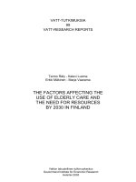
Altogether, 84 different CWPs were identified and
distributed into eight functional classes (Fig. 2 and
Table 1): proteins acting on carbohydrates, proteins
possibly related to lipid metabolism; proteins with
interaction domains; oxido-reductases; proteases; miscellaneous proteins; signaling and proteins of unknown function. From these 84 CWPs, 24 (29 %) were
identified using both the DT and ND Methods. It should be
noted that no structural protein was identified. Besides, 16
CWPs (18 %) were previously identified in the cell wall
proteome of sugarcane cell suspension cultures [28]. Consequently, 68 sugarcane CWPs were newly identified in this
study.
Regarding the DT Method, the oxido-reductases (31 %),
mainly peroxidases (Prxs) and two blue copper binding
proteins, constituted the most represented class, followed
by proteins related to lipid metabolism (18 %), all being
lipid transfer proteins (LTPs). Asp proteases (16 %) and
miscellaneous proteins (7.5 %), comprising thaumatin, germins and dirigent protein, were also identified (Table 1).
Surprisingly, only one glycoside hydrolase (GH) of the GH3
family, as well as a single pectin methylesterase (PME) were
identified from the proteins acting on carbohydrates class
(5 %). Proteins with interaction domains (2.5 %) were represented by one serine protease inhibitor. Proteins of yet unknown function (20 %) were numerous and it was possible
to highlight the presence of proteins with DUF642 domains, already found in other cell wall proteomes [38, 39],
and proteins homologous to phloem filament protein 1.
The most represented functional class using the ND
Method was that of proteins acting on carbohydrates
(25 %), mostly GHs (families 1, 3, 19, 28, 17, 18, 35) and
two carbohydrate esterases. Proteins related to lipid
metabolism (20 %) comprised LTPs and one GDSL-lipase.
Oxido-reductases (14 %) were mostly Prxs. Miscellaneous
proteins (13 %) were mainly represented by thaumatins and
germins. Proteases (12 %) were Asp, Ser or Cys proteases.
Proteins with interaction domains were represented by one
Ser protease inhibitor and signaling proteins by one
leucine-rich repeat receptor kinase. Finally, proteins of unknown function comprised proteins with DUF642 and
DUF568 domains.
We have also performed a quantitative analysis of the
CWPs identified by both methods (Table 1). Only the proteins present in amounts higher than 100 femtomoles,
calculated by averaging the results of the two biological
repeats, have been listed in Table 2. When a protein has
been identified using both methods, its quantification
could be the same or different if either of the two methods
could extract it more efficiently. These differences could,
(i) result from the loss of proteins during the washings
steps required to purify cell walls using the DT Method
or, (ii) due to different types of interactions with cell wall
components. Among the proteins present in high amount
in culm cell walls, LTPs are well represented with 10 out
of 17 proteins. One GH3, three Asp proteases and two
DUF642 proteins were also found in the top17 list.
Two approaches were used to statistical analysis: a
multivariate analysis, the Scores plot and Vip scores
(Fig. 3b, c, respectively), and a univariate one, the Volcano plot, as shown in Fig. 3a. In Fig. 3a, three proteins
could be considered as those contributing the most to
the distinction between the DT and ND Methods.
Figure 3b indicates that the DT and ND Methods differ
statistically from each other, since it is possible to separate two distinct groups of proteins regarding the quantity of proteins extracted in each technique. In addition,
the two first components (vectors) contributed positively
to the model (value of Q2 positive = 66.5 %), and the
variation of the proteins was 97.5 % (R2). Values of Q2 >
0.08 indicates that a model is better than chance, and
scores of 0.7 or higher, demonstrate a very robust trend
or separation [40]. The protein SCCCRZ3002G10 of
unknown function was the one that contributed the
most to the separation of the groups, being found in
higher amount using the ND Method (Fig. 3a, c). The
SCCCAM2002F12 and SCEPLB1044H04 LTPs, in turn,
were the third and the fourth proteins that contributed to
the separation of the two groups in Partial-Least Squares
Discriminant Analysis - PLS-DA2, being found in higher
amount in the ND and DT Methods, respectively.
As presented in Fig. 3c, using the average of the quantitative data obtained for each method, the statistical analysis
showed that from the 15 proteins that most contributed to
Calderan-Rodrigues et al. BMC Plant Biology (2016) 16:14
Page 8 of 17
Fig. 2 Distribution of CWPs identified in 2-month-old sugarcane culms. Proteins were distributed in functional classes according to
bioinformatics predictions: PAC stands for proteins acting on carbohydrates; OR, for oxido-reductases; LM, for proteins possibly involved
in lipid metabolism; P, for proteases; ID, for proteins with interaction domains (with proteins or polysaccharides); S, for proteins possibly
involved in signaling; M, for miscellaneous; UF, for unknown function
distinguish the DT and ND Methods, nine of them showed
a much higher amount using the ND Method. Additional
file 7 shows important features identified by Volcano Plot.
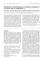
Comparison of the CWPs of sugarcane young culms to
those of stems of other plants
Previous cell wall proteomics studies were performed
on B. distachyon basal and apical internodes [26],
Medicago sativa basal and apical stems [22] and
Linum usitatissimum young stems [23]. All these
data have been collected in the WallProtDB database
[39] and annotated in the same way, thus allowing
comparisons [41]. These CWPs were compared to
the newly identified CWPs of sugarcane stems
(Fig. 4). In B. distachyon, a protocol very similar to
the DT Method was used, but the LC-MS/MS analysis were done with 1-D gel pieces [26]. L. usitatissimum stem CWPs were extracted using a protocol
similar to the DT Method and 1-D gel pieces corresponding to stained protein bands were used as
starting material for FT-ICR MS analysis [23]. On
the other hand, in alfalfa stems, EGTA tretament
Calderan-Rodrigues et al. BMC Plant Biology (2016) 16:14
Page 9 of 17
Table 2 Most abundant CWPs in the cell wall proteome of
sugarcane young stems. Proteins with average amounts between
the two biological repeats higher than 100 femtomols using
either method are listed (see Table 1)
SUCEST accession
number
Functional annotation
Methoda
Proteins acting on carbohydrates
SCCCCL4009F05
GH3
ND
Proteins related to lipid metabolism
SCCCAM2002F12
LTP
DT < < ND
SCBFLR1046E09
LTP
DT < < ND
SCEPLB1044H04
LTP
DT > > ND
SCEZLB1006F09
LTP
DT > ND
SCCCLR1048F06
LTP
DT ~ ND
SCUTST3131G03
LTP
DT > ND
SCRUFL4024B04
LTP
ND
SCCCLR1076D05
LTP
ND
SCBGLR1097G03
Asp protease
DT
SCMCLR1123H12
Asp protease
DT > > ND
SCVPLR2012E01
Asp protease
DT ~ ND
Expressed protein (DUF642)
DT ~ ND
Proteases
Unknown function
SCCCCL4009G04
SCSGLR1084A12
Expressed protein (DUF642)
DT ~ ND
SCEZLB1013B06
Homologous to phloem
filament protein 1
DT
SCSFST1066G10
Expressed protein
DT < < ND
SCRUFL4024B08.b
Expressed protein
DT < < ND
a
The relative amount of proteins quantified using either method is indicated
(see Table 1)
and LiCl were used for protein extraction, and 1-D
gel pieces were digested prior to analysis using a
nanoAcquity UPLC system [22]. Although different
strategies for protein extraction and MS analyses
have been used, all the protocols used the same salts
to extract proteins from cell walls: CaCl2 and/or
LiCl.
The stem cell wall proteomes of all the above species showed very similar percentages of proteins acting on carbohydrates. An outstanding observation
was that sugarcane had a much higher percentage of
proteins related to lipid metabolism (17 %) than all
the other species (0–9 %). The dicot M. sativa presented a much higher proportion of proteins with
interaction domains in comparison with the monocots (14 % vs less than 5 %). The monocots showed
a higher proportion of oxido-reductases in comparison with the dicots (about 20 % vs about 15 %). A
much smaller proportion of proteases was found in
L. usitatissimum stems [23].
Discussion
In this work, 84 different sugarcane CWPs were identified
in young culms using two different strategies. Together
with the cell wall proteome of cell suspension cultures
[28], 137 different CWPs of sugarcane have been identified. In this study alone, 68 CWPs were newly identified
and 16 CWPs were identified in both culms and cell suspension cultures, among which 5 Prxs. Besides, the proportion of proteins predicted to be intracellular in culm
extracts (33 % and 44 %) was lower than in sugarcane cell
suspension culture extracts (81.6 %) [28], being quite the
same as in B. distachyon young internodes [26]. This is
probably inherent to the type of material, since a lot of cell
debris are present in the culture medium [28].
Interestingly, the proportion of intracellular proteins
was higher in leaves than in stems in B. distachyon [26];
the same case has been observed for sugarcane (unpublished observations). The ND Method has lead to the
identification of about 75 % more CWPs than the DT
Method (69 CWPs vs 39), and around 81 % of the CWPs
(68 CWPs out of 84) have been identified using one
method of extraction only. These results show the importance of using different strategies to enlarge the
coverage of a cell wall proteome. The ND Method has
allowed the recovery of more CWPs of sugarcane culms,
and much more GHs than the DT method. If the objective
of the study is to get an overview of CWPs or of glycosidases, this strategy should be considered. In addition, if
the goal is especifically to recover GHs, perhaps a total
protein extraction followed by affinity chromatography on
Concanavalin A is the best option [25]. However, if the
aim is to go deeper into Prxs, the DT Method looks more
appropriate. Besides, both methods showed a good reproducibility since between 65 % and 82 % of CWPs were
identified in both biological replicates. Although rarely
discussed in cell wall proteomics paper, this result is consistent with those of previous studies [26].
The ND Method could recover both a higher number
of CWPs and a higher amount of those contributing to
the discrimination between the two methods through
the statistical analysis. Additionally, the three proteins
highlighted in the univariate analysis were also present
in the multivariate analysis, being numbers 1, 3 and 4
from the 15 CWPs considered to be the most important
for the discrimination between the two methods. The
major difference between the two ND and DT methods
regards proteins acting on carbohydrates: only one CWP
has been identified using the DT Method whereas one
fourth of the CWPs belongs to this class using the ND
Method. Since the same organs were analyzed, this difference has to be related to the strategy used for protein
extraction. Some proteins could have been lost during
the washing steps required to clean cell wall fragments
in the case of the DT Method [9]. This could explain
Calderan-Rodrigues et al. BMC Plant Biology (2016) 16:14
Fig. 3 (See legend on next page.)
Page 10 of 17
Calderan-Rodrigues et al. BMC Plant Biology (2016) 16:14
Page 11 of 17
(See figure on previous page.)
Fig. 3 a. Volcano plot: Univariate Statistical analysis of the quantified proteins in both methods. Axis x: Fold Change. Axis y: p value. b. Scores plot:
separation of two groups based on the statistical analysis of the amount of the proteins. c. VIP scores. Multivariate Statistical analysis showing the
15 proteins that contributed the most to the separation of the two groups: DT (T1) and ND Method (T2), regarding quantitative data. Black
squares mean higher amounts of proteins and gray ones lower amounts. Since two replicates were used for each treatment, the median was
calculated from both of them and named T1R3 and T2R3 for DT and ND Methods, respectively
why more CWPs were found using the ND Method.
However, the use of the DT Method with sugarcane cell
suspension cultures allowed the recovery of several GHs
[28]. Then, the low number of GH identified in this
study using the DT Method could be related to the
structure of the sugarcane culm cell walls. In the case of
grasses, cell walls contain different matrix polysaccharides and protein components, when compared to dicot
cell walls. As an example, grass cell walls present cellulose microfibrils interlocked by glucuronoarabinoxylans
instead of xyloglucans. In addition, they contain a substantial proportion of non-cellulosic polymers wired on
cellulose microfibrils by alkali-resistant phenolic linkages [7].
As found with the ND Method, most previous cell wall
proteomics studies showed that proteins acting on carbohydrates were the most represented [29, 42]. The role of such
proteins in cell walls points to the rearrangements of polysaccharides during development [11, 43–45]. These modifications can occur through the hydrolysis of glycosidic
bonds within polysaccharides or between a carbohydrate
and a non-carbohydrate moiety [46]. Not surprisingly, they
can play important roles during germination [47], defense
against herbivory [48], lignification [49] and regulation of
phytohormones [50]. In this functional class, all but two
were GHs, represented by one acyl-esterase and one PME.
GH1, 3, 17, 19 and 28 were also found as the major GH
families present in the cell wall proteomes of B. distachyon,
Oryza sativa and A. thaliana [26, 51]. One member of the
A. thaliana GH1 family has been shown to degrade βmannosides, suggesting that it could hydrolyze mannans,
galactomannans, or glucogalactomannans in muro [46].
Proteins of the GH3 family could have α-L-arabinofuranosidase and/or β-xylosidase activities [52]. One GH3 is among
the most abundant CWPs identified in sugarcane culms.
GH1 and GH3 were also identified in termite stomach,
being characterized as β-glucosidases, i.e. cellulases that
preferentially hydrolyze β-1-4 glycosidic linkages [53]. However, the overexpression of a rice β-glucosidase and an
endo-glucanase was null and led to deleterious effects,
respectively [54]. This may indicate that perhaps these enzymes should not be altered if the goal is to achieve a less
Fig. 4 Comparison of the percentage of proteins identified. CWPs present in this study were compared with known cell wall proteomes of stems
from different species: B. distachyon [26], L. usitatissimum [23], and M. sativa [22]. Proteins were distributed in functional classes – according to the
legend of Fig. 2
Calderan-Rodrigues et al. BMC Plant Biology (2016) 16:14
recalcitrant plant. However, by altering the expression of
exo-glucanases, it was possible to increase saccharification
in rice, besides negative effects on plant development [54].
In B. distachyon culms, no GH35 was identified and a
higher proportion of GH17 and 18 were found in comparison to GH1 and 3 [26], an opposite finding to sugarcane
culms. Another CWP-to-watch is the PME, since the
expression of a fungal pectin methylesterase inhibitor
(PMEI) in wheat and Arabidopsis could increase the efficiency of enzymatic saccharification [55].
The proportion of oxido-reductases was almost the
same as that found in the cell wall proteome of sugarcane cell suspension cultures [28]. In B. distachyon
culms, the percentage of oxido-reductases was closer to
that found using the ND Method [26], although the
work was performed with a protocol very similar to
the DT Method. So it is not possible to conclude that the
method itself was more likely to extract these proteins. As
found in B. distachyon [26], Prxs and blue copper binding
proteins were more numerous in the sugarcane than in
the A. thaliana cell wall proteome. Different populations
of Prxs were extracted by the ND and DT Methods. This
could be related to their different abilities to interact with
pectins as shown for a zucchini and an A. thaliana Prxs
[56, 57]. Although Prxs are numerous (14 out of 84
CWPs) in the sugarcane culm cell wall proteome, none of
them was found amongst the most abundant CWPs. Prxs
are well-known cell wall enzymes, identified in many cell
wall proteomics studies [58]. They could be involved in
cell wall polysaccharide rearrangements during development, defense reactions or signaling [58]. Their activity is
versatile. During the hydroxylic cycle, Prxs can produce
ROS that break cell wall polysaccharides in a nonenzymatic way, promoting wall extension, whereas during
the peroxidative cycle, Prxs can favor cross-linking of cell
wall components such as structural proteins or lignins
[59]. So, Prxs are also a class of proteins to be watched
when searching for proteins that could potentially facilitate the production of cellulosic ethanol. The blue copper
binding proteins have already been found in cell wall proteomes [42, 60]. They have been associated to redox processes such as electron transfer proteins with small
molecular mass compounds [61]. Blue copper binding
proteins were not found in the cell wall proteome of sugarcane cell suspension culture [28].
LTPs were already identified in many cell wall proteomes [29, 60]. They have been assumed to bind hydrophobic molecules in cell walls which could be essential
for cell wall loosening, thus facilitating wall extension
[62]. LTPs could also be involved in cuticle formation
[63]. Since sugarcane culms have a thick cuticle, this
could explain the high number of identified LTPs in
their cell wall proteome. This number is much higher
than in any other species studied before [10, 22, 25, 26].
Page 12 of 17
This explanation is consistent with the fact that a much
lower number of lipid-related proteins was found in sugarcane cell suspension cultures which are undifferentiated
cells [28]. LTPs are also the family that embraced the highest number of proteins with an average quantity higher
than 100 femtomoles (8 out of 17 proteins). Additionally,
five LTPs were among the 15 proteins who contributed
the most to the discrimination between the ND and DT
Methods.
Proteases can participate in various processes of the
plant life cycle, such as development, defense, stress response and adaptation to the environment [64]. In sugarcane culms, mostly Asp proteases have been identified.
Asp proteases were also numerous in the B. distachyon
cell wall proteome [26]. Three Asp protease were found
among the 17 most abundant CWPs of the sugarcane
culm cell wall proteome. Asp proteases may be linked to
disease resistance signaling, being accumulated in the
extracellular matrix under pathogen attack [65]. Besides,
two Ser proteases of the subtilisin family were identified.
Proteins of this family have been shown to display
various functions in plant development and signaling
[14, 64, 66, 67]. Finally, one Cys protease was identified,
a type of protease that can be related to the regulation of
senescence and seed germination, as well as to defense
roles [65, 68]. Cys proteases are known to be secreted in
the apoplast [65]. It should be noted that Ser and Cys proteases were only found using the ND Method.
Several thaumatins have been identified, mainly using
the ND Method. Thaumatins are pathogenesis-related
proteins. Several of them have been shown to be β-1,3glucanases showing anti-fungal activity [69]. However,
one thaumatin has been shown to exhibit a polyphenol
oxidase activity [70]. Finally, some proteins of unknown
function were found, especially members of the DUF642
and DUF568 families. DUF642 proteins present a conserved region found in a number of plant proteins [71],
and have been identified in all the cell wall proteomes
studied so far [63]. One A. thaliana DUF642 protein has
been shown to interact with cellulose in vitro [38]. In
sugarcane culms, two DUF642 proteins were among the
most abundant proteins. Thus, these proteins probably
take part in important processes in the cell wall. On the
other hand, one DUF568 is known as an auxinresponsive protein, AIR12, that may interact with other
redox partners within the plasma membrane to constitute a redox link between the cytoplasm and the apoplast [72].
Some protein families were under-represented in the
cell wall proteome of sugarcane culms when compared to
other cell wall proteomes. Only one protease inhibitor has
been identified. It belonged to the Bowman-Birk family. It
has been characterized as a trypsin inhibitor associated
with the regulation of endogenous seed proteinases,
Calderan-Rodrigues et al. BMC Plant Biology (2016) 16:14
storage of sulfur amino acids and defense against insects
and pathogens [73]. In sugarcane cell suspension cultures,
different families of proteins with interaction domains
have been identified, and in B.distachyon, proteins of the
Bowman-Birk family were found both in leaves and internodes [26]. Regarding proteins possibly involved in signaling, the LRR receptor kinase family was commonly found
in other cell wall proteomics studies [23, 26, 28]. Such
proteins probably play roles in signal perception during
development or in response to environmental cues [74].
One dirigent protein has been identified in sugarcane
culms. Such proteins have been assumed to play a role in
lignification through the control of monolignol coupling
affecting wall flexibility and its mechanical strength [75].
Members of this family have been identified in B. distachyon stems [26]. No structural protein has been found in
the sugarcane cell wall proteome, as in previous studies
[23, 25, 26, 28, 29, 42]. This is probably because these
proteins are difficult to extract when they are covalently cross-linked [59]. Usually, they cannot be extracted by salts [35], thus, different strategies should be
used if structural proteins, such as extensins, are the
focus [76].
Conclusions
This work has contributed to three main aspects: (i)
characterize CWPs from sugarcane young stems, (ii)
compare the CWPs found, regarding type and amount,
using two different methods of extraction and (iii) point
at candidate CWPs to be used in future research to enhance 2G ethanol production. This study also offered a
glimpse to the quantification of CWPs, providing help
for the decision of which method is more suitable for
the efficient extraction of different types of CWPs from
sugarcane culms. If the focus is on GHs or getting an
overview of the cell wall proteome, then the ND
Method could be used. Otherwise, if looking for Prxs,
the DT Method is the more adequate. Our results
highlight the importance of using different strategies
to isolate CWPs.
Future studies that could explain how these proteins
interact with cell wall components, and use these GHs
to obtain a custom-made plant to enhance 2G ethanol
production will bring new perspectives to an old problem: the viability of this biofuel. In addition to GHs,
attention should be paid to other proteins such as Prxs
and dirigent proteins, since Prxs can favor cross-linking
of the cell wall components such as proteins or lignins
[58]. Therefore, they could be used in genetic engineering since lignin is a cell wall barrier preventing the
access of cellulose to enzyme attack in order to break
these sugars into fermentable ones [77]. Lowering the
lignin content or modifying lignin linkages to facilitate
its removal are two possible ways to enhance the
Page 13 of 17
efficiency of biomass deconstruction [1]. Finally, some
proteins of yet unknown function could be interesting
candidates.
Methods
Plant material
Sugarcane plants from variety SP80-3280 were used in
all the experiments, provided by Dr. Maria Cristina Falco
from the Sugarcane Technology Center (CTC, http://
www.ctcanavieira.com.br/). This sugarcane variety was
chosen as the one having available sequenced ESTs [36].
Pieces of culms of 7 cm each containing lateral buds
were planted in pots, containing a mixture of vermiculite
1:1 compost (Plantmax, Eucatex Indústria e Comércio
SA, São Paulo, Brazil) and acclimated in a greenhouse at
26 °C. Sugarcane plants were watered daily and nutrient solution (Plant-Prod 4 g/L, Master Plant-Prod Inc, Brampton,
ON, Canada) was added every 15 days. Since the plants
were obtained after only two months of growth, all the
portions of the culms were collected. For both methods,
the plants were collected and the proteins were immediately extracted.
Extraction of proteins from cell walls and separation by
1D-electrophoresis
Two different strategies were used, respectively called
DT and ND Method. Two biological replicates were
performed in each case. For each experiment, material
from 2 different plants randomly picked was used. The
DT Method was a destructive one [35], whereas the ND
Method was a non-destructive one [29].
To perform the extraction of proteins with the DT
Method, culms were collected and cut into small pieces,
washed with Ultra High Quality (UHQ) water and transferred to a blender containing 500 mL of a sodium acetate
buffer 5 mM, pH 4.6, with 0.4 M sucrose, polyvinylpolypyrrolidone (PVPP) (1 g per 10 g of fresh tissue, Sigma
Chemical, St Louis, MO, USA) and 3.3 % (v/v) antiprotease cocktail (P9599, Sigma). The plant material was
ground in the blender for 8 min at maximum speed. Cell
walls were separated from the soluble cytoplasmic fluid
through centrifugation for 15 min, at 1000 g and 4 °C. The
resulting pellet was submitted to two successive centrifugations in 500 mL of sodium acetate buffer 5 mM, pH 4.6,
plus 0.6 M and 1 M sucrose, respectively. The final pellet
was washed with 3 L of 5 mM sodium acetate buffer,
pH 4.6, on a Nylon net (pore size = 50 μm) (Nitex,
Dominique Dutscher, Brumath, France). The resulting
cell wall fraction was ground with liquid nitrogen, and
freeze dried for 48 h. The extraction of proteins from
purified cell walls with 0.2 M CaCl2 and 2 M LiCl solutions was conducted as described [59].
In the case of the ND Method, the culms were collected, washed with UHQ water, cut in small pieces
Calderan-Rodrigues et al. BMC Plant Biology (2016) 16:14
(about 7 cm in length) and then immersed in a beaker
with a buffer solution containing 5 mM sodium acetate,
pH 4.6, 0.3 M mannitol, 0.2 M CaCl2 and 0.1 % (v/v)
anti-protease cocktail (P9599, Sigma). The beaker was
placed in a desiccator attached to a vacuum pump and
the culm pieces were infiltrated under vacuum for
10 min. Thereafter, the infiltrated material was centrifuged (200 g for 20 min at 4 °C) in swinging buckets
(CTR429, Jouan centrifuge). The resulting fluids were
collected at the bottom of the tubes. The processes of
vacuum infiltration and centrifugation were repeated
once. Finally, the pieces of culms were infiltrated again
and centrifuged, as in the previous step, in a solution
containing 2 M LiCl instead of CaCl2. The protein
extracts were desalted on EconoPac® 10DG column
(BIO-RAD, Hercules, CA, USA) as described [42]. Proteins were then solubilized in UHQ water and quantified by the CooAssay Protein Assay kit (Interchim,
Montluçon, France) according to a modified Bradford
method [78].
In order to verify the quality of the extractions, 40 μg of
proteins were separated by 1D-electrophoresis as described
[79]. After that, the staining of the bands was carried out
with a Coomassie Brilliant Blue (CBB)-based method [80].
The image of the gel was obtained through a scanner (GEIII Image scanner, GE Healthcare, Ramonville Saint-Agne,
France).
MSE analysis
Sample preparation was performed as described [28].
However, after increasing pH by adding 5 μL of 1 N
NH4OH, an additional step was performed: the addition
of phosphorylase B-rabbit (Waters, Manchester, UK) as
an internal standard (2.5 μL of 1 pmol.μL−1) to the
digested aliquot (80 μL). Consequently, 17.5 μL of
20 mM ammonium formate was added to the vials,
reaching a final volume of 100 μL.
For each extract, 5 μL of the total protein digest
(containing 3 μg of proteins) were fractionated by
reverse-phase ultraperformance liquid chromatography
(2D- nanoACQUITY UPLC®, Waters®, Manchester, UK).
Separation in two dimensions, elution and trapping were
performed as described [28]. Acquisition of MS data
used a Synapt G2 HDMS equipped with an ion mobility
cell and a NanoLockSpray source in the positive ion
and ‘V’mode (Waters®), with the same parameters as
described [28]. MS experiments were performed by
switching between low (3 eV) and high collision energies (15–50 eV) applied to the ‘T-wave’ cell trap, filled
with argon. The low and high energy scans from m/z
50 to 2000 used a scan time of 0.8 s. The intensities of
the spectra were calculated using the stoichiometric
method during MS experiments, according to the internal
standard, to identify and quantify the proteins [81].
Page 14 of 17
The doubly-charged ion ([M + 2H] 2+) was used for
initial single-point calibration and MS/MS fragment ions
of GFP [Glu 1]-Fibrinopeptide B m/z 785,84,206 ([M +
2H] 2+) (Waters, Corp., Milford, USA) were used as lock
masses and instrument calibration. Data-independent
scanning (MSE) experiments were performed by switching
between low (3 eV) and elevated collision energies (15–
50 eV), applied to the trap ‘T-wave’ cell filled with argon.
Scan time of 0.8 s were used for low and high energy scans
from m/z 50 to 2000 [81].
Identification and annotation of proteins
The bioinformatics analysis was performed as described
[28]. However, since phosphorylase B-rabbit (Waters) was
used as an internal standard to quantify peptides in the
present study, its sequence was added to the SUCESTtranslated EST database. The quantification of the proteins, in femtomoles, was obtained as an average from
the biological replicates. Proteins were noted as “identified” when quantification was not possible due to
low abundance (Table 1). The PGLS 2.5.1 expression
data values of p < 0.05 and p > 0.95 were considered
as statistically significant for down or up-regulation,
respectively, considering the quantitative protein ratio
DT method/ND Method.
Two biological replicates were performed in this study,
and only proteins presented in both replicates were considered. Proteins were considered to be secreted and
named CWPs when it was possible to predict a signal peptide with at least two bioinformatics programs, when no
intracellular retention signal was predicted and when no
more than one trans-membrane domain was predicted
[28]. This work was done either manually for sugarcane
translated ESTs or using ProtAnnDB for S. bicolor sequences [82]. In order to find S. bicolor (the closest species
with a fully sequenced genome) homologous genes for the
identified sugarcane ESTs, a blastp search was performed
[83], as described [28]. Only proteins showing at least one
specific peptide were considered.
CWPs were distributed into eight functional classes according to their annotation using InterPro [84] and PFAM
[85]. All the data have been included in the WallProtDB
database [43].
The median of the quantified proteins identified was
calculated, being considered as T1R3 and T2R3, for the
DT and ND Methods, respectively. Statistical processing
was performed with MetaboAnalyst software 2.0 [86].
The quantitative data were normalized by the median,
followed by a logarithmic transformation (Log2) and
Pareto Scaling. The Partial-Least Squares Discriminant
Analysis (PLS-DA) was used for the data analysis. In
PLS-DA, R2 values were observed, which indicate how
much of the total variation in the dataset is described by
the analysis components, and Q2 values, which indicate
Calderan-Rodrigues et al. BMC Plant Biology (2016) 16:14
Page 15 of 17
how accurately the model can predict class membership.
Both of them, therefore, are performance indicators [87].
The PLS-DA models were constructed and the importance of the variable in the projection (VIP) was used to
identify the 15 ions that had a higher discrimination between the groups in the component with the highest
power projection.
Besides the multivariate approaches, the univariate
method (Student’s t- test and fold change) was performed to measure the significance of each protein in
distinguishing the DT and ND Methods groups. The fold
change threshold (x 4) and t-tests threshold 0.05 were
adopted. To assess whether the proteins highlighted in
the loading scores were statistically significant, a Volcano
analysis was performed.
Acknowledgements
Financial support was provided by a USP-COFECUB project (Process number:
10.1.1947.11.9), a BIOEN/FAPESP PRONEX project (Process number: 2008/56100-5)
as well as by INCT - Instituto Nacional de Ciência e Tecnologia do Bioetanol, CNPq
(Process number: 142784/2007-9) and FAPESP (Process number: 2007/59327-8). EJ
is thankful to Paul Sabatier University (Toulouse, France) and CNRS for supporting
the research work of her team. The authors are thankful to Dr MC Falco and CTC
for providing the sugarcane plants. They thank GM Souza and Dr M Nishiyama,
for kindly providing sugarcane protein sequences from SUCEST. MJCR thanks LM
Franceschini for the bioinformatics assistance, ER Alexandrino for the
help with the figures and S Guidetti for handling the mass spectrometer.
The authors are grateful to Dr C Albenne for stimulating discussions.
Availability of supporting data
Received: 10 August 2015 Accepted: 5 December 2015
The proteomics data have been included in the WallProtDB public database ( />
Additional files
Additional file 1: Raw MS data from sugarcane culms using Method
1, biological replicate1. (XLS 2.26 MB)
Additional file 2: Raw MS data from sugarcane culms using Method
1, biological replicate 2. (XLS 3.02 MB)
Additional file 3: Raw MS data from sugarcane culms using Method
2, biological replicate1. (XLS 2.33 MB)
Additional file 4: Raw MS data from sugarcane culms using Method
2, biological replicate2. (XLS 1.39 MB)
Additional file 5: CWPs identified in 2 month-old sugarcane culms.
(XLSX 30.3 kb)
Additional file 6: Proteins of sugarcane young culms identified
using Method 1 or 2 and predicted to be intracellular. (XLSX 21.2 kb)
Additional file 7: Important features identified by Volcano plot.
(XLSX 10.0 kb)
Abbreviations
CWP: cell wall protein; DT: Destructive; EST: expressed sequenced tag;
FC: fold change; GH: glycoside hydrolase; LRR: leucine-rich repeat; LTP: lipid
transfer protein; MS: mass spectrometry; ND: non-destructive; PME: pectin
methylesterase; Prx: class III peroxidase; UHQ: ultra high quality; UPLC: ultra
Performance Liquid Chromatography.
Competing interests
The authors have declared no conflict of interest.
Authors’ contributions
MJCR participated in the conception of the study, performed the extractions,
the preparation for sequencing, data analysis and drafted the manuscript. EJ
participated in the design of the study, data analysis, discussion and revision
of the manuscript. TD gave supervision for protein extractions and data
analysis. MBCR assisted in the data analysis and draft of the manuscript. TRC
assisted in protein quantification and revision of the manuscript. JFG assisted
in data analysis and revision of the manuscript. HSC assisted in the
bioinformatics analysis and updating of WallProtDB. RPL assisted in the
conception of the study, the extraction procedures and discussion of the
results. CAL participated in the conception of the study, data analysis,
discussion and revision of the manuscript. All authors have read and
approved the final version of the manuscript.
Author details
1
Departamento de Genética, Laboratório Max Feffer de Genética de Plantas,
Escola Superior de Agricultura “Luiz de Queiroz”, Universidade de São Paulo,
Av. Pádua Dias 11, CP 83, 13400-970 Piracicaba, SP, Brazil. 2Université de
Toulouse; UPS; UMR 5546, Laboratoire de Recherche en Sciences Végétales,
BP 42617, F-31326 Castanet-Tolosan, France. 3CNRS; UMR 5546, BP 42617,
F-31326 Castanet-Tolosan, France.
References
1. Boaretto LF, Mazzafera P. The proteomes of feedstocks used for the
production of second-generation ethanol: a lacuna in the biofuel era. Ann
Appl Biol. 2013;163:12–22.
2. Sorek N, Yeats TH, Szemenyei H, Youngs H, Somerville C. The implications of
lignocellulosic biomass chemical composition for the production of
advanced biofuels. BioScience. 2014;64:192–201.
3. Abramson M, Shoseyov O, Shani Z. Plant cell reconstruction toward improved
lignocellulosic production and processability. Plant Sci. 2010;178:61–72.
4. Carpita N, Tierney M, Campbell M. Molecular biology of the plant cell wall:
searching for the genes that define structure, architecture and dynamics.
Plant Mol Biol. 2001;47:1–5.
5. Somerville C, Bauer S, Brininstool G, Facette M, Hamman T, Milne J, et al.
Toward a systems approach to understanding plant cell walls. Science.
2004;306:2206–11.
6. Chundawat SPS, Beckham GT, Himmel ME, Dale BE. Deconstruction of
lignocellulosic biomass to fuels and chemicals. Annu Rev Chem Biomol Eng.
2011;2:121–45.
7. Carpita NC, Gibeaut DM. Structural models of primary-cell walls in flowering
plants - consistency of molecular-structure with the physical-properties of
the walls during growth. Plant J. 1993;3:1–30.
8. Albenne C, Canut H, Jamet E. Plant cell wall proteomics: the leadership of
Arabidopsis thaliana. Front Plant Sci. 2013;4:111.
9. Albenne C, Canut H, Hoffmann L, Jamet E. Plant cell wall proteins: A large
body of data, but what about runaways? Proteomes. 2014;2:224–42.
10. Rose JKC, Lee SJ. Straying off the highway: Trafficking of secreted plant
proteins and complexity in the plant cell wall proteome. Plant Physiol.
2010;153:433–36.
11. Cassab G, Varner JE. Cell wall proteins. Ann Rev Plant Phys. 1988;39:321–53.
12. Vogel J. Unique aspects of the grasses cell wall. Curr Opin Plant Biol.
2008;11(3):301–7.
13. Jamet E, Canut H, Boudart G, Pont-Lezica RF. Cell wall proteins: a new insight
through proteomics. Trends Plant Sci. 2006;11:33–9.
14. Von Groll U, Berger D, Altmann T. The subtilisin-like serine protease SDD1
mediates cell-to-cell signaling during Arabidopsis stomatal development.
Plant Cell. 2002;14:1527–39.
15. DeBono A, Yeats TH, Rose JKC, Bird D, Jetter R, Kunst L, et al. Arabidopsis
LTPG is a glycosylphosphatidylinositol-anchored lipid transfer protein
required for export of lipids to the plant surface. Plant Cell. 2009;21:1230–38.
16. Fry SC. Primary cell wall metabolism: tracking the careers of the wall
polymers in living plant cells. New Phytol. 2004;161:641–75.
17. Han Y, Chen H. Improvement of corn stover bioconversion efficiency by
using plant glycosidase hydrolase. Bioresour Technol. 2011;102:4787–92.
18. Rapoport TA. Transport of proteins across the endoplasmic reticulum
membrane. Science. 1992;258:931–36.
19. Vitale A, Denecke J. The endoplasmic reticulum - Gateway of the secretory
pathway. Plant Cell. 1999;11:615–28.
Calderan-Rodrigues et al. BMC Plant Biology (2016) 16:14
20. Chivasa S, Ndimba BK, Simon WJ, Robertson D, Yu XL, Knox JP, et al. Proteomic
analysis of the Arabidopsis thaliana cell wall. Electrophoresis. 2002;23:1754–65.
21. Emanuelsson O, Nielsen H, Brunak S, von Heijne G. Predicting subcellular
localization of proteins based on their N-terminal amino acid sequence.
J Mol Biol. 2000;300:1005–16.
22. Verdonk JC, Hatfield RD, Sullivan ML. Proteomic analysis of cell walls of two
developmental stages of alfafa stems. Front Plant Sci. 2012;3:279.
23. Day A, Fénart S, Neutelings G, Hawkins S, Rolando C, Tokarski C. Identification
of cell wall proteins in the flax (Linum usitatissimum) stem. Proteomics.
2013;13:812–25.
24. Lim S, Crisholm K, Coffin RH, Peters RD, Al-Mughrabi KI, Wang-Pruski G, et al.
Protein profiling in potato (Solanum tuberosum L.) leaf tissues by differential
centrifugation. J Proteome Res. 2012;11:2594–601.
25. Minic Z, Jamet E, Negroni L, der Garabedian PA. A subproteome of
Arabidopsis thaliana trapped on Concanavalian A is enriched in cell wall
glycoside hydrolases. J Exp Bot. 2007;58:2503–12.
26. Douché T, San Clemente H, Burlat V, Roujol D, Valot B, Zivy M, et al.
Brachypodium distachyon as a model plant toward improved biofuel crops:
Search for secreted proteins involved in biogenesis and disassembly of cell
wall polymers. Proteomics. 2013;13:2438–54.
27. Núñez-López L, Aguirre-Cruz A, Barrera-Figueroa B, Peña-Castro JM.
Improvement of enzymatic saccharification yield in Arabidopsis thaliana by
ectopic expression of the rice SUB1A-1 transcript factor. PeerJ. 2015;3:e817.
28. Calderan-Rodrigues MJ, Jamet E, Bonassi MBCR, Guidetti-Gonzalez S, Begossi
AC, Setem LV, et al. Cell wall proteomics of sugarcane cell suspension
culture. Proteomics. 2014;14:738–49.
29. Boudart G, Jamet E, Rossignol M, Lafitte C, Borderies G, Jauneau A, et al. Cell
wall proteins in apoplastic fluids of Arabidopsis thaliana rosettes: Identification
by mass spectrometry and bioinformatics. Proteomics. 2005;5:212–21.
30. Bakker H. Sugar cane cultivation and management. 1st ed. Horsham:
Springer Science & Business Media; 2012.
31. Diola V, Santos F. Fisiologia. In: Santos F, Borém A, Caldas C, editors. Canade-açúcar: bioenergia, açúcar e álcool: tecnologias e perspectivas. Viçosa:
Editora UFV; 2010. p. 25–49.
32. Marafon AC. Análise Quantitativa de Crescimento em Cana-de-açúcar: uma
Introdução ao Procedimento Prático. Embrapa Tabuleiros Costeiros. 2012.
Accessed 28
Oct 2015.
33. Angyal SJ, Craig DC, Kuszmann J. The composition and conformation of
D-Threo-3,4-Hexodiulose in solution, and the X-ray crystal-structure of its
beta-anomer. Carbohydr Res. 1989;194:21–9.
34. Jamet E, Canut H, Albenne C, Boudart G, Pont-Lezica R. Cell wall. In: Agrawal
GK, Rakwal R, editors. Plant Proteomics: Technologies, Strategies, and
Applications. London: Wiley; 2008. p. 293–307.
35. Feiz L, Irshad M, Pont-Lezica RF, Canut H, Jamet E. Evaluation of cell wall
preparations for proteomics: a new procedure for purifying cell walls from
Arabidopsis hypocotyls. Plant Methods. 2006;2:10–23.
36. Vettore AL. Analysis and functional annotation of an expressed sequence
tag collection for tropical crop sugarcane. Genome Res. 2003;13:2725–35.
37. Ming R, Liu S, Lin Y, da Silva J, Wilson W, Braga D, et al. Detailed
alignment of saccharum and sorghum chromosomes: comparative
organization of closely related diploid and polyploid genomes. Genetics.
1998;150:1663–82.
38. Vázquez-Lobo A, Roujol D, Zuñiga-Sánchez E, Albenne C, Piñero D, Gamboa
de Buen A, et al. The highly conserved spermatophyte cell wall DUF642
protein family: phylogeny and first evidence of interaction with cell wall
polysaccharides in vitro. Mol Phylogenet Evol. 2012;63:510–20.
39. WallProtDB Laboratoire de Recherche en Sciences Végétales, Toulouse.
Accessed 21 Aug 2015.
40. Salek RM, Xia J, Innes A, Sweatman BC, Adalbert R, Randle S, et al. A
metabolomic study of the CRND8 transgenic mouse model of Alzheimer’s
disease. Neurochem Int 2010, 56:937–47.
41. San Clemente H, Jamet E. WallProtDB, a database resource for plant cell
wall proteomics. Plant Methods. 2015;11:2.
42. Ligat L, Lauber E, Albenne C, San Clemente H, Valot B, Zivy M, et al. Analysis of
the xylem sap proteome of Brassica oleracea reveals a high content in secreted
proteins. Proteomics. 2011;11:1798–813.
43. Freshour G, Clay RP, Fuller MS, Albersheim P, Darvill AG, Hahn MG.
Developmental and tissue-specific structural alterations of the cellwall polysaccharides of Arabidopsis thaliana roots. Plant Physiol.
1996;110:1413–29.
Page 16 of 17
44. Nicol F, Hofte H. Plant cell expansion: scaling the wall. Cur Opin Plant Biol.
1998;1:12–7.
45. Rose JC, Bennett AB. Cooperative disassembly of the cellulose-xyloglucan
network of plant cell walls: Parallels between cell expansion and fruit
ripening. Trends Plant Sci. 1999;4:176–83.
46. Xu Z, Escamilla-Treviño LL, Zeng L, Lalgondar M, Bevan DR, Winkel BSJ, et al.
Functional genomic analysis of Arabidopsis thaliana glycoside hydrolase
family 1. Plant Mol Biol. 2004;55:343–67.
47. Leah R, Kigel J, Svendsen I, Mundy J. Biochemical and molecular characterization
of a barley seed β-glucosidase. J Biol Chem. 1995;270:15789–97.
48. Rask L, Andréasson E, Ekbom B, Eriksson S, Pontoppidan B, Meijer J.
Myrosinase: gene family evolution and herbivore defense in Brassicaceae.
Plant Mol Biol. 2000;42:93–113.
49. Dharmawardhana DP, Ellis BE, Carlson JE. A β-glucosidase from lodgepole pine
specific for the lignin precursor coniferin. Plant Physiol. 1995;107:331–39.
50. Falk A, Rask L. Expression of a zeatin-O-glucosidedegrading β-glucosidase in
Brassica napus. Plant Physiol. 1995;108:1369–77.
51. Darley CP, Forrester A, McQueen-Mason S. The molecular basis of plant cell
wall extension. Plant Mol Biol. 2001;47:179–95.
52. Tateishi A, Kamiyoshihara Y, Matsuno J, Miyohashi F, Shiba H, Kanayama Y,
et al. Heterologous expression of tomato glycoside hydrolase family 3 α-Larabinofuranosidase/β-xylosidases in tobacco suspension cultured cells and
synergic action of a family 51 isozyme under antisense suppression of the
enzyme. Physiol Plantarum. 2013;150:238–51.
53. Franco-Cairo JPL, Leonardo FC, Alvarez TM, Ribeiro DA, Bϋchli F,
Costa-Leonardo AM, et al. Functional characterization and target
discovery of glycoside hydrolases from the digestome of the lower
termite Coptotermes gestroi. Biotechnol Biofuels. 2011;4:50.
54. Nigorikawa M, Watanabe A, Furukawa K, Sonoki T, Ito Y. Enhanced
saccharification of rice straw by overexpression of rice exo-glucanase. Rice J.
2012;5:14.
55. Lionetti V, Francocci F, Ferrari S, Volpi C, Bellincampi D, Galletti R, et al.
Engineering the cell wall by reducing de-methyl-esterified homogalacturonan
improves saccharification of plant tissues for bioconversion. PNAS.
2010;107(2):617–21.
56. Carpin S, Crèvecoeur M, de Meyer M, Simon P, Greppin H, Penel C.
Identification of a Ca2 + −pectate binding site on an apoplastic
peroxidase. Plant Cell. 2001;13:511–20.
57. Shah K, Penel C, Gagnon J, Dunand C. Purification and identification of a Ca
2 + −pectate binding peroxidase from Arabidopsis leaves. Phytochemistry.
2004;65:307–12.
58. Francoz E, Ranocha P, Nguyen-Kim H, Jamet E, Burlat V, Dunand C. Roles of
cell wall peroxidases in plant development. Phytochemistry. 2015;112:15–21.
59. Irshad M, Canut H, Borderies G, Pont-Lezica R, Jamet E. A new picture
of cell wall protein dynamics in elongating cells of Arabidopsis thaliana:
Confirmed actors and newcomers. BMC Plant Biol. 2008;8:16–32.
60. Chen XY, Kim ST, Cho WK, Rim Y, Kim S, Kim SW, et al. Proteomics of
weakly bound cell wall proteins in rice calli. J Plant Physiol.
2009;166:675–85.
61. Nersissian AM, Shipp EL. Blue copper-binding domains. Adv Protein Chem.
2002;60:271–340.
62. Nieuwland J, Feron R, Huisman BAH, Fasolino A, Derksen J, Mariani C. Lipid
transfer proteins enhance cell wall extension in tobacco. Plant Cell.
2005;17:2009–19.
63. Yeats TH, Rose JKC. The formation and function of plant cuticles. Plant
Physiol. 2013;163:5–20.
64. Figueiredo A, Monteiro F, Sebastiana M. Subtilisin-like proteases in plant–
pathogen recognition and immune priming: a perspective. Front Plant Sci.
2014;5:739.
65. van der Hoorn RAL. Plant proteases: from phenotypes to molecular mechanisms.
Ann Rev Plant Biol. 2008;59:191–223.
66. Rautengarten C, Steinhauser D, Bussis D, Stintzi A, Schaller A, Kopka J, et
al. Inferring hypotheses on functional relationships of genes: analysis of
the Arabidopsis thaliana subtilase gene family. PLoS Comput Biol. 2005;
1:297–312.
67. Tanaka H, Onouchi H, Kondo M, Hara-Nishimura I, Nishimura M, Machida
C, et al. A subtilisin-like serine protease is required for epidermal surface
formation in Arabidopsis embryos and juvenile plants. Development.
2001;128:4681–89.
68. Martinez M. Plant protein-coding gene families: emerging bioinformatics
approaches. Trends Plant Sci. 2011;16:558–67.
Calderan-Rodrigues et al. BMC Plant Biology (2016) 16:14
Page 17 of 17
69. Liu JJ, Sturrock R, Ekramoddoullah AK. The superfamily of thaumatin-like
proteins: its origin, evolution, and expression towards biological function.
Plant Cell Rep. 2010;29:419–36.
70. Li R, Wang Y, Hu W, Liao X. Changes in the activity, dissociation,
aggregation, and the secondary and tertiary structures of a thaumatin-like
protein with a high polyphenol oxidase activity induced by high pressure
CO2. Innov Food Sci Emerg Technol. 2014;23:68–78.
71. Bayer EM, Bottrill AR, Walshaw J, Vigouroux M, Naldrett MJ, Thomas CL, et al.
Arabidopsis cell wall proteome defined using multidimensional protein
identification technology. Proteomics. 2006;6:301–11.
72. Preger V, Tango N, Marchand C, Lemaire SD, Carbonera D, Di Valentin M, et
al. Auxin-responsive genes AIR12 Ccde for a new family of plasma
membrane b-type cytochromes specific to flowering plants. Plant Physiol.
2009;150:606–20.
73. Mello MO, Tanaka AS, Silva-Filho MC. Molecular evolution of Bowman-Birk type
proteinase inhibitors in flowering plants. Mol Phylogenet Evol. 2003;27:103–12.
74. Park S, Moon J-C, Park YC, Kim JH, Kim DS, Jang CS. Molecular dissection of
the response of a rice leucine-rich repeat receptor-like kinase (LRR-RLK)
gene to abiotic stresses. J Plant Physiol. 2014;171:1645–53.
75. Wu R, Wang L, Wang Z, Shang H, Liu X, Zhu Y, et al. Cloning and expression
analysis of a dirigent protein gene from the resurrection plant Boea
hygrometrica. Prog Nat Sci. 2009;19:347–52.
76. Chen Y, Ye D, Held MA, Cannon MC, Ray T, Saha P, et al. Identification of
the abundant hydroxyproline-rich glycoproteins in the root walls of wildtype Arabidopsis, an ext3 mutant line, and its phenotypic revertant. Plants.
2015;4:85–111.
77. Wang T-Y, Chen H-L, Li W-H, Sung H-M, Shih M-C. Omics applications to
biofuel research. In: Hou CT, Shaw J-F, editors. Biocatalysis and Biomolecular
Engineering. Hoboken: Wiley; 2010. p. 265–76.
78. Bradford MM. A rapid and sensitive method for the quantitation of
microgram quantities of protein utilizing the principle of protein-dye
binding. Anal Biochem. 1976;72:248–54.
79. Laemmli UK. Cleavage of structural proteins during the assembly of the
head of bacteriophage T4. Nature. 1970;227:680–93.
80. Scheler C, Lamer S, Pan Z, Li XP, Salnikow J, Jangblut P. Peptide mass
fingerprint sequences coverage from differentially stained proteins in twodimensional electrophoresis patterns by matrix assisted laser desorption/
ionization-mass spectrometry (MALDI-MS). Electrophoresis. 1998;19:918–27.
81. Silva JC, Gorenstein MV, Li GZ, Vissers JP, Geromanos SJ. Absolute
quantification of proteins by LCMSE: a virtue of parallel MS acquisition. Mol
Cell Proteomics. 2006;5:144–56.
82. ProtAnnDB. Laboratoire de Recherche em Sciences Végétales. 2015.
/>Accessed 15 Aug 2015.
83. Altschul SF, Gish W, Miller W, Myers EW, Lipman DJ. Basic local alignment
search tool. J Mol Biol. 1990;1990(215):403–10.
84. Mitchell A, Hsin-Yu C, Daugherty L, Fraser M, Hunter S, Lopez R, et al. The
InterPro protein families database: the classification resource after 15 years.
Nucleic Acids Res. 2015;D43(Database issue):D213–21.
85. Finn RD, Bateman A, Clements J, Coggill P, Eberhardt RY, Eddy SR, et al.
The Pfam protein families database. Nucleic Acids Res.
2014;D42(Database issue):D222–30.
86 Xia J, Sinelnikov I, Han B,Wishart DS. MetaboAnalyst 3.0 - making
metabolomics more meaningful. Nucl Acids Res (doi:10.1093/nar/gkv380).
87. Salek RM, Xia J, Innes A, Sweatman BC, Adalbert R, Randle S, et al. A
metabolomics study of the CRND8 transgenic mouse model of Alzheimer’s
disease. Neurochem Int. 2010;56:937–47.
Submit your next manuscript to BioMed Central
and we will help you at every step:
• We accept pre-submission inquiries
• Our selector tool helps you to find the most relevant journal
• We provide round the clock customer support
• Convenient online submission
• Thorough peer review
• Inclusion in PubMed and all major indexing services
• Maximum visibility for your research
Submit your manuscript at
www.biomedcentral.com/submit